SlideShare uma empresa Scribd logo
1 de 20
Small footprint – giant steps.
MAGNETOM C! with syngo MR C12


www.siemens.com/healthcare
                                 1
Expanding
    the scope of
    mid-field MRI


            Work that flows, image quality that convinces and patient comfort
            that satisfies: these are just the beginning of the advantages
            of MAGNETOM C!

            Field-proven technology and extensive experience are distilled into
            the surprisingly powerful MAGNETOM C! with the most compact,
            C-shaped magnet and smallest pole diameter of 137 cm (54 inches).

            4 x 4. True multi channel imaging with 4 element coils and
            4 channels allows simultaneous placement of up to 4 coils for faster
            head-to-toe scanning.

            Optimized component integration, high-field technology and
            superior workflow support deliver superb image quality and high
            diagnostic confidence in an excellent price/performance package.


2
The generous, open feeling and inviting appearance contribute
substantially to patient acceptance. Easy 270° accessibility and patient-
friendly side loading add to optimum comfort and will make a crucial
difference to your patients as well.

You will benefit from shorter scan times and higher throughput.
Increased healthcare quality, seamless workflow and low operating costs
all promote a high return-on-investment.

Discover the changing face of mid-field MRI.


                                                                            3
High-field competence
    goes mid-field.
    MAGNETOM C!

    Experience and competence work together in          Unique, trendsetting syngo applications
    the most efficient and compact form. Through the
    excellent integration and synchronization of        Expanding new horizons to mid-field open MRI
    advanced components, orchestrated by the very       with applications formerly considered exclusively
    latest of software techniques, MAGNETOM C!          high-field:
    moves beyond existing MRI mid-field boundaries.     • syngo GRAPPA – parallel acquisition technique for
                                                           higher spatial and temporal resolution.
                                                        • syngo SPACE – isotropic 3D imaging in
    Superior components                                    different contrasts e.g. for patients with spinal
                                                           abnormalities.
    True multi channel imaging with fast and easy,      • syngo REVEAL – diffusion weighted imaging in
    simultaneous placement of up to 4 coils permits        the abdomen e.g. for follow-up of liver cancer
    the largest anatomical coverage in mid-field MRI.      treatment
    Benefit almost instantly from extraordinary image   • syngo BLADE – motion correction reproducible,
    quality in head-to-toe applications through the        sharp images even for uncooperative patients
    exceptional gradient system.                           (e.g. with trauma)
                                                        Experience high-field competence in mid-field.


                                                        Seamless workflow

                                                        syngo, intuitive and easy-to-use software integrates
                                                        all patient related information, physiological and
4                                                       imaging data across your entire clinical workflow.
Competence


Phoenix gives superb reproducibility based on
your image. Extract MR protocols from images by
drag & drop. This means increased comfort for
patients and technologists, as well as shorter exam
times.


True multi channel RF system

• Complete range of 4-element coils
• Simultaneous placement of up to 4 coils
  (up to 13 elements)
• iPAT (integrated parallel acquisition technique)
  for high spatial and temporal resolution


High-field technology

• Water cooled gradient system with 24mT/m
                                                      High resolution isotropic 3D imaging for multiplanar reconstruction with syngo SPACE
  and 55T/m/s slew rate


                                                                                                                                             5
Applied high-field
                         competence
                         MAGNETOM C!                                                               High resolution imaging with good
                                                                                                   delineation of cartilage with unique
                                                                                                   3D CISS.


                         Strong gradients –                                Advanced applications –              Advanced applications –
                         advanced applications                             syngo SPACE                          syngo REVEAL

                         Most powerful gradients in class                  Isotropic 3D imaging in              Diffusion weighted imaging
                                                                           different contrasts e.g.             in the abdomen e.g. for
                         • Gradient 24 mT/m                                for patients with spinal             follow-up of liver cancer
                         • Slew rate 55 T/m/s                              abnormalities.                       treatment


High-field gradients for faster     High-field gradients to catch motion   syngo SPACE for isotropic 3D         Advanced abdominal imaging with
scans like contrast enhanced MR     in cardiac cine studies                imaging in various contrasts.        syngo REVEAL diffusion imaging
angiography in the abdomen                                                                                      in all body regions including liver.


6
“Higher field strength means higher signal-                                                               flexibility
to-noise ratio – but it is not the only pre-                                                              Unprecedented coil combinations.
                                                                                                          13 x 4 for coverage up to whole CNS.
requisite for good image quality. Gradient
and RF technology of the MAGNETOM C!                                                                      accuracy
enable us to achieve mid-field images with a                                                              Excellent image quality.
                                                                                                          Superb SNR with comprehensive
quality comparable to high-field MR systems.“                                                             family of 4 element coils.

Prof. H.-M. Klein of Radiological Center – Ev. Jung-Stilling Hospital, Siegen, Germany                    speed
                                                                                                          Better results, faster. iPAT ensures fast
                                                                                                          acquisition with high resolution.


Advanced applications –                 Advanced Applications –                                           Flexibility –
syngo GRAPPA                            syngo BLADE                                                       true multi-channel RF system

Parallel acquisition techniques         Correction of motion in all                                       • Complete range of 4 element coils
(iPAT) for higher spacial and           3 directions for sharp images                                     • Up to four coils can be positioned
temporal resolution and larger          even with uncooperative                                             simultaneously = 13 elements for one
coverage                                patients and patients under                                         examination
                                        stress                                                            • Posterior parts of the coils can stay
                                                                                                            on the table for more than 95 % of the
                                                                                                            exams


                                             without                with
                                           syngo BLADE          syngo BLADE


syngo GRAPPA for larger coverage        Trauma, claustrophobic or pediatric       Up to 4 coils with 13 elements can be positioned simultaneously for
and higher resolution. 120 slices of    patients may move during the exa-         coverage of up to 100 cm field of view.
1.5 mm and 512 matrix in the head       mination. syngo BLADE corrects for
in 6:30 min.                            motion in all three directions.


                                                                                                                                                        7
Confidence in diagnosis.
    From a clinical perspective.

    Expand your expectations. Expect superb image           • Exceptional image quality
    quality from the true multi channel RF system with        Best SNR at 0.35 T
    the best SNR at 0.35 T and the largest anatomical
    coverage in mid-field. MAGNETOM C! offers excep-
    tional gradients for rapid acquisition and highly       • Multi-channel application suite
    differentiated image details. Iso-center imaging          • Broad range of sequences and techniques
    assures excellent image quality for all anatomical          for all clinical applications
    areas.                                                    • Included in the standard package

    Advanced techniques such as comprehensive
    cardiac imaging and the unique syngo REVEAL revo-       • Outstanding syngo MR Applications
    lutionize mid-field capabilities. syngo REVEAL for        • syngo MR Neuro
    body diffusion increases metastasis conspicuity and       • syngo MR Ortho
    permits virtually instant diagnosis. syngo GRAPPA         • syngo MR Vascular
    parallel acquisition technique for higher resolu-         • syngo MR Cardiac
    tion, larger coverage and faster scans, significantly     • syngo MR Pediatric
    speeds workflow. Referring physicians will notice         • syngo MR Oncology
    and appreciate the improved service.                      • syngo MR Body

    From now on, with MAGNETOM C! high-field diag-
    nostic confidence at mid-field can be your standard.


8
Confidence


             9
1   2   3
syngo MR Neuro

Comprehensive state of the art applications
for neuro imaging including 3D FLASH,
3D HASTE, 3D TSE Restore, iPAT, Water
Excitation, DIXON and Spectral Fat Satura-
tion techniques.

Coverage up to whole CNS in one examina-
tion without patient repositioning [1].
Dark Fluid protocols for evaluation of lesions       4   5
in the brain [2].

HASTE sequence for superior diffusion
imaging with ADC maps [3] as well as EPI
diffusion and perfusion. MR myelography
[5] and Turbo SE sequence with Restore [4]
for excellent T2 contrast with short acqui-
sition times.


                                                 1   3
syngo MR Ortho

Comprehensive state of the art applications
for orthopedic imaging including Siemens
unique 3D DESS sequence for cartilage
imaging. High resolution 3D protocols for
imaging of smallest anatomies like TMJ
[1] (512 matrix, 1.75 mm slice thickness
with contiguous coverage).

Fat saturation to match every application        2
with water excitation, spectral suppression
[2], DIXON or inversion recovery.

Excellent image quality with 40 cm field of
view for coverage of the hips [3] and thighs.


                                                 1   3
syngo MR Oncology

Comprehensive oncological applications for
high-field diagnostic confidence.

High-resolution imaging in a short 4 second
breath-hold for maximum patient accep-
tance in oncologic imaging [1].

4 element coils for highest SNR and
resolution: e.g. 4 element Breast Array coil     2
for bilateral breast imaging [2].

T1 Spin Echo with DIXON Fat Saturation for
excellent visualization of contrast enhance-
ment in a sarcoma [3].


10
1   2
        syngo MR Pediatric

        Excellent image quality for pediatric appli-
        cations. Special protocols which match the
        special T1 and T2 values of infants and
        children are available for comprehensive
        applications including neuro [1, 2], ortho-
        pedic, oncological and abdominal imaging.*

        No dose lung imaging with TrueFISP
    3   technique to avoid frequent exposure to
        x-rays during follow up studies e.g. of
        tuberculosis [3].


        * The safety of imaging fetuses/infants has not
          been established.


1   2
        syngo MR Body

        Comprehensive set of applications for quick
        and easy body imaging.

        2D PACE motion correction facilitates
        abdominal imaging at mid-field by enabling
        free-breathing examinations without
        respiratory belt and motion monitored
        [1 small image] and anatomically reordered
    3   multi-breath-hold studies [1].

        3D TSE [2] and 3D HASTE sequences for
        visualization of fluid in the biliary system
        and urinary tract.


1   2
        syngo MR Vascular and syngo MR Cardiac

        Comprehensive vascular applications with
        and without contrast media facilitated by
        the high-field gradient system.

        Time of flight [2, showing recurring hem-
        orrhage], phase contrast and contrast en-
        hanced MRA with Care Bolus bolus timing
        technique [1] are easy to use with Inline
    3   Technology.

        The MAGNETOM C! also offers comprehen-
        sive cardiac applications at mid-field includ-
        ing e.g. cine TrueFISP for evaluation of
        cardiac function [3].


                                                     11
Cardiac Imaging.
                                               Comprehensive cardiac
                                               applications at mid-field.


                                                    1                       3
syngo MR Cardiac

Comprehensive cardiac imaging at 0.35 T
including cardiac late enhancement for
assessment of myocardial viability. Eva-
luation of cardiac perfusion or myocardial
wall thickening with a bulls eye plot.
Dark blood technique for morphological
imaging [2], cineTrueFISP for evaluation
of cardiac function [1] and left ventricular
volume [4] supported by 4D ventricular
function visualization [3] and evaluation.
For easier patient positioning and better
patient comfort MAGNETOM C! can be
equipped with a wireless patient monitoring
unit with ECG, pulse and respiratory gating.        2                       4


12
Women’s Health imaging.
Comprehensive applications
at mid-field.


             1               2
                                 syngo MR Women’s Health

                                 Superb gradient and RF systems enable
                                 high-resolution breast imaging with
                                 high signal-to-noise ratio. Bilateral breast
                                 imaging with 4 element Breast Array coil
                                 including dynamic imaging with inline
                                 calculation of wash-in and wash-out maps
                                 [1] and visualization of contrast media
                                 with fat saturation and subtraction tech-
                                 niques [2].

                                 Homogeneous fat saturation over large
                                 field of views for imaging of the breast
                                 and cervix [3].
                             3


                                                                         13
MAGNETOM C! makes it
easy from start to finish.
Get ready for comprehensive workflow improvements:


        Workflow Innovations

        • syngo Inline Technology: Allows you to perform certain processing steps
          as early as during image reconstruction and display them immediately after
          completion. That means processing instead of post-processing. Subtraction
          images, Maximum Intensity Projection, etc., are displayed immediately after
          completion of the scan.

        • syngo Phoenix: Enable simple exchange of MR protocols, quickly retrieving
          the entire protocol information in one easy drag-and-drop step. This allows
          reproducible follow-up examinations.

        • syngo Expert-i: Gives physicians and experts the ability to remotely access
          the MRI suite from virtually anywhere within the network, making it possi-
          ble to address difficult clinical questions while a patient is being scanned.

        • Multi Channel Array coils: Makes patient positioning easy. The posterior
          parts of the head an spine coils can stay on the table for almost all exami-
          nations to safe time during patient setup. Multiple coils for extended field
          of view can be placed simultaneously to avoid patient repositioning and
          facilitate image composition.


                                                                                          15
Comfort

      Patient comfort at its best.
      From a patient perspective.


      More space means less anxiety. A unique C-shaped        • Patient friendly appearance
      magnet for easy side loading and a friendly, compact      Most open, compact C-shaped magnet
      design cooperate to create an open and free atmo-
      sphere. The smallest pole diameter – 137 cm (54“)       • Patient friendly exams
      ensures optimal patient comfort. For up to 2/3 of         Side loading
      all exams, the patient’s head remains outside the
      magnet, giving a feeling of spaciousness and relax-     • Most open feeling
      ing claustrophobic tensions. Easy 270° accessibility,     Smallest pole diameter – 137 cm (54 inches)
      the patients always feel close to assistance.
                                                              • Easy set up
                                                                Posterior parts of the coils remain on table
      Design defines acceptance
                                                              • Close to assistance
      The patients will appreciate the quick and com-           270° accessibility
      fortable examinations. Posterior coil parts remain
      on table for more than 95 % of the exams. Obese
      patients appreciate the comfortable side loading. For
      these patients, MAGNETOM C! comes with unique
      extra large body and knee coils. These advantages,
      combined with the open, patient-friendly design,
      attract more patients, who will recommend your
      site.


16
Easy 270° accessibility.             Smallest pole diameter of           Complete range of anatomically   Open on three sides for comfortable
Children feel relaxed, parents can   137 cm (54 inches). The patient’s   optimized coils, including       side loading and optimum patient
be close during the examination.     head remains outside the magnet     175 cm (69‘’) XXL body and XL    comfort.
                                     for most exams.                     79 cm (31.1‘’) knee arrays for
                                                                         obese patients.


                                                                                                                                          17
High patient throughput supported by fast sequences. T1 sagittal,
      T2 sagittal and axial and myelogram in less than 9 minutes.


     Cost-
     Efficiency

         From a business perspective.
         Move up to cost efficient quality care.


         Comprehensive workflow from start to finish. Every               • Excellent price performance ratio
         step is optimized to streamline your examinations:                 Comprehensive application suite
         beginning with quick and easy patient positioning
         and posterior coil parts that remain on the table. New           • Minimal siting requirements
         protocols and scan speed close to high-field per-                  Less than 30 m² (325 sq.ft)
         formance assure increased throughput and higher
         image quality. A wide choice of applications opens               • Low operating costs
         opportunities for more referrals. Structured report-               Permanent magnet – no helium
         ing and DVD burning accelerate distribution because
         time is essential.                                               • Inline technology
                                                                            Processing instead of Post-Processing
         And with Life, Siemens’ integrated customer
         care solution, you’ll know that your ever-evolving               • Phoenix
         business needs are being met.                                      What you see is what you get.
                                                                            Drag & drop images to scan.
         Evolve, the obsolescence protection program helps
         you to stay at the leading edge of technology and                • syngo Expert-i
         advancement. You will benefit more from the latest                 Allows remote access into the
         workflow improvements, clinical applications and                   MR console by an expert user
         the most advanced computer technology.                             within the network.

                                                                          • Excellent Return-On-Investment
                                                                            Decreased costs – optimized profitability
18
Images courtesy of:                                  Local Contact Information

Salmaniya Medical Complex, Manama, Bahrain           In the USA
Eduardus Hospital, Cologne, Germany                  Siemens Medical Solutions USA, Inc.
Ev. Jung-Stilling Hospital, Siegen, Germany          51 Valley Stream Parkway
Mamata General Hospital, Khammam, India              Malvern, PA 19355
Diagnostic Centre Kenya Ltd., Nairobi, Kenya         Phone: +1 888-826-9702
KL SMC Sports Imaging, Kuala Lumpur, Malaysia        Phone: 610-448-4500
HaiNan NongKen Hospital, SanYa, P.R. China           Fax: 610-448-2254
Sir Run Run Shaw Hospital, HangZhou, P.R. China
IMADIA, Gava, Spain                                  In China:
Cleveland Community Hospital, Cleveland, USA         Siemens Medical Park, Shanghai
Lone Star Imaging, Mission, USA                      278, Zhouzhu Road
Praxis Dr. Sens, Frankfurt, Germany                  SIMZ, Nanhui District
Horsens Regional Hospital, Horsens, Denmark          Shanghai, 201318,
LY HeDong People’s Hospital, PRC                     P.R.China
MRI Arlberg, Lech, Austria                           Phone: +86-21-38895000
                                                     Fax: +86-10-28895001

                                                     In Japan:
                                                     Siemens-Asahi
                                                     Medical Technologies Ltd.
The information in this document contains            Takanawa Park Tower 14 F
general descriptions of the technical options        20-14, Higashi-Gotanda 3-chome
available, which do not always have to be present    Shinagawa-ku
in individual cases. The required features should    Tokyo 141-8644
therefore be specified in each individual case at    Phone: +81 3 5423 8489
the time of closing the contract.
                                                     In Asia:
Siemens reserves the right to modify the design      Siemens Pte Ltd
and specifications contained herein without prior    Healthcare Sector
notice. Please contact your local Siemens Sales      Regional Headquarters
representative for the most current information.     The Siemens Center
                                                     60 MacPherson Road
syngo Evolve Package: In the event that upgrades     Singapore 348615
require FDA approval, Siemens cannot predict         Phone: +65 6490-6000
whether or when the FDA will issue its approval.     Fax: +65 6490-6001
Therefore, if regulatory clearance is obtained
and is applicable to this package, it will be made
available according to the termsof this offer.       Global Business Unit

Note: Any technical data contained in this           Siemens AG
document may vary within defined tolerances.         Medical Solutions
Original images always lose a certain amount of      Magnetic Resonance
detail when reproduced.                              Henkestr. 127
                                                     DE-91052 Erlangen
Please find fitting accessories:                     Germany
www.siemens.com/medical-accessories Medical          Phone: +49 9131 84-0


Global Siemens Headquarters                          Global Siemens                                Global Siemens Headquarters
                                                     Healthcare Headquarters
Siemens AG                                                                                         Siemens AG
Wittelsbacherplatz 2                                 Siemens AG                                    Wittelsbacherplatz 2
80333 Muenchen                                       Healthcare Sector                             DE-80333 Muenchen
Germany                                              Henkestr. 127                                 Germany
                                                     91052 Erlangen
                                                     Germany
                                                     Phone: +49 9131 84-0
                                                     www.siemens.com/healthcare


www.siemens.com/healthcare

Order No. A91MR-300-8C-7600 | Printed in Germany | CC MR 00300 WS 06092. | © 06.2009, Siemens AG

Mais conteúdo relacionado

Mais procurados

New Techniques in Radiotherapy
New Techniques in RadiotherapyNew Techniques in Radiotherapy
New Techniques in RadiotherapySantam Chakraborty
 
history of VMAT
history of VMAThistory of VMAT
history of VMATRajesh R
 
Screen Film Radiographic Technique
Screen Film Radiographic TechniqueScreen Film Radiographic Technique
Screen Film Radiographic TechniqueMuhammad Arif Afridi
 
Imaging modalities
Imaging modalitiesImaging modalities
Imaging modalitiesRad Tech
 
Imrt delivery
Imrt deliveryImrt delivery
Imrt deliverymakhhi
 
Special Procedures: TBI, TSET and IORT
Special Procedures: TBI, TSET and IORTSpecial Procedures: TBI, TSET and IORT
Special Procedures: TBI, TSET and IORTMiami Cancer Institute
 
Radiologia digital: Por Gisele Sena (Flor Radioativa)
Radiologia digital: Por Gisele Sena (Flor Radioativa)Radiologia digital: Por Gisele Sena (Flor Radioativa)
Radiologia digital: Por Gisele Sena (Flor Radioativa)Gisele Sena
 
Aspectos técnicos em radioterapia de tórax e mama (2011)
Aspectos técnicos em radioterapia de tórax e mama (2011)Aspectos técnicos em radioterapia de tórax e mama (2011)
Aspectos técnicos em radioterapia de tórax e mama (2011)Lucas Radicchi
 
A-Z of AI in Radiology
A-Z of AI in RadiologyA-Z of AI in Radiology
A-Z of AI in RadiologyDr Hugh Harvey
 
Mamografia aula
Mamografia aulaMamografia aula
Mamografia aularadiomed
 
The vmat vs other recent radiotherapy techniques
The vmat vs other recent radiotherapy techniquesThe vmat vs other recent radiotherapy techniques
The vmat vs other recent radiotherapy techniquesM'dee Phechudi
 
GAMMA KNIFE , X-KNIFE AND CYBERKNIFE
GAMMA KNIFE , X-KNIFE AND CYBERKNIFE GAMMA KNIFE , X-KNIFE AND CYBERKNIFE
GAMMA KNIFE , X-KNIFE AND CYBERKNIFE Pooja Gupta
 
Essentials of radiation therapy and cancer immunotherapy by Dr. Basil Tumaini
Essentials of radiation therapy and cancer immunotherapy by Dr. Basil TumainiEssentials of radiation therapy and cancer immunotherapy by Dr. Basil Tumaini
Essentials of radiation therapy and cancer immunotherapy by Dr. Basil TumainiBasil Tumaini
 

Mais procurados (20)

Pacs
PacsPacs
Pacs
 
New Techniques in Radiotherapy
New Techniques in RadiotherapyNew Techniques in Radiotherapy
New Techniques in Radiotherapy
 
history of VMAT
history of VMAThistory of VMAT
history of VMAT
 
TomoTherapy
TomoTherapyTomoTherapy
TomoTherapy
 
Screen Film Radiographic Technique
Screen Film Radiographic TechniqueScreen Film Radiographic Technique
Screen Film Radiographic Technique
 
Imaging modalities
Imaging modalitiesImaging modalities
Imaging modalities
 
Imrt delivery
Imrt deliveryImrt delivery
Imrt delivery
 
Special Procedures: TBI, TSET and IORT
Special Procedures: TBI, TSET and IORTSpecial Procedures: TBI, TSET and IORT
Special Procedures: TBI, TSET and IORT
 
MSCT & DECT.pptx
MSCT & DECT.pptxMSCT & DECT.pptx
MSCT & DECT.pptx
 
Radiologia digital: Por Gisele Sena (Flor Radioativa)
Radiologia digital: Por Gisele Sena (Flor Radioativa)Radiologia digital: Por Gisele Sena (Flor Radioativa)
Radiologia digital: Por Gisele Sena (Flor Radioativa)
 
Aspectos técnicos em radioterapia de tórax e mama (2011)
Aspectos técnicos em radioterapia de tórax e mama (2011)Aspectos técnicos em radioterapia de tórax e mama (2011)
Aspectos técnicos em radioterapia de tórax e mama (2011)
 
A-Z of AI in Radiology
A-Z of AI in RadiologyA-Z of AI in Radiology
A-Z of AI in Radiology
 
HISTÓRIA DA MAMOGRAFIA
HISTÓRIA DA MAMOGRAFIAHISTÓRIA DA MAMOGRAFIA
HISTÓRIA DA MAMOGRAFIA
 
Aula 06 densitometria
Aula 06 densitometriaAula 06 densitometria
Aula 06 densitometria
 
Mamografia aula
Mamografia aulaMamografia aula
Mamografia aula
 
The vmat vs other recent radiotherapy techniques
The vmat vs other recent radiotherapy techniquesThe vmat vs other recent radiotherapy techniques
The vmat vs other recent radiotherapy techniques
 
GAMMA KNIFE , X-KNIFE AND CYBERKNIFE
GAMMA KNIFE , X-KNIFE AND CYBERKNIFE GAMMA KNIFE , X-KNIFE AND CYBERKNIFE
GAMMA KNIFE , X-KNIFE AND CYBERKNIFE
 
Essentials of radiation therapy and cancer immunotherapy by Dr. Basil Tumaini
Essentials of radiation therapy and cancer immunotherapy by Dr. Basil TumainiEssentials of radiation therapy and cancer immunotherapy by Dr. Basil Tumaini
Essentials of radiation therapy and cancer immunotherapy by Dr. Basil Tumaini
 
Ct image artifacts
Ct image artifactsCt image artifacts
Ct image artifacts
 
Radiographic Exposure.pptx
Radiographic Exposure.pptxRadiographic Exposure.pptx
Radiographic Exposure.pptx
 

Semelhante a Magnetom C¡ 0.35 T - Siemens- OPEN

COMPUTER APPLICATIONS IN DENTISTRY.pptx
COMPUTER APPLICATIONS IN DENTISTRY.pptxCOMPUTER APPLICATIONS IN DENTISTRY.pptx
COMPUTER APPLICATIONS IN DENTISTRY.pptxParkhiBhatngar
 
Go 2 d3d ceph_cataloge
Go 2 d3d ceph_catalogeGo 2 d3d ceph_cataloge
Go 2 d3d ceph_catalogeYenDoan19
 
Cephalometry (2) /certified fixed orthodontic courses by Indian dental academy
Cephalometry (2) /certified fixed orthodontic courses by Indian dental academy Cephalometry (2) /certified fixed orthodontic courses by Indian dental academy
Cephalometry (2) /certified fixed orthodontic courses by Indian dental academy Indian dental academy
 
NEWER HORIZONS IN MINIMAL ACCESS / LAPAROSCOPIC SURGERY
NEWER HORIZONS IN MINIMAL ACCESS / LAPAROSCOPIC SURGERYNEWER HORIZONS IN MINIMAL ACCESS / LAPAROSCOPIC SURGERY
NEWER HORIZONS IN MINIMAL ACCESS / LAPAROSCOPIC SURGERYJohn Thanakumar
 
whitepaper-advanced-ultrasound-imaging-201507
whitepaper-advanced-ultrasound-imaging-201507whitepaper-advanced-ultrasound-imaging-201507
whitepaper-advanced-ultrasound-imaging-201507Ajay Anand
 
Carestream Whitepaper Advanced Ultrasound Imaging
Carestream Whitepaper Advanced Ultrasound ImagingCarestream Whitepaper Advanced Ultrasound Imaging
Carestream Whitepaper Advanced Ultrasound ImagingCarestream
 
Mri d stream brochure_final_16 nov 12
Mri d stream brochure_final_16 nov 12Mri d stream brochure_final_16 nov 12
Mri d stream brochure_final_16 nov 12Jhon Arriaga Cordova
 
Recent Advances in Radiotherapy Techniques
Recent Advances in Radiotherapy TechniquesRecent Advances in Radiotherapy Techniques
Recent Advances in Radiotherapy TechniquesSubrata Roy
 
INTERACTIVE ANALYTICAL TOOL FOR CORNEAL CONFOCAL IMAGING
INTERACTIVE ANALYTICAL TOOL FOR CORNEAL CONFOCAL IMAGINGINTERACTIVE ANALYTICAL TOOL FOR CORNEAL CONFOCAL IMAGING
INTERACTIVE ANALYTICAL TOOL FOR CORNEAL CONFOCAL IMAGINGMadhavi Tippani
 
SRT AND SRS - Arun.pdf
SRT AND SRS - Arun.pdfSRT AND SRS - Arun.pdf
SRT AND SRS - Arun.pdfBBAdhikari
 

Semelhante a Magnetom C¡ 0.35 T - Siemens- OPEN (20)

COMPUTER APPLICATIONS IN DENTISTRY.pptx
COMPUTER APPLICATIONS IN DENTISTRY.pptxCOMPUTER APPLICATIONS IN DENTISTRY.pptx
COMPUTER APPLICATIONS IN DENTISTRY.pptx
 
Go 2 d3d ceph_cataloge
Go 2 d3d ceph_catalogeGo 2 d3d ceph_cataloge
Go 2 d3d ceph_cataloge
 
Rf juno drf_product_overview
Rf juno drf_product_overviewRf juno drf_product_overview
Rf juno drf_product_overview
 
Rf juno drf_product_overview
Rf juno drf_product_overviewRf juno drf_product_overview
Rf juno drf_product_overview
 
SIEMENS - MAGNETOM Aera 1.5T
SIEMENS - MAGNETOM Aera 1.5TSIEMENS - MAGNETOM Aera 1.5T
SIEMENS - MAGNETOM Aera 1.5T
 
Magnetom Spectra RM 3T -SIEMENS
Magnetom Spectra RM 3T -SIEMENSMagnetom Spectra RM 3T -SIEMENS
Magnetom Spectra RM 3T -SIEMENS
 
Cephalometry (2) /certified fixed orthodontic courses by Indian dental academy
Cephalometry (2) /certified fixed orthodontic courses by Indian dental academy Cephalometry (2) /certified fixed orthodontic courses by Indian dental academy
Cephalometry (2) /certified fixed orthodontic courses by Indian dental academy
 
NEWER HORIZONS IN MINIMAL ACCESS / LAPAROSCOPIC SURGERY
NEWER HORIZONS IN MINIMAL ACCESS / LAPAROSCOPIC SURGERYNEWER HORIZONS IN MINIMAL ACCESS / LAPAROSCOPIC SURGERY
NEWER HORIZONS IN MINIMAL ACCESS / LAPAROSCOPIC SURGERY
 
Symbia T Spect-CT
Symbia T  Spect-CTSymbia T  Spect-CT
Symbia T Spect-CT
 
whitepaper-advanced-ultrasound-imaging-201507
whitepaper-advanced-ultrasound-imaging-201507whitepaper-advanced-ultrasound-imaging-201507
whitepaper-advanced-ultrasound-imaging-201507
 
Carestream Whitepaper Advanced Ultrasound Imaging
Carestream Whitepaper Advanced Ultrasound ImagingCarestream Whitepaper Advanced Ultrasound Imaging
Carestream Whitepaper Advanced Ultrasound Imaging
 
Mri d stream brochure_final_16 nov 12
Mri d stream brochure_final_16 nov 12Mri d stream brochure_final_16 nov 12
Mri d stream brochure_final_16 nov 12
 
Recent advances in mri
Recent advances in mriRecent advances in mri
Recent advances in mri
 
FieldStrength 50 advanced neuro
FieldStrength 50 advanced neuroFieldStrength 50 advanced neuro
FieldStrength 50 advanced neuro
 
Recent Advances in Radiotherapy Techniques
Recent Advances in Radiotherapy TechniquesRecent Advances in Radiotherapy Techniques
Recent Advances in Radiotherapy Techniques
 
Brochure fona xpan_dg_plus_en
Brochure fona xpan_dg_plus_enBrochure fona xpan_dg_plus_en
Brochure fona xpan_dg_plus_en
 
Brochure FONA XPAN DG PLUS
Brochure FONA XPAN DG PLUSBrochure FONA XPAN DG PLUS
Brochure FONA XPAN DG PLUS
 
INTERACTIVE ANALYTICAL TOOL FOR CORNEAL CONFOCAL IMAGING
INTERACTIVE ANALYTICAL TOOL FOR CORNEAL CONFOCAL IMAGINGINTERACTIVE ANALYTICAL TOOL FOR CORNEAL CONFOCAL IMAGING
INTERACTIVE ANALYTICAL TOOL FOR CORNEAL CONFOCAL IMAGING
 
FieldStrength 49
FieldStrength 49FieldStrength 49
FieldStrength 49
 
SRT AND SRS - Arun.pdf
SRT AND SRS - Arun.pdfSRT AND SRS - Arun.pdf
SRT AND SRS - Arun.pdf
 

Mais de Jhon Arriaga Cordova (20)

Resonancia no ecoplanar en colesteatoma.pptx
Resonancia no ecoplanar en colesteatoma.pptxResonancia no ecoplanar en colesteatoma.pptx
Resonancia no ecoplanar en colesteatoma.pptx
 
Oído.pptx
Oído.pptxOído.pptx
Oído.pptx
 
Toshiba Visions 23
Toshiba Visions 23Toshiba Visions 23
Toshiba Visions 23
 
Ct somatom sessions-33
Ct somatom sessions-33Ct somatom sessions-33
Ct somatom sessions-33
 
Somatom sessions 32
Somatom sessions 32Somatom sessions 32
Somatom sessions 32
 
Somatom sessions 31
Somatom sessions 31Somatom sessions 31
Somatom sessions 31
 
Somatom sessions 30
Somatom sessions 30Somatom sessions 30
Somatom sessions 30
 
Somatom sessions 26
Somatom sessions 26Somatom sessions 26
Somatom sessions 26
 
Somatom sessions 25
Somatom sessions 25Somatom sessions 25
Somatom sessions 25
 
Somatom sessions 24
Somatom sessions 24Somatom sessions 24
Somatom sessions 24
 
Somatom session 29
Somatom session 29Somatom session 29
Somatom session 29
 
Somatom session 29
Somatom session 29Somatom session 29
Somatom session 29
 
Somatom session 28
Somatom session 28Somatom session 28
Somatom session 28
 
Somatom session 27
Somatom session 27Somatom session 27
Somatom session 27
 
Somatom session 27
Somatom session 27Somatom session 27
Somatom session 27
 
Neurography Supplement to Issue 50
Neurography Supplement to Issue 50Neurography Supplement to Issue 50
Neurography Supplement to Issue 50
 
ISMRM Edition - Issue 49
ISMRM Edition - Issue 49ISMRM Edition - Issue 49
ISMRM Edition - Issue 49
 
Pediatric Imaging - Issue 47
Pediatric Imaging - Issue 47Pediatric Imaging - Issue 47
Pediatric Imaging - Issue 47
 
ISMRM Edition – Issue 58
ISMRM Edition – Issue 58ISMRM Edition – Issue 58
ISMRM Edition – Issue 58
 
Pediatric SPR Edition – Issue 57
Pediatric SPR Edition – Issue 57Pediatric SPR Edition – Issue 57
Pediatric SPR Edition – Issue 57
 

Último

History and Development of Pharmacovigilence.pdf
History and Development of Pharmacovigilence.pdfHistory and Development of Pharmacovigilence.pdf
History and Development of Pharmacovigilence.pdfSasikiranMarri
 
Culture and Health Disorders Social change.pptx
Culture and Health Disorders Social change.pptxCulture and Health Disorders Social change.pptx
Culture and Health Disorders Social change.pptxDr. Dheeraj Kumar
 
Lippincott Microcards_ Microbiology Flash Cards-LWW (2015).pdf
Lippincott Microcards_ Microbiology Flash Cards-LWW (2015).pdfLippincott Microcards_ Microbiology Flash Cards-LWW (2015).pdf
Lippincott Microcards_ Microbiology Flash Cards-LWW (2015).pdfSreeja Cherukuru
 
Report Back from SGO: What’s New in Uterine Cancer?.pptx
Report Back from SGO: What’s New in Uterine Cancer?.pptxReport Back from SGO: What’s New in Uterine Cancer?.pptx
Report Back from SGO: What’s New in Uterine Cancer?.pptxbkling
 
LUNG TUMORS AND ITS CLASSIFICATIONS.pdf
LUNG TUMORS AND ITS  CLASSIFICATIONS.pdfLUNG TUMORS AND ITS  CLASSIFICATIONS.pdf
LUNG TUMORS AND ITS CLASSIFICATIONS.pdfDolisha Warbi
 
Wessex Health Partners Wessex Integrated Care, Population Health, Research & ...
Wessex Health Partners Wessex Integrated Care, Population Health, Research & ...Wessex Health Partners Wessex Integrated Care, Population Health, Research & ...
Wessex Health Partners Wessex Integrated Care, Population Health, Research & ...Wessex Health Partners
 
POST NATAL EXERCISES AND ITS IMPACT.pptx
POST NATAL EXERCISES AND ITS IMPACT.pptxPOST NATAL EXERCISES AND ITS IMPACT.pptx
POST NATAL EXERCISES AND ITS IMPACT.pptxvirengeeta
 
Presentation on General Anesthetics pdf.
Presentation on General Anesthetics pdf.Presentation on General Anesthetics pdf.
Presentation on General Anesthetics pdf.Prerana Jadhav
 
Music Therapy's Impact in Palliative Care| IAPCON2024| Dr. Tara Rajendran
Music Therapy's Impact in Palliative Care| IAPCON2024| Dr. Tara RajendranMusic Therapy's Impact in Palliative Care| IAPCON2024| Dr. Tara Rajendran
Music Therapy's Impact in Palliative Care| IAPCON2024| Dr. Tara RajendranTara Rajendran
 
Glomerular Filtration and determinants of glomerular filtration .pptx
Glomerular Filtration and  determinants of glomerular filtration .pptxGlomerular Filtration and  determinants of glomerular filtration .pptx
Glomerular Filtration and determinants of glomerular filtration .pptxDr.Nusrat Tariq
 
call girls in aerocity DELHI 🔝 >༒9540349809 🔝 genuine Escort Service 🔝✔️✔️
call girls in aerocity DELHI 🔝 >༒9540349809 🔝 genuine Escort Service 🔝✔️✔️call girls in aerocity DELHI 🔝 >༒9540349809 🔝 genuine Escort Service 🔝✔️✔️
call girls in aerocity DELHI 🔝 >༒9540349809 🔝 genuine Escort Service 🔝✔️✔️saminamagar
 
Presentation on Parasympathetic Nervous System
Presentation on Parasympathetic Nervous SystemPresentation on Parasympathetic Nervous System
Presentation on Parasympathetic Nervous SystemPrerana Jadhav
 
Informed Consent Empowering Healthcare Decision-Making.pptx
Informed Consent Empowering Healthcare Decision-Making.pptxInformed Consent Empowering Healthcare Decision-Making.pptx
Informed Consent Empowering Healthcare Decision-Making.pptxSasikiranMarri
 
Case Report Peripartum Cardiomyopathy.pptx
Case Report Peripartum Cardiomyopathy.pptxCase Report Peripartum Cardiomyopathy.pptx
Case Report Peripartum Cardiomyopathy.pptxNiranjan Chavan
 
Statistical modeling in pharmaceutical research and development.
Statistical modeling in pharmaceutical research and development.Statistical modeling in pharmaceutical research and development.
Statistical modeling in pharmaceutical research and development.ANJALI
 
97111 47426 Call Girls In Delhi MUNIRKAA
97111 47426 Call Girls In Delhi MUNIRKAA97111 47426 Call Girls In Delhi MUNIRKAA
97111 47426 Call Girls In Delhi MUNIRKAAjennyeacort
 
Apiculture Chapter 1. Introduction 2.ppt
Apiculture Chapter 1. Introduction 2.pptApiculture Chapter 1. Introduction 2.ppt
Apiculture Chapter 1. Introduction 2.pptkedirjemalharun
 
SWD (Short wave diathermy)- Physiotherapy.ppt
SWD (Short wave diathermy)- Physiotherapy.pptSWD (Short wave diathermy)- Physiotherapy.ppt
SWD (Short wave diathermy)- Physiotherapy.pptMumux Mirani
 
Presentació "Real-Life VR Integration for Mild Cognitive Impairment Rehabilit...
Presentació "Real-Life VR Integration for Mild Cognitive Impairment Rehabilit...Presentació "Real-Life VR Integration for Mild Cognitive Impairment Rehabilit...
Presentació "Real-Life VR Integration for Mild Cognitive Impairment Rehabilit...Badalona Serveis Assistencials
 
PULMONARY EMBOLISM AND ITS MANAGEMENTS.pdf
PULMONARY EMBOLISM AND ITS MANAGEMENTS.pdfPULMONARY EMBOLISM AND ITS MANAGEMENTS.pdf
PULMONARY EMBOLISM AND ITS MANAGEMENTS.pdfDolisha Warbi
 

Último (20)

History and Development of Pharmacovigilence.pdf
History and Development of Pharmacovigilence.pdfHistory and Development of Pharmacovigilence.pdf
History and Development of Pharmacovigilence.pdf
 
Culture and Health Disorders Social change.pptx
Culture and Health Disorders Social change.pptxCulture and Health Disorders Social change.pptx
Culture and Health Disorders Social change.pptx
 
Lippincott Microcards_ Microbiology Flash Cards-LWW (2015).pdf
Lippincott Microcards_ Microbiology Flash Cards-LWW (2015).pdfLippincott Microcards_ Microbiology Flash Cards-LWW (2015).pdf
Lippincott Microcards_ Microbiology Flash Cards-LWW (2015).pdf
 
Report Back from SGO: What’s New in Uterine Cancer?.pptx
Report Back from SGO: What’s New in Uterine Cancer?.pptxReport Back from SGO: What’s New in Uterine Cancer?.pptx
Report Back from SGO: What’s New in Uterine Cancer?.pptx
 
LUNG TUMORS AND ITS CLASSIFICATIONS.pdf
LUNG TUMORS AND ITS  CLASSIFICATIONS.pdfLUNG TUMORS AND ITS  CLASSIFICATIONS.pdf
LUNG TUMORS AND ITS CLASSIFICATIONS.pdf
 
Wessex Health Partners Wessex Integrated Care, Population Health, Research & ...
Wessex Health Partners Wessex Integrated Care, Population Health, Research & ...Wessex Health Partners Wessex Integrated Care, Population Health, Research & ...
Wessex Health Partners Wessex Integrated Care, Population Health, Research & ...
 
POST NATAL EXERCISES AND ITS IMPACT.pptx
POST NATAL EXERCISES AND ITS IMPACT.pptxPOST NATAL EXERCISES AND ITS IMPACT.pptx
POST NATAL EXERCISES AND ITS IMPACT.pptx
 
Presentation on General Anesthetics pdf.
Presentation on General Anesthetics pdf.Presentation on General Anesthetics pdf.
Presentation on General Anesthetics pdf.
 
Music Therapy's Impact in Palliative Care| IAPCON2024| Dr. Tara Rajendran
Music Therapy's Impact in Palliative Care| IAPCON2024| Dr. Tara RajendranMusic Therapy's Impact in Palliative Care| IAPCON2024| Dr. Tara Rajendran
Music Therapy's Impact in Palliative Care| IAPCON2024| Dr. Tara Rajendran
 
Glomerular Filtration and determinants of glomerular filtration .pptx
Glomerular Filtration and  determinants of glomerular filtration .pptxGlomerular Filtration and  determinants of glomerular filtration .pptx
Glomerular Filtration and determinants of glomerular filtration .pptx
 
call girls in aerocity DELHI 🔝 >༒9540349809 🔝 genuine Escort Service 🔝✔️✔️
call girls in aerocity DELHI 🔝 >༒9540349809 🔝 genuine Escort Service 🔝✔️✔️call girls in aerocity DELHI 🔝 >༒9540349809 🔝 genuine Escort Service 🔝✔️✔️
call girls in aerocity DELHI 🔝 >༒9540349809 🔝 genuine Escort Service 🔝✔️✔️
 
Presentation on Parasympathetic Nervous System
Presentation on Parasympathetic Nervous SystemPresentation on Parasympathetic Nervous System
Presentation on Parasympathetic Nervous System
 
Informed Consent Empowering Healthcare Decision-Making.pptx
Informed Consent Empowering Healthcare Decision-Making.pptxInformed Consent Empowering Healthcare Decision-Making.pptx
Informed Consent Empowering Healthcare Decision-Making.pptx
 
Case Report Peripartum Cardiomyopathy.pptx
Case Report Peripartum Cardiomyopathy.pptxCase Report Peripartum Cardiomyopathy.pptx
Case Report Peripartum Cardiomyopathy.pptx
 
Statistical modeling in pharmaceutical research and development.
Statistical modeling in pharmaceutical research and development.Statistical modeling in pharmaceutical research and development.
Statistical modeling in pharmaceutical research and development.
 
97111 47426 Call Girls In Delhi MUNIRKAA
97111 47426 Call Girls In Delhi MUNIRKAA97111 47426 Call Girls In Delhi MUNIRKAA
97111 47426 Call Girls In Delhi MUNIRKAA
 
Apiculture Chapter 1. Introduction 2.ppt
Apiculture Chapter 1. Introduction 2.pptApiculture Chapter 1. Introduction 2.ppt
Apiculture Chapter 1. Introduction 2.ppt
 
SWD (Short wave diathermy)- Physiotherapy.ppt
SWD (Short wave diathermy)- Physiotherapy.pptSWD (Short wave diathermy)- Physiotherapy.ppt
SWD (Short wave diathermy)- Physiotherapy.ppt
 
Presentació "Real-Life VR Integration for Mild Cognitive Impairment Rehabilit...
Presentació "Real-Life VR Integration for Mild Cognitive Impairment Rehabilit...Presentació "Real-Life VR Integration for Mild Cognitive Impairment Rehabilit...
Presentació "Real-Life VR Integration for Mild Cognitive Impairment Rehabilit...
 
PULMONARY EMBOLISM AND ITS MANAGEMENTS.pdf
PULMONARY EMBOLISM AND ITS MANAGEMENTS.pdfPULMONARY EMBOLISM AND ITS MANAGEMENTS.pdf
PULMONARY EMBOLISM AND ITS MANAGEMENTS.pdf
 

Magnetom C¡ 0.35 T - Siemens- OPEN

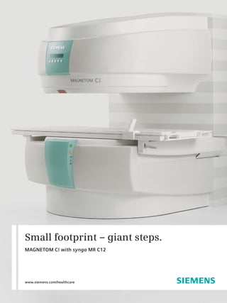
  • 1. Small footprint – giant steps. MAGNETOM C! with syngo MR C12 www.siemens.com/healthcare 1
  • 2. Expanding the scope of mid-field MRI Work that flows, image quality that convinces and patient comfort that satisfies: these are just the beginning of the advantages of MAGNETOM C! Field-proven technology and extensive experience are distilled into the surprisingly powerful MAGNETOM C! with the most compact, C-shaped magnet and smallest pole diameter of 137 cm (54 inches). 4 x 4. True multi channel imaging with 4 element coils and 4 channels allows simultaneous placement of up to 4 coils for faster head-to-toe scanning. Optimized component integration, high-field technology and superior workflow support deliver superb image quality and high diagnostic confidence in an excellent price/performance package. 2
  • 3. The generous, open feeling and inviting appearance contribute substantially to patient acceptance. Easy 270° accessibility and patient- friendly side loading add to optimum comfort and will make a crucial difference to your patients as well. You will benefit from shorter scan times and higher throughput. Increased healthcare quality, seamless workflow and low operating costs all promote a high return-on-investment. Discover the changing face of mid-field MRI. 3
  • 4. High-field competence goes mid-field. MAGNETOM C! Experience and competence work together in Unique, trendsetting syngo applications the most efficient and compact form. Through the excellent integration and synchronization of Expanding new horizons to mid-field open MRI advanced components, orchestrated by the very with applications formerly considered exclusively latest of software techniques, MAGNETOM C! high-field: moves beyond existing MRI mid-field boundaries. • syngo GRAPPA – parallel acquisition technique for higher spatial and temporal resolution. • syngo SPACE – isotropic 3D imaging in Superior components different contrasts e.g. for patients with spinal abnormalities. True multi channel imaging with fast and easy, • syngo REVEAL – diffusion weighted imaging in simultaneous placement of up to 4 coils permits the abdomen e.g. for follow-up of liver cancer the largest anatomical coverage in mid-field MRI. treatment Benefit almost instantly from extraordinary image • syngo BLADE – motion correction reproducible, quality in head-to-toe applications through the sharp images even for uncooperative patients exceptional gradient system. (e.g. with trauma) Experience high-field competence in mid-field. Seamless workflow syngo, intuitive and easy-to-use software integrates all patient related information, physiological and 4 imaging data across your entire clinical workflow.
  • 5. Competence Phoenix gives superb reproducibility based on your image. Extract MR protocols from images by drag & drop. This means increased comfort for patients and technologists, as well as shorter exam times. True multi channel RF system • Complete range of 4-element coils • Simultaneous placement of up to 4 coils (up to 13 elements) • iPAT (integrated parallel acquisition technique) for high spatial and temporal resolution High-field technology • Water cooled gradient system with 24mT/m High resolution isotropic 3D imaging for multiplanar reconstruction with syngo SPACE and 55T/m/s slew rate 5
  • 6. Applied high-field competence MAGNETOM C! High resolution imaging with good delineation of cartilage with unique 3D CISS. Strong gradients – Advanced applications – Advanced applications – advanced applications syngo SPACE syngo REVEAL Most powerful gradients in class Isotropic 3D imaging in Diffusion weighted imaging different contrasts e.g. in the abdomen e.g. for • Gradient 24 mT/m for patients with spinal follow-up of liver cancer • Slew rate 55 T/m/s abnormalities. treatment High-field gradients for faster High-field gradients to catch motion syngo SPACE for isotropic 3D Advanced abdominal imaging with scans like contrast enhanced MR in cardiac cine studies imaging in various contrasts. syngo REVEAL diffusion imaging angiography in the abdomen in all body regions including liver. 6
  • 7. “Higher field strength means higher signal- flexibility to-noise ratio – but it is not the only pre- Unprecedented coil combinations. 13 x 4 for coverage up to whole CNS. requisite for good image quality. Gradient and RF technology of the MAGNETOM C! accuracy enable us to achieve mid-field images with a Excellent image quality. Superb SNR with comprehensive quality comparable to high-field MR systems.“ family of 4 element coils. Prof. H.-M. Klein of Radiological Center – Ev. Jung-Stilling Hospital, Siegen, Germany speed Better results, faster. iPAT ensures fast acquisition with high resolution. Advanced applications – Advanced Applications – Flexibility – syngo GRAPPA syngo BLADE true multi-channel RF system Parallel acquisition techniques Correction of motion in all • Complete range of 4 element coils (iPAT) for higher spacial and 3 directions for sharp images • Up to four coils can be positioned temporal resolution and larger even with uncooperative simultaneously = 13 elements for one coverage patients and patients under examination stress • Posterior parts of the coils can stay on the table for more than 95 % of the exams without with syngo BLADE syngo BLADE syngo GRAPPA for larger coverage Trauma, claustrophobic or pediatric Up to 4 coils with 13 elements can be positioned simultaneously for and higher resolution. 120 slices of patients may move during the exa- coverage of up to 100 cm field of view. 1.5 mm and 512 matrix in the head mination. syngo BLADE corrects for in 6:30 min. motion in all three directions. 7
  • 8. Confidence in diagnosis. From a clinical perspective. Expand your expectations. Expect superb image • Exceptional image quality quality from the true multi channel RF system with Best SNR at 0.35 T the best SNR at 0.35 T and the largest anatomical coverage in mid-field. MAGNETOM C! offers excep- tional gradients for rapid acquisition and highly • Multi-channel application suite differentiated image details. Iso-center imaging • Broad range of sequences and techniques assures excellent image quality for all anatomical for all clinical applications areas. • Included in the standard package Advanced techniques such as comprehensive cardiac imaging and the unique syngo REVEAL revo- • Outstanding syngo MR Applications lutionize mid-field capabilities. syngo REVEAL for • syngo MR Neuro body diffusion increases metastasis conspicuity and • syngo MR Ortho permits virtually instant diagnosis. syngo GRAPPA • syngo MR Vascular parallel acquisition technique for higher resolu- • syngo MR Cardiac tion, larger coverage and faster scans, significantly • syngo MR Pediatric speeds workflow. Referring physicians will notice • syngo MR Oncology and appreciate the improved service. • syngo MR Body From now on, with MAGNETOM C! high-field diag- nostic confidence at mid-field can be your standard. 8
  • 10. 1 2 3 syngo MR Neuro Comprehensive state of the art applications for neuro imaging including 3D FLASH, 3D HASTE, 3D TSE Restore, iPAT, Water Excitation, DIXON and Spectral Fat Satura- tion techniques. Coverage up to whole CNS in one examina- tion without patient repositioning [1]. Dark Fluid protocols for evaluation of lesions 4 5 in the brain [2]. HASTE sequence for superior diffusion imaging with ADC maps [3] as well as EPI diffusion and perfusion. MR myelography [5] and Turbo SE sequence with Restore [4] for excellent T2 contrast with short acqui- sition times. 1 3 syngo MR Ortho Comprehensive state of the art applications for orthopedic imaging including Siemens unique 3D DESS sequence for cartilage imaging. High resolution 3D protocols for imaging of smallest anatomies like TMJ [1] (512 matrix, 1.75 mm slice thickness with contiguous coverage). Fat saturation to match every application 2 with water excitation, spectral suppression [2], DIXON or inversion recovery. Excellent image quality with 40 cm field of view for coverage of the hips [3] and thighs. 1 3 syngo MR Oncology Comprehensive oncological applications for high-field diagnostic confidence. High-resolution imaging in a short 4 second breath-hold for maximum patient accep- tance in oncologic imaging [1]. 4 element coils for highest SNR and resolution: e.g. 4 element Breast Array coil 2 for bilateral breast imaging [2]. T1 Spin Echo with DIXON Fat Saturation for excellent visualization of contrast enhance- ment in a sarcoma [3]. 10
  • 11. 1 2 syngo MR Pediatric Excellent image quality for pediatric appli- cations. Special protocols which match the special T1 and T2 values of infants and children are available for comprehensive applications including neuro [1, 2], ortho- pedic, oncological and abdominal imaging.* No dose lung imaging with TrueFISP 3 technique to avoid frequent exposure to x-rays during follow up studies e.g. of tuberculosis [3]. * The safety of imaging fetuses/infants has not been established. 1 2 syngo MR Body Comprehensive set of applications for quick and easy body imaging. 2D PACE motion correction facilitates abdominal imaging at mid-field by enabling free-breathing examinations without respiratory belt and motion monitored [1 small image] and anatomically reordered 3 multi-breath-hold studies [1]. 3D TSE [2] and 3D HASTE sequences for visualization of fluid in the biliary system and urinary tract. 1 2 syngo MR Vascular and syngo MR Cardiac Comprehensive vascular applications with and without contrast media facilitated by the high-field gradient system. Time of flight [2, showing recurring hem- orrhage], phase contrast and contrast en- hanced MRA with Care Bolus bolus timing technique [1] are easy to use with Inline 3 Technology. The MAGNETOM C! also offers comprehen- sive cardiac applications at mid-field includ- ing e.g. cine TrueFISP for evaluation of cardiac function [3]. 11
  • 12. Cardiac Imaging. Comprehensive cardiac applications at mid-field. 1 3 syngo MR Cardiac Comprehensive cardiac imaging at 0.35 T including cardiac late enhancement for assessment of myocardial viability. Eva- luation of cardiac perfusion or myocardial wall thickening with a bulls eye plot. Dark blood technique for morphological imaging [2], cineTrueFISP for evaluation of cardiac function [1] and left ventricular volume [4] supported by 4D ventricular function visualization [3] and evaluation. For easier patient positioning and better patient comfort MAGNETOM C! can be equipped with a wireless patient monitoring unit with ECG, pulse and respiratory gating. 2 4 12
  • 13. Women’s Health imaging. Comprehensive applications at mid-field. 1 2 syngo MR Women’s Health Superb gradient and RF systems enable high-resolution breast imaging with high signal-to-noise ratio. Bilateral breast imaging with 4 element Breast Array coil including dynamic imaging with inline calculation of wash-in and wash-out maps [1] and visualization of contrast media with fat saturation and subtraction tech- niques [2]. Homogeneous fat saturation over large field of views for imaging of the breast and cervix [3]. 3 13
  • 14.
  • 15. MAGNETOM C! makes it easy from start to finish. Get ready for comprehensive workflow improvements: Workflow Innovations • syngo Inline Technology: Allows you to perform certain processing steps as early as during image reconstruction and display them immediately after completion. That means processing instead of post-processing. Subtraction images, Maximum Intensity Projection, etc., are displayed immediately after completion of the scan. • syngo Phoenix: Enable simple exchange of MR protocols, quickly retrieving the entire protocol information in one easy drag-and-drop step. This allows reproducible follow-up examinations. • syngo Expert-i: Gives physicians and experts the ability to remotely access the MRI suite from virtually anywhere within the network, making it possi- ble to address difficult clinical questions while a patient is being scanned. • Multi Channel Array coils: Makes patient positioning easy. The posterior parts of the head an spine coils can stay on the table for almost all exami- nations to safe time during patient setup. Multiple coils for extended field of view can be placed simultaneously to avoid patient repositioning and facilitate image composition. 15
  • 16. Comfort Patient comfort at its best. From a patient perspective. More space means less anxiety. A unique C-shaped • Patient friendly appearance magnet for easy side loading and a friendly, compact Most open, compact C-shaped magnet design cooperate to create an open and free atmo- sphere. The smallest pole diameter – 137 cm (54“) • Patient friendly exams ensures optimal patient comfort. For up to 2/3 of Side loading all exams, the patient’s head remains outside the magnet, giving a feeling of spaciousness and relax- • Most open feeling ing claustrophobic tensions. Easy 270° accessibility, Smallest pole diameter – 137 cm (54 inches) the patients always feel close to assistance. • Easy set up Posterior parts of the coils remain on table Design defines acceptance • Close to assistance The patients will appreciate the quick and com- 270° accessibility fortable examinations. Posterior coil parts remain on table for more than 95 % of the exams. Obese patients appreciate the comfortable side loading. For these patients, MAGNETOM C! comes with unique extra large body and knee coils. These advantages, combined with the open, patient-friendly design, attract more patients, who will recommend your site. 16
  • 17. Easy 270° accessibility. Smallest pole diameter of Complete range of anatomically Open on three sides for comfortable Children feel relaxed, parents can 137 cm (54 inches). The patient’s optimized coils, including side loading and optimum patient be close during the examination. head remains outside the magnet 175 cm (69‘’) XXL body and XL comfort. for most exams. 79 cm (31.1‘’) knee arrays for obese patients. 17
  • 18. High patient throughput supported by fast sequences. T1 sagittal, T2 sagittal and axial and myelogram in less than 9 minutes. Cost- Efficiency From a business perspective. Move up to cost efficient quality care. Comprehensive workflow from start to finish. Every • Excellent price performance ratio step is optimized to streamline your examinations: Comprehensive application suite beginning with quick and easy patient positioning and posterior coil parts that remain on the table. New • Minimal siting requirements protocols and scan speed close to high-field per- Less than 30 m² (325 sq.ft) formance assure increased throughput and higher image quality. A wide choice of applications opens • Low operating costs opportunities for more referrals. Structured report- Permanent magnet – no helium ing and DVD burning accelerate distribution because time is essential. • Inline technology Processing instead of Post-Processing And with Life, Siemens’ integrated customer care solution, you’ll know that your ever-evolving • Phoenix business needs are being met. What you see is what you get. Drag & drop images to scan. Evolve, the obsolescence protection program helps you to stay at the leading edge of technology and • syngo Expert-i advancement. You will benefit more from the latest Allows remote access into the workflow improvements, clinical applications and MR console by an expert user the most advanced computer technology. within the network. • Excellent Return-On-Investment Decreased costs – optimized profitability 18
  • 19.
  • 20. Images courtesy of: Local Contact Information Salmaniya Medical Complex, Manama, Bahrain In the USA Eduardus Hospital, Cologne, Germany Siemens Medical Solutions USA, Inc. Ev. Jung-Stilling Hospital, Siegen, Germany 51 Valley Stream Parkway Mamata General Hospital, Khammam, India Malvern, PA 19355 Diagnostic Centre Kenya Ltd., Nairobi, Kenya Phone: +1 888-826-9702 KL SMC Sports Imaging, Kuala Lumpur, Malaysia Phone: 610-448-4500 HaiNan NongKen Hospital, SanYa, P.R. China Fax: 610-448-2254 Sir Run Run Shaw Hospital, HangZhou, P.R. China IMADIA, Gava, Spain In China: Cleveland Community Hospital, Cleveland, USA Siemens Medical Park, Shanghai Lone Star Imaging, Mission, USA 278, Zhouzhu Road Praxis Dr. Sens, Frankfurt, Germany SIMZ, Nanhui District Horsens Regional Hospital, Horsens, Denmark Shanghai, 201318, LY HeDong People’s Hospital, PRC P.R.China MRI Arlberg, Lech, Austria Phone: +86-21-38895000 Fax: +86-10-28895001 In Japan: Siemens-Asahi Medical Technologies Ltd. The information in this document contains Takanawa Park Tower 14 F general descriptions of the technical options 20-14, Higashi-Gotanda 3-chome available, which do not always have to be present Shinagawa-ku in individual cases. The required features should Tokyo 141-8644 therefore be specified in each individual case at Phone: +81 3 5423 8489 the time of closing the contract. In Asia: Siemens reserves the right to modify the design Siemens Pte Ltd and specifications contained herein without prior Healthcare Sector notice. Please contact your local Siemens Sales Regional Headquarters representative for the most current information. The Siemens Center 60 MacPherson Road syngo Evolve Package: In the event that upgrades Singapore 348615 require FDA approval, Siemens cannot predict Phone: +65 6490-6000 whether or when the FDA will issue its approval. Fax: +65 6490-6001 Therefore, if regulatory clearance is obtained and is applicable to this package, it will be made available according to the termsof this offer. Global Business Unit Note: Any technical data contained in this Siemens AG document may vary within defined tolerances. Medical Solutions Original images always lose a certain amount of Magnetic Resonance detail when reproduced. Henkestr. 127 DE-91052 Erlangen Please find fitting accessories: Germany www.siemens.com/medical-accessories Medical Phone: +49 9131 84-0 Global Siemens Headquarters Global Siemens Global Siemens Headquarters Healthcare Headquarters Siemens AG Siemens AG Wittelsbacherplatz 2 Siemens AG Wittelsbacherplatz 2 80333 Muenchen Healthcare Sector DE-80333 Muenchen Germany Henkestr. 127 Germany 91052 Erlangen Germany Phone: +49 9131 84-0 www.siemens.com/healthcare www.siemens.com/healthcare Order No. A91MR-300-8C-7600 | Printed in Germany | CC MR 00300 WS 06092. | © 06.2009, Siemens AG