2. 1402 Inorganic Chemistry, Vol. 38, No. 7, 1999 Inscore et al.
Magnetic circular dichroism (MCD) spectroscopy has been
utilized in the study of desulfo-inhibited xanthine oxidase,24
DMSO reductase,25 and glycerol-inhibited DMSO reductase,26
and the MCD spectra of the DMSO reductases have been
interpreted in the context of out-of-plane S pπ f Mo charge
transfer transitions. Additionally, resonance Raman spectros-
copy27-29 has been used to probe the lowest energy absorption
features observed in the Mo(VI) and Mo(IV) oxidation states
of the R. sphaeroides enzyme and the vibrational spectra display
a complicated series of vibrational bands in the low-frequency
region between 250-450 cm-1 where metal-ligand vibrations
are expected to occur. These vibrational bands were shown to
be sensitive to 34S isotopic substitution, indicating that the
observed modes possess a significant Mo-S stretching contri-
bution, indicative of the S f Mo charge transfer nature of the
low-energy absorption features. Curiously, the multitude of low-
frequency vibrations found in DMSO reductase are absent in
the resonance Raman spectra of human sulfite oxidase and its
C207S mutant.30 Regardless, the observation of S f Mo charge
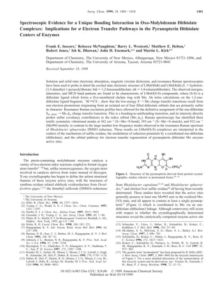
Figure 2. Consensus active site structures for the oxidized Mo(VI) transfer transitions associated with the Mo-ene-dithiolate portion
state of the three families of pyranopterin Mo enzymes according to of the active site has provided the impetus for synthetic and
Hille1; DMSO reductase family; xanthine oxidase family; sulfite oxidase electronic structure studies of model compounds which mimic
family. geometric, spectroscopic, and electron transfer aspects of the
coordination geometry, the X-ray studies have provided pro- active site. Most notable among the spectroscopic work has been
found insight toward the development of consensus structures a seminal MCD and electronic absorption study which deter-
which correlate the crystallographic results with available mined that S f Mo LMCT transitions dominate the low-energy
EXAFS18-21 and spectroscopic data. This has allowed for absorption and MCD spectra of oxomolybdenum ene-1,2-
categorizing these Mo enzymes into three main families on the dithiolate model complexes.31 The results of this study suggested
basis of the geometric structure of the oxidized active sites that the oxomolybdenum ene-1,2-dithiolate moiety is a uniquely
(Figure 2).1,7 designed catalytic unit, allowing for MotO bond destabilization
The crystallographic studies have concluded that the pyran- and concomitant activation in oxygen atom transfer catalysis
opterin adopts a distinctly nonplanar conformation with an as well as providing an electron transfer conduit coupling Mo
approximately 40° twist of the pyran ring,10-16,22 strongly with the pyranopterin.
favoring a reduced pyranopterin which lacks extensive π-de- X-ray crystallographic and XAS studies have defined the
localization over the tricyclic ring. Although the pyranopterin salient structural features for the pyranopterin centers of Mo
is well known to anchor the pyranopterin to the protein via enzymes. This allows for detailed spectroscopic studies to be
extensive hydrogen bonding,10-16,22 its precise functional role initiated on relevant model complexes in order to develop insight
in catalysis remains undetermined. The intimate association of into the electronic origin of observed spectroscopic features and
the pyranopterin dithiolate with Mo suggests a variety of roles their contributions to reactivity. We have employed a combina-
for the pyranopterin in catalysis, including modulating the tion of electronic absorption, MCD, and resonance Raman
reduction potential and acting as an electron transfer pathway spectroscopies to understand the electronic structure of first
to other endogenous or exogenous redox partners.1,7 There is generation mono-oxo Mo(V) ene-1,2-dithiolate models for
structural and reactivity evidence for the direct involvement of
enzymes in the sulfite oxidase family. The results of this
the pyranopterin in electron transfer. The structure of D. gigas
spectroscopic study are significant with respect to understanding
aldehyde oxidoreductase has the amino group of the pterin
the mechanism and determining the stereochemical location of
hydrogen bonded to a cysteinyl sulfur of a 2Fe2S cluster.12 Also,
the catalytically labile oxo group in sulfite oxidase; providing
sulfite oxidase is inactivated by ferricyanide oxidation of the
an emerging picture of how Mo redox potentials may be
pyranopterin to the fully oxidized state.23 This study conclusively
modulated by a coordinated dithiolate; and describing the orbital
showed that the presence of a fully oxidized pterin compromised
pathway for electron transfer regeneration of these Mo enzyme
the ability of the enzyme to reduce the physiological oxidant
active sites.
(cytochrome c) but did not significantly affect the ability of
the enzyme to oxidize sulfite. Therefore, it appears that the
oxidized pyranopterin severely retards the egress of reducing (24) Peterson, J.; Godfrey, C.; Thomson, A. J.; George, G. N.; Bray, R. C.
Biochem. J. 1986, 233, 107-110.
equivalents to the endogenous b-type heme. (25) Benson, N.; Farrar, J. A.; McEwan, A. G.; Thompson, A. J. FEBS
Lett. 1992, 307, 169-172.
(18) Cramer, S. P.; Gray, H. B.; Rajagopalan, K. V. J. Am. Chem. Soc. (26) Finnegan, M. G.; Hilton, J.; Rajagopalan, K. V.; Johnson, M. K. Inorg.
1979, 101, 2772-2774. Chem. 1993, 32, 2616-2617.
(19) George, G. N.; Kipke, C. A.; Prince, R. C.; Sunde, R. A.; Enemark, (27) Gruber, S.; Kilpatrick, L.; Bastian, N. R.; Rajagopalan, K. V.; Spiro,
J. H.; Cramer, S. P. Biochemistry 1989, 28, 5075-5080. T. G. J. Am. Chem. Soc. 1990, 112, 8179-8180.
(20) Cramer, S. P.; Wahl, R.; Rajagopalan, K. V. J. Am. Chem. Soc. 1981, (28) Kilpatrick, L.; Rajagopalan, K. V.; Hilton, J.; Bastian, N. R.; Steifel,
103, 7721-7727. E. I.; Pilato, R. S.; Spiro, T. G. Biochemistry 1995, 34, 3032-3039.
(21) George, G. N.; Garrett, R. M.; Prince, R. C.; Rajagopalan, K. V. J. (29) Garton, S. D.; Hilton, J.; Oku, H.; Crouse, B. R.; Rajagopalan, K. V.;
Am. Chem. Soc. 1996, 118, 8588-8592. Johnson, M. K. J. Am. Chem. Soc. 1997, 119, 12906-12916.
(22) Chan, M. K.; Mukund, S.; Kletzin, A.; Adams, M. W. W.; Rees, D. (30) Garton, S. D.; Garrett, R. M.; Rajagopalan, K. V.; Johnson, M. K. J.
C. Science 1995, 267, 1463-1469. Am. Chem. Soc. 1997, 119, 2590-2591.
(23) Gardlik, S.; Rajagopalan, K. V. J. Biol. Chem. 1991, 266, 4889- (31) Carducci, M. D.; Brown, C.; Solomon, E. I.; Enemark, J. H. J. Am.
4895. Chem. Soc. 1994, 116, 11856-11868.
3. Unique Bonding in Oxo-Molybdenum Dithiolates Inorganic Chemistry, Vol. 38, No. 7, 1999 1403
Experimental Section either S-1 or S-20 response, an Oxford Instruments SM4000-7T
superconducting magneto-optical cryostat (0-7 Tesla and 1.4-300 K),
General. Unless otherwise noted, all reactions were carried out in and an Oxford Instruments ITC503 temperature controller. The
an inert atmosphere of nitrogen using Schlenk techniques. All solvents spectrometer was calibrated for CD intensity and wavelength using
were dried by distillation, and deoxygenated prior to use. Purification camphorsulfonic acid and a Nd-doped reference glass sample (Schott
of solvents was accomplished using the following methodologies: Glass). Solid-state MCD spectra were obtained by dispersing finely
pyridine and triethylamine from potassium hydroxide; toluene from ground samples in poly(dimethylsiloxane) and compressing the suspen-
sodium benzophenone. Other solvents were used without further sion between two 1 mm thick Infrasil quartz discs (ESCO). Depolar-
purification. The compounds LMoOCl2,32 LMoVO(tdt),32 LMoVO- ization of the incident radiation was checked by comparing the
(bdt),33,34 and LMoVO(edt)32 were prepared as previously described. difference in CD intensity of a standard Ni (+)-tartrate solution
Abbreviations. L, hydrotris(3,5-dimethyl-1-pyrazolyl)borate; bdt, positioned before and then after the sample. Samples which depolarized
1,2-benzenedithiolate; tdt, 3,4-toluenedithiolate; edt, 1,2-ethanedithio- the light by <5% were deemed suitable. The MCD spectra in the 250-
late; H2qdt, quinoxaline-2,3-dithiol; qdt, 2,3-dithioquinoxaline). 800 nm range were obtained at 2.0 nm resolution, and data between
Preparation of LMoO(qdt). The reagents 2,3-dihydroxyquinoxaline 400 and 1050 nm were collected at a fixed slit width of 150 µ. All
and phosphorus pentasulfide were purchased from Aldrich Chemical MCD spectra were collected in an applied magnetic field of 7 Tesla.
Co. Quinoxaline-2,3-dithiol was prepared by a modified version of Vibrational and Resonance Raman Spectroscopy. Infrared spectra
Morrison.35,36 To a dry toluene solution of LMoOCl2 (0.5 g, 1.05 mmol) were recorded on a BOMEM MB-100 FT-IR spectrometer as pressed
was added a toluene solution containing 0.41 g (2.1 mmol) of H2qdt KBr disks. The infrared spectra were utilized to monitor the purity of
and 150 µL (2.1 mmol) of triethylamine, dropwise by cannula at 70 the compounds, as indicated by the absence of the 962 cm-1 MotO
°C. This solution was allowed to react for approximately 5 h, and during stretch associated with the LMoOCl2 precursor complex.32
this time the color of the solution changed from lime green to dark Resonance Raman spectra were collected in a 135° backscattering
red. The resulting dark red solution was filtered and concentrated at geometry. A Coherent Innova 70-5 (5W) Ar+ ion laser was the photon
reduced pressure to give a dark red powder, which was subsequently source (457.9-528.7 nm, 9 discrete lines) for inducing Raman
redissolved in a minimum amount of toluene and chromatographed on scattering. The scattered radiation was dispersed onto a liquid N2 cooled
silica gel. The compound eluted in a binary mixture of toluene/1,2- 1′′ Spex Spectrum One CCD detector using a Spex 1877E triple grating
dichloroethane (9:1) as a red band. Yield ) 15%. Anal. Calcd for monochromator equipped with 600, 1200, and 1800 gr/mm holographic
C23H26N8OS2BMo: C, 45.93; H, 4.36. Found: C, 45.04; H, 4.33. IR gratings at the spectrographic stage. The laser power at the sample was
(KBr, cm-1): ν(ModO) 940, ν(B-H) 2551. MS (FAB): m/z 602.1 kept between 40 and 100 mW in order to prevent possible photo- and
(parent ion), 507 (parent - 3,5-dimethylpyrazole), 410 (parent - thermal degradation of the sample. Solid samples were prepared as
dithiolate). finely ground powders and dispersed in a NaCl(s) matrix with Na2SO4
Physical Characterization. Elemental analysis was performed at added as an internal standard. These samples were subsequently sealed
The University of New Mexico using a Perkin-Elmer 2400 CHN in an NMR tube and Raman spectra were obtained by spinning the
elemental analyzer equipped with a P-E AD-6 Autobalance. Mass sample in a modified NMR sample holder/spinner. The samples were
Spectra were collected at The Nebraska Center for Mass Spectrometry maintained at ∼140 ( 10 K by the use of a custom designed cold N2
in the Department of Chemistry at the University of NebraskasLincoln. gas flow system. The sample temperature was periodically monitored
Electronic Absorption Spectroscopy. Mull and solution electronic with a Lakeshore silicon diode (PT-470) enclosed in a separate NMR
absorption spectra were collected on a double beam Hitachi U-3501 tube. The construction of resonance Raman profiles was accomplished
UV-vis-NIR spectrophotometer capable of scanning a wavelength by comparing the integrated intensity of a Raman band at a given
region between 185 and 3200 nm. All absorption spectra were collected excitation wavelength relative to that of the 992.4 cm-1 band of Na2-
at 2.0 nm resolution in a single-beam configuration. The instrument SO4. All data were scan averaged, and any individual data set with
was calibrated with reference to the 656.10 nm deuterium line. vibrational bands compromised by cosmic events was discarded.
Immediately following acquisition of the sample spectra, background Solution Raman spectra were obtained in degassed benzene and spun
spectra were collected to correct for residual absorption due to the in a sealed NMR tube at room temperature. Depolarization ratios were
solvent or mulling agent and to correct for light scattering effects. obtained by placing a rotatable polarizer before the polarization
Solution samples were prepared by dissolving the compounds in scrambler and monochromator entrance slit. Relative Raman intensities
degassed dichloroethane. The electronic absorption spectra were (perpendicular and parallel to incident radiation) for a given Raman
subsequently collected in 1 cm pathlength Helma quartz cells (black- band were measured relative to the 992 cm-1 band of benzene.
masked Suprasil II, equipped with a Teflon stopper). Mull samples Ab Initio Calculations. Ab initio calculations were performed using
were prepared by grinding the solid sample into a fine powder before the Gaussian 94 suite of programs.37 A 6-31G** basis set was employed
dispersing it into poly(dimethylsiloxane). The prepared mull was in calculating the energies and wavefunctions for the model 1,2-ene-
subsequently placed between two 1 mm thick Infrasil quartz discs dithiolate (-SCHdCHS-).
(ESCO) and secured in a custom designed sample holder. A Janis
STVP-100 continuous flow cryostat mounted in a custom designed Results
cradle assembly was used for acquisition of the low-temperature (∼5
K) spectra. The sample temperature was continuously monitored with Solution Electronic Absorption Spectra. Figure 3 depicts
a Lakeshore silicon-diode (PT-470) and regulated by a combination of the room temperature electronic absorption spectrum of LMoO-
helium flow and dual heater assemblies. Gaussian resolution of spectral (bdt) between 6000 and 35 000 cm-1 in dichloroethane. The
bands and corrections for light scattering were accomplished with spectrum is very similar to that previously reported for LMoO-
KaleidaGraph and programs incorporated within the Hitachi version (tdt).31 However, the transitions observed for LMoO(tdt) are
of the Grams software package. generally shifted to slightly lower energies relative to the
Magnetic Circular Dichroism Spectroscopy. Low-temperature corresponding bands in LMoO(bdt). The low-energy region of
MCD data were collected on a system consisting of a Jasco J600 CD the spectrum consists of three distinct spectral features (bands
spectropolarimeter employing Hamamatsu photomultiplier tubes of
1, 2, and 4) below ∼20 000 cm-1. We have found these bands
to be characteristic of LMoO(S-S) compounds, where S-S is a
(32) Cleland, W. E., Jr.; Barnhart, K. M.; Yamanouchi, K.; Collison, D.;
Mabbs, F. E.; Ortega, R. B.; Enemark, J. H. Inorg. Chem. 1987, 26, dithiolene or dithiolate ligand which forms a five-membered
1017-1025. chelate ring with Mo. Band 3 is very weak, and only discernible
(33) Dhawan, I. K.; Enemark, J. H. Inorg. Chem. 1996, 35, 4873-4882. in the low-temperature MCD spectra (Vide infra). The transition
(34) Dhawan, I. K.; Pacheco, A.; Enemark, J. H. J. Am. Chem. Soc. 1994,
116, 7911-7912.
energies and molar extinction coefficients for four LMoO(S-S)
(35) Morrison, D. C.; Furst, A. J. Org. Chem. 1956, 21, 470-471.
(36) Helton, M. E.; Kirk, M. L. Submitted for publication. (37) Gaussian Incorporated, Pittsburgh, PA.
4. 1404 Inorganic Chemistry, Vol. 38, No. 7, 1999 Inscore et al.
Table 1. Summary of Electronic Absorption Data for LMoO(S-S) Complexes in Dichloroethane
Emax, cm-1 ( , M-1 cm-1)
LMoO(bdt) LMoO(tdt) LMoO(qdt)36 LMoO(edt)
band 1 9 100 (360) 9 100 (490) 11 300 (170) 11 800 (160)
band 2 13 100 (270) 13 000 (270) 13 700 (130) 15 500 (220)
band 4 19 400 (sh,1220) 19 600 (sh,1320) 19 100 (1050) 20 000 (sh, 570)
Table 2. Calculated Oscillator Strengths of LMoO(bdt) and the
Relationship between MCD and Electronic Absorption Bands
band E(soln)max oscillator E(mull)max E(MCD)max MCD
no. (cm-1) strength (cm-1) (cm-1)a terma
1 9 100 5.6 × 10⁻³ 8 500 ---- ----
2 13 100 3.3 × 10⁻³ 12 700 12 400 -C
3 15 800 ---- ---- 15 700 +C
4 19 400 1.6 × 10⁻² 19 200 19 300 +C
5 22 100 1.7 × 10⁻² 21 500 21 000 +pseudo A
6 25 100 9.2 × 10⁻² 24 600 24 300 +pseudo A
a A positive pseudo A-term is a derivative shaped MCD feature with
the positive component at higher energy. Emax represents the point at
which the pseudo A-term changes sign.
Figure 3. Gaussian-resolved 293 K electronic absorption spectrum of
LMoO(bdt) in dichloroethane (2.45 × 10⁻⁴ M). The dashed lines
represent the individual Gaussians used in the fit.
Figure 5. 4.86 K MCD spectra of LMoO(bdt) (solid line) and LMoO-
(tdt) (dotted line) dispersed in poly(dimethylsiloxane). Note the overall
similarity of the spectral features.
derivative-shaped dispersions which possess zero intensity at
Figure 4. 5 K electronic absorption (heavy line) and 4.86 K MCD an energy corresponding to an absorption maximum. The
(light line) spectra of LMoO(bdt) dispersed in poly(dimethylsiloxane). relationship between observed MCD and electronic absorption
bands in LMoO(bdt) is given in Table 2.
complexes are presented in Table 1 for comparative purposes. Figure 5 compares the 5 K/7 T MCD mull spectra of LMoO-
Of particular interest is band 4, which is the first absorption (bdt) and LMoO(tdt) in the spectral region between 10 000 and
feature possessing appreciable intensity characteristic of a charge 40 000 cm-1. The MCD spectra are seen to be quite similar,
transfer transition. and this band pattern is characteristic of LMoO(S-S) complexes
Solid-State Electronic Absorption and MCD Spectra. The where the MotO bond is oriented cis to a single dithiolate
5 K mull MCD/absorption overlay of LMoO(bdt) is shown in ligand which forms a five-membered chelate ring with Mo.31
Figure 4. The electronic absorption spectrum exhibits five However, noticeable differences in MCD sign exist for these
distinct features in the solid state with weak to significant compounds in the 14 000-17 000 cm-1 range, where no
intensity. The 21 500 cm-1 band is observed as a reproducible discernible maxima occur in the absorption spectra (see Figure
shoulder in the mull absorption of LMoO(bdt) but is conspicu- 3). Close inspection of Figure 5 reveals the reason the 21 500
ously absent in the corresponding absorption spectrum of cm-1 transition in LMoO(bdt) is not resolved in the MCD or
LMoO(tdt). The general similarity of the solution and mull absorption spectra of LMoO(tdt). The positive C-term at 19 300
absorption spectra for LMoO(bdt) indicate that only minor cm-1 and the low-energy negatively signed component of the
structural changes accompany solvation. This is true for all of 24 300 cm-1 positive pseudo A-term observed in LMoO(bdt)
the LMoO(S-S) compounds listed in Table 1. are energetically compressed in the MCD spectrum of LMoO-
The MCD spectrum of LMoO(bdt) is composed of both (tdt), effectively masking the 21 500 cm-1 spectral feature. The
C-terms and pseudo A-terms.38 MCD C-terms possess absorptive MCD spectrum of LMoO(qdt) shows this transition as a clearly
bandshapes with intensity maxima at the same energy as resolved positive pseudo A-term.36
corresponding absorption features, while pseudo A-terms possess Vibrational Spectra. The IR data for LMoO(bdt) and LMoO-
(tdt) display intense peaks at 932 and 926 cm-1, respectively.
(38) Piepho, S. B.; Schatz, P. N. Group Theory in Spectroscopy with
Applications to Magnetic Circular Dichroism; Wiley-Interscience: Strong IR bands in the 910-965 cm-1 range have been reported
New York, 1983. for a variety of LMoOX2 complexes,39-43 and this band has
5. Unique Bonding in Oxo-Molybdenum Dithiolates Inorganic Chemistry, Vol. 38, No. 7, 1999 1405
Figure 7. 140 K solid-state resonance Raman excitation profiles for
Figure 6. 293 K resonance Raman spectrum of LMoO(bdt) in benzene. LMoO(bdt). The incident laser power measured at the sample was ∼50
The spectrum was obtained with 514.5 nm excitation and the incident mW. The profiles are superimposed on the Gaussian-resolved 5 K mull
laser power at the sample was ∼75 mW. Unmarked bands are those of absorption spectrum of Figure 4. The dashed lines represent the
the solvent. individual Gaussians used in the fit. ν1 (diamonds), ν3 (squares),
ν4 (circles).
been assigned as the MotO stretching vibration. Vibrational
studies on related compounds possessing the {MoVtO}3+ unit
also reveal the presence of a band in this region assignable as
the MotO stretch.44-47 The IR spectra are also useful for
detecting very small quantities of LMoOCl2 precursor complex
that may be present in the sample as a contaminant. No 961
cm-1 MotO stretch characteristic of LMoOCl2 was observed
in the IR spectra of the LMoO(S-S) complexes used in this
study.32
Figure 6 shows the 514.5 nm Raman spectrum of LMoO-
(bdt) in benzene. The solution spectrum contains only three
observable vibrational modes between 250 and 1000 cm-1, with
frequencies of 362, 393, and 932 cm-1. Vibrational bands in
the 300-400 cm-1 region of transition metal 1,2-ene-dithiolates
have been collectively assigned as Mo-S stretching vibrations.48-50 Figure 8. Highest occupied molecular orbitals of the model 1,2-ene-
A qualitative depolarization study was performed on LMoO- dithiolate, -SCHdCHS-. The small amplitudes of the wavefunction
(bdt) in benzene using 496.5 nm excitation which yielded on the dithiolate carbon atoms are not shown for clarity.
depolarization ratios of 0.40 (362 cm-1), 0.22 (393 cm-1), and
0.01(932 cm-1). Since the symmetry of the LMoO(S-S) providing strong evidence that the structural integrity of these
complexes is very close to Cs, the depolarization ratios for these complexes is maintained in solution.
bands are characteristic of totally symmetric a′ modes. Raman Resonance Raman Excitation Profiles. Resonance Raman
spectra for LMoO(bdt) and LMoO(tdt) were also collected in excitation profiles were collected in the solid state at 140 K
the solid-state at 140 K using laser excitation at wavelengths using laser excitation wavelengths between 457.9 and 528.7 nm.
between 528.7 and 457.9 nm. As was the case in solution, three This wavelength range encompasses the absorption envelopes
vibrational bands were observed. These occured at 362, 393, of bands 4 and 5. All three observed vibrational bands for
and 931 cm-1 for LMoO(bdt) and at 342, 376, and 926 cm-1 LMoO(bdt) were found to be resonantly enhanced, and the
for LMoO(tdt). Therefore, no significant ground-state vibrational resonance Raman profiles have been superimposed upon the
frequency shifts occur between the solid and solution spectra, Gaussian resolved 5 K mull absorption spectrum in Figure 7.
Extremely selectiVe resonance Raman enhancement patterns are
(39) Chang, C. S. J.; Collison, D.; Mabbs, F. E.; Enemark, J. H. Inorg. evident for excitation into band 4 (362 and 393 cm-1 modes)
Chem. 1990, 29, 2261-2267.
(40) Chang, C. S. J.; Enemark, J. H. Inorg. Chem. 1991, 30, 683-688. and band 5 (931 cm-1 mode). Interestingly, the resonance
(41) Nipales, N.; Westmoreland, T. D. Inorg. Chem. 1995, 34, 3374-3377. Raman profile for the corresponding 926 cm-1 mode of LMoO-
(42) Lincoln, S. E.; Loehr, T. M. Inorg. Chem. 1990, 29, 1907-1915. (tdt) shows no high-energy turnover when pumping into band
(43) Chang, C. S. J.; Pecci, T. J.; Carducci, M. D.; Enemark, J. H. Inorg.
Chem. 1993, 32, 4106-4110. 5. This is consistent with the low-temperature mull absorption
(44) Bradbury, J. R.; Mackay, M. F.; Wedd, A. G. Aust. J. Chem. 1978, and MCD spectra of LMoO(tdt), which indicate that band 6 is
31, 2423-2430. lowered in energy and overlaps band 5. The result implies that
(45) Ellis, S. R.; Collison, D.; Garner, C. D. J. Chem. Soc. Dalton Trans.
1989, 413-417.
the 931 cm-1 mode of LMoO(bdt) is also resonantly enhanced
(46) Burt, R. J.; Dilworth, J. R.; Leigh, G. J.; Zubieta, J. A. J. Chem. Soc. when pumping into band 6.
Dalton Trans. 1982, 2295-2298. Ab Initio Calculations. Ab initio calculations on the model
(47) Ueyama, N.; Okamura, T.; Nakamura, A. J. Am. Chem. Soc. 1992,
114, 8129-8137.
ethenedithiolate (-SCHdCHS-) fragment resulted in an isolated
(48) Spiro, T. (Ed.) Molybdenum Enzymes; John Wiley and Sons: New set of four filled dithiolate orbitals that are primarily sulfur in
York, 1985. character, and these are depicted in Figure 8. These are the
(49) Subramanian, P.; Burgmayer, S.; Richards, S.; Szalai, V.; Spiro, T. ligand wavefunctions which can energetically mix and form
G. Inorg. Chem. 1990, 29, 3849-3853.
(50) Oku, H.; Ueyama, N.; Nakamura, A. Inorg. Chem. 1995, 34, 3667- symmetry-adapted linear combinations (SALC’s) with the d
3676. orbitals of appropriate symmetry localized on Mo. The calcula-
6. 1406 Inorganic Chemistry, Vol. 38, No. 7, 1999 Inscore et al.
tions indicate that the electron density is largely localized on
the sulfur atoms, in agreement with experimental results from
photoelectron spectroscopy.51 The four orbitals may be divided
into two sets with respect to their orientation to the plane
containing the constituent atoms of the ene-dithiolate. These
are the in-plane components, φa′ and φa′′, and the out-of-plane
ip ip
components φa′ and φa′′ . The superscripts a′ and a′′ refer to the
op op
symmetry of the wavefunction with respect to the Cs mirror
plane. This mirror plane is oriented perpendicular to the ene-
dithiolate plane, bisecting the CdC bond.
Analysis
Origin of the S f Mo Charge Transfer. The LMoO(S-S)
compounds in Table 1 possess nearly perfect Cs symmetry with
respect to the donor atoms which comprise the first coordination
sphere. However, an argument can be made that effective C4V
symmetry may be utilized due to the presence of the strong
field terminal oxo ligand. This is apparent in the nearly axial A
tensors determined from single-crystal EPR studies on LMoOCl252
and EPR simulations of LMoO(bdt)33,34 and LMoO(edt)32 frozen
solution spectra. From a ligand-field perspective, this would
imply nearly degenerate dxz,yz orbitals in the ground state.
Recognizing this fact we will utilize C4V f C2V f Cs descent
in symmetry in the analysis of the LMoO(S-S) compounds,
keeping in mind the strong axial nature of the ligand field and Figure 9. Spectroscopically effective molecular orbital diagram for
the anticipated small anisotropy within the equatorial plane. the LMoO(S-S) complexes. The molecular orbital energies are not to
The oscillator strength of an electronic absorption band is scale. The ψxz,yz orbitals are shown as degenerate due to the dominance
of the strongly bound oxo ligand on the z axis.
proportional to |〈ψG|r|ψE〉|2, where ψG and ψE are the ground
and excited state wavefunctions, and r is the position vector.53-55
In a one-electron approximation, |〈ψG|r|ψE〉|2 ) |〈ψa|r|ψb〉|2, where Cn and C′ are the sulfur atomic orbital coefficients in ψa
n
where ψa and ψb are the two molecular orbitals involved in the and ψb, and 〈χn|χ′ 〉 is an overlap integral. Thus, the same type
n
one electron promotion ψa f ψb. Metal-ligand covalency of dithiolate molecular orbital (e.g., 〈φip|φip〉 or 〈φop|φop〉) must
results in molecular orbital wavefunctions that can be expanded be present in the ground and excited wavefunctions for enhanced
in terms of metal- and ligand-centered functions: charge transfer transition intensity, and this intensity is a direct
consequence of the Mo d-dithiolate S covalency.
ψa ) C1φM + C2φL + ... The ligand field splitting of the d-orbital manifold in LMoO-
(S-S) and other high-valent metal oxo compounds is dominated
ψb ) C′ φM + C′ φL + ...
1 2 by the presence of a short 1.7-1.9 Å MtO bond.56-59 The
terminal oxo ligand is an extremely strong σ- and π-donor, and
where φL ) ∑Cnχn
n
in the presence of a moderate to weak equatorial ligand field
the d-orbital splitting diagram in Figure 9 results. Here, the Mo
Here, the Ci are the coefficients of the metal (φM) and ligand ψz2 and ψa′′,ψa′ orbitals are strongly destabilized by σ and π
a′
xz yz
(φL) orbitals which constitute the ψa and ψb ground state antibonding interactions with the terminal oxo ligand. In d1
molecular orbital wavefunctions. Considering the LMoO(S-S) LMoO(S-S) complexes, all of the d orbitals may act as acceptor
compounds in this study, φM are the Mo d orbitals (e.g., φa′ orbitals in low-energy LMCT interactions involving the coor-
xy
dinated dithiolate. Therefore, the energy of these charge transfer
etc.); φL are the four orthogonal ene-dithiolate orbitals (φa′ ,ip
transitions will be strongly affected by the oxo-mediated
φa′ , φa′′, and φa′′ ) of Figure 8, to which the primary contribu-
op ip op destabilization of these acceptor orbitals. Figure 9 also depicts
tors are the χn for the atomic sulfur p orbital functions. It
the anticipated relative energy of the lowest ligand field and
has been shown that the dominant contributor to the oscil-
charge-transfer excitations. As a result of the Mo d orbital
lator strengths of all transitions are integrals of the form
splitting pattern, the predicted lowest energy charge transfer
〈χn|r|χ′ 〉.53-55 In the limit of no overlap between the two sulfur
n
p orbitals on different atoms, 〈χn|r|χ′ 〉 reduces to53 transitions will be to the Mo ψa′ orbital followed by transitions
xy
n
to the ψa′′,ψa′ orbital set. These LMCT transitions originate
xz yz
from the four dithiolate based φL molecular orbitals of Figure
∑CnC′nr〈χn|χ′n〉
n
8, two of which are oriented within the dithiolate plane (φa′ ip
and φa′′) and two which are orthogonal to it (φa′ and φa′′ ). It
ip op op
(51) Gleiter, R.; Spanget-Larsen, J. Top. Curr. Chem.: Spectrosc. 1979,
can be seen from this diagram that the stabilization of these
86, 139-196. dithiolate molecular orbitals will result from specific Mo
(52) Collison, D.; Eardley, D. R.; Mabbs, F. E.; Rigby, K.; Bruck, M. A.; d-dithiolate S bonding interactions.
Enemark, J. H.; Wexler, P. A. J. Chem. Soc. Dalton Trans. 1994,
1003-1011.
(53) Avoird, A.; Ros, P. Theoret. Chim. Acta (Berlin) 1966, 4, 13-21. (56) Ballhausen, C. J.; Gray, H. B. Inorg. Chem. 1962, 1, 111-122.
(54) Solomon, E. I. Comm. Inorg. Chem. 1984, 3, 227-320. (57) Gray, H. B.; Hare, C. R. Inorg. Chem. 1962, 1, 363-368.
(55) LaCroix, L. B.; Shadle, S. E.; Wang, Y.; Averill, B. A.; Hedman, B.; (58) Collison, D. J. Chem. Soc. Dalton Trans. 1990, 2999-3006.
Hodgson, K. O.; Solomon, E. I. J. Am. Chem. Soc. 1996, 118, 7755- (59) Winkler, J. R.; Gray, H. B. Comments Inorg. Chem. 1981, 1, 257-
7768. 263.
7. Unique Bonding in Oxo-Molybdenum Dithiolates Inorganic Chemistry, Vol. 38, No. 7, 1999 1407
MotO and Mo-S bonds, and it is these excited state distortions
which form the basis for A-term resonance Raman enhancement
of totally symmetric normal modes.60
Only three vibrations were observed in the solid-state Raman
spectra of LMoO(bdt) (362.0, 393.0, and 931.0 cm-1) and
LMoO(tdt) (342.0, 376.0, and 926.0 cm-1), and all are
resonantly enhanced. The solution Raman depolarization results
for LMoO(bdt) show that all three of these vibrations are
polarized, providing strong evidence for assigning the vibrational
bands as totally symmetric a′ modes. Inspection of Figure 10
reveals that four of the six modes predicted for a Cs MoO(S,S)
chromophore are of a′ symmetry. The high-frequency ν3 mode
is easily assigned as the totally symmetric MotO stretch on
the basis of its depolarization ratio (F|/F⊥ ) 0.01), high-
frequency, strong IR absorption, and analogy to similar oxo-
molybdenum compounds. There are two vibrational bands found
in the 300-400 cm-1 region, where Mo-S stretching vibrations
are anticipated to occur.48-50 Since there exists only one totally
symmetric a′ S-Mo-S stretch, we assign the 393 cm-1 mode
Figure 10. The six vibrational symmetry coordinates for a simple four as the S-Mo-S stretch (ν2) and the 362 cm-1 as the symmetric
atom model of the LMoO(S-S) complexes. The true normal modes will S-Mo-S bend (ν6). The ν6 bending mode is anticipated to
be a linear combination of symmetry coordinates possessing the same occur at much higher frequency in LMoO(bdt) than in LMoOCl2,
symmetry. since free S‚‚‚S movement is impeded by the presence of the
dithiolate chelate ring. We anticipate a high degree of mode
Gaussian Resolution of the Absorption Spectra. A com- mixing between ν6 and ν1 due to their close energy spacing
bination of electronic absorption and MCD spectroscopy has (∆ν ) 31 cm-1) and the fact that both modes possess a′
been utilized in order to quantitate the number of distinct symmetry. We have not been able to observe the low frequency
electronic transitions below ∼28 000 cm-1. A key feature to a′ dithiolate bend (ν4). This is probably due to poor resonant
be recognized is the similarity in energy and intensity of the enhancement as a result of a very small excited state displace-
three lowest energy absorption features for the LMoO(S-S) ment along this normal coordinate.
compounds listed in Table 1. This is strongly indicative of a
There is a marked difference in the LMoO(bdt) resonance
common electronic origin for each of the three electronic
Raman enhancement patterns for in-plane (ν6 and ν1) and out-
transitions. The low energy transitions (bands 1, 2, and 4)
of-plane (ν3) modes. The two in-plane modes, ν6 and ν1, are
observed in the low-temperature electronic absorption spectra
resonantly enhanced within the envelope of band 4, while ν3 is
correspond with MCD C-terms. As a result, the solution
resonantly enhanced within the envelope of band 5. When these
absorption spectra in this region were fitted with three individual
profiles are superimposed on the Gaussian-resolved mull
Gaussians. Interestingly, the MCD in the 14 000-17 000 cm-1
absorption spectrum in Figure 7, it becomes clear that the
range (band 3) is quite complex. This region is dominated by
electronic origins of bands 4 and 5 are radically different from
positive MCD intensity in LMoO(bdt) and negative intensity
one another. These remarkably orthogonal resonance Raman
in LMoO(tdt). The absorption intensity here is very weak, and
enhancement patterns are indicative of LMCT transitions
no discernible band maximum is observed. Therefore, band 3
strongly localized in the dithiolate-Mo plane (band 4) and
was fit with a single small Gaussian. The two MCD pseudo
perpendicular to it along the MotO bond (band 5). The
A-terms (bands 5 and 6) have been approximated as single
resonance enhancement of ν6 and ν1 provide direct evidence
unresolved Gaussians in the solution absorption spectra. These
for band 4 being an in-plane LMCT transition. This is the first
MCD pseudo A-terms necessarily derive from 2B2 f 2E
direct evidence for the involvement of the φa′ dithiolate orbital
transitions in C4V (2A′ f 2A′,2A′′ in Cs). The small low- ip
in the low-energy charge transfer spectra of biomimetic oxo-
symmetry splitting of these 2E states support the results of
molybdenum/oxotungsten dithiolate compounds. Finally, the
single-crystal EPR studies on LMoOCl2,52 which have shown
presence of the obscured band 5 has been definitively confirmed
that C4V to Cs symmetry lowering does not have a dramatic effect
by the ν3 profile. This is significant, since it is a new band not
on the energy splitting of the Mo dxz,yz orbital set. The Gaussian
observed in the MCD or absorption spectra of LMoO(tdt) due
resolution of the LMoO(bdt) solution absorption spectrum was
to overlapping transitions.
presented in Figure 3, and the results were summarized in Table
2. Band Assignments. A combination of descent in symmetry
Resonance Raman Probes of the S f Mo Charge (C4V f C2V f Cs) and orbital overlap considerations have been
Transfer. Resonance Raman spectroscopy can be used to utilized in order to assign bands 1-6. The inherently low
directly probe the origin of charge-transfer transitions through oscillator strengths of the Mo ligand field bands in oxomolyb-
the construction of resonance Raman excitation profiles. The denum complexes61,62 ( < 100 M-1 cm-1), coupled with the
spectroscopic innocence of the hydrotris(3,5-dimethyl-1-pyra- presence of low-energy LMCT excitations, render these transi-
zolyl)borate ligand results in all LMCT transitions below tions difficult to observe in LMoO(S-S) compounds. As a result,
∼28 000 cm-1 being S f Mo in origin.31 This allows us to LMCT bands are anticipated to dominate the absorption spectra.
focus on a simple, effective four atom MoO(S,S) chromophore. Bands 1 and 2. These are the two lowest energy absorption
This four atom vibrational model yields the 3N - 6 ) 6
symmetry coordinates listed in Figure 10, which form a basis (60) Albrecht, A. C. J. Chem. Phys. 1961, 34, 1476-1484.
(61) Garner, C. D.; Lambert, P.; Mabbs, F. E.; King, T. J. J. Chem. Soc.
for the six normal modes of vibration. In this model, S f Mo Dalton Trans. 1977, 1191-1198.
charge transfer transitions result in excited-state distortions along (62) Pence, H. E.; Selbin, J. Inorg. Chem. 1969, 8, 353-358.
8. 1408 Inorganic Chemistry, Vol. 38, No. 7, 1999 Inscore et al.
features observed in all four of the LMoO(S-S) compounds listed sity borrowing mechanism for enhanced oscillator strength of
in Table 1. Band 1 is too low in energy to be assigned as the the ligand field band. However, the ψa′ f ψx2-y2 transition is
op
a′′
ψa′ f ψa′′,ψa′ LF transition, and thus it is assigned as a LMCT
xy xz yz not anticipated to provide an efficient intensity gaining mech-
transition. The oscillator strengths for these two low energy anism for the ψa′ f ψx2-y2 LF transition since it is an ψop f
xy
a′′
transitions are relatively low ( ∼ 133-449 M-1 cm-1), ψip transition and is predicted to possess low intensity (see bands
allowing for their assignment as ψa′′ f ψa′ or ψa′ f ψa′ .
op xy op xy 1 and 2). Furthermore, intensity borrowing is expected to be
Band 1 corresponds to an single MCD C-term which is positive minimal due to the high energy of the ψa′ f ψx2-y2 and ψa′ f
op
a′′
ip
for LMoO(tdt),31 LMoO(edt),31 and LMoO(qdt).36 However, the a′′
ψx2-y2 states, which are predicted to occur at energies greater
sign of the MCD C-term is variable for band 2, being positive than 30 000 and 40 000 cm-1, respectively. As a result, the ψa′ xy
for LMoO(edt) and negative for LMoO(bdt), LMoO(tdt), and a′′
f ψx2-y2 LF transition is most likely buried under the envelope
LMoO(qdt). Since the sign of the C-term for these transitions of the intense ψa′ f ψa′ LMCT transition.
ip xy
is governed by spin-orbit coupling with other excited states,38
Band 5. The band is assigned as the ψa′′ f ψa′′,ψa′ LMCT
op xz yz
the variation for band 2 most reasonably results from differences
transition. This is consistent with the resonance Raman en-
in out-of-state spin-orbit coupling among the four LMoO(S-
hancement of ν3, since the ψa′′ f ψa′′,ψa′ transition formally
op xz yz
S) complexes of Table 1. The definitive energy ordering of the
results in the promotion of an electron from an out-of-plane
ψa′′ f ψa′ and ψa′ f ψa′ transitions is unknown and requires
op xy op xy dithiolate molecular orbital to a Mo-based orbital which is
the explicit evaluation of out-of-state spin-orbit coupling matrix
strongly antibonding with respect to the MotO bond. This
elements. In summary, these ψop f ψxy transitions are antici-
transition is observed as a weak shoulder in the low-temperature
pated to occur at low energy and possess relatively low
mull absorption spectrum of LMoO(bdt), and is not discernible
absorption intensity due to poor orbital overlap.
in the absorption spectra of LMoO(tdt), LMoO(qdt), or LMoO-
Band 3. This band is assigned as the ψa′ f ψa′′,ψa′ LF
xy xz yz (edt). Gaussian resolution of the LMoO(bdt) absorption spectrum
band, which has been observed in the 13 000-16 000 cm-1 yields a molar extinction coefficient of ∼1100 M-1 cm-1. The
region for a variety of oxomolybdenum(V) compounds.63 For ψa′′ f ψa′′,ψa′ transition is observed as a poorly resolved
op xz yz
example, this transition occurs at 16 000 cm-1 ( ) 17 M-1 pseudo A-term feature in the MCD of LMoO(bdt) but is clearly
cm-1) in [MoOCl4]- (C4V symmetry) and at 14 180 cm-1 ( ) resolved in the LMoO(qdt) spectrum.36 The pseudo A-term
50 M-1 cm-1) for LMoOCl2 (Cs symmetry).31 The transition is bandshape is a direct manifestation of dominant in-state spin-
very weak, displaying no discernible absorption feature that orbit coupling governing the magnitude of the MCD intensity.
corresponds with the positive C-term in the MCD spectrum of Band 6. This transition is easily assignable as the ψa′ f op
LMoO(bdt), and the negative C-term for LMoO(tdt) and LMoO- a′′ a′
ψxz ,ψyz counterpart to band 5, due to its pseudo A-term
(qdt). No MCD C-term was observed in this region for LMoO-
character. This transition is higher in energy than ψa′′ f ψa′′,op xz
(edt), presumably due to a combination of weak intensity and
ψa′ since ψa′ is stabilized relative to ψa′′ by virtue of its
masking by the more intense band 4. The ψa′ f ψa′′,ψa′
xy xz yz
yz op op
transition should manifest itself as a pseudo A-term in the MCD interaction with ψa′ . xy
spectrum, but this is not observed experimentally. Again, this In summary, the combination of electronic absorption, MCD,
is most likely due to complications arising from low, inequiva- and resonance Raman spectra have been interpreted in the
context of ligand field and group theoretical arguments, allowing
lent oscillator strengths for the individual ψa′ f ψa′′ and ψa′
xy xz xy
for a reasonable assignment of the six lowest energy transitions
f ψa′ components coupled with out-of-state spin-orbit cou-
yz in LMoO(S-S) compounds. However, definitive assignment of
pling.
the electronic transitions below 18,000 cm-1 is difficult due to
Band 4. This is the first intense ( ) 1220 M-1 cm-1 for complications arising from inherently low oscillator strength
LMoO(bdt)) LMCT transition in the LMoO(S-S) complexes and and out-of-state spin-orbit coupling.
is assigned as the in-plane ψa′ f ψa′ LMCT transition based
ip xy Covalency Contributions to ψa′ . The electronic structure
xy
on the resonance Raman enhancement of both ν6 and ν1 in- of these LMoO(S-S) complexes derives from a combination of
plane vibrational modes within the envelope of this band in two factors. The first is the strong axial σ- and π-donor
LMoO(bdt). The intensity of this band directly probes the degree properties of the terminal oxo ligand, which dominates the ligand
of φa′ -φa′ orbital mixing since it is a bonding f antibonding
xy ip field and predetermines the energy of the Mo based acceptor
transition. orbitals. The second is the equatorial dithiolate sulfur donors,
The ψa′ f ψx2-y2 ligand field transition is also anticipated to
xy
a′′
which are the origin of the low energy LMCT features.
occur in this region. Where this transition has been clearly Dithiolate covalency contributions to ψa′ can be directly
xy
observed as an isolated band, the intensity has been found to probed via the relative oscillator strengths of the ψa′ f ψa′ op xy
be very low.61,62 This is partially due to the fact that this and ψa′ f ψa′ transitions. These three wavefunctions may be
ip xy
transition is orbitally forbidden in the parent C4V symmetry31 ( expanded below in terms of Mo- and dithiolate-based functions:
) 9 M-1 cm-1 for [MoOCl4]-), as well as being forbidden in
C2V symmetry. The effects of symmetry lowering can be found ψa′ ) c1φa′ + c2φa′ + c3φa′
xy xy ip op
in [MoOCl3{P(NMe2)3O}2], which possesses Cs symmetry with
three Cl- ligands in the equatorial plane.61 The extinction ψa′ ) c′ φa′ + c′ φa′ + c′ φa′
ip 1 xy 2 ip 3 op
coefficient of the ψa′ f ψx2-y2 transition for this compound is
xy
a′′
only 20 M -1 cm-1. Therefore, intensity enhancement of ψa′ f
xy
ψa′ ) c′′φa′ + c′′φa′ + c′′φa′
op 1 xy 2 ip 3 op
a′′
ψx2-y2 due to symmetry lowering appears to be minor. Only the
ψa′ f ψx2-y2 and ψa′ f ψx2-y2 LMCT excited states can
ip
a′′
op
a′′ The oscillator strength (f) of the LMoO(S-S) ψa′ f ψa′ LMCT
ip xy
a′ a′′
configurationally mix with ψxy f ψx2-y2 to provide an inten- transition is approximately 2.6-6.0 times more intense than the
lower energy ψa′ f ψa′ transitions. The square root of the
op xy
(63) Lever, A. B. P. Inorganic Electronic Spectroscopy, 2nd ed.; Elsevier oscillator strength for the ψa′ f ψa′ transition is proportional
op xy
Science Publishing: New York, 1986; p 241. to c2c′′〈φa′ |φa′ 〉 + c3c′′〈φa′ |φa′ 〉, since 〈φa′ |φa′ 〉 ) 0 due to the
2 ip ip 3 op op ip op
9. Unique Bonding in Oxo-Molybdenum Dithiolates Inorganic Chemistry, Vol. 38, No. 7, 1999 1409
the Mo φa′ and in-plane S φa′ orbitals. The Mo φa′ orbital
xy ip xy
derives from the t2g set in Oh and is a π-type orbital with respect
to bonding interactions with simple monodentate donor ligands.
However, the φa′ orbital of the dithiolate ligand is positioned
ip
for good overlap with the Mo φa′ orbital to give a special
xy
three-center pseudo-σ type bonding interaction (Figure 11). The
energy of the ψa′ f ψa′ LMCT transition (band 4) reflects the
ip xy
strength of this bonding interaction, and the intensity of the band
directly probes the pseudo-σ mediated Mo-dithiolate cova-
lency.65
The energy of ψa′ is primarily affected by the nature of the
xy
equatorial ligand field, and strong pseudo-σ Mo-dithiolate
bonding raises the energy of this orbital. Additionally, the
effective nuclear charge (Z′eff) of the metal is a property of the
donor atoms, and appreciable changes in Z′eff for Mo may be
anticipated as a result of the highly polarizable dithiolate sulfur
donor ligands.66 The relatively high intensity of the ψa′ f ψa′′,
op xz
ψa′ and ψa′′ f ψa′′,ψa′ LMCT transitions in the LMoO(S-S)
yz op xz yz
complexes reflects the π-donor character of the dithiolates and
the concomitant reduction in Z′eff due to π-mediated charge
donation to Mo. Thus, the relative π-donor properties of the
dithiolate ligand can modulate the reduction potential of the
Figure 11. Molecular orbital diagram showing the psuedo-σ bonding Mo center by affecting the valence ionization energy of the ψa′ xy
and antibonding combinations of the Mo φa′ and dithiolate φa′
xy ip orbital through changes in Z′eff on Mo.
orbitals.
The observed oscillator strength of the ψa′ f ψa′ transition
ip xy
orthogonality of the S pz and px,y atomic orbitals. Similarly, f1/2 for the LMoO(S-S) compounds depends on the ligand backbone.
The intensity of this transition for LMoO(edt), which has a C-C
for the ψa′ f ψa′ transition is proportional to c2c′ 〈φa′ |φa′ 〉 +
ip xy 2 ip ip
single bond, is a factor of two less than that observed for the
c3c′ 〈φa′ |φa′ 〉. Clearly, the oscillator strength ratio indicates
3 op op other LMoO(S-S) compounds that possess an aromatic dithiolate
that c2 > c3 in the ground state, and covalency contributions to
C-C bond. The large ψa′ -ψa′ in-plane covalency for ene-
ψa′ are dominated by φa′ . If we make the approximation64 that ip xy
xy ip dithiolates suggests that this interaction, in concert with π-de-
the oscillator strengths of ψa′ f ψa′ and ψa′ f ψa′ are
ip xy op xy localization, may play a major role in the unusual ability of
determined solely by the ligand coefficients in ψa′ , then the
xy these ligands to support multiple redox states67 and to electroni-
c22/c32 ratio is ∼2.6-6.0. This seems reasonable and provides cally buffer metal centers to large changes in charge.68 The
an estimate of anisotropic (in-plane vs out-of-plane) covalency possible role of in-plane covalency in electron transfer in pterin-
contributions to Mo-dithiolate bonding in the ground state ψa′ xy containing molybdenum enzymes is discussed below.
HOMO wavefunction. Implications for Enzymes. The spectroscopic studies of the
LMoO(S-S) compounds discussed above provide a framework
Discussion
for discussing the electronic structures and properties of cis-
The results of this study have provided for a reasonable MoO(ene-dithiolate) units in enzymes. The domination of the
assignment of the low-energy dithiolate f Mo charge transfer ligand field by the MotO group isolates ψa′ as the redox
xy
transitions, which are the dominant spectral features in LMoO- orbital and restricts its spatial orientation to the plane perpen-
(S-S) compounds. In particular, resonance Raman spectroscopy dicular to the MotO vector. The interactions of φa′ with the
xy
has been extremely useful in the definitive assignment of the φa′ and φa′ orbitals of an ene-dithiolate oriented cis to MotO
ip op
ψa′′ f ψa′′,ψa′ and ψa′ f ψa′ LMCT transitions by virtue of
op xz yz ip xy are depicted in Figure 12. These bonding interactions further
their radically different excitation profiles. A significant distor- perturb the energy of ψa′ and hence the reduction potential of
xy
tion along the ν1 and ν6 Mo-S modes accompanies the ψa′ f ip the Mo center. The interactions of Figure 12 also affect the
ψa′ transition, indicative of a substantial change in in-plane
xy electron density distribution in ψa′ and are thereby expected to
xy
Mo-dithiolate bonding accompanying this one-electron promo- play a significant role in providing for efficient coupling into
tion to the Mo-dithiolate antibonding ψa′ orbital. The ψa′′ f
xy op protein mediated superexchange pathways.69 As a result, we
ψa′′,ψa′ transition results in an excited state distortion along ν3
xz yz have formulated an “oxo-gate” hypothesis whereby a single
and a concomitant weakening of the MotO bond. Although MotO bond oriented cis to the dithiolate plane is a necessary
resonance Raman enhancement of ν4 may have been anticipated,
it was not observed. Presumably, the distortion along this out- (65) The pseudo-σ interaction refers to the three-center bonding interaction
of-plane Mo-S symmetric bending mode is very small. that occurs between the Mo based φa′ orbital and the φa′ LCAO of
xy ip
the dithiolate chelate.
The definitive assignment of band 4 as ψa′ f ψa′ , a
ip xy (66) The exceptionally strong σ and π donor oxo ligand also plays a
bonding-to-antibonding transition is extremely important. Figure profound role in modulating Z′eff. Changes in the nature of the
11 depicts the bonding and antibonding combinations between pyranopterin-dithiolate may play a greater role in affecting changes
in Z′eff during the course of catalysis and/or determining differences
among the various pyranopterin Mo enzymes.
(64) The approximation that the oscillator strengths of the ψa′ f ψa′ and
ip xy (67) Burns R. P.; McAuliffe C. A. AdV. Inorg. Chem. 1979, 22, 303-348.
ψa′ f ψa′ transitions are determined solely by the ligand coefficients
op xy (68) Westcott, B. L.; Gruhn, N. E.; Enemark, J. H. J. Am. Chem. Soc. 1998,
in ψa′ is supported by density functional calculations on [MoO-
xy 120, 3382-3386.
(ethenedithiolate)2]-. McNaughton, R.; Kirk, M. L. Manuscript in (69) Holm, R.; Kennepohl, P.; Solomon, E. I. Chem. ReV. 1996, 96, 2239-
preparation. 2314.
10. 1410 Inorganic Chemistry, Vol. 38, No. 7, 1999 Inscore et al.
Efficient electron transfer rates should also be facilitated by
minimimal reorganizational energy of the active site. Our
resonance Raman profiles conclusively show that one-electron
promotions into the monooxo Mo ψa′ redox orbital (band 4)
xy
result in no appreciable distortion along the high-frequency
MotO stretching mode.
The consensus active site structure of sulfite oxidase derived
from X-ray crystallography and EXAFS (Figure 2c) raises the
question as to which oxo ligand is directly involved in the
catalytic oxidation of sulfite to sulfate. Crystallography has
shown that the active site is deeply buried within the Mo binding
domain, and that substrate has access only to the oxo ligand
Figure 12. Orbital interaction diagram detailing how the φa′ orbital
residing in the equatorial plane.16 The equatorial oxo ligand is
xy
mixes with φa′ and φa′ as a function of the OtMo-S angle. The also anticipated to be the reactive oxo ligand from an electronic
ip op
φa′ -φa′ interaction is maximized at θ ) 90° and decreases with structure viewpoint. Formal oxygen atom transfer results in a
xy ip
increasing OtMo-S angle. The φa′ -φa′ overlap is minimized at θ ) reduced mono-oxo Mo(IV) site with the apical MotO bond
xy op
90° and increases as the OtMo-S angle increases. oriented perpendicular to the dithiolate plane, allowing for
efficient pyranopterin-dithiolate-mediated electron transfer
condition for efficient electron transfer regeneration of pyran-
regeneration of the active site. In the reduced state, a H2O
opterin Mo enzyme active sites following formal oxygen atom
molecule or OH- occupies this equatorial site. According to
transfer.
our oxo-gate hypothesis, H2O/OH- only becomes fully depro-
Facile electron transfer regeneration requires that there be a
tonated following the transfer of the second electron, resulting
high degree of electronic communication between donor and
in the fully oxidized dioxo Mo(VI) state which is then fully
acceptor sites. The magnitude of the electron transfer coupling
competent for substrate oxidation. The Mo(VI) redox orbital is
matrix element (HDA), between donor and acceptor sites, is
now no longer oriented in a manner conducive with favorable
affected by the degree of metal-ligand covalency;69 anisotropy
interactions involving the dithiolate portion of the pyranoterin.
in this covalency (dictated by the nature of the ligand donor
This should have the effect of lowering the Mo reduction
set, σ vs π bonding, in-plane vs out-of-plane bonding contribu-
potential and decoupling the Mo site from the electron transfer
tions); and electron tunneling through the protein matrix.70,71
pathway, thereby preventing accidental reduction of the active
Although there has been considerable effort expended in
site prior to catalysis.
understanding the contributions of electron tunneling through
proteins,70,71 the effects of metal-ligand covalency are in general Summary
much less understood, having been studied in detail only for Detailed spectroscopic studies on LMoO(S-S) model com-
blue copper proteins72 and rubredoxin.73 If the pyranopterin- plexes have been undertaken in order to develop insight into
dithiolate is directly involved in an effective electron transfer the electronic origin of observed spectroscopic features and
regeneration pathway, good overlap must exist between the Mo relate these to electron transfer and oxo transfer reactivity in
φa′ orbital and at least one of the dithiolate orbitals of the same
xy the pyranopterin Mo enzymes. The results of this work are
symmetry (φa′ and φa′ ). In fact, the magnitude of HDA is a
ip op significant, detailing a highly covalent interaction between the
function of this overlap and is proportional to the square of the redox active Mo φa′ atomic orbital and the dithiolate φa′
xy ip
molecular orbital coefficients c2 and c3.74 The spectroscopically SALC. This is a remarkable three-center pseudo-σ type bonding
determined c22/c32 ratio for LMoO(bdt) is ∼2.6-6.0. Thus, the interaction which couples the φa′ orbital directly into the in-
xy
electron-transfer rate, which is proportional to HDA2, is antici- plane orbitals of the pyranopterin. This effective in-plane
pated to be approximately 7-36 times more efficient via the covalency is anticipated to play an important role in modulating
pseudo-σ φa′ -φa′ pathway than through the φa′ -φa′ pathway.
xy ip xy op the reduction potential of the active site by destabilizing ψa′ .xy
This is important because the structure of the pyranopterin
The MotO group controls the orientation of φa′ , and φa′ -φa′
xy xy ip
favored by current evidence (Figure 1) has a reduced (formally
overlap is maximized when the dithiolate chelate is oriented
tetrahydro) pyrazine ring located between the Mo-dithiolate
cis to the MotO bond. This has resulted in the development
fragment and the π-system of the pyrimidine ring of the
of an oxo-gate hypothesis, whereby the Mo reduction potential
pyranopterin. Thus, the pyranopterin structure of Figure 1
presents a poor π electron transfer pathway; efficient π-mediated and the coupling of φa′ to electron transfer pathways involving
xy
electron transfer for a reduced pyranopterin would require the σ system of the pyranopterin are dictated by the orientation
scission of the pyran ring to give a specific dihydropterin as of the MotO bond(s) relative to the dithiolate chelate. This
oxo-gate hypothesis has important implications for the mech-
well as a more covalent φa′ -φa′ interaction to couple φa′ into
xy op xy anism of sulfite oxidase, where the catalytically labile oxo ligand
the π-system.75 Therefore, we propose that the pathway of
resides in the equatorial plane, and the axial oxo ligand appears
pyranopterin mediated electron transfer most likely involves the
to be an essential requirement for facile electron transfer to
σ-orbitals.
regenerate the active site of the enzyme.
(70) Beratan, D. N.; Betts, J. N.; Onuchic, J. N. Science 1991, 252, 1285- Acknowledgment. The authors thank Prof. Partha Basu for
1288. substantive comments. We gratefully acknowledge the generous
(71) Beratan, D. N.; Onuchic, J. N.; Betts, J. N.; Bowler, B. E.; Gray, H.
B. J. Am. Chem. Soc. 1990, 112, 7915-7921. financial support of the The National Institutes of Health (Grant
(72) Solomon, E. I. Baldwin, M. J.; Lowery, M. D. Chem. ReV. 1992, 92, No. GM-057378 to M.L.K. and Grant No. GM-37773 to J.H.E.).
521-542.
(73) Lowery, M. D.; Guckert, J. A.; Gebhard, M. S.; Solomon, E. I. J. IC981126O
Am. Chem. Soc. 1993, 115, 3012-3013.
(74) Balaji, V.; Ng, L.; Jordan, K. D.; Paddon-Row, M. N.; Patney, H. K. (75) Enemark, J. H.; Garner, C. D. JBIC, J. Biol. Chem. 1997, 2, 817-
J. Am. Chem. Soc. 1987, 109, 6957-6969. 822.