40120130405013
•
1 gostou•373 visualizações

This document summarizes a research paper on computer-based automatic detection and classification of liver tumors using multilevel wavelet transformation and neural networks. The paper presents an algorithm that segments MRI images using k-means clustering to detect liver tumors at early stages. Feature extraction is performed on the images and a probabilistic neural network is used to classify tumors as benign, malignant, or normal. Experimental results showed clustering-based segmentation was more accurate than thresholding methods. The algorithm was able to automatically detect and analyze liver tumors in MRI/CT images to help clinicians.
![International Journal of Electronics and Communication Engineering & Technology (IJECET), ISSN 0976 –
6464(Print), ISSN 0976 – 6472(Online) Volume 4, Issue 5, September – October (2013), © IAEME
whereas malignant tumours can be dangerous. Hence, it is necessary to detect and diagnose
malignant tumours,discussed by S.S.Kumar,R.S.Monietal [1].so that early treatment can save many
lives. Segmentation of liver tissues in nervous tissue, nerve tissue and growth on medical pictures
isn't solely of high interest in serial treatment observation of “disease burden” in medicine
imaging,The manual analysis of the tumor samples is time overwhelming, inaccurate and needs
intensive trained person to avoid diagnostic errors E-Liang Chen, Pau-Choo Chung etal [2]. Taking
the parameters in to considerations we propose an automatic detection algorithm consists of effective
segmentation techniques and database implementation. Focusing on this two parameters we aim a
automating the liver tumor using multilevel wavelet and neural networks in matlab.
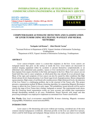
ALGORITHM DESIGN
The automated disease identification system is not a single process. This system consists of
various modules the success rate of each and every step is highly important to ensure the overall high
accurate outputs. the rest of the work is organized as follows
QUERY IMAGE
DATA BASE
IMAGE
MULTI LEVEL DWT
MULTI LEVEL DWT
FEATURE
EXTRACTION
FEATURE
EXTRACTION
TRAINED PROBABILISTIC NEURAL NETWORK
Classification
IFBENIGANORMALIGNANT
CLUSTERINGTECHNIQUE
TUMORDETECTION
Fig:1 Algorithm processing steps
127](data:image/gif;base64,R0lGODlhAQABAIAAAAAAAP///yH5BAEAAAAALAAAAAABAAEAAAIBRAA7)
Recomendados
Recomendados
Mais conteúdo relacionado
Mais procurados
Mais procurados (19)
Destaque
Destaque (17)
Semelhante a 40120130405013
Semelhante a 40120130405013 (20)
Mais de IAEME Publication
Mais de IAEME Publication (20)
Último
Último (20)
40120130405013
- 1. International Journal of Electronics and JOURNALEngineering & Technology (IJECET), ISSN 0976 – INTERNATIONAL Communication OF ELECTRONICS AND 6464(Print), ISSN 0976 – 6472(Online) Volume 4, Issue 5, September – October (2013), © IAEME COMMUNICATION ENGINEERING & TECHNOLOGY (IJECET) ISSN 0976 – 6464(Print) ISSN 0976 – 6472(Online) Volume 4, Issue 5, September – October, 2013, pp. 126-131 © IAEME: www.iaeme.com/ijecet.asp Journal Impact Factor (2013): 5.8896 (Calculated by GISI) www.jifactor.com IJECET ©IAEME COMPUTER BASED AUTOMATIC DETECTION AND CLASSIFICATION OF LIVER TUMOR USING MULTILEVEL WAVELET AND NEURAL NETWORKS Neelapala Anil Kumar1, Alluri Harish Varma2 1 Assistant Professor in Department of ECE, Vignan's Institute of Information Technology, Visakhapatnam 2 Department of ECE, Vignan's Institute of Information Technology, Visakhapatnam ABSTRACT Liver cancer or hepatic cancer is a cancer that originates in the liver. Liver cancers are malignant tumors that grow on the surface or inside the liver. Liver tumors are discovered on medical imaging equipment (often by accident) or present themselves symptomatically as an abdominal mass, abdominal pain, jaundice, nausea or liver dysfunction. Liver cancers are cancers that originate from organs elsewhere in the body and migrate to the liver. Many liver cancers are not found until they start to cause symptoms, at which point they may already be at an advanced stage. Many of the signs and symptoms of liver cancer can also be caused by other conditions like High blood calcium levels (hypocalcaemia), Low blood sugar levels (hypoglycaemia), Breast enlargement (gynecomastia), High counts of red blood cells (erythrocytosis) High cholesterol levels, the detection of the liver Tumor is a challenging problem, due to the structure of the Tumor cells. This project presents a segmentation method, K-Means clustering algorithm, for segmenting Magnetic Resonance images to detect the liver Tumor in its early stages. The probabilistic neural network will be used to classify the stage of liver Tumor that is benign, malignant or normal. The experimental result shows that the Clustering based segmentation results are more accurate and reliable than segmentation through thresholding methods in all cases. Probabilistic Neural Network with image and data processing techniques was employed to implement an automated liver Tumor classification. Key Words: Gray level co-occurrence matrix(GLCM), K-mean clustering, Magnetic resonance imaging(MRI), Probabilistic neural networks(PNN). INTRODUCTION Liver cancer is life threatening and occurs without pre-warning, considered one of the most common internal malignancies worldwide. Abnormal growths on the liver are called liver tumours, which could be both benign and malignant. Benign tumour do not really cause harm to one's health 126
- 2. International Journal of Electronics and Communication Engineering & Technology (IJECET), ISSN 0976 – 6464(Print), ISSN 0976 – 6472(Online) Volume 4, Issue 5, September – October (2013), © IAEME whereas malignant tumours can be dangerous. Hence, it is necessary to detect and diagnose malignant tumours,discussed by S.S.Kumar,R.S.Monietal [1].so that early treatment can save many lives. Segmentation of liver tissues in nervous tissue, nerve tissue and growth on medical pictures isn't solely of high interest in serial treatment observation of “disease burden” in medicine imaging,The manual analysis of the tumor samples is time overwhelming, inaccurate and needs intensive trained person to avoid diagnostic errors E-Liang Chen, Pau-Choo Chung etal [2]. Taking the parameters in to considerations we propose an automatic detection algorithm consists of effective segmentation techniques and database implementation. Focusing on this two parameters we aim a automating the liver tumor using multilevel wavelet and neural networks in matlab. ALGORITHM DESIGN The automated disease identification system is not a single process. This system consists of various modules the success rate of each and every step is highly important to ensure the overall high accurate outputs. the rest of the work is organized as follows QUERY IMAGE DATA BASE IMAGE MULTI LEVEL DWT MULTI LEVEL DWT FEATURE EXTRACTION FEATURE EXTRACTION TRAINED PROBABILISTIC NEURAL NETWORK Classification IFBENIGANORMALIGNANT CLUSTERINGTECHNIQUE TUMORDETECTION Fig:1 Algorithm processing steps 127
- 3. International Journal of Electronics and Communication Engineering & Technology (IJECET), ISSN 0976 – 6464(Print), ISSN 0976 – 6472(Online) Volume 4, Issue 5, September – October (2013), © IAEME QUERY IMAGE The Query Image or Input Image is the image on which we will perform the search using the models in the database. The data base images can be MRI or CT scan images. So our algorithm focus on the liver tumor images which can be of either of the types used for analysis of liver tumor. DATA BASE IMAGE The database is the collection of various image samples of CT or MRI of different stages of liver tumor images. It includes various severity levels of liver tumors Yu-Len Huang, Jeon-Hor Chen etal [3,4]. livetumor samples are collected from Indo American cancer hospital from Oncology department, Banjara Hills, Hyderabad. This images are considered as reference images for the analysis of liver tumor. The effective tumor analysis depends upon the number of data base images. MULTI LEVEL DWT For images, there exist an algorithm similar to the one-dimensional case for two-dimensional wavelets and scaling functions obtained from one- dimensional ones by tensorial product. This kind of two-dimensional DWT leads to a decomposition of approximation coefficients at level j in four components: the approximation at level j + 1, and the details in three orientations (horizontal, vertical, and diagonal)[5].The discrete wavelet transform is obtained by applying complementary low-pass and high-pass filters and subsequent decimation (H and L). Both H and L are applied to data vector x1, x2, ...,x8. The output of H is the four wavelet coefficients for the first resolution; the output of L is the four coefficients of the scaling function. The wavelet coefficients of the other resolution levels are obtained by iterating the low- and high-pass filtering steps on the coefficients of the scaling function FEATURES EXTRACTION Feature extraction is a special form of dimensionality reduction. When the input data to an algorithm is too large to be processed and it is suspected to be notoriously redundantV.Subbiah, L.Ganesanetal [6]. Then the input data will be transformed into a reduced representation set of features, named features vector. Transforming the input data into the set of features is called feature extraction. If the features extracted are carefully chosen it is expected that the features set will extract the relevant information from the input data in order to perform the desired task using this reduced representation instead of the full size input. By using feature extraction we can estimate the parameters of liver tumour like entropy, energy, contrast and correlation. PROBABILISTIC NEURAL NETWORKS (PNN) Probabilistic (PNN) and General Regression Neural Networks (GRNN) have similar architectures, but there is a fundamental difference Probabilistic networks perform classification where the target image is categorical, whereas general regression neural networks perform regression where the target image is continuousLuyao Wang, Zhi Zhang etal [7]. If you select a PNN/GRNN network, DTREG will automatically select the correct type of network based on the type of target image. CLASSIFICATION OF TYPE OF CANCER After applying probabilistic neural networks the cancer samples are classified According to the severity Miltiades Gletsos, Stavroula G etal [8] and they are named as benign (not harmful) and malignant( harmful).this classification is done with comparison of data base images . 128
- 4. International Journal of Electronics and Communication Engineering & Technology (IJECET), ISSN 0976 – 6464(Print), ISSN 0976 – 6472(Online) Volume 4, Issue 5, September – October (2013), © IAEME SEGMENTATION Image segmentation is the process of partitioning a digital image into multiple segments (sets of pixels, also known as super pixels)R. Adams, L .Bischofetal [9]. The goal of segmentation is , pixels) . to simplify and/or change the representation of an image into something that is more meaningful and easier to analyse. Image segmentation is typically used to locate objects and boundaries in images. r More precisely, image segmentation is the process of assigning a label to every pixel in an image such that pixels with the same label share certain visual characteristics.The result of image characteristics.The segmentation is a set of segments that collectively cover the entire image, or a set of contours extracted from the image L. L. Wu, M. S. Yangetal [10]. Each of the pixels in a region is [10]. similar with respect to some characteristic or computed property, such as color, intensity or texture. intensity, Adjacent regions are significantly different with respect to the same characteristics. It is applied to a . stack of images, typically in medical imaging. imaging RESULT The following figures shows the results by specifying detection, classification and area calculation to detect and analyze the liver tumor. Table1: The table shows various samples of performance graphs, area of tumor and type of cancer for liver tumor TEST IMAGES PERFORMANCE GRAPH TYPE OF CANCER 5.3910 benign 4.3460 benign 0.5280 benign No tumor area detected normal No tumor area detected normal No tumor area detected normal No tumor area detected 129 AREA Of TUMOR IN mm.sq normal
- 5. International Journal of Electronics and Communication Engineering & Technology (IJECET), ISSN 0976 – 6464(Print), ISSN 0976 – 6472(Online) Volume 4, Issue 5, September – October (2013), © IAEME CONCLUSION In summary a medical system for the automatic detection of primary signs of liver tumor has been developed by maintaining the effective database which addresses the area of tumor and type of severity. The results demonstrated with various samples of CT and MRI liver tumor images and this algorithm proven to be well suited in compliment the screening of liver tumor helping the oncologists in their daily practice. ACKNOWLEDGEMENTS The satisfaction that accompanies the successful completion of task would be put incomplete without the mention of the people who made it possible, whose constant guidance and encouragement crown all the efforts with success. It would not have been possible without the kind support and help of many individuals and organizations. We would like to extend our sincere thanks to all of them. We would like to express our special gratitude and thanks to Dr.P.V.RamaRaju, senior professor, department of ECE, SRKR ENGINEERING COLLEGE, BHIMAVARAM and we also thankful to Dr.RavinderRaju, MBBS, DCHfor his medical guidance and encouragement for completion of this paper. REFERENCES [1] [2] [3] [4] [5] [6] [7] [8] [9] S. S. Kumar, R. S. Moni, 1. Rajeesh, "Automatic liver and lesion segmentation: a primary step in diagnosis of liver diseases", Signal, Image and Video Processing DOl: I 0.1007/s11760-011-0223-y, March 31, 2011 E-Liang Chen, Pau-CHoo Chung, Ching-Liang Chen, Hong-Ming Tsai, Chein I Chang, "An Automatic Diagnostic system for CT Liver Image Classification", IEEE Transactions Biomedical Engineering,Vol. 45, pp. 783-794,1 998 Yu-Len Huang, Jeon-Hor Chen, Wu-Chung Shen, "Diagnosis of hepatic tumours with texture analysis in non enhanced computed tomography images", Academic Radiology,Vol. 13, p p 713-720,2006. Yu-Len Huang, Jeon-Hor Chen, Wu-Chung Shen, "Computer-Aided Diagnosis of Liver Tumours in Non-enhanced CT Images", Journal of Medical Physics,Vol. 9,p p 141-150,2004. K. Mala,V . Sadasivam, "Wavelet based texture analysis of Liver tumour from computed tomography images for characterization using linear vector quantization neural network", International Conference on Advanced Computing and Communications-ADCOM 2006, pp. 267- 270,2006. V. SubbiahBharathi, L. Ganesan, "Orthogonal moments based texture analysis of CT liver Images", Pattern Recognition Letters, Vol. 29, pp. I8 68-1872, 2008 Luyao Wang, Zhi Zhang, Jingjing Liu, Bo Jiang, XiyaoDuan, QingguoXie, Daoyu Hu, Zhen Li, "Classification of Hepatic Tissues from CT Images Based on Texture Features and Multiclass Support Vector Machines", Proceedings of the 6th International Symposium on Neural Networks: Advances in Neural Networks, p p 374-381,2 009. Miltiades Gletsos, Stavroula G. Mougiakakou, George K. Matsopoulos, Konstantina S. Nikita, Alexandra S. Nikita, DimitriosKelekis, "A Computer-Aided Diagnostic System to Characterize CT Focal Liver Lesions: Design and Optimization of a Neural Network Classifier", IEEE Transactions on Information Technology in Bio Medicine, Vol. 7, pp. 153162,2003. R. Adams,L . Bischof, "Seeded Region Growing", IEEE Transactions on Pattern Analysis and Machine Intelligence, Vol. 16, p p. 641-647, 1994. 130
- 6. International Journal of Electronics and Communication Engineering & Technology (IJECET), ISSN 0976 – 6464(Print), ISSN 0976 – 6472(Online) Volume 4, Issue 5, September – October (2013), © IAEME [10] L. L. Wu, M. S. Yang, "Alternative c means clustering algorithms", Pattern Recognition Vol. c-means 35,p p. 2267-2278,2 002. [11] Mayur V. Tiwari and D. S. Chaudhari, “An Overview of Automatic Brain Tumor Detection nd f from Magnetic Resonance Images”, International Journal of Advanced Research in rom Images Engineering & Technology (IJARET), Volume 4, Issue 2, 2013, pp. 61 - 68, ISSN Print: 0976-6480, ISSN Online: 0976-6499. 6480, 0976 [12] B.Venkateswara Reddy, Dr.P.Satish Kumar, Dr.P.Bhaskar Reddy and B.Naresh Kumar nd Reddy, “Identifying Brain Tumour from MRI Image using Modified FCM and Support Identifying Vector Machine”, International Journal of Computer Engineering & Technology (IJCET), ournal Volume 4, Issue 1, 2013, pp. 244 - 262, ISSN Print: 0976 – 6367, ISSN Online: 0976 – 6375. , [13] Selvaraj.D and Dhanasekaran.R “MRI Brain Tumour Detection by Histogram and nd Dhanasekaran.R, y Segmentation by Modified GVF Model”, International Journal of Electronics and y ournal Communication Engineering & Technology (IJECET), Volume 4, Issue 1, 201 pp. 55 - 68, , 2013, ISSN Print: 0976- 6464, ISSN Online: 0976 –6472. AUTHORS’ INFORMATION NEELAPALA. ANIL KUMAR has Obtained B. Tech. in ECE Department from JNT University, Hyderabad and ME in Electronic Instrumentation (EI) from Andhra University, Visakhapatnam. He h eight has years of teaching experience, presently working at Vignan's Institute of Information Technology, Visakhapatnam, as Assistant Professor in Department of ECE. He has added two book for his account. His Areas of interests are bio medical instrumentation and image processing. HARISH VARMAALLURI is pursuing his M. Tech degree in the Department of Electronics & Communications, Vignan's institute of Information Communications, and Technology, Duvvada. His Areas of interests are bio-medical and image bio medical processing. 131