

Recomendados
Mais conteúdo relacionado
Mais procurados
Mais procurados (20)
Destaque
Semelhante a 4embryonic period...
Semelhante a 4embryonic period... (20)
Mais de 2nd year MBBS student
Último
Último (20)
4embryonic period...
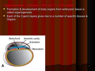
- 1. Organogenesis / Embryonic Period / 3 rd to 8 th weeks Formation & development of body organs from embryonic tissue is called organogenesis Each of the 3 germ layers gives rise to a number of specific tissues & Organs 1
- 2. Formation of the Neural tube The neuro-ectoderm (neural plate) is derived from the ectoderm overlying the notochord and is induced by it during the third week. Then neural plate begins to fold. It is 1st converted into neural groove . The neural groove deepens and eventually forms a neural tube . Two masses of ectoderm at edges of neural plate, form neural crest . 2
- 3. Initially the neural crest separates neuro-ectoderm from skin ectoderm. As folding of the neural tube occurs, the neural crest cells detach from the ectoderm and form clusters that migrate into the mesoderm. 3
- 4. There are three derivatives of the ectoderm in this region: Skin ectoderm – gives rise to the epidermis of the skin Neural crest ectoderm – are cells that migrate widely and give rise to a large variety of structures, to be listed later. Neural ectoderm – gives rise to the central nervous system, including the neurones and neuroglia. 4
- 5. Derivaties of Ectoderm 1. Surface Ectoderm A. Lining epithelia Skin epidermis Mucous membrane of lips ,cheeks , gums, part of floor of mouth , part of the palate, nasal cavity & sinuses Lower part of anal canal ( below pectinate line) Terminal parts of male urethra Outer surface of labia minora & whole of labia majora Anterior epithelium of cornea, epithelium of conjunctiva, epithelial layers of ciliary body & iris Outer layer of tympanic membrane, epithelial lining of membranous labyrinth including the special end organs Lacrimal canaliculi, sac, nasolacrimal duct 5
- 6. B. Glands : Sweat glands, sebaceous glands parotid gland, mammary glands, pars anterior of pituitary gland C. Other derivatives tooth enamel, Hair Nails 6
- 7. 2. Neuroectoderm 1. Neural tube CNS Retina & Optic nerve & musculature of iris Pineal & pituitary gland Neurons Neuroglia – Ependymal cells , Macroglia(Astrocytes, oligodendrocytes) 2. Neural crest Schwann cells Chromaffin cells (adrenal medulla) Dorsal root ganglia & dorsal root of spinal nerve Sympathetic ganglia Sensory ganglia of V, VII, IX & X cranial nerves Melanocytes of skin Leptomeninges Bones & connective tissues of cranio-fascial structues Parts of heart 7
- 8. 3. Ectodermal Placode:Ectodermal thickenings which have important roles in development of special sensory systems. Otic placode – gives rise to structures needed for hearing & balance Lens placode – forms lens Nasal placode – nasal cavities & para nasal sinus 8
- 9. APUD-Amine-precursor uptake and decarboxylation cells (Neuroendocrine cells) 9
- 10. Mesoderm forms several distinct masses : Mesoderm in the lateral part of the embryo is divided into three distinct longitudinal masses: Paraxial mesoderm - a longitudinal column of cells that lies next to the notochord - it gives rise to the axial skeleton and skeletal muscle - Intermediate cell mass - it gives rise to the genitourinary system Lateral plate mesoderm - gives rise to body wall structures - is continuous with the extra-embryonic mesoderm - splits into two layers enclosing the intra-embryonic coelom 10
- 11. Two important masses of mesoderm The trilaminar embryo has two important masses of mesoderm: At the cranial end is the transverse mesoderm, in which are situated the pericardial cavity and the cardiogenic mesoderm . At the caudal end is the connecting stalk that contains the allantoic diverticulum (allantoises) , a small outgrowth from the roof of the yolk sac and projecting into the connecting stalk. The allantoic diverticulum will later give rise to the greater part of the urinary bladder 11
- 12. Folding of the Embryo. Folding occurs by differential growth of tissues. Neural ectoderm grows faster than the surrounding skin ectoderm and consequently fold to form a neural tube. Similarly, skin ectoderm grows faster than the underlying mesoderm and endoderm, and this differential growth causes folding of the trialminar disc and gives shape to the embryo. Folding occurs mainly at the edges of the embryonic disc and forms three main folds: Head fold Tail fold Lateral folds - convert the embryo into a tubular structure. These are not three separate folds but occur simultaneously and merge into one another. The notochord, neural tube and somites stiffen the dorsal axis of the embryo. 12
- 13. As a result of the formation of the head fold: The foregut is formed by folding of the endoderm The stomodaeum is an invagination of ectoderm, and has the buccopharyngeal membrane separating it from the foregut It opens into the amniotic cavity. The pericardial cavity and cardiogenic mesoderm are shifted to the ventral aspect of the embryo and lie ventral to the foregut. The part of the transverse mesoderm between the pericardial cavity and the yolk sac is the septum transversum proper. In it the liver will develop. The amniotic cavity extends ventral to the cranial end of the embryo. The yolk sac is constricted from the cranial aspect. 13
- 14. As a result of the formation of the tail fold: the hindgut is formed The cloaca is an invagination of ectoderm and has the cloacal membrane separating it from the hindgut. The connecting stalk is shifted ventrally The allantoic diverticulum is shifted ventrally. It is an invagination of hindgut endoderm into the yolk sac. The amniotic cavity extends ventral to the caudal end of the embryo. The yolk sac is constricted from the caudal end 14
- 15. Transverse folding of the embryo Transverse folding Converts the endoderm into a primitive gut tube The intra-embryonic coelom surrounds the gut tube (Transverse plate mesoderm) The communication between the intra- and extra- embryonic coeloms becomes constricted and eventually obliterated 15
- 16. Note that drastic and important changes occur in the embryonic cavities as a consequence of folding: The amniotic cavity surrounds the embryo completely on all aspects and becomes the predominant cavity. It enlarges progressively. The yolk sac becomes constricted on all sides, and becomes a small sac connected to the midgut by a narrow vitelline duct. It becomes progressively smaller. The extra-embryonic coelom is gradually obliterated by the expanding amnion and eventually disappears completely 16
- 17. Somites During the fourth week the embryo is segmented. Each segment consists of a somite innervated by a segmental nerve derived from a segment of the neural tube. A somite is divided into two parts: The sclerotome is the ventro-medial part of the somite. It contains a “cavity” of loose cells. Cells from the sclerotome migrate medially to surround the notochord and neural tube and form the axial skeleton. 17
- 18. The dermomyotome is the dorso-lateral part of the somite. Cells from the dermomyotome migrate laterally and, as its name implies, gives rise to (i) skeletal muscle and (ii) the dermis of the skin. The concept of the myotome in gross anatomy is an embryological concept. Each anatomical myotome is derived from the embryological dermomyotome that is innervated by a segmental nerve and forms a goroup of skeletal muscle cells and the dermis of the corresponding segment of ectoderm. 18
- 19. The neural tube induces the formation of the neural arches and their fusion across the midline. Def ects of closure of the neural tube will also cause failure of fusion of the overlying neural arches. This anomaly is termed a meningomyelocoele . As illustrated in the adjacent diagram. 19
- 20. The vertebral bodies are formed from two adjacent somites Note that the segmental spinal nerves emerge at the level of the corresponding somite, between adjacent vertebrae. The intervertebral discs correspond to the original somites and remain unossified. 20
- 21. Blood vessels develop throughout the mesoderm Mesodermal cells differentiate into endothelial cells surrounding a central group of erythroblasts. These are blood islands that coalesce to form blood vessels. Almost all parts of the mesoderm gives rise to blood vessels. Differentiation of blood vessels Blood islands and eventually blood vessels appear: in the extra-embryonic mesoderm in the second week in the intra-embryonic mesoderm in the third week the primitve heart tube develops in the cardiogenic mesoderm (in the transverse mesoderm) at the beginning of the fourth week and a primitive circulation is established 21
- 22. Three structures develop in the transverse mesoderm: cardiogenic mesoderm – in which the primitive heart tubes form pericardial cavity into which the heart tubes invaginate the septum transversum forms part of the diaphragm, fibrous pericardium and connective tissue of the liver. Note that the cardiogenic mesoderm and septum transversum are situated in the cervical region and so are innervated from cervical segmental nerves. 22
- 23. 23
- 24. 24
- 25. 25