![Group activity ,[object Object],[object Object],[object Object],[object Object],[object Object]](data:image/gif;base64,R0lGODlhAQABAIAAAAAAAP///yH5BAEAAAAALAAAAAABAAEAAAIBRAA7)
Recomendados
Mais conteúdo relacionado
Mais procurados
Mais procurados (20)
Destaque
Destaque (20)
Semelhante a Inservice presentation/Age-related macular degeneration
Semelhante a Inservice presentation/Age-related macular degeneration (20)
Último
Último (20)
Inservice presentation/Age-related macular degeneration
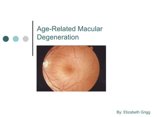
- 1. Age-Related Macular Degeneration By: Elizabeth Grigg
- 8. What does age-related macular degeneration look like? Healthy, normal retina Early AMD - drusen appearing on retina Macula Optic disc Yellow spots are drusen Images from http://www.agingeye.net/maculardegen/maculardegeninformation.php
- 9. As the disease progresses… Late or end-stage dry AMD (geographic atrophy) Geographic atrophy (may lead to) Wet or exudative AMD Sub-retinal hemorrhage Images from http://www.agingeye.net/maculardegen/maculardegeninformation.php
- 26. The Amsler grid Taken from: http://www.stlukeseye.com/eyeq/amslerprint.html
- 28. How might the Amsler grid look to a person who has AMD? First image from http:// www.nei.nih.gov/photo/keyword.asp?conditions =Age-Related+Macular+Degeneration+%28AMD%29 Second image from http://www.cnib.ca/en/your-eyes/eye-conditions/amd/diagnosing/diagnostic-tests/Default.aspx
- 30. More examples… Clockwise from top: First image from http://www.dukehealth.org/eye_center/specialties/macular_degeneration/care_guides/macular _degeneration_frequently_asked_questions Second image from http://www.stlukeseye.com/conditions/MacularDegeneration.html Third image from http://www.nei.nih.gov/photo/sims/images/ESD11.jpg
- 33. How eccentric viewing can help Advanced vision Magnifier alone Eccentric viewing defect + magnifier Images from http://www.agingeye.net/maculardegen/maculardegensymptoms.php
Notas do Editor
- Using the eye model I made earlier in the semester, I would begin the presentation by asking the audience if they know where the macula is in the eye. I would show them on the eye model how it is a small point on the retina in the very back of the eye.
- What you are seeing, or not seeing, is what the vision of an individual who has age-related macular degeneration might be like. You’ll notice that you cannot see anything directly in front of you, but you can still see the surrounding area. By moving your fist closer to your eye and than farther away, you can see the range of impact that age-related macular degeneration can have on vision.
- The macula is on the center of the retina and is made mostly of cones, which are cells that help us see fine detail and colors. The rest of the surrounding area on the retina is made mostly of rods, which are responsible for night vision, peripheral vision and are better at detecting movement than the cones.
- Drusen may be present on the retina of younger people as well, but these are typically hard drusen, meaning that the deposits have definable edges. As you age, they become known as soft drusen because the edges become less defined.
- Just the presence of drusen alone on the retina and pigment changes is known as early-stage AMD. The end-stage, or late-stage of the dry form is known as geographic atrophy.
- The wet form is also known as neovascular age-related macular degeneration.
- This form is much more severe than dry AMD.
- Notice how the normal retina appears pink and has no drusen. The picture on the right has soft drusen throughout the retina. At this point, vision would not be affected too much.
- The picture on the right shows the large mass of drusen known as geographic atrophy. This area would cause a scotoma, or blind spot in the central vision. The picture on the right shows a sub-retinal hemorrhage, which left untreated will cause permanent vision loss. The size, location and number of these hemorrhages varies from person to person.
- Peripheral vision is not affected because rods are responsible for that area of vision. The macula is responsible for central and color vision.
- While geographic atrophy is worse than early AMD, if it is not located in the area of finest central vision, some central vision may be retained.
- Dry AMD may progress over several years, but wet AMD progresses much more quickly, sometimes within several weeks or days. Because of the speed in which neovascularization can occur, patients are encouraged to monitor their vision with an Amsler grid regularly so they can detect any changes in their vision.
- AMD is a genetic or hereditary disease, but lifestyle may increase or decrease the risk of developing AMD, especially the wet form.
- You’ll notice that many of the risk factors are associated with cardiovascular disease.
- Many of these activities are things that depend on having fine central vision or color vision. Any activities that use one or both of these may be affected (sewing, eating off a plate, etc).
- Even if individuals are genetically predisposed to AMD, they should make lifestyle changes that reduce the amount of risk. They should modify their behavior as much as possible based on the previously mentioned risk factors.
- The AREDS study did not show that people who did not have AMD or even early AMD benefitted by taking this formulation. Only those in the intermediate or later stages were shown to benefit.
- This study is currently ongoing, with a scheduled end date of around 2014. Studies have shown that lutein and zeaxanthin have a protective effect on the macula. These pigments are both already present in the macula, and this study is examining whether or not consuming more of them increases their impact.
- Laser photocoagulation uses a high-intensity laser to destroy leaky blood vessels, but it causes indiscriminate damage to blood vessels in the retina. Photodynamic therapy is more controlled: it uses a medication that is injected intravenously and then activated by an infrared laser that destroys neovascularization. However, both of them can damage healthy blood vessels and some patients develop disciform scars as a result of the treatment. These two treatments are older and not used as commonly as more recently developed treatments.
- VEGF is the protein responsible for the growth of abnormal blood vessels. These drugs bind to the VEGF protein and inhibit it’s activity. They are shown to be safe and effective in treating AMD. Anti-VEGF treatments are the most recent FDA approved treatments for AMD. However, Avastin is not currently FDA approved for treating AMD but it is approved for treating colon cancer.
- There have been concerns in the past that cataract surgery increased the risk of AMD, but recent studies are showing that this is not the case.
- Some individuals with AMD will develop this syndrome – it should be explained so that if hallucinations do occur, the individual realizes that it is due to physical changes, and not psychosis.
- Individuals at risk for AMD or who have been diagnosed with AMD are encouraged to use an Amsler grid at home on a regular basis. Even with frequent use of an Amsler grid, individuals should continue to see their eye doctor on a regular basis.
- The Amsler grid may also be black with white lines.
- An individual might see distortions or blind spots of the Amsler grid, which they are already aware of and have seen their doctor about. However, if they notice any new changes, then they need to contact their doctor as soon as possible, as this could be an indication of the formation of new blood vessels under the retina.
- These pictures demonstrate what the vision of someone who has AMD might look like. Note that in the picture on the right a central scotoma, or blind spot, exists, and the surrounding area appears wavy and distorted. The remaining area in the peripheral vision also appears blurry as a result of decreased visual acuity. The size and location of central scotomas will vary from person to person.
- These pictures show how the effects of AMD may vary from person to person.
- I would show examples of writing guides (check writing and letter writing) here. Some great resources for ideas on how to adjust are available at the American Foundation for Blindness website; afb.org. Their Senior Site also has a list of local resources and agencies that may be contacted for additional assistance.
- A low vision specialist can detect where scotomas, or blind spots are located, and then suggest appropriate assistive devices to use, and provide training on how to use them. This specialist would also provide eccentric viewing training. A rehabilitation therapist can teach individuals with AMD independent living skills.
- Magnifiers help individuals with AMD read more easily, but for those with more severe vision loss, learning eccentric viewing can be beneficial, as the example in this slide shows.
- I would close by saying that while AMD can greatly reduce central vision, there is a wide variety of aids that can be used and living skills that can be learned to make life much easier. Having AMD does not mean that you can no longer do any of the things you used to do before.