SlideShare uma empresa Scribd logo
1 de 35
Baixar para ler offline
I.B. Biology S.L.    Éanna OBoyle


         Human Health 

         & Physiology
Topic 5: Human Health and Physiology


5.1 Digestion
5.1.1        Explain why digestion of large food molecules is essential.

Digestion refers to the mechanical and chemical breakdown of complex

food molecules into smaller molecules so that they can be absorbed. Once

absorbed, the products of digestion are used

i)      as an energy source.

ii)     for making other molecules required by the living body.


The human digestive system is rather representative of the mammalian

system in many respects. We will follow the path of food through the human

digestive system.


5.1.2        Explain the need for enzymes in digestion.

We have seen that carbohydrates, lipids, and proteins are often very large,

complex molecules composed of repeating molecular subunits (see section

2). Except for certain triglycerides (see section on lipids), these

“macromolecules” can’t be absorbed through the walls of the digestive

system.


So in the digestive process, enzymes help break down these large

molecules into smaller molecules, which can be then absorbed through walls
of the digestive system.


The bonds are broken through hydrolysis, the opposite of condensation, and

the process in which water is added to the complex molecules. In other

words, enzymes help break the bonds present in these large molecules.


During digestion, carbohydrates are hydrolyzed (remember... the opposite of

condensation) into simple sugars, fats into fatty acids and glycerol, proteins

into their various amino acids, and nucleic acids into free nucleotides.


5.1.3 	   State the source, substrate, products and optimum pH
          conditions for one amylase, one protease and one lipase.

a) Carbohydrate Digestion:

Starch ( a polysaccharide carbohydrate) is a typical large complex molecule

that we eat (e.g. in potatoes, rice, and bread). Any enzyme that digests

starch is called an amylase.


One example of an amylase is salivary amylase, which is produced by the

salivary gland in the mouth. This enzyme acts on starch (the substrate),

producing the disaccharide maltose (remember ... 2 glucose units linked

together) and many longer polysaccharide (starch) fragments.
maltose

                           amylase

H2O      +


                 Amylose                short-chain starch

             (type of starch)              fragment


[Note that many molecules of water react with 1 molecule of amylose].


Later, in the intestine, these maltose molecules are broken down further into

glucose molecules by the enzyme maltase (see section 2 notes for the

reaction).


With the joint action of amylase and maltase, starches like amylose are

broken down into glucose which can be absorbed through the wall of the

intestines in the digestive system.


b) Protein digestion:

Proteins are among the largest and most complex molecules known. Not

surprisingly, their digestion is also complex, requiring a number of different

enzymes that form a family called proteases.
The first step in protein digestion ocurs in the stomach. Here the enzyme
pepsin, produced by the gastric glands, acts upon proteins (the substrate),
by attacking and breaking peptide bonds (remember section 2 for what a
peptide bond is!). Pepsin can only attack 4 of the 20 amino acids that occur
on earth, and in all 4 they are broken down into smaller peptide lengths.


Further protein breakdown occurs in the small intestine by for example
trypsin.
The final steps in protein digestion are carried out by the enzymes
aminopeptidases and dipeptidases (which belong to the peptidase group of
enzymes). Dipeptidases attack peptide bonds in dipeptides, yielding two
amino acids; e.g. starting with pepsin, & eventually ending with dipeptidase:
                                  Pepsin                                  Pepsin

           serine     glutamine       proline      glycine      leucine            serine


                              H 2O                                          H2 O


             Dipeptidase


           H2O


                                    Serine and glutamine are two free amino acids,
                                    which can be absorbed through the wall of the
                      glutamine
                                    digestive system.
           serine
c) Lipids:

Fat digestion generally occurs in the small intestine. Fats are digested by the
enzyme pancreatic lipase. Lipase breaks down neutral fats (triglycerides)
into fatty acids, glycerol, monoglycerides and diglycerides.

              o
                                                                           OH
                                                       OH
                                 Lipase                                      OH
              o                                        OH

                                                    OH
              o

                                                                             OH
               + 3 H2O

          Tryglyceride 	                    glycerol        3 ‘free’ fatty acids


5.1.4 	  Draw a diagram of the digestive system.
See end of these notes and textbook!


5.1.5 	       Outline the functions of the stomach, small intestine, and
              large intestine.

The stomach temporarily stores food and begins its digestion. In addition, its 

acids and enzymes kill many of the organisms we swallow.


The small intestine has 2 roles - digestion and absorption.


The large intestine (also called colon or bowel) main functons are:

a) to absorb water and minerals into the blood 

b) to prepare the faeces to leave the digestive tract.
Very little, if any, digestive activity takes place in the large intestine itself,
although a rapid digestion of food residues is carried on by huge
populations of microrganisms (e.gs. Escherichia coli and methane-
producing bacteria).


5.1.6 	    Distinguish between absorption and assimilation.
Digestive enzymes help break down complex carbohydrates into simple
sugars, lipids into glycerol and fatty acids, and proteins into amino acids.
These end products of digestion along with the other nutrients, vitamins,
minerals, and water, are of no value to your body until they leave the
digestive system. They must pass into your blood for distribution to your
cells.


The process by which nutrients pass from the digestive system into the body
fluids (blood and lymph) is called absorption.


Assimilation describes the process by which energy is released by these
absorbed molecules.


5.1.7 	    Explain how the structure of the villus is related to its role in
           absorption of the end products of digestion.

The small intestine has special structures - villi (sing.: villus) - which help
absorption. These fingerlike projections greatly increase the surface area of
the small intestine. The surface area is further increased by the microvilli -
tiny folds in the villi’s surfaces. The large surface area allows nutrients to
pass easily into the blood vessels and lymph system. See further notes in
text ...
Digestive System


                    Villi
Digestive System


            mouth
                                            larynx

           trachea
(or air tube; not part                      (o)esophagus
of digestive system!)


               liver
                                            stomach
      gall bladder


  small intestine                           pancreas


  large intestine


      appendix

              anus                          rectum
5.2 - The Transport System
It beats day and night every day of our lives, but what does the heart

really do for us?


Your heart, quite simply, is a pump. But it is one of the most fascinating

pumps you'll ever learn about. First you must understand that every cell

in our body - from the cells in our hair to the cells in our toes need

OXYGEN and NUTRIENTS to survive and keep us alive. Since they can't

get these nutrients themselves, blood is used to deliver them right to the

cells. (sort of like room service)

The basic job of the heart is pump that blood through your body so that

the blood can deliver the oxygen and nutrients right to the cells. The

heart keeps your blood pumping at all times as it picks up oxygen from

your lungs and nutrients from your digestive system and sends them to

all cells of your body. Your heart is the power behind the delivery system.

Inside the heart is four chambers. Two of those chambers send the blood

up to your lungs to get oxygen, then the other two chambers send that

oxygen rich blood to the rest of your body. Valves inside of the heart

make sure that the blood only moves in one direction.

The younger you are, the faster your heart beats. A baby's heart beats

about 90 times a minute. A twelve year old heart beats about 78 times a

minute and an adult heart beats about 70 times a minute. Of course the

more active you are, the more your heart beats, since the cells need

oxygen faster to keep you moving.
5.2.1 Draw a diagram of the heart showing all four chambers,
associate blood vessels and valves.

                                               semi-lunar
                                                 valves


5.2.2 Describe the action of the heart in terms of collecting blood,
pumping blood and opening and closing valves.

5.2.3 Outline the control of the heartbeat in terms of the
pacemaker, nerves and adrenalin.
5.2.4 Explain the relationship between the structure and function
of arteries, capillaries and veins.


5.2.5 State that blood is compose of plasma, erythrocytes, 

leucocytes (phagocytes and lymphocytes) and platelets.


5.2.6 State that the following are transported by the blood:
nutrients, oxygen, carbon dioxide, hormones, antibodies and
urea.
The Heart and the Circulatory System
Glossary

Anterior: adjective referring to front end of an animal or organism.


Aorta: the main systemic artery of the body, emerging directly from the left ventricle.


Arteriole: a small arterial branch that delivers blood directly to a capillary bed.


Artery: a muscular blood vessel that carries blood away from the heart.


Atrium: one of the chambers of the heart that receives blood directly from a vein.


Circulatory system: the system of the body responsible for internal transport. Composed of the heart, blood 

vessels, lymphatic vessels, lymph, and the blood.


Closed circulatory system: a type of circulatory system where the blood is contained within a system of 

vessels and the heart.


Coronary artery: one of the arteries that supply blood to the heart.


Coronary vein: one of the veins that receive blood from the heart muscle and empty directly into the right 

atrium.


Deoxygenated blood: blood that is low in oxygen concentration.


Dorsal: adjective referring to the top or upper surface of an organism.


Heart: the muscular organ composed of cardiac muscle that is responsible for pumping blood throughout the 

body.


Heart attack: a condition occurring when a section of the heart is deprived of oxygenated blood and dies.


Interstitial fluid: the fluid filling the microscopic spaces between cells of the body.


Open circulatory system: a type of circulatory system where the blood is not contained within a system of 

vessels and the heart. Blood empties from vessels into sinuses and then returns through other vessels to a 

"heart.".


Peristalsis: wave-like muscular contractions in the walls of tubular organs, e.g. the alimentary canal. 

Peristalsis serves to push material contained within the organ along its length.


Posterior: adjective referring to the hind end of an animal.


Pulmonary artery: one of the arteries carrying deoxygenated blood from the heart to the lungs.


Septum: the wall dividing the two ventricles.


Sinus: a cavity into which blood flows and baths the internal organs in organisms with an open circulatory 

system.


Spiracle: opening which leads to a trachea in an insect, arachnid, isopod, centipede or millipede.


Vein: one of the blood vessels that carries blood to the heart.


Ventral: adjective referring to the lower surface of an animal or organism.


Ventricle: one of the muscular chambers of the heart that is responsible for pumping blood from the heart into 

the arteries.


Venule : a small venous branch that carries blood from a capillary bed to a vein.
The Blood Vessels


Exp
TTlojLeading up to a heart attack

ing


Vess
       From World Book © 2001 World Book, Inc., 233 N. Michigan Avenue, Suite 2000, Chicago, IL
       60601. All rights reserved.
World Book illustrations by Paul Peck, M.D. and Charles Wellek
A Coronary Bypass


From World Book © 2001 World Book, Inc., 233 N. Michigan Avenue, Suite 2000, Chicago, IL 60601. All rights reserved.
World Book illustrations by Virginia Samter


       Heart-Lung Machine (during bypass operation)


From World Book © 2001 World Book, Inc., 233 N. Michigan Avenue, Suite 2000, Chicago, IL
60601. All rights reserved.
World Book illustration by Sarah Woodward and John Eggert
Red Blood Cell (erythrocyte)


From World Book © 2001 World Book, Inc., 233 N. Michigan Avenue, Suite
2000, Chicago, IL 60601. All rights reserved.
© Dr. Tony Brain/SPL from
Photo Researchers

                        A White Blood Cell


                From World Book © 2001 World Book, Inc.,
                233 N. Michigan Avenue, Suite 2000, Chicago,
                IL 60601. All rights reserved.
NIBSC/SPL
                from Photo Researchers
There are three types of vessels - arteries, veins and capillaries. Arteries take
oxygenated blood from the heart out to all areas of the body. The walls of the
arteries are too thick for oxygen and nutrients to pass through, so arteries lead to
smaller vessels called capillaries. The walls of the capillaries are thin enough for
red and white blood cells to squeeze through and enter other body tissues. The
circulation of blood can easily be observed moving through arteries, capillaries and
veins.

Arteries, veins and capillaries are not anatomically the same. They are not just
tubes through which the blood flows. Both arteries and veins have layers of smooth
muscle surrounding them. Arteries have a much thicker layer, and many more
elastic fibers as well.

The largest artery, the aorta leaving the heart, also has cardiac muscle fibers in its
walls for the first few cm of its length immediately leaving the heart. Arteries have to
expand to accept the blood being forced into them from heart, and then squeeze
this blood onto the veins when the heart relaxes. Arteries have the property of
elasticity, meaning that they can expand to accept a volume of blood and then
contract and squeeze back to their original size after the pressure is released.

A good way to think of them is like a balloon. When you blow into the balloon, it
inflates to hold the air. When you release the opening, the balloon squeezes the air
back out. It is the elasticity of the arteries that maintains the pressure on the blood
when the heart relaxes, and keeps it flowing forward. Arteries branch into arterioles
as they get smaller. Arterioles eventually become capillaries, which are very thin
and branching.

Capillaries are really more like a web than a branched tube. It is in the capillaries
that the exchange between the blood and the cells of the body takes place. Here
the blood gives up its carbon dioxide and takes on oxygen.
In the special capillaries of the kidneys, the blood gives up many waste products in
the form of urine.

Capillary beds are also the sites where white blood cells are able to leave the
blood and defend the body against harmful invaders. Capillaries are so small that
when you look at blood flowing through them under a microscope, the cells have to
pass through in single file.

As the capillaries begin to thicken and merge, they become venules. Venules
eventually become veins and head back to the heart. Veins do not have as many
elastic fibers as arteries. Veins do have valves, which keep the blood from pooling
and flowing back to the legs under the influence of gravity.
When these valves breakdown, as often happens in older or inactive people, the
blood does flow back and pool in the legs. The result is varicose veins, which often
appear as large purplish tubes in the lower legs.
Topic 5.3 - Pathogens and Disease
5.3.1 Define pathogen.
Pathogen - an organism or virus that causes a disease. Pathogens cause
disease in the host.


5.3.2 State one example of a disease caused by members of each of
the following groups: viruses, bacteria, fungi, protozoa, flatworms
and roundworms.
Viruses: Influenza, mumps, measles, chicken pox, common cold. 

Bacteria: Cholera, tuberculosis, tetanus, diphtheria. 

Fungi: Athlete's foot, Candida (thrush)

Protozoa: Malaria. 

Roundworms: Ascaris eggs contained in contaminated food are 

swallowed, circulate through the blood stream, reach the lungs, grow into 

larvae in the nasal cavities, swallowed into the stomach where they grow 

into adult worms and start the cycle again. Hookworm.

Flatworms: Pork tapeworm, Bilharzia.
5.3.3 List six methods by which pathogens are transmitted and gain
entry to the body.
1) From the air - pathogen enters through respiratory system (e.g. droplets)

2) Direct contact - pathogen enters through the skin.

3) Through food or water - pathogen enters through the digestive system.

4) Through cuts in the skin.

5) Blood transfusion. 

6) Infected animals and insects - pathogen enters through bites.

7) Sexual contact - pathogen enters through reproductive system.


5.3.4 Describe the cause, transmission and effects of one human
bacterial disease.
Tuberculosis is caused by Mycobacterium tuberculosis, a rod-shaped
bacterium. See text.

5.3.5 Explain why antibiotics are effective against bacteria but not
viruses.
Antibiotics block specific metabolic pathways found in bacteria, but not in
eukaryotic cells. Viruses reproduce using the host cell metabolic pathways
that are not affected by antibiotics.

5.3.6 Explain the cause, transmission and social implications of
AIDS.
AIDS is a retrovirus having RNA as its genetic material and not DNA. It
transcribes its RNA into DNA using an enzyme called reverse
transcriptase. It is transmitted by sexual intercourse, sharing of needles,
blood transfusions, accidents causing blood contamination, cuts in the
skin, tattoos and ear piercing with infected needles. Social implications
are that people don't feel very comfortable with a person who has AIDS.
People with AIDS can find it difficult to buy health insurance plans, find
jobs, have friends, and build normal social relations. People have changed
their sexual lifestyles due to awareness and education about AIDS. See
h/o.
Topic 5.4 - Defense Against Infectious Disease
5.4.1 Explain how skin and mucous membranes act as barriers
against pathogens.

The skin and mucous membranes are the first lines of defense against
disease. The skin has a thick keratin layer on the surface which helps
prevent organisms from entering the body. Where there is no skin, such as
the mouth cavity, epithelial cells there form a mucous membrane that
produces sticky mucous which traps and stops the action of many
organisms. Other barriers to pathogens include acid in the stomach,
resident bacteria, and tears.


5.4.2 Outline how phagocytic leucocytes ingest pathogens in the
blood and in body tissues.

When the phagocytes meet the pathogens, they ingest the organisms by
phagocytosis. Once they are in the phagocytes, the pathogens go into the
vesicles which fuse with the lysosomes, which then release hydrolytic
enzymes on them and destroy them.


5.4.3 State the difference between antigens and antibodies.

An antigen is a foreign macromolecule that does not belong to the host
organism and that elicits an immune resonse. An antibody is a protein and
is called an immunoglobulin. It is made of 4 polypeptides, 2 heavy chains
and 2 light chains. It sticks to antigens and to lymphocytes.


5.4.4 Explain antibody production.

Many different types of lympocyte exist. Each type recognizes one specific
antigen and responds by dividing to form a clone. This clone then secretes
a specific antibody agaist the antigen.
5.4.5 Outline the effects of HIV on the immune system.

HIV attacks helper T cells, which are part of the immune system that are
important for the function of B lymphocytes. The virus enters the helper T
cells and replicates there. The cells burst and release new viruses, these
viruses infect other helper T cells and possibly other cells such as
phagocytes as well. The destruction of helper T cells paralyses the immune
system since they communicate between different cells of the immune
system and activate them. This enables any other parasite or organism
usually kept under control by the immune system to be able to affect the
body.
Topic 5.5 - Gas Exchange

5.5.1 List the features of alveoli that adapt them to gas exchange.
There is a large surface area, a wall consisting of a single layer of flattened
cells, a moist lining, and a dense network of capillaries. See text, p.51.


5.5.2 State the difference between ventilation, gas exchange, and
cell respiration.
*Ventilation in humans involves bringing fresh air to the alveoli and
removing stale air which is low in oxygen, O2 and high in CO2. It maintains a
high concentration of oxygen in the alveoli and low carbon dioxide as we
breathe in and out.
*Gas exchange is the process where one gas (e.g. CO2) is swapped with
another (e.g. oxygen). In humans, it occurs between the alveoli and the
capillaries by diffusion - O2 passes from the alveoli to the capillaries and
CO2 passes from the capillaries to the alveoli. This happens because there
are concentration gradients of O2 and CO2 between the air (in alveoli) and
the blood (in capillaries).
*Cell respiration is the chemical reaction that occurs inside the cell
(cytoplasm and mitochondria) and that results in the controlled release of
energy in the form of ATP. In humans O2 is used up and CO2 is produced.

5.5.3 Explain the necessity for a ventilation system.
It is needed to maintain concentration gradients in the alveoli.

5.5.4 Draw a diagram of the ventilation system including trachea,
bronchi, bronchioles, and lungs.
See notes...

5.5.5 Explain the mechanism of ventilation in human lungs
including the action of the internal and external intercoastal
muscles, the diaphragm and the abdominal muscles.

See notes
Topic 5.6 - Homeostasis and Excretion
5.6.1 State that homeostasis involves maintaining the internal
environment at a constant level or between narrow limits, including
blood pH, oxygen and carbon dioxide concentrations, blood
glucose, body temperature and water balance.

                                     Here the internal environment refers
                                     to blood and tissue inside our bodies.
                                     Homeostasis can also be seen as the
                                     ability of an organism to maintain
                                     stable conditions inside its body even
                                     as outside conditions change
                                     dramatically. Cells as well as
                                     organisms have this ability of
                                     homeostasis.

5.6.2 Explain that homeostasis involves monitoring levels of
variables and correcting changes in levels by negative feedback
mechanisms.

If body temperature falls below 37 degrees Celsius, then messages are
sent by the hypothalamus to different parts of the body so temperature is
increased to normal. Conversely, if body temperature rises above 37
degrees Celsius, messages sent decrease body temperature to normal.
Therefore, a change in a variable is counteracted by the opposite change
to return the body to a normal temperature. Other variables in the blood
include the pH, pressure, O2/CO2 & glucose concentrations.
Negative feedback mechanisms are very common in organisms and
simply refer to how a change in levels (eg tºC, pressure, ...) always causes
the opposite change. In the example above, the body temperature
increase caused the body to react so that the temperature decreased.
5.6.3 State that the nervous and the endocrine systems are both
involved in homeostasis.

The endocrine system is made up of glands that produce (‘secrete’)
hormones (‘chemical messengers’) into the blood.


5.6.4 State that the nervous system consists of the central nervous
system (CNS) and peripheral nerves and is composed of special
cells called neurons that can carry electrical impulses rapidly.
The nervous system consists of neurons. The nervous system enables us
to adjust to changes in our surroundings. Such neurons (nerve cells) as the
receptors in the eyes translate information from the environment into
electrical nerve impulses. Sensory neurons carry the electrical impulses to
neurons in the ‘brain and spinal cord’ (the central nervous system or
CNS). Motor neurons then carry instructions from the brain to muscles,
internal organs, and other body parts via the peripheral nervous system
(PNS), which connects body parts to the CNS.


5.6.5 Describe the control of body temperature including the
transfer of heat in blood, the role of sweat glands and skin
arterioles, and shivering.

First, the nerve cells beneath the skin, thermoreceptors, detect a change in
the environment surrounding the human. These thermoreceptors send
messages that are received by the hypothalamus. The hypothalamus is
made of nerve cells and is considered a part of the nervous and endocrine
systems. Hormones are released from the hypothalamus and they travel to
the pituitary gland. The pituitary gland then releases a hormone bound for
the thyroid-gland which in turn releases thyroxine. The release of thyroxine
increases the metabolic rate of the body and in turn releases more heat.
For example, when the weather is hot, less thyroxine is released and less
heat is produced. The hypothalamus also plays a role in transmitting nerve
messages to muscles, blood capillaries and sweat glands. The effect of
this is the occurrence of responses such as shivering, vasoconstriction or
vasodilatation and sweating.

Cold weather: More thyroxine produced. Skin arterioles become
narrower (vasoconstriction) and carry less blood so the skin gets colder (to
prevent heat loss). Shivering occurs as muscles contract rapidly to
generate heat. Sweat glands do not produce sweat and the skin dries.


Hot weather: Less thyroxine produced. Arterioles widen (vasodilation) so
skin gets hotter and loses heat to environment. Muscles inactive. Sweat
glands release sweat which has cooling effect on body.


5.6.6 State that the endocrine system consists of glands which
release hormones that are transported in the blood. See p.52 (Allott)


5.6.7 Explain the control of blood glucose concentration, including
the roles of glucagon, insulin, and alpha and beta cells in the
pancreatic islets.

Insulin and glucagon regulate the sugar (glucose) level in the body. These
two hormones are manufactured in the pancreas. Insulin stimulates
enzymes in the liver and muscles that convert glucose to glycogen, and so
decrease glucose levels while at the same time producing glucose storage
in the form of glycogen. Glucagon stimulates enzymes in the liver that
hydrolyze glycogen to glucose, which then enter the blood and so increase
glucose levels.

Receptors in the pancreas are sensitive to the changes in glucose level,
thus releasing the necessary requirements of insulin and glucagon
depending on the needs of the body. The beta (β) cells found in the islets
of the pancreas make insulin and the alpha (α) cells make glucagon.
5.6.8 Define excretion
Excretion is the removal of metabolic waste from the body.


5.6.9 Outline the role of the kidney in excretion and the maintenance
of water balance.

See diagram in h.o.
Excretion: The kidneys remove the waste products from the blood and
make urine as a result, which is stored in the bladder before it is excreted
through the urethra. Urea is one of the main waste products.

Water balance (an example of homeostasis): By varying the composition
and volume of urine, the kidneys help to keep the water and salt
concentrations in the blood and tissue fluids constant. For example, if the
body has excess water or too little salt, then more urine with a low salt
concentration is produced.
Topic 5.7 - Reproduction

5.7.1 Draw diagrams of the adult male and female reproductive
systems.


From World Book © 2001 World Book, Inc., 233 N. Michigan Avenue, Suite 2000, Chicago, IL 60601. All rights reserved.
World Book diagram by George Suyeoka


Also see Allott (p. 24) for an alternative angle of the female system.


5.7.2 Explain the role of hormones in regulating the changes of
puberty (testosterone, estrogen) in boys and girls, and in the
menstrual cycle (follicle stimulation hormone (FSH), luteinizing
hormone (LH), estrogen, and progesterone).

The reproductive activities of humans are under the influence of hormones
(and genes!).
Boys: From birth to the age of ten or so, testosterone level is very low. It
increases sharply after that and begins puberty in males. This is when
sperm production takes place. Testosterone stays at high levels until the
age of 40-50, then it gradually decreases. It is also responsible for male
secondary sexual charactheristics.
Girls: Oestrogen (or estrogen - USA) production is responsible for puberty
in girls (often beginning 9-12 years of age). It leads to the maturty of eggs
which leads to the menstrual cycle, as well as to the female secondary
sexual charactheristics.

Between puberty and menopause, women who are not pregnant follow the
menstrual cycle. This cycle is mainly controlled by 4 hormones:
   • two produced in the pituitary gland - follicle stimulating hormone
    (FSH) and the luteinizing hormone (LH).
   • two produced in the ovary - oestrogen and progesterone.
The levels of these hormones vary during the menstrual cycle.

The beginning of the cycle is usually considered as the maturing of an egg
in the ovary. Simply put, FSH levels increase and this is responsible for
the growth of an oocyte (an immature egg) and it's follicle (surrounding
cells). Two weeks after the start of menstruation, ovulation (release of egg
caused by rupturing of follicle - tºC increase by < 1ºC) occurs due to a
sudden and sharp increase in LH. It also causes the empty follicle to
develop into the corpus luteum (a yellow body) which starts releasing the
hormone progesterone as well as continuing to release oestrogen. These
are responsible for maintaining and thickening the endometrium (thick
lining of spongy, blood rich tissue of the uterus in preperation for
implantation). Progesterone production therefore begins the preparation of
the uterus for the zygote or fertilized egg - if it happens! If fertilization does
occur, progesterone will help maintain the endometrium throughout the
pregnancy. Progesterone also thickens and increases secretion in the
vaginal lining, and the growth of milk ducts in the breasts.


Effects of menstruation (not on IB): Some women have mild to moderate
abdominal cramps a few days before or during menstruation. This
discomfort, called dysmenorrhea, results from contractions of the uterus
and is usually normal. During the days before menstruation begins, some
women experience emotional or physical symptoms that may include
depression, anxiety, fatigue, headache, body swelling, or pain in the
breasts. This condition is called premenstrual syndrome (PMS).


Menstruation signifies good health if it occurs regularly and without
excessive pain, fatigue, or blood loss. Most women carry on their usual
activities. Menstrual discharge can be absorbed either by a sanitary
napkin, a disposable pad that covers the vaginal opening, or by a tampon,
a roll of absorbent material worn inside the vagina.


The most common reason for a young woman to miss a menstrual period
is pregnancy. Other reasons include emotional stress, weight loss, and
abnormal hormonal balance. If a woman frequently misses her period or if
it occurs less often than every 35 days, she should consult a doctor.


5.7.3 List the secondary sexual characteristics in both sexes.
Males:
   • voice change due to enlargement of larynx
   • hair growth in the pubic area, under the armpits, and in the face
   • the building of muscles (skeletal)
   • the penis and testes grow larger
   • the prostrate gland and seminal vesicles begin to secrete fluid
   • sperm is produced and released during ejaculation


Females:,
   • hair growth in the pubic area and under the armpits
   • beginning of the menstrual cycle
   • vagina and uterus enlarge
   • vagina begins to secrete fluid
• breasts enlarge
   • pelvis enlarges
   • fat develops in buttocks and thighs


5.7.4 State the difference between copulation and fertilization.

Copulation (or sexual intercourse) occurs when the penis enters the
vagina. If the male ejaculates, semen containing up to 10 million sperm
travel through the vagina - cervix - uterus - oviducts (or called fallopian
tubes). Fertilization occurs when the nucleus of the sperm fuses with the
nucleus of the egg, and the woman then is pregnant.


5.7.5 Describe early embryo development up to the implantation of
the blastocyst.

Fertilization occurs and results in the formation of the one-celled zygote
which starts a series of cell divisions (mitosos) in the oviduct called the
cleavage stage. This results in the formation of a ball of cells. When there
are 16 cells the structure is called a morula , and this becomes a hollow
ball (blastocyst). After 7 days the developing blastocyst implants itself into
the uterine wall where it can develop into a baby.


5.7.6 State that the fetus is supported and protected by the amniotic
sac and amniotic fluid.

The fetus is supported and protected by the amniotic sac and amniotic
fluid. The fetus floats in this fluid which acts as a shock absorber for
sudden movements of the fetus. See diagram on page 55 in Allott.


5.7.7 State that materials are exchanged between the maternal and
fetal blood in the placenta.

Materials are exchanged between the maternal and fetal blood in the
placenta. For example oxygen from mother to fetus and carbon dioxide
from fetus to mother.


5.7.8 Outline the process of birth and its hormonal control,
including progesterone and oxytocin.


From World Book © 2001 World Book, Inc., 233 N. Michigan Avenue, Suite 2000, Chicago, IL 60601. All
rights reserved.
World Book illustration by Charles Wellek


Progesterone is increasingly secreted throughout pregnancy. After 9
months or so, progesterone production falls which in turn allows another
hormone oxytocin to be produced. Oxytocin causes contractions of the
uterus, which stimulates the production of even more oxytocin, and so the
contractions become stronger and stronger (e.g. of positive feedback).
This ‘labor’, along with delivery and afterbirth mark the three stages of
birth. Dilation of the cervix also occurs at this time. Later, the cervix
becomes fully dilated. The most powerful contractions are during the next
stage, delivery. The placenta, along with blood and other fluids come out
after the baby. The placenta that comes out marks the afterbirth.
5.7.9 Describe four methods of family planning and contraception.

There is sterilization. In this, the female gets a tube legation where the
oviducts are tied so the sperm can't reach the egg. Or the male gets a
vasectomy where the sperm ducts are cut and prevents the release of
sperm. Another method is pills. These prevent ovulation by inhibiting FSH
and LH. The use of a male condom prevents the release of sperm into
vagina. The last method is intrauterine device (IUD) which prevents
fertilization or i
mplantation.


5.7.10 Discuss the ethical issues of family planning and
contraception.


People may find this unethical due to religious, traditional, or other beliefs
that families are supposed to be large. But, if all families were that large,
there would be a large popluation growth and more limiting factors would
set in and the death rate would increase. There would also be more
starvation, disease, and competition for survival. It is debated as to what is
more ethical.


5.7.11 Outline the technique of amniocentesis.


Amniocentesis is where some amniotic fluid is drawn by a syringe through
the abdomen of the mother. The cells are then grown on a tissue culture to
be studied to create a karyotype and are then studied to find out if the are
any abnormalties.


5.7.12 Outline the process of in vitro fertilization (IVF).


Eggs are removed from the ovaries of a woman by suction through the
vagina. They are sucked into a syringe and placed in a glass dish. The
eggs are then cleaned to remove blood and other unwanted material. The
egg is then incubated. Then, sperms are added and fertilization takes
places and the embryo is then transferred through the vagina to the uterus.


5.7.13 Discuss the ethical issues of IVF.


This once again can be a religious unethical idea. Someone that is not
meant to reproduce has a child.
Phagocytosis -
From World Book © 2001 World Book, Inc., 233 N. Michigan Avenue, Suite 2000, Chicago, IL
60601. All rights reserved.
World Book illustrations by Mary Ann Olson and Dr. Paul Peck

Mais conteúdo relacionado

Mais procurados

Membrane digestion & absorption
Membrane digestion & absorptionMembrane digestion & absorption
Membrane digestion & absorptionEneutron
 
C26 Digestion In Human
C26 Digestion In HumanC26 Digestion In Human
C26 Digestion In HumanCarolyn Khoo
 
Anatomy of liver
Anatomy of liverAnatomy of liver
Anatomy of liverShekharTeli
 
Digestion and Absorption of LIpids
Digestion and Absorption of LIpidsDigestion and Absorption of LIpids
Digestion and Absorption of LIpidsNamrata Chhabra
 
M&C Digestion Assignment Part 1
M&C Digestion Assignment Part 1M&C Digestion Assignment Part 1
M&C Digestion Assignment Part 1haziah21695
 
DIGESTION & ABSORPTION OF CARBOHYDRATES
DIGESTION & ABSORPTION OF CARBOHYDRATESDIGESTION & ABSORPTION OF CARBOHYDRATES
DIGESTION & ABSORPTION OF CARBOHYDRATESYESANNA
 
BIOLOGY FORM 4 CHAPTER 4 - CHEMICAL COMPOSITION OF THE CELL PART 1
BIOLOGY FORM 4 CHAPTER 4 - CHEMICAL COMPOSITION OF THE CELL PART 1BIOLOGY FORM 4 CHAPTER 4 - CHEMICAL COMPOSITION OF THE CELL PART 1
BIOLOGY FORM 4 CHAPTER 4 - CHEMICAL COMPOSITION OF THE CELL PART 1Nirmala Josephine
 
Digestion and absorption of food
Digestion and absorption of food Digestion and absorption of food
Digestion and absorption of food ganeshbond
 
Absorption in small Intestine
Absorption in small IntestineAbsorption in small Intestine
Absorption in small Intestineirfan khursheed
 
Digestion and absorption of carbohydrates and proteins
Digestion and absorption of carbohydrates and proteinsDigestion and absorption of carbohydrates and proteins
Digestion and absorption of carbohydrates and proteinsenamifat
 
Digestive system (Carbohydrate, Protein, fat)
Digestive system (Carbohydrate, Protein, fat) Digestive system (Carbohydrate, Protein, fat)
Digestive system (Carbohydrate, Protein, fat) Pharmacy Universe
 
Digestion, Mobilization & Transport of Fats
Digestion, Mobilization & Transport of FatsDigestion, Mobilization & Transport of Fats
Digestion, Mobilization & Transport of FatsSyed Muhammad Khan
 
Sports Nutrition_Chapter 2_Nutrients
Sports Nutrition_Chapter 2_NutrientsSports Nutrition_Chapter 2_Nutrients
Sports Nutrition_Chapter 2_Nutrientsjklein4736
 
Carbohydrate digestion
Carbohydrate digestionCarbohydrate digestion
Carbohydrate digestionranirani92
 
Digestion, absorption and metabolism
Digestion, absorption and metabolismDigestion, absorption and metabolism
Digestion, absorption and metabolismkaryllemae
 
2 digestion and absorption of lipid 2015
2 digestion and absorption of lipid 20152 digestion and absorption of lipid 2015
2 digestion and absorption of lipid 2015Dhiraj Trivedi
 
Digestion and absorption of carbohydrate
Digestion and absorption of carbohydrateDigestion and absorption of carbohydrate
Digestion and absorption of carbohydrateHerat Soni
 

Mais procurados (20)

Membrane digestion & absorption
Membrane digestion & absorptionMembrane digestion & absorption
Membrane digestion & absorption
 
C26 Digestion In Human
C26 Digestion In HumanC26 Digestion In Human
C26 Digestion In Human
 
Anatomy of liver
Anatomy of liverAnatomy of liver
Anatomy of liver
 
Digestion and Absorption of LIpids
Digestion and Absorption of LIpidsDigestion and Absorption of LIpids
Digestion and Absorption of LIpids
 
M&C Digestion Assignment Part 1
M&C Digestion Assignment Part 1M&C Digestion Assignment Part 1
M&C Digestion Assignment Part 1
 
DIGESTION & ABSORPTION OF CARBOHYDRATES
DIGESTION & ABSORPTION OF CARBOHYDRATESDIGESTION & ABSORPTION OF CARBOHYDRATES
DIGESTION & ABSORPTION OF CARBOHYDRATES
 
Biology form 4: water
Biology form 4: waterBiology form 4: water
Biology form 4: water
 
BIOLOGY FORM 4 CHAPTER 4 - CHEMICAL COMPOSITION OF THE CELL PART 1
BIOLOGY FORM 4 CHAPTER 4 - CHEMICAL COMPOSITION OF THE CELL PART 1BIOLOGY FORM 4 CHAPTER 4 - CHEMICAL COMPOSITION OF THE CELL PART 1
BIOLOGY FORM 4 CHAPTER 4 - CHEMICAL COMPOSITION OF THE CELL PART 1
 
Digestion and absorption of food
Digestion and absorption of food Digestion and absorption of food
Digestion and absorption of food
 
Absorption in small Intestine
Absorption in small IntestineAbsorption in small Intestine
Absorption in small Intestine
 
Digestion and absorption of carbohydrates and proteins
Digestion and absorption of carbohydrates and proteinsDigestion and absorption of carbohydrates and proteins
Digestion and absorption of carbohydrates and proteins
 
Digestive system (Carbohydrate, Protein, fat)
Digestive system (Carbohydrate, Protein, fat) Digestive system (Carbohydrate, Protein, fat)
Digestive system (Carbohydrate, Protein, fat)
 
Human nutrition
Human nutritionHuman nutrition
Human nutrition
 
Digestion, Mobilization & Transport of Fats
Digestion, Mobilization & Transport of FatsDigestion, Mobilization & Transport of Fats
Digestion, Mobilization & Transport of Fats
 
Sports Nutrition_Chapter 2_Nutrients
Sports Nutrition_Chapter 2_NutrientsSports Nutrition_Chapter 2_Nutrients
Sports Nutrition_Chapter 2_Nutrients
 
Digestive system
Digestive systemDigestive system
Digestive system
 
Carbohydrate digestion
Carbohydrate digestionCarbohydrate digestion
Carbohydrate digestion
 
Digestion, absorption and metabolism
Digestion, absorption and metabolismDigestion, absorption and metabolism
Digestion, absorption and metabolism
 
2 digestion and absorption of lipid 2015
2 digestion and absorption of lipid 20152 digestion and absorption of lipid 2015
2 digestion and absorption of lipid 2015
 
Digestion and absorption of carbohydrate
Digestion and absorption of carbohydrateDigestion and absorption of carbohydrate
Digestion and absorption of carbohydrate
 

Semelhante a Ib fiology01

Human digestion teacher
Human digestion teacherHuman digestion teacher
Human digestion teacherJiapei Tan
 
5 d00712b260-2.-digestive-system
5 d00712b260-2.-digestive-system5 d00712b260-2.-digestive-system
5 d00712b260-2.-digestive-systemArosek Padhi
 
Digestive System Yatendra Singh
Digestive System Yatendra SinghDigestive System Yatendra Singh
Digestive System Yatendra SinghYatendra Singh
 
Digestive system in detail
Digestive system in detailDigestive system in detail
Digestive system in detailJ. Priyanka
 
Nutrition in humans
Nutrition in humansNutrition in humans
Nutrition in humanschenghong03
 
6.1 digestion
6.1 digestion6.1 digestion
6.1 digestioncartlidge
 
Human digestive system
Human digestive systemHuman digestive system
Human digestive systemAroma noori
 
Mechanical and chemical digestion in human
Mechanical and chemical digestion in humanMechanical and chemical digestion in human
Mechanical and chemical digestion in humanhaziah21695
 
Digestive system of poultry (avian physiology)
Digestive system of poultry (avian physiology)Digestive system of poultry (avian physiology)
Digestive system of poultry (avian physiology)mithu mehr
 
Digestive system
Digestive systemDigestive system
Digestive systemKomal Kp
 
Digestive System Parts And Function
Digestive System Parts And FunctionDigestive System Parts And Function
Digestive System Parts And FunctionF Blanco
 

Semelhante a Ib fiology01 (20)

Human digestion teacher
Human digestion teacherHuman digestion teacher
Human digestion teacher
 
5 d00712b260-2.-digestive-system
5 d00712b260-2.-digestive-system5 d00712b260-2.-digestive-system
5 d00712b260-2.-digestive-system
 
Digestive System Yatendra Singh
Digestive System Yatendra SinghDigestive System Yatendra Singh
Digestive System Yatendra Singh
 
6.1 digestion
6.1 digestion6.1 digestion
6.1 digestion
 
Digestion!
Digestion!Digestion!
Digestion!
 
Human digestive system
Human digestive systemHuman digestive system
Human digestive system
 
Human digestive system
Human digestive systemHuman digestive system
Human digestive system
 
Biology.sumepro
Biology.sumeproBiology.sumepro
Biology.sumepro
 
Digestive enzymes
Digestive enzymesDigestive enzymes
Digestive enzymes
 
Digestive system in detail
Digestive system in detailDigestive system in detail
Digestive system in detail
 
Nutrition in humans
Nutrition in humansNutrition in humans
Nutrition in humans
 
6.1 digestion
6.1 digestion6.1 digestion
6.1 digestion
 
Digestive system iii
Digestive system iiiDigestive system iii
Digestive system iii
 
Digestive ii
Digestive iiDigestive ii
Digestive ii
 
Human digestive system
Human digestive systemHuman digestive system
Human digestive system
 
Mechanical and chemical digestion in human
Mechanical and chemical digestion in humanMechanical and chemical digestion in human
Mechanical and chemical digestion in human
 
Unit ix
Unit ixUnit ix
Unit ix
 
Digestive system of poultry (avian physiology)
Digestive system of poultry (avian physiology)Digestive system of poultry (avian physiology)
Digestive system of poultry (avian physiology)
 
Digestive system
Digestive systemDigestive system
Digestive system
 
Digestive System Parts And Function
Digestive System Parts And FunctionDigestive System Parts And Function
Digestive System Parts And Function
 

Ib fiology01

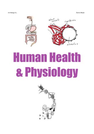
  • 1. I.B. Biology S.L. Éanna OBoyle Human Health & Physiology
  • 2. Topic 5: Human Health and Physiology 5.1 Digestion 5.1.1 Explain why digestion of large food molecules is essential. Digestion refers to the mechanical and chemical breakdown of complex food molecules into smaller molecules so that they can be absorbed. Once absorbed, the products of digestion are used i) as an energy source. ii) for making other molecules required by the living body. The human digestive system is rather representative of the mammalian system in many respects. We will follow the path of food through the human digestive system. 5.1.2 Explain the need for enzymes in digestion. We have seen that carbohydrates, lipids, and proteins are often very large, complex molecules composed of repeating molecular subunits (see section 2). Except for certain triglycerides (see section on lipids), these “macromolecules” can’t be absorbed through the walls of the digestive system. So in the digestive process, enzymes help break down these large molecules into smaller molecules, which can be then absorbed through walls
  • 3. of the digestive system. The bonds are broken through hydrolysis, the opposite of condensation, and the process in which water is added to the complex molecules. In other words, enzymes help break the bonds present in these large molecules. During digestion, carbohydrates are hydrolyzed (remember... the opposite of condensation) into simple sugars, fats into fatty acids and glycerol, proteins into their various amino acids, and nucleic acids into free nucleotides. 5.1.3 State the source, substrate, products and optimum pH conditions for one amylase, one protease and one lipase. a) Carbohydrate Digestion: Starch ( a polysaccharide carbohydrate) is a typical large complex molecule that we eat (e.g. in potatoes, rice, and bread). Any enzyme that digests starch is called an amylase. One example of an amylase is salivary amylase, which is produced by the salivary gland in the mouth. This enzyme acts on starch (the substrate), producing the disaccharide maltose (remember ... 2 glucose units linked together) and many longer polysaccharide (starch) fragments.
  • 4. maltose amylase H2O + Amylose short-chain starch (type of starch) fragment [Note that many molecules of water react with 1 molecule of amylose]. Later, in the intestine, these maltose molecules are broken down further into glucose molecules by the enzyme maltase (see section 2 notes for the reaction). With the joint action of amylase and maltase, starches like amylose are broken down into glucose which can be absorbed through the wall of the intestines in the digestive system. b) Protein digestion: Proteins are among the largest and most complex molecules known. Not surprisingly, their digestion is also complex, requiring a number of different enzymes that form a family called proteases.
  • 5. The first step in protein digestion ocurs in the stomach. Here the enzyme pepsin, produced by the gastric glands, acts upon proteins (the substrate), by attacking and breaking peptide bonds (remember section 2 for what a peptide bond is!). Pepsin can only attack 4 of the 20 amino acids that occur on earth, and in all 4 they are broken down into smaller peptide lengths. Further protein breakdown occurs in the small intestine by for example trypsin. The final steps in protein digestion are carried out by the enzymes aminopeptidases and dipeptidases (which belong to the peptidase group of enzymes). Dipeptidases attack peptide bonds in dipeptides, yielding two amino acids; e.g. starting with pepsin, & eventually ending with dipeptidase: Pepsin Pepsin serine glutamine proline glycine leucine serine H 2O H2 O Dipeptidase H2O Serine and glutamine are two free amino acids, which can be absorbed through the wall of the glutamine digestive system. serine
  • 6. c) Lipids: Fat digestion generally occurs in the small intestine. Fats are digested by the enzyme pancreatic lipase. Lipase breaks down neutral fats (triglycerides) into fatty acids, glycerol, monoglycerides and diglycerides. o OH OH Lipase OH o OH OH o OH + 3 H2O Tryglyceride glycerol 3 ‘free’ fatty acids 5.1.4 Draw a diagram of the digestive system. See end of these notes and textbook! 5.1.5 Outline the functions of the stomach, small intestine, and large intestine. The stomach temporarily stores food and begins its digestion. In addition, its acids and enzymes kill many of the organisms we swallow. The small intestine has 2 roles - digestion and absorption. The large intestine (also called colon or bowel) main functons are: a) to absorb water and minerals into the blood b) to prepare the faeces to leave the digestive tract.
  • 7. Very little, if any, digestive activity takes place in the large intestine itself, although a rapid digestion of food residues is carried on by huge populations of microrganisms (e.gs. Escherichia coli and methane- producing bacteria). 5.1.6 Distinguish between absorption and assimilation. Digestive enzymes help break down complex carbohydrates into simple sugars, lipids into glycerol and fatty acids, and proteins into amino acids. These end products of digestion along with the other nutrients, vitamins, minerals, and water, are of no value to your body until they leave the digestive system. They must pass into your blood for distribution to your cells. The process by which nutrients pass from the digestive system into the body fluids (blood and lymph) is called absorption. Assimilation describes the process by which energy is released by these absorbed molecules. 5.1.7 Explain how the structure of the villus is related to its role in absorption of the end products of digestion. The small intestine has special structures - villi (sing.: villus) - which help absorption. These fingerlike projections greatly increase the surface area of the small intestine. The surface area is further increased by the microvilli - tiny folds in the villi’s surfaces. The large surface area allows nutrients to pass easily into the blood vessels and lymph system. See further notes in text ...
  • 9. Digestive System mouth larynx trachea (or air tube; not part (o)esophagus of digestive system!) liver stomach gall bladder small intestine pancreas large intestine appendix anus rectum
  • 10. 5.2 - The Transport System It beats day and night every day of our lives, but what does the heart really do for us? Your heart, quite simply, is a pump. But it is one of the most fascinating pumps you'll ever learn about. First you must understand that every cell in our body - from the cells in our hair to the cells in our toes need OXYGEN and NUTRIENTS to survive and keep us alive. Since they can't get these nutrients themselves, blood is used to deliver them right to the cells. (sort of like room service) The basic job of the heart is pump that blood through your body so that the blood can deliver the oxygen and nutrients right to the cells. The heart keeps your blood pumping at all times as it picks up oxygen from your lungs and nutrients from your digestive system and sends them to all cells of your body. Your heart is the power behind the delivery system. Inside the heart is four chambers. Two of those chambers send the blood up to your lungs to get oxygen, then the other two chambers send that oxygen rich blood to the rest of your body. Valves inside of the heart make sure that the blood only moves in one direction. The younger you are, the faster your heart beats. A baby's heart beats about 90 times a minute. A twelve year old heart beats about 78 times a minute and an adult heart beats about 70 times a minute. Of course the more active you are, the more your heart beats, since the cells need oxygen faster to keep you moving.
  • 11. 5.2.1 Draw a diagram of the heart showing all four chambers, associate blood vessels and valves. semi-lunar valves 5.2.2 Describe the action of the heart in terms of collecting blood, pumping blood and opening and closing valves. 5.2.3 Outline the control of the heartbeat in terms of the pacemaker, nerves and adrenalin.
  • 12. 5.2.4 Explain the relationship between the structure and function of arteries, capillaries and veins. 5.2.5 State that blood is compose of plasma, erythrocytes, leucocytes (phagocytes and lymphocytes) and platelets. 5.2.6 State that the following are transported by the blood: nutrients, oxygen, carbon dioxide, hormones, antibodies and urea.
  • 13. The Heart and the Circulatory System Glossary Anterior: adjective referring to front end of an animal or organism. Aorta: the main systemic artery of the body, emerging directly from the left ventricle. Arteriole: a small arterial branch that delivers blood directly to a capillary bed. Artery: a muscular blood vessel that carries blood away from the heart. Atrium: one of the chambers of the heart that receives blood directly from a vein. Circulatory system: the system of the body responsible for internal transport. Composed of the heart, blood vessels, lymphatic vessels, lymph, and the blood. Closed circulatory system: a type of circulatory system where the blood is contained within a system of vessels and the heart. Coronary artery: one of the arteries that supply blood to the heart. Coronary vein: one of the veins that receive blood from the heart muscle and empty directly into the right atrium. Deoxygenated blood: blood that is low in oxygen concentration. Dorsal: adjective referring to the top or upper surface of an organism. Heart: the muscular organ composed of cardiac muscle that is responsible for pumping blood throughout the body. Heart attack: a condition occurring when a section of the heart is deprived of oxygenated blood and dies. Interstitial fluid: the fluid filling the microscopic spaces between cells of the body. Open circulatory system: a type of circulatory system where the blood is not contained within a system of vessels and the heart. Blood empties from vessels into sinuses and then returns through other vessels to a "heart.". Peristalsis: wave-like muscular contractions in the walls of tubular organs, e.g. the alimentary canal. Peristalsis serves to push material contained within the organ along its length. Posterior: adjective referring to the hind end of an animal. Pulmonary artery: one of the arteries carrying deoxygenated blood from the heart to the lungs. Septum: the wall dividing the two ventricles. Sinus: a cavity into which blood flows and baths the internal organs in organisms with an open circulatory system. Spiracle: opening which leads to a trachea in an insect, arachnid, isopod, centipede or millipede. Vein: one of the blood vessels that carries blood to the heart. Ventral: adjective referring to the lower surface of an animal or organism. Ventricle: one of the muscular chambers of the heart that is responsible for pumping blood from the heart into the arteries. Venule : a small venous branch that carries blood from a capillary bed to a vein.
  • 14. The Blood Vessels Exp TTlojLeading up to a heart attack ing Vess From World Book © 2001 World Book, Inc., 233 N. Michigan Avenue, Suite 2000, Chicago, IL 60601. All rights reserved. World Book illustrations by Paul Peck, M.D. and Charles Wellek
  • 15. A Coronary Bypass From World Book © 2001 World Book, Inc., 233 N. Michigan Avenue, Suite 2000, Chicago, IL 60601. All rights reserved. World Book illustrations by Virginia Samter Heart-Lung Machine (during bypass operation) From World Book © 2001 World Book, Inc., 233 N. Michigan Avenue, Suite 2000, Chicago, IL 60601. All rights reserved. World Book illustration by Sarah Woodward and John Eggert
  • 16. Red Blood Cell (erythrocyte) From World Book © 2001 World Book, Inc., 233 N. Michigan Avenue, Suite 2000, Chicago, IL 60601. All rights reserved. © Dr. Tony Brain/SPL from Photo Researchers A White Blood Cell From World Book © 2001 World Book, Inc., 233 N. Michigan Avenue, Suite 2000, Chicago, IL 60601. All rights reserved. NIBSC/SPL from Photo Researchers
  • 17. There are three types of vessels - arteries, veins and capillaries. Arteries take oxygenated blood from the heart out to all areas of the body. The walls of the arteries are too thick for oxygen and nutrients to pass through, so arteries lead to smaller vessels called capillaries. The walls of the capillaries are thin enough for red and white blood cells to squeeze through and enter other body tissues. The circulation of blood can easily be observed moving through arteries, capillaries and veins. Arteries, veins and capillaries are not anatomically the same. They are not just tubes through which the blood flows. Both arteries and veins have layers of smooth muscle surrounding them. Arteries have a much thicker layer, and many more elastic fibers as well. The largest artery, the aorta leaving the heart, also has cardiac muscle fibers in its walls for the first few cm of its length immediately leaving the heart. Arteries have to expand to accept the blood being forced into them from heart, and then squeeze this blood onto the veins when the heart relaxes. Arteries have the property of elasticity, meaning that they can expand to accept a volume of blood and then contract and squeeze back to their original size after the pressure is released. A good way to think of them is like a balloon. When you blow into the balloon, it inflates to hold the air. When you release the opening, the balloon squeezes the air back out. It is the elasticity of the arteries that maintains the pressure on the blood when the heart relaxes, and keeps it flowing forward. Arteries branch into arterioles as they get smaller. Arterioles eventually become capillaries, which are very thin and branching. Capillaries are really more like a web than a branched tube. It is in the capillaries that the exchange between the blood and the cells of the body takes place. Here the blood gives up its carbon dioxide and takes on oxygen. In the special capillaries of the kidneys, the blood gives up many waste products in the form of urine. Capillary beds are also the sites where white blood cells are able to leave the blood and defend the body against harmful invaders. Capillaries are so small that when you look at blood flowing through them under a microscope, the cells have to pass through in single file. As the capillaries begin to thicken and merge, they become venules. Venules eventually become veins and head back to the heart. Veins do not have as many elastic fibers as arteries. Veins do have valves, which keep the blood from pooling and flowing back to the legs under the influence of gravity. When these valves breakdown, as often happens in older or inactive people, the blood does flow back and pool in the legs. The result is varicose veins, which often appear as large purplish tubes in the lower legs.
  • 18. Topic 5.3 - Pathogens and Disease 5.3.1 Define pathogen. Pathogen - an organism or virus that causes a disease. Pathogens cause disease in the host. 5.3.2 State one example of a disease caused by members of each of the following groups: viruses, bacteria, fungi, protozoa, flatworms and roundworms. Viruses: Influenza, mumps, measles, chicken pox, common cold. Bacteria: Cholera, tuberculosis, tetanus, diphtheria. Fungi: Athlete's foot, Candida (thrush) Protozoa: Malaria. Roundworms: Ascaris eggs contained in contaminated food are swallowed, circulate through the blood stream, reach the lungs, grow into larvae in the nasal cavities, swallowed into the stomach where they grow into adult worms and start the cycle again. Hookworm. Flatworms: Pork tapeworm, Bilharzia.
  • 19. 5.3.3 List six methods by which pathogens are transmitted and gain entry to the body. 1) From the air - pathogen enters through respiratory system (e.g. droplets) 2) Direct contact - pathogen enters through the skin. 3) Through food or water - pathogen enters through the digestive system. 4) Through cuts in the skin. 5) Blood transfusion. 6) Infected animals and insects - pathogen enters through bites. 7) Sexual contact - pathogen enters through reproductive system. 5.3.4 Describe the cause, transmission and effects of one human bacterial disease. Tuberculosis is caused by Mycobacterium tuberculosis, a rod-shaped bacterium. See text. 5.3.5 Explain why antibiotics are effective against bacteria but not viruses. Antibiotics block specific metabolic pathways found in bacteria, but not in eukaryotic cells. Viruses reproduce using the host cell metabolic pathways that are not affected by antibiotics. 5.3.6 Explain the cause, transmission and social implications of AIDS. AIDS is a retrovirus having RNA as its genetic material and not DNA. It transcribes its RNA into DNA using an enzyme called reverse transcriptase. It is transmitted by sexual intercourse, sharing of needles, blood transfusions, accidents causing blood contamination, cuts in the skin, tattoos and ear piercing with infected needles. Social implications are that people don't feel very comfortable with a person who has AIDS. People with AIDS can find it difficult to buy health insurance plans, find jobs, have friends, and build normal social relations. People have changed their sexual lifestyles due to awareness and education about AIDS. See h/o.
  • 20. Topic 5.4 - Defense Against Infectious Disease 5.4.1 Explain how skin and mucous membranes act as barriers against pathogens. The skin and mucous membranes are the first lines of defense against disease. The skin has a thick keratin layer on the surface which helps prevent organisms from entering the body. Where there is no skin, such as the mouth cavity, epithelial cells there form a mucous membrane that produces sticky mucous which traps and stops the action of many organisms. Other barriers to pathogens include acid in the stomach, resident bacteria, and tears. 5.4.2 Outline how phagocytic leucocytes ingest pathogens in the blood and in body tissues. When the phagocytes meet the pathogens, they ingest the organisms by phagocytosis. Once they are in the phagocytes, the pathogens go into the vesicles which fuse with the lysosomes, which then release hydrolytic enzymes on them and destroy them. 5.4.3 State the difference between antigens and antibodies. An antigen is a foreign macromolecule that does not belong to the host organism and that elicits an immune resonse. An antibody is a protein and is called an immunoglobulin. It is made of 4 polypeptides, 2 heavy chains and 2 light chains. It sticks to antigens and to lymphocytes. 5.4.4 Explain antibody production. Many different types of lympocyte exist. Each type recognizes one specific antigen and responds by dividing to form a clone. This clone then secretes a specific antibody agaist the antigen.
  • 21. 5.4.5 Outline the effects of HIV on the immune system. HIV attacks helper T cells, which are part of the immune system that are important for the function of B lymphocytes. The virus enters the helper T cells and replicates there. The cells burst and release new viruses, these viruses infect other helper T cells and possibly other cells such as phagocytes as well. The destruction of helper T cells paralyses the immune system since they communicate between different cells of the immune system and activate them. This enables any other parasite or organism usually kept under control by the immune system to be able to affect the body.
  • 22. Topic 5.5 - Gas Exchange 5.5.1 List the features of alveoli that adapt them to gas exchange. There is a large surface area, a wall consisting of a single layer of flattened cells, a moist lining, and a dense network of capillaries. See text, p.51. 5.5.2 State the difference between ventilation, gas exchange, and cell respiration. *Ventilation in humans involves bringing fresh air to the alveoli and removing stale air which is low in oxygen, O2 and high in CO2. It maintains a high concentration of oxygen in the alveoli and low carbon dioxide as we breathe in and out. *Gas exchange is the process where one gas (e.g. CO2) is swapped with another (e.g. oxygen). In humans, it occurs between the alveoli and the capillaries by diffusion - O2 passes from the alveoli to the capillaries and CO2 passes from the capillaries to the alveoli. This happens because there are concentration gradients of O2 and CO2 between the air (in alveoli) and the blood (in capillaries). *Cell respiration is the chemical reaction that occurs inside the cell (cytoplasm and mitochondria) and that results in the controlled release of energy in the form of ATP. In humans O2 is used up and CO2 is produced. 5.5.3 Explain the necessity for a ventilation system. It is needed to maintain concentration gradients in the alveoli. 5.5.4 Draw a diagram of the ventilation system including trachea, bronchi, bronchioles, and lungs. See notes... 5.5.5 Explain the mechanism of ventilation in human lungs including the action of the internal and external intercoastal muscles, the diaphragm and the abdominal muscles. See notes
  • 23. Topic 5.6 - Homeostasis and Excretion 5.6.1 State that homeostasis involves maintaining the internal environment at a constant level or between narrow limits, including blood pH, oxygen and carbon dioxide concentrations, blood glucose, body temperature and water balance. Here the internal environment refers to blood and tissue inside our bodies. Homeostasis can also be seen as the ability of an organism to maintain stable conditions inside its body even as outside conditions change dramatically. Cells as well as organisms have this ability of homeostasis. 5.6.2 Explain that homeostasis involves monitoring levels of variables and correcting changes in levels by negative feedback mechanisms. If body temperature falls below 37 degrees Celsius, then messages are sent by the hypothalamus to different parts of the body so temperature is increased to normal. Conversely, if body temperature rises above 37 degrees Celsius, messages sent decrease body temperature to normal. Therefore, a change in a variable is counteracted by the opposite change to return the body to a normal temperature. Other variables in the blood include the pH, pressure, O2/CO2 & glucose concentrations. Negative feedback mechanisms are very common in organisms and simply refer to how a change in levels (eg tºC, pressure, ...) always causes the opposite change. In the example above, the body temperature increase caused the body to react so that the temperature decreased.
  • 24. 5.6.3 State that the nervous and the endocrine systems are both involved in homeostasis. The endocrine system is made up of glands that produce (‘secrete’) hormones (‘chemical messengers’) into the blood. 5.6.4 State that the nervous system consists of the central nervous system (CNS) and peripheral nerves and is composed of special cells called neurons that can carry electrical impulses rapidly. The nervous system consists of neurons. The nervous system enables us to adjust to changes in our surroundings. Such neurons (nerve cells) as the receptors in the eyes translate information from the environment into electrical nerve impulses. Sensory neurons carry the electrical impulses to neurons in the ‘brain and spinal cord’ (the central nervous system or CNS). Motor neurons then carry instructions from the brain to muscles, internal organs, and other body parts via the peripheral nervous system (PNS), which connects body parts to the CNS. 5.6.5 Describe the control of body temperature including the transfer of heat in blood, the role of sweat glands and skin arterioles, and shivering. First, the nerve cells beneath the skin, thermoreceptors, detect a change in the environment surrounding the human. These thermoreceptors send messages that are received by the hypothalamus. The hypothalamus is made of nerve cells and is considered a part of the nervous and endocrine systems. Hormones are released from the hypothalamus and they travel to the pituitary gland. The pituitary gland then releases a hormone bound for the thyroid-gland which in turn releases thyroxine. The release of thyroxine increases the metabolic rate of the body and in turn releases more heat. For example, when the weather is hot, less thyroxine is released and less heat is produced. The hypothalamus also plays a role in transmitting nerve
  • 25. messages to muscles, blood capillaries and sweat glands. The effect of this is the occurrence of responses such as shivering, vasoconstriction or vasodilatation and sweating. Cold weather: More thyroxine produced. Skin arterioles become narrower (vasoconstriction) and carry less blood so the skin gets colder (to prevent heat loss). Shivering occurs as muscles contract rapidly to generate heat. Sweat glands do not produce sweat and the skin dries. Hot weather: Less thyroxine produced. Arterioles widen (vasodilation) so skin gets hotter and loses heat to environment. Muscles inactive. Sweat glands release sweat which has cooling effect on body. 5.6.6 State that the endocrine system consists of glands which release hormones that are transported in the blood. See p.52 (Allott) 5.6.7 Explain the control of blood glucose concentration, including the roles of glucagon, insulin, and alpha and beta cells in the pancreatic islets. Insulin and glucagon regulate the sugar (glucose) level in the body. These two hormones are manufactured in the pancreas. Insulin stimulates enzymes in the liver and muscles that convert glucose to glycogen, and so decrease glucose levels while at the same time producing glucose storage in the form of glycogen. Glucagon stimulates enzymes in the liver that hydrolyze glycogen to glucose, which then enter the blood and so increase glucose levels. Receptors in the pancreas are sensitive to the changes in glucose level, thus releasing the necessary requirements of insulin and glucagon depending on the needs of the body. The beta (β) cells found in the islets of the pancreas make insulin and the alpha (α) cells make glucagon.
  • 26. 5.6.8 Define excretion Excretion is the removal of metabolic waste from the body. 5.6.9 Outline the role of the kidney in excretion and the maintenance of water balance. See diagram in h.o. Excretion: The kidneys remove the waste products from the blood and make urine as a result, which is stored in the bladder before it is excreted through the urethra. Urea is one of the main waste products. Water balance (an example of homeostasis): By varying the composition and volume of urine, the kidneys help to keep the water and salt concentrations in the blood and tissue fluids constant. For example, if the body has excess water or too little salt, then more urine with a low salt concentration is produced.
  • 27. Topic 5.7 - Reproduction 5.7.1 Draw diagrams of the adult male and female reproductive systems. From World Book © 2001 World Book, Inc., 233 N. Michigan Avenue, Suite 2000, Chicago, IL 60601. All rights reserved. World Book diagram by George Suyeoka Also see Allott (p. 24) for an alternative angle of the female system. 5.7.2 Explain the role of hormones in regulating the changes of puberty (testosterone, estrogen) in boys and girls, and in the menstrual cycle (follicle stimulation hormone (FSH), luteinizing hormone (LH), estrogen, and progesterone). The reproductive activities of humans are under the influence of hormones (and genes!). Boys: From birth to the age of ten or so, testosterone level is very low. It increases sharply after that and begins puberty in males. This is when sperm production takes place. Testosterone stays at high levels until the age of 40-50, then it gradually decreases. It is also responsible for male secondary sexual charactheristics.
  • 28. Girls: Oestrogen (or estrogen - USA) production is responsible for puberty in girls (often beginning 9-12 years of age). It leads to the maturty of eggs which leads to the menstrual cycle, as well as to the female secondary sexual charactheristics. Between puberty and menopause, women who are not pregnant follow the menstrual cycle. This cycle is mainly controlled by 4 hormones: • two produced in the pituitary gland - follicle stimulating hormone (FSH) and the luteinizing hormone (LH). • two produced in the ovary - oestrogen and progesterone. The levels of these hormones vary during the menstrual cycle. The beginning of the cycle is usually considered as the maturing of an egg in the ovary. Simply put, FSH levels increase and this is responsible for the growth of an oocyte (an immature egg) and it's follicle (surrounding cells). Two weeks after the start of menstruation, ovulation (release of egg caused by rupturing of follicle - tºC increase by < 1ºC) occurs due to a sudden and sharp increase in LH. It also causes the empty follicle to develop into the corpus luteum (a yellow body) which starts releasing the hormone progesterone as well as continuing to release oestrogen. These are responsible for maintaining and thickening the endometrium (thick lining of spongy, blood rich tissue of the uterus in preperation for implantation). Progesterone production therefore begins the preparation of the uterus for the zygote or fertilized egg - if it happens! If fertilization does occur, progesterone will help maintain the endometrium throughout the pregnancy. Progesterone also thickens and increases secretion in the vaginal lining, and the growth of milk ducts in the breasts. Effects of menstruation (not on IB): Some women have mild to moderate abdominal cramps a few days before or during menstruation. This discomfort, called dysmenorrhea, results from contractions of the uterus
  • 29. and is usually normal. During the days before menstruation begins, some women experience emotional or physical symptoms that may include depression, anxiety, fatigue, headache, body swelling, or pain in the breasts. This condition is called premenstrual syndrome (PMS). Menstruation signifies good health if it occurs regularly and without excessive pain, fatigue, or blood loss. Most women carry on their usual activities. Menstrual discharge can be absorbed either by a sanitary napkin, a disposable pad that covers the vaginal opening, or by a tampon, a roll of absorbent material worn inside the vagina. The most common reason for a young woman to miss a menstrual period is pregnancy. Other reasons include emotional stress, weight loss, and abnormal hormonal balance. If a woman frequently misses her period or if it occurs less often than every 35 days, she should consult a doctor. 5.7.3 List the secondary sexual characteristics in both sexes. Males: • voice change due to enlargement of larynx • hair growth in the pubic area, under the armpits, and in the face • the building of muscles (skeletal) • the penis and testes grow larger • the prostrate gland and seminal vesicles begin to secrete fluid • sperm is produced and released during ejaculation Females:, • hair growth in the pubic area and under the armpits • beginning of the menstrual cycle • vagina and uterus enlarge • vagina begins to secrete fluid
  • 30. • breasts enlarge • pelvis enlarges • fat develops in buttocks and thighs 5.7.4 State the difference between copulation and fertilization. Copulation (or sexual intercourse) occurs when the penis enters the vagina. If the male ejaculates, semen containing up to 10 million sperm travel through the vagina - cervix - uterus - oviducts (or called fallopian tubes). Fertilization occurs when the nucleus of the sperm fuses with the nucleus of the egg, and the woman then is pregnant. 5.7.5 Describe early embryo development up to the implantation of the blastocyst. Fertilization occurs and results in the formation of the one-celled zygote which starts a series of cell divisions (mitosos) in the oviduct called the cleavage stage. This results in the formation of a ball of cells. When there are 16 cells the structure is called a morula , and this becomes a hollow ball (blastocyst). After 7 days the developing blastocyst implants itself into the uterine wall where it can develop into a baby. 5.7.6 State that the fetus is supported and protected by the amniotic sac and amniotic fluid. The fetus is supported and protected by the amniotic sac and amniotic fluid. The fetus floats in this fluid which acts as a shock absorber for sudden movements of the fetus. See diagram on page 55 in Allott. 5.7.7 State that materials are exchanged between the maternal and fetal blood in the placenta. Materials are exchanged between the maternal and fetal blood in the
  • 31. placenta. For example oxygen from mother to fetus and carbon dioxide from fetus to mother. 5.7.8 Outline the process of birth and its hormonal control, including progesterone and oxytocin. From World Book © 2001 World Book, Inc., 233 N. Michigan Avenue, Suite 2000, Chicago, IL 60601. All rights reserved. World Book illustration by Charles Wellek Progesterone is increasingly secreted throughout pregnancy. After 9 months or so, progesterone production falls which in turn allows another hormone oxytocin to be produced. Oxytocin causes contractions of the uterus, which stimulates the production of even more oxytocin, and so the contractions become stronger and stronger (e.g. of positive feedback). This ‘labor’, along with delivery and afterbirth mark the three stages of birth. Dilation of the cervix also occurs at this time. Later, the cervix becomes fully dilated. The most powerful contractions are during the next stage, delivery. The placenta, along with blood and other fluids come out after the baby. The placenta that comes out marks the afterbirth.
  • 32. 5.7.9 Describe four methods of family planning and contraception. There is sterilization. In this, the female gets a tube legation where the oviducts are tied so the sperm can't reach the egg. Or the male gets a vasectomy where the sperm ducts are cut and prevents the release of sperm. Another method is pills. These prevent ovulation by inhibiting FSH and LH. The use of a male condom prevents the release of sperm into vagina. The last method is intrauterine device (IUD) which prevents fertilization or i mplantation. 5.7.10 Discuss the ethical issues of family planning and contraception. People may find this unethical due to religious, traditional, or other beliefs that families are supposed to be large. But, if all families were that large, there would be a large popluation growth and more limiting factors would set in and the death rate would increase. There would also be more starvation, disease, and competition for survival. It is debated as to what is more ethical. 5.7.11 Outline the technique of amniocentesis. Amniocentesis is where some amniotic fluid is drawn by a syringe through the abdomen of the mother. The cells are then grown on a tissue culture to be studied to create a karyotype and are then studied to find out if the are any abnormalties. 5.7.12 Outline the process of in vitro fertilization (IVF). Eggs are removed from the ovaries of a woman by suction through the
  • 33. vagina. They are sucked into a syringe and placed in a glass dish. The eggs are then cleaned to remove blood and other unwanted material. The egg is then incubated. Then, sperms are added and fertilization takes places and the embryo is then transferred through the vagina to the uterus. 5.7.13 Discuss the ethical issues of IVF. This once again can be a religious unethical idea. Someone that is not meant to reproduce has a child.
  • 35. From World Book © 2001 World Book, Inc., 233 N. Michigan Avenue, Suite 2000, Chicago, IL 60601. All rights reserved. World Book illustrations by Mary Ann Olson and Dr. Paul Peck