All on 4 v4 modification
•
2 gostaram•1,142 visualizações

Ole Jensen Article on all on 4 V4 modification for dental implant placement in atrophic maxilla and mandible

Recomendados
Mais conteúdo relacionado
Mais procurados
Mais procurados (20)
Semelhante a All on 4 v4 modification
Semelhante a All on 4 v4 modification (20)
Último
Último (20)
All on 4 v4 modification
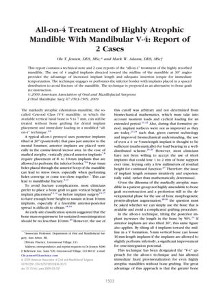
- 1. J Oral Maxillofac Surg 67:1503-1509, 2009 All-on-4 Treatment of Highly Atrophic Mandible With Mandibular V-4: Report of 2 Cases Ole T. Jensen, DDS, MSc,* and Mark W. Adams, DDS, MSc† This report contains a technical note and 2 case reports of the “all-on-4” treatment of the highly resorbed mandible. The use of 4 angled implants directed toward the midline of the mandible at 30° angles provides the advantage of increased implant length and adequate insertion torque for immediate temporization. The technique engages or perforates the inferior border with implants placed in a spaced distribution to avoid fracture of the mandible. The technique is proposed as an alternative to bone graft reconstruction. © 2009 American Association of Oral and Maxillofacial Surgeons J Oral Maxillofac Surg 67:1503-1509, 2009 The markedly atrophic edentulous mandible, the so- called Cawood Class IV-V mandible, in which the available vertical basal bone is 5 to 7 mm, can still be treated without bone grafting for dental implant placement and immediate loading in a modified “all- on-4” technique.1-6 A typical all-on-4 protocol uses posterior implants tilted at 30° (posteriorly) that pass just anterior to the mental foramen; anterior implants are placed verti- cally in the canine-lateral incisor area. In the case of marked atrophy, vertically placed anterior implants7,8 require placement of 8- to 10-mm implants that are allowed to perforate the inferior border.5,6 Four 4-mm holes placed through the anterior hoop of the mandible can lead to stress risers, especially when perforating holes converge or come too close together.7 This can lead to mandibular fracture.9-11 To avoid fracture complications, most clinicians prefer to place a bone graft to gain vertical height at implant placement12-14 or before implant surgery15-17 to have enough bone height to sustain at least 10-mm implants, especially if a favorable anterior-posterior spread is difficult to obtain.18,19 An early site classification system suggested that the bone mass requirement for sustained osseointegration should be no less than 10 mm.20 However, the use of this cutoff was arbitrary and not determined from biomechanical mathematics, which must take into account moment loads and cyclical loading for an extended period.21,22 Also, during that formative pe- riod, implant surfaces were not as improved as they are today,23,24 such that, given current technology and improved biomechanical understanding, the use of even a 4- or 5-mm-length implant is thought to be sufficient (mathematically) for load bearing in a well- distributed scheme.25,26 However, most clinicians have not been willing to accept the use of short implants that could lose 1 to 2 mm of bone support over time, leaving only a few millimeters of residual height for continued function.27 Therefore, the issue of implant length remains intuitively and experien- tially valid, rather than mathematically determined. Given the dilemma of the markedly atrophic man- dible in a patient group not highly amendable to bone graft reconstruction and a profession still in the de- velopmental phase for the use of bone morphogenetic protein-alloplast augmentation,28-30 the question must be asked whether we can simply use the bone that is available and avoid a complicated grafting procedure. In the all-on-4 technique, tilting the posterior im- plant increases the length in the bone by 50%.31 If anterior implants are also tilted 30°, this same figure also applies. By tilting all 4 implants toward the mid- line in a V formation, 5-mm vertical bone can house 10-mm-length implants if the implants are allowed to slightly perforate inferiorly, a significant improvement for osseointegration potential. This technique has been designated the “V-4” ap- proach for the all-on-4 technique and has allowed immediate fixed provisionalization for even highly atrophic mandibles without bone grafting. The great advantage of this approach is that the greater bone *Asssociate Professor, Department of Oral and Maxillofacial Sur- gery, Ann Arbor, MI. †Private Practice, Greenwood Village, CO. Address correspondence and reprint requests to Dr Jensen: 8200 E Belleview Ave, Suite 520, Greenwood Village, CO 80111; e-mail: Ole.jensen@Clearchoice.com © 2009 American Association of Oral and Maxillofacial Surgeons 0278-2391/09/6707-0023$36.00/0 doi:10.1016/j.joms.2009.03.031 1503
- 2. mass generally found more toward the midline can be taken advantage of, and, despite implant convergence toward the midline, the holes perforating the inferior cortex remain well distributed and relatively far apart from each other, reducing the fracture potential. The parasymphyseal area, where a mandibular fracture is mostly likely to occur, is avoided altogether.32,33 Case Reports CASE 1 A 72-year-old woman had worn dentures for longer than 30 years and presented with severe mandibular atrophy with 8 to 10 mm of alveolar height as viewed on the Panorex (Fig 1). However, because of reverse architecture only 4 to 5 mm of vertical dimension was present in the mid-alveolar (axial) dimension. The mental foramina were dehisced and relatively forward in the arch. The nerves were partially exposed poste- riorly. A full-thickness crestal incision was made anteriorly but only through the mucosa posteriorly to avoid cutting the nerves. Using blunt dissection, the nerves were located and deflected laterally, leaving the fora- men free of neural tissue. Anteriorly, the mentalis muscle attachment was left undisturbed to prevent ptosis. The mandible appeared very fragile overall, but it had been especially resorbed in the parasym- physeal regions. Although all-on-4 fixture placement had been planned on the computer, the surgical placement criteria dictated placing the implant visu- ally to not fracture the mandible. The first fixture was placed directly into the right mental foramen (Fig 2A) and angled forward 30°. The anterior implants were evenly spaced and also directed toward the midline at 30° (Fig 2B). This created an overall V-shape place- ment appearance on Panorex designated a “V-4” all- on-4 placement (Fig 3). Additionally, the implant placement angles were tilted anteriorly to avoid lin- gual plate perforation (Fig 4). Finally, 30° abutments were placed to compensate for implant angulation for immediate prosthetic rehabilitation. CASE 2 An 81-year-old female patient with a history of wearing full dentures for 35 years who had been FIGURE 1. Preoperative Panorex view of 72-year-old woman who presented with severe alveolar atrophy. Jensen et al. Treatment With Mandibular V-4. J Oral Maxillofac Surg 2009. FIGURE 2. A, Using the all-on-4 technique, posterior fixture was placed directly through mental foramen after deflecting dehisced inferior alveolar nerve laterally. B, This was done bilaterally. Jensen et al. Treatment With Mandibular V-4. J Oral Maxillofac Surg 2009. 1504 TREATMENT WITH MANDIBULAR V-4
- 3. taking an oral bisphosphonate (Fosamax; Merck, Whitehouse Station, NJ) for 7 years for osteoporosis presented for dental implant rehabilitation. The re- sults from a fasting C-terminal telopeptide study were satisfactory (315 pg/mL). The mandible was highly atrophic with 3 to 4 mm of vertical bone in the right symphysis and 5 to 6 mm in the left. In preparing the implant sites, the vertical available bone was a maximum of 5 to 7 mm (Fig 5). After reflection of a flap, taking care to avoid nerve injury and preserving the mentalis muscle attach- ment, posterior implants were placed through the foramen sites after deflection of the dehisced nerves. Because the mandible resorbs, the mental foramen often presents in a mid-crestal location. These im- plants were placed at 30°, angling forward (Fig 6). The front implants were well-distributed and placed at somewhat less than 30° but still angled toward the midline (Fig 7). The overall distribution and display on the Panorex was a V shape (Fig 8). The patient was immediately provided, after placement of the 30° abutments, with a fixed provisional bridge. Discussion Patients who have worn dentures for 3 or more decades may seek implant reconstruction because of the pain from exposed inferior alveolar nerves owing to complete alveolar loss from atrophy. Denture com- pression of exposed nerves is best treated in this setting by dental implants; however, the lack of jaw bone height is a concern. Although a 10-mm vertical height may be present mid-symphysis, the parasym- physeal areas are often one half the height of the symphysis. Also, in this setting, the mid-alveolar area is often of a reverse architecture, such that the actual mid-axial alveolar height is much less than seems apparent on a Panorex. Although the lateral bone height can be relatively high, it cannot be accessed for implant placement; thus, often implants must be placed where bone is relatively deficient. Therefore, most experienced clinicians prefer to place implants with a careful minimal torque technique but still per- forating through the inferior border. Using this ap- proach, an 8- or 10-mm fixture is still placed into a 5- to 7-mm site. The value of angulation of implants in a V-4 distri- bution strategy is that bone grafting can be avoided, because fixtures are favorably directed toward the location of maximal bone mass. This approach is also excellent to use without inferior border perforation if somewhat greater bone mass is available. The V-4 technique is biomechanically favorable in 3 ways: 1) mandibular continuity preservation; 2) a greater length of implants; and 3) the V-shape is very stable biomechanically. FIGURE 3. Placement of 2 anterior implants angled at 30° to midline created a V shape for “all-on-4” placement, designated V-4. Jensen et al. Treatment With Mandibular V-4. J Oral Maxillofac Surg 2009. FIGURE 5. View of 81-year-old woman who presented with severe mandibular atrophy with 5 to 7 mm of bone available in desired implant sites. Jensen et al. Treatment With Mandibular V-4. J Oral Maxillofac Surg 2009. FIGURE 4. All implants angled slightly anteriorly to avoid perfo- ration of lingual plate. Jensen et al. Treatment With Mandibular V-4. J Oral Maxillofac Surg 2009. JENSEN ET AL 1505
- 4. MANDIBULAR CONTINUITY PRESERVATION A 4-mm hole drilled into the anterior tibia, a weight- bearing bone, reduces bone strength by 40%.34 A 4-mm hole drilled into the mandible, especially into a low-bone-volume atrophic mandible, may consider- ably weaken the jaw, even though it is not a weight- bearing bone.35 The placement of 4 holes through the hoop of the mandible, especially if they are not centrally placed, risks a discontinuity fracture intraoperatively36 or during the demineralization phase of healing.37 At about 3 weeks after surgery, it is possible for a jaw fracture to occur under normal functional loading38 ow- ing to the relative weakening of the jaw caused by the regional acceleratory phenomenon.39 However, the area at the greatest risk of this is the parasymphysis, which is avoided using V-4 angulation. The implants should be placed using a screw tap method, even using self-tapping implant protocols to decrease insertion torque values and not overload the bone.38,40 FIGURE 6. A, B, Posterior implants placed at 30° angulation. Jensen et al. Treatment With Mandibular V-4. J Oral Maxillofac Surg 2009. FIGURE 7. Anterior implants angled forward at approximately 30° such that adjacent implants are parallel to each other and do not converge at inferior border. Jensen et al. Treatment With Mandibular V-4. J Oral Maxillofac Surg 2009. FIGURE 8. Overall presentation on Panorex was a V-4 display. Jensen et al. Treatment With Mandibular V-4. J Oral Maxillofac Surg 2009. 1506 TREATMENT WITH MANDIBULAR V-4
- 5. GREATER LENGTH OF IMPLANTS What is important is not simply to have a greater implant length, but also to have the implant primarily fixated into compact bone.41 Bone grafting to gain implant length is an alternative strategy; however, vertical bone grafts are the earliest to fail under stress, and implants secured mainly by bone grafts can some- times fail with time.42 The incidental elevation of inferior border periosteum to gain periosteal prolifer- ative bone must also be considered as potential sec- ondary support, although it does not always occur43 (Fig 9). Therefore, the most dependable bone for long-term osseointegration is compact bone, more of which is encountered by implant angulation using a V-4 strategy. These compromised sites should probably use 4-mm diameter implants or less rather than trying to gain more surface osseointegration using shorter, wider (5-mm) implants, which considerably increases the risk of jaw fracture. FIGURE 9. A, Implant insertion through inferior border. B, Periosteal bone apposition observed 6 months later. C, Panorex demonstrating vertical bone growth of 2 to 3 mm compared with preoperative view after 6 months of function. D, Occlusal scheme with anteriorized occlusion and posterior disclusion during 6-month provisional loading phase. Jensen et al. Treatment With Mandibular V-4. J Oral Maxillofac Surg 2009. JENSEN ET AL 1507
- 6. V-SHAPE BIOMECHANICS The reason the V shape is favorable biomechani- cally is the greater length of implants into more dense bone. Also, the angulated implant pull-out strength in a splinted configuration is intuitively greater for an- gled implants. In the V-4 strategy, this is multiplied by a factor of 4, although this has not been studied experimentally. Finite element analysis of the tilted implants that are splinted in a full-fixed prosthesis revealed a decreased peri-implant “bone strain” com- pared with vertical implants, supporting a cantile- vered prosthesis and implying better load-bearing bi- omechanics.44 The highly atrophic mandible in the elderly patient can be treated with an all-on-4 technique without bone grafting with an immediate loading protocol by distributing the implants in a V shape, desig- nated the V-4 technique. The V-4 is protective of mandibular continuity, derives increased implant length with acceptable insertion torque values, and maintains a standard all-on-4 pattern of prosthetic distri- bution despite the angulated placement. A splinted V-4 distribution has highly favorable biomechanics. Overall, the V-4 permits the use of a conservative nongrafting approach in what might otherwise require significant bone graft reconstruction in a commonly elderly popu- lation. References 1. Cawood JI, Howell RA: A classification of the edentulous jaws. Int J Oral Maxillofac Surg 17:232, 1988 2. Cawod JI: Reconstructive preprosthetic surgery. I. Anatomical considerations. Int J Oral Maxillofac Surg 20:75, 1991 3. Chan MF, Johnston C, Howell RA, et al: Prosthetic management of the atrophic mandible using endosseous implants and over- dentures: A six year review. Br Dent J 179:329, 1995 4. Eufinger H, Gellrich NC, Sandmann D, et al: Descriptive and metric classification of jaw atrophy: An evaluation of 104 man- dibles and 96 maxillae of dried skulls. Int J Oral Maxillofac Surg 26:23, 1997 5. Cawood JI: Arnhem consensus on preprosthetic surgery, May 1989. Int J Oral Maxillofac Surg 19:10, 1990 6. Cawood JI, Stoelinga PJ; International Academy for Oral and Facial Rehabilitation: Int J Oral Maxillofac Surg 35:195, 2006 7. Stellingsma C, Vissink A, Meijer HJ, et al: Implantology and the severely resorbed edentulous mandible. Crit Rev Oral Biol Med 15:240, 2004 8. Perry RT: Reconstruction of advanced mandibular resorption with both subperiosteal and root-form implants. Implant Dent 7:94, 1998 9. Meijer HG, Raghoebar GM, Visser A: Mandibular fracture caused by peri-implant bone loss: Report of a case. J Periodon- tol 74:1067, 2003 10. Kan JY, Lozada JL, Boyne PJ, et al: Mandibular fractures after endosseous implant placement in conjunction with inferior alveolar nerve transposition: A patient treatment report. Int J Oral Maxillofac Implants 12:466, 1997 11. Mason ME, Triplett RG, Van Sickels JE, et al: Mandibular frac- tures through endosseous cylinder implants: Report of cases and review. J Oral Maxillofac Surg 48:311, 1990 12. Gutta R, Waite PD: Cranial bone grafting and simultaneous implants: A submental technique to reconstruct the atrophic mandible. Br J Oral Maxillofac Surg 46:477, 2008 13. van der Meij EH, Bankestijn J, Berns RM, et al: Int J Oral Maxillofac Surg 34:152, 2005 14. Quinn PD, Kent K, MacAfee KA II: Reconstructing the atrophic mandible with inferior border grafting and implants: A prelim- inary report. Int J Oral Maxillofac Implants 7:87, 1992 15. Felice P, Iezzi G, Lizio G, et al: Reconstruction of atrophied posterior mandible with inlay technique and mandibular ramus block graft for implant prosthetic rehabilitation. J Oral Maxil- lofac Surg 67:372, 2009 16. Bell RB, Blakey GH, White RP, et al: Staged reconstruction of the severely atrophic mandible with autogenous bone graft and endosteal implants. J Oral Maxillofac Surg 60:1135, 2002 17. Stellingsma K, Raghoebar GM, Meijer HJ, et al: The extremely resorbed mandible: A comparative prospective study of 2-year results with 3 treatment strategies. Int J Oral Maxillofac Im- plants 19:563, 2004 18. Rodriguez AM, Aguilino SA, Lund PS, et al: Evaluation of strain at the terminal abutment site of a fixed mandibular implant prosthesis during cantilever loading. J Prosthodont 2:93, 1993 19. Krennmair G, Furhauser R, Krainhofner M, et al: Clinical out- come and prosthodontic compensation of tiled interforaminal implants for mandibular overdentures. Int J Oral Maxillofac Implants 20:923, 2005 20. Rody AR: The atrophic mandible. J Okla Dent Assoc 97:26, 2005 21. Stamenkovic D: The biomechanics of dental implants and den- tures. Srp Arh Celok Lek 136(Suppl. 2):73, 2008 22. Quek HC, Tan KB, Nicholls JI: Load fatigue performance of four implant-abutment interface designs: Effect of torque level and implant system. Int J Oral Maxillofac Implants 23: 253, 2008 23. Mendonca G, Mendoca DB, Aragao FJ, et al: Advancing dental implant surface technology—From micron to nanotopography. Biomaterials 28:3822, 2008 24. Bhatavadekar N: Assessing the evidence supporting the claims of select dental implant surfaces: A systematic review. Int Dent J 58:363, 2008 25. Georgiopoulos B, Kalioras K, Provatidis C, et al: The effects of implant length and diameter prior to and after osseointegra- tion: a 2-D finite element analysis. J Oral Implantol 33:243, 2007 26. Motoyoshi M, Inaba M, Ono A, et al: The effect of cortical bone thickness on the stability of orthodontic mini-implants and on the stress distribution in surrounding bone. Int J Oral Maxillo- fac Surg 38:13, 2009 27. Jung YC, Han CH, Lee KWA: 1-Year radiographic evaluation of marginal bone around dental implants. Int J Oral Maxillofac Implants 11:811, 1996 28. Carter TJ, Brar PS, Tolas A, et al: Off-label use of recombinant human bone morphogenetic protein-2(rhBMP-2) for recon- struction of mandibular bone defects in humans. J Oral Maxil- lofac Surg 66:616, 2008 29. Herford AS, Boyne PJ: Reconstruction of mandibular continuity defects with bone morphogenetic protein-2(rhBMP-2). J Oral Maxillofac Surg 66:618, 2008 30. Boyne PJ: Application of bone morphogenetic proteins in the treatment of clinical oral and maxillofacial osseous defects. J Bone Joint Surg Am 83-A:S146, 2001 (suppl 1) 31. Malo P, Ranger B, Nobre M: “All-on-four” immediate-function concept with Branemark system implants for completely eden- tulous mandibles: A retrospective clinical study. Clin Implant Dent Relat Res 5:31, 2003 32. King RE, Scianna JM, Petruzzelli GJ: Mandible fracture patterns: A suburban trauma center experience. Am J Otolaryngol 25: 301, 2004 33. Kulak Burun Bogaz Ihtis Derg. The relationship between the fracture site and etiology in mandibular fractures. Kulak Burun Bogaz Ihtis Derg 14:25, 2005 34. Tommasini SM, Nasser P, Schaffler MB, et al: Relationship between bone morphology and bone quality in male tibias; implications for stress fracture risk. J Bone Miner Res 20:1372, 2005 1508 TREATMENT WITH MANDIBULAR V-4
- 7. 35. Stellingsma C, Vissink A, Raghoebar GM: Surgical dilemmas. Choice of treatment in cases of extremely atrophic mandibles. Ned Tijdschr Tandheelkd 115:665, 2008 36. Lamas Pelayo J, Penarrocha Diago M, Marti Bowen E, et al: Intraoperative complications during oral implantology. Med Oral Pathol Oral Cir Bucal 13:E239, 2008 37. Wagner KW, Schoen R, Wongchuensoontorn C, et al: Compli- cated late mandibular fracture following third molar removal. Quinessence Int 38:63, 2007 38. Schilling T, Mueller M, Minne HW, et al: Influence of inflam- mation-mediated osteopenia on the regional acceleratory phe- nomenon and the systemic acceleratory phenomenon during healing of a bone defect in the rat. Calcif Tissue Int 63:160, 1998 39. Cehreli MC, Akkocaoglu M, Comert A, et al: Strains around apically free versus grafted implants in the posterior maxilla of human cadavers. Med Biol Engl Comput 45:395, 2007 40. Al-Nawas B, Wagner W, Grotz KA: Insertion torque and reso- nance frequency analysis of dental implant systems in an ani- mal model with loaded implants. Int J Oral Maxillofac Implants 21:726, 2006 41. Cleek TM, Reynolds KJ, Hearn TC: Effect of screw torque level on cortical bone pullout strength. J Orthop Trauma 21:117, 2007 42. Barone A, Covani U: Maxillary alveolar ridge reconstruction with nonvascularized autogenous block bone: Clinical results. J Oral Maxillofac Surg 65:2039, 2007 43. Botticelli D, Berglundh T, Buser D, et al: Appositional bone formation in marginal defects at implants. Clin Oral Implants Res 14:1, 2003 44. Heckmann SM, Karl M, Wichmann MG, et al: Loading of bone surrounding implants through three-unit fixed partial denture fixation: A finite-element analysis based on in vitro and in vivo strain measurements. Clin Oral Implants Res 17:345, 2006 JENSEN ET AL 1509