
Recomendados
Mais conteúdo relacionado
Mais procurados
Mais procurados (20)
Destaque
Destaque (13)
Semelhante a Icm renal 234
Semelhante a Icm renal 234 (20)
Mais de frederickrose
Último
Último (20)
Icm renal 234
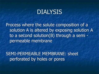
- 1. DIALYSIS Process where the solute composition of a solution A is altered by exposing solution A to a second solution(B) through a semi - permeable membrane SEMI-PERMEABLE MEMBRANE: sheet perforated by holes or pores
- 5. Mechanisms of solute transport Diffusion-random molecular motion concentration gradient molecular weight membrane resistance Ultrafiltration-water is pushed through membrane hydrostatic pressure osmotic pressure
- 9. Sand is swept away
- 16. PERMCATH
- 20. Portable hemodialysis Wearable hemodialysis Hemodialysis machine machine apparatus
- 23. Dialyzer
- 25. Dialyzers Low- efficiency-surface area:0.8-2.1 m²,small pores High- efficiency-bigger surface area, small or big pores High- flux-big pores (pass middle molecules)
- 26. Dialyzers
- 35. Dialysis Adequacy: Urea Kinetics
- 37. Dialysis Adequacy Indices of urea removal Kt/V Reflects urea removal Population studies suggest Kt/V should be >1.2 URR Also reflects urea removal Current goal is URR>65%
- 39. Kt/V K is the dialyzer blood urea clearance (Liters per hour) t is the dialysis session length [In a typical dialysis session:4 hs ] (hours) [Water content:~60% of dry body weight: If BW=70 kg then water V is the volume of distribution content is ( 0.6 X 70) 42 L ] of urea ( Liters )
- 47. NO BREAK
- 48. Indications for Dialysis-acute GI: nausea ; vomiting (morning) ; poor appetite Symptoms: Mental status alteration; fatigued ; weakness Signs : asterixis ; pericardial friction rub ; fluid overload Lab : Hyperkalemia, Severe Metabolic Acidosis
- 49. Case 1 22 years old male, cocaine abuser, with a known obstructive uropathy presented to hospital with severe sepsis secondary to pneumonia. LAB: Hgb-9.6 g/dl ;WBC-25800 ;PLT-603000 Na-132meq/l; K-3.1meq/l; Cl-107; Glucose-90 ; BUN-130 mg/dl Creatinine-4.7 ; Osm-330 ; pH-6.95; pO2-109; pCO2- 10 ;HCO3-2; L.A.-0.6 ( Severe metabolic Acidosis with elevated Anion Gap ) Chest X ray: Middle lobe+lingular pneumonia.U.S. Bilat. moderate hydronephrosis Follow-up: empiric Cephtriaxone and Vanco Blood culture were positive for Staph. aureus and E.Coli Admitted to ICU: despite IV Bicarbonate and 4 liters of crystalloids remained acidotic and oliguric therefore a regular standard dialysis was prescribed. 2 ½ hours after starting dialysis became rapidly unresponsive and intubation was done At completion of HD and over the subsequent 4 hs the neurologic status deteriorated
- 50. Case 1 Computerized Tomography (CT) head showing diffuse cerebral edema with effacement of basal cisterns and generalized loss of gray-white differentiation LAB after dialysis: pH-7.36; HCO3-19 ; Na-132 ; K-2 ; BUN-37 (URR- 71%) AUTOPSY: Diffuse cerebral edema
- 51. Dialysis Disequilibrium Syndrome Dialysis disequilibrium syndrome (DDS), a complication of haemodialysis, is characterized by neurological symptoms including headache, disorientation, nausea, seizures and coma. This syndrome is assumed to result from brain swelling occurring as a consequence of a rapid haemodialysis process. PATHOGENESIS: In uremic state there is a reduced expression of urea transporters and an increased expression of AQP in brain cells – consequently Acute urea removal occurs more slowly across BBB than from plasma generating a “reverse osmotic gradient” promoting water movement into brain. AVOID DDS initiating dialysis “gently”: less efficient dialyzer, reduce session length ,reduce blood flow rate , run blood and dialysate in the same direction ( less diffusion) , add osmols to dialysate
- 52. Dialysis Disequilibrium Syndrome Dialysis disequilibrium syndrome (DDS), a complication of haemodialysis, is characterized by neurological symptoms including headache, disorientation, nausea, seizures and coma. This syndrome is assumed to result from brain swelling occurring as a consequence of a rapid haemodialysis process. PATHOGENESIS: In uremic state there is a reduced expression of urea transporters and an increased expression of AQP in brain cells – consequently Acute urea removal occurs more slowly across BBB than from plasma generating a “reverse osmotic gradient” promoting water movement into brain. AVOID DDS initiating dialysis “gently”: less efficient dialyzer, reduce session length ,reduce blood flow rate , run blood and dialysate in the same direction ( less diffusion) , add osmols to dialysate
- 54. הגיע הזמן לנוח... אבל קודם לחדר … דיאליזה
- 73. Principles of peritoneal dialysis
- 74. Principles of peritoneal dialysis Continuous Ambulatory Peritoneal Dialysis-CAPD
- 75. Principles of peritoneal dialysis Continuous Cycling Peritoneal Dialysis or Automated Peritoneal Dialysis ( APD ) Cycler Nightly Intermittent Peritoneal Dialysis
- 87. PERITONEAL FIBROSIS : SIMPLE SCLEROSIS AND SCLEROSING PERITONITIS Simple Sclerosis Sclerosing Peritonitis Frequency very common very rare poor biocompatibility of peritoneal dialysis unknown, only risk factors due to osmotic agents, peritoneal dialysis-dependent risk hyperosmolarity, low pH, factors: buffer duration of dialysis poor biocompatibility acetate buffer Etiology disinfectants catheter in-line bacterial filters particles of plastics plasticizers peritonitis peritoneal dialysis-independent risk factors: beta-blockers tumors genetic predisposition Reproducibility yes with dialysis no with dialysis in animal models no without dialysis yes without dialysis Clinical severe absent manifestations high mortality
- 88. Simple sclerosis Sclerosing Peritonitis of macrophagic origin Giant cells sclerotic tissue limited to visceral and Fibroblasts and mesoblasts occur throughout the parietal peritoneum sclerotic tissue, but are often more frequent in deeper layers. In sclerosing peritonitis unlike simple the thickness of sclerotic tissue in sclerosis, the muscle layer is compressed. The simple sclerosis does not exceed thickness of the sclerotic tissue is not uniform in a given patient but normally reaches very high values 40-50 µm between 1,000 and 4,000 µm
- 89. In sclerosing peritonitis, unlike simple sclerosis, a dramatic progression of the sclerosis occurs. This is combined with aspects not found in simple sclerosis, such as inflammatory infiltrates, calcifications and typical vascular alterations. The peritoneal surface is reduced to a rough thickened membrane similar to the sole of a shoe .In extreme cases of sclerosing encapsulating peritonitis, the sclerotic process completely fixes groups of intestinal loops, almost completely preventing their movement. Often the sclerosis is not homogeneous, but one area of the abdomen may be more affected than others, forming a mass. This situation has been described with the term "abdominal cocoon“. The cocoon may be perfectly palpable, like a tumor; the sclerotic tissue of the cocoon usually contains loops of the small intestine and sacs of ascites, and often calcifications.
Notas do Editor
- \n
- \n
- \n
- \n
- \n
- \n
- \n
- \n
- \n
- \n
- \n
- \n
- \n
- \n
- \n
- -catheter in Int jug vein or in int femoral vein\n-can be in the body for 1 yr \n-do this when have no time to prepare patient\n-\n
- \n
- -subclavian vein can promote stenosis of subclavian vein and all the veins of the arm collapse\n-\n
- -dialysis – wash machine\n-blood passes through cylinder- have diffusion and convection\n-need to put in dialysis on other side of machine – pure water \n
- \n
- -dialyzer w/ 1000s of microtubules inside \n-blood comes into each capillary of the dialyzer, the diaslysis is coming btwn the capilaries and exits the other side \n
- \n
- -blood and dialysis go in opp directions \n-can’t be in same direction b/c dialysis will be saturated - max diffusion \n
- -waste products come from blood to dialysis \n-at other end substances from dialysis go to the blood \n
- -depends on area of pore size \n-urea- small molecule \n-uremic – misnomer, symptoms not due to urea, uremia can be treatment for some patients eg. malaria \n-\n
- \n
- \n
- -used to have cellulose membranes\n-now use poly… membrane \n
- \n
- -give some heparin – so blood doesn’t coag as exit body to dialyzer \n-comes out of body – called arterial blood (even though from vein)\n-blood entering body called venous blood \n-if apply venous clamp – not complete block\n-P will build up in the dialyzer, as blood pump continues to go\n-P is important in chronic dialysis patients (or ppl in acute renal failure – everything stays in their body, can can easily have excess of water in body – pulm edema) \n-put venous clamp, to make more convection to remove excess water\n
- \n
- \n
- \n
- \n
- \n
- \n
- \n
- \n
- \n
- \n
- \n
- \n
- \n
- \n
- \n
- \n
- \n
- \n
- \n
- \n
- \n
- \n
- \n
- \n
- \n
- \n
- \n
- \n
- \n
- \n
- \n
- \n
- \n
- \n
- \n
- \n
- \n
- \n
- \n
- \n
- \n
- \n
- \n
- \n
- \n
- \n
- \n
- \n
- \n
- \n
- \n
- \n
- \n
- \n
- \n
- \n
- \n
- \n
- \n
- \n
- \n