SlideShare uma empresa Scribd logo
1 de 15
Baixar para ler offline
REVIEWS
Excess body weight and obesity—the link
with gastrointestinal and hepatobiliary cancer
Prashant Kant and Mark A. Hull
Abstract | Excess body weight (EBW) is an independent risk factor for many human malignancies, including
cancers throughout the gastrointestinal and hepatobiliary tract from the esophagus to the colorectum. The
relative risk of gastrointestinal cancer in obese individuals is approximately 1.5–2.0 times that for normal
weight individuals, with organ-specific and gender-specific differences for specific cancers. The association
between EBW and risk of premalignant stages of gastrointestinal carcinogenesis, such as colorectal
adenoma and Barrett esophagus, is similar, implying a role for EBW during the early stages of carcinogenesis
that could be relevant to preventative strategies. EBW also impacts negatively on gastrointestinal cancer
outcomes. The mechanistic basis of the association between EBW and carcinogenesis remains incompletely
understood. Postulated mechanisms include increased insulin and insulin-like growth factor signaling and
chronic inflammation (both linked to the metabolic syndrome), as well as signaling via adipokines, such as
leptin. The role of obesity-related changes in the intestinal microbiome in gastrointestinal carcinogenesis
deserves further attention. Whether weight loss leads to reduced future gastrointestinal and liver cancer risk
has yet to be fully explored. There is some support for the idea that weight loss negatively regulates colorectal
carcinogenesis. In addition, data suggest a reduction in risk of several cancers in the first 10 years after
bariatric surgery.
Kant, P & Hull, M. A. Nat. Rev. Gastroenterol. Hepatol. 8, 224–238 (2011); published online 8 March 2011; doi:10.1038/nrgastro.2011.23
.

Introduction

Leeds Institute of
Molecular Medicine,
University of Leeds,
Wellcome Trust Brenner
Building, St James’s
University Hospital,
Beckett Street, Leeds
LS9 7TF, UK (P. Kant,
M. A. Hull).

A large body of epidemiological evidence links excess
body weight (EBW) with an increased risk of many
cancers, including those of the breast, endometrium,
kidney and gastrointestinal tract.1,2 Given the high prevalence of EBW and obesity (defined as a BMI ≥30 kg/m2)
in most populations throughout the world, amounting
to an estimated 1.6 billion adults globally,3 EBW is now
considered to be an important, potentially preventable
cause of cancer.4,5 It has been estimated that 3.2% of all
incident cancers in men and 8.6% of all incident cancers
in women in Europe are attributable to EBW.6
Several gastrointestinal and hepatobiliary cancers are
represented in the list of recognized obesity-associated
cancers, including colon, rectal, esophageal (adenocarcinoma), gastric, gallbladder, liver (hepatocellular)
and pancreatic cancer (Table 1). Other gastrointestinal
cancers, including small intestinal cancer, may also have
an association with EBW.7 Importantly, some gastro­
intestinal cancers develop via a clinically recognized
premalignant precursor phase, such as the colorectal
adenoma (also known as a polyp) and Barrett esophagus.
Evidence is now emerging that EBW is also associated
with increased risk of these premalignant precursor stages
(Table 1).8–11 These findings provide an excellent stimulus to intervene with individual and population-level
antiobesity measures prior to malignant change.

Correspondence to:
M. A. Hull
m.a.hull@leeds.ac.uk

Competing interests
The authors declare no competing interests.

224  |  APRIL 2011  |  VOLUME 8


By contrast, current understanding of the effect that
the reduction of EBW has on future gastrointestinal
and hepatobiliary cancer risk is rudimentary despite
the potential importance of this knowledge for cancer
prevention in overweight individuals. Evidence also suggests that EBW can alter gastrointestinal cancer outcomes
after diagnosis.
This Review discusses the association between EBW
and cancer risk in the gastrointestinal and hepatobiliary
tract, with particular emphasis on the premalignant
phases of gastrointestinal and liver carcinogenesis that
are most relevant to cancer prevention. Current understanding of the pathophysiological mechanisms linking
EBW and carcinogenesis in the gastrointestinal and
hepatobiliary tract is explored, and existing evidence
that body weight reduction impacts on gastro­ ntestinal
i
carcino­ enesis is summarized. Finally, the current litg
erature describing the association between EBW and
gastrointestinal cancer outcomes is discussed.

Carcinogenesis associated with EBW
Colorectal carcinogenesis
Colorectal cancer
Considerable observational evidence has established that
EBW is an independent risk factor for colorectal cancer
(CRC). In Europe, it is estimated that EBW accounts
for 16% (12,700 incident cases) and 2.6% (3,200 incident cases) of CRC cases in men and women, respectively.6 Similar estimates in the USA suggest that 14.2%
www.nature.com/nrgastro

© 2011 Macmillan Publishers Limited. All rights reserved
REVIEWS
of all incident CRC cases are attributable to a BMI of
over 25 kg/m2.12 A meta-analysis by Moghaddam et al.1
reported an overall age-standardized relative risk (RR) of
CRC of 1.4 (95% CI 1.2–1.5) for obese individuals versus
individuals with normal BMI.1 There are gender-specific
and site-specific variations in CRC risk associated with
EBW. A meta-analysis of 29 cohort studies reported that a
step-wise increase in BMI of 5 kg/m2 is associated with
a RR of colon cancer of 1.2 (1.2–1.3) for men and 1.09
(1.04–1.14) for women.2 The RR of rectal cancer was
attenuated and was only increased significantly in obese
men (RR 1.09 [1.06–1.12]), whilst no association between
BMI and rectal cancer was shown in women.
Whereas BMI is a generalized measure of adiposity,
other more specific anthropometric measures of body fat
distribution have led to a more refined appreciation of the
risk associated with CRC. There is a growing apprecia­
tion that the pathophysiological effects of visceral (or
abdominal) adipose tissue (VAT) are more potent than
those of subcutaneous adipose tissue (SAT) during
gastro­ ntestinal carcinogenesis (see Supplementary
i
Box 1 online). The waist circumference or waist–hip
ratio are generally accepted as simple measures of VAT.
There are currently no globally accepted cut-off values
for waist circum­ erence or waist–hip ratio, although
f
an NIH report concluded that men and women with
a waist circum­ erence of 102 cm and 88 cm, respecf
tively, should be considered at ‘high risk’ of developing
obesity-related disease.13
Meta-analyses have demonstrated an increased risk of
CRC associated with increasing waist circumference and
waist–hip ratio.1,14,15 One pooled meta-analysis observed
a RR of all CRC amongst men and women of 1.5 (1.4–
1.7) for the highest versus the lowest categories of waist
circumference.1 When subdivided by gender and site, it
is clear that this relationship is similar to that with BMI.
Each 10 cm increase in waist circumference results in a
33% and 16% increased risk of colon cancer in males
and females, respectively.14 Corresponding estimates for

Key points
■■ Excess body weight (EBW) is a potentially preventable cause of several
gastrointestinal and hepatobiliary malignancies, including colorectal,
esophageal, liver and pancreatic cancer
■■ A positive association between EBW and premalignant conditions, such as
colorectal adenoma and Barrett esophagus, implies that EBW promotes the
early stages of carcinogenesis (which could be relevant for prevention)
■■ The mechanisms linking excess adiposity (particularly visceral or abdominal
fat) and gastrointestinal carcinogenesis are unclear but may include insulin and
insulin-like growth factor signaling, chronic inflammation, adipokine signaling
and changes to the intestinal microbiome
■■ Whether weight loss reduces future gastrointestinal and liver cancer risk has
not been addressed fully and requires further study
■■ EBW is also associated with worse outcomes from gastrointestinal cancer
following diagnosis

each 0.1 unit increase in waist–hip ratio were 43% and
20%.14 For rectal cancer, the association was only apparent in males (12% increased risk per 0.1 waist–hip ratio
unit increase).14 Evidence that VAT has an important
pathophysiological role comes from a study in which a
twofold increased risk of CRC in men and women with
a waist circumference of 99.1 cm, compared with a waist
circumference of 83.8 cm, persisted despite adjustment
for BMI.16
Colorectal adenoma
The colorectal adenoma is the recognized premalignant
precursor of the majority of CRCs. A positive association between EBW and risk of colorectal adenoma would
imply that EBW can drive colorectal tumor initiation and
colorectal adenoma promotion separate from malignant
progression and CRC growth. Several studies have thus
investigated the association between body weight and
risk of colorectal adenoma (Table 2).8,10,17–37 It is apparent that there is considerable heterogeneity in study
design and analysis, which makes direct comparison
of indivi­ ual studies difficult. Unlike CRC, increased
d

Table 1 | Excess body weight and cancer of the gastrointestinal and hepatobiliary tract
Site

Cancer

Recognized premalignant condition

Increased risk associated
with excess body weight?

Oral cavity/pharynx

Squamous cell carcinoma

Leukoplakia

No data

Esophagus

Adenocarcinoma

Barrett esophagus

Yes

Squamous cell carcinoma

No

No

Stomach

Adenocarcinoma

Intestinal metaplasia and gastric atrophy

Yes

Biliary tree

Gallbladder adenocarcinoma

Adenoma; chronic cholecystitis

Yes

Intrahepatic cholangiocarcinoma

No

Yes

Liver

Hepatocellular carcinoma

Nonalcoholic steatohepatitis (nonalcoholic
fatty liver disease)

Yes

Pancreas

Adenocarcinoma

No

Yes

Small intestine

Adenocarcinoma

No

Yes

Lymphoma

No

No data

Colon and rectum

Adenocarcinoma

Adenoma

Yes*

Anus

Squamous cell carcinoma

No

No data

*Gender-specific differences are apparent (risk greater for males than females).

NATURE REVIEWS | GASTROENTEROLOGY  HEPATOLOGY 	
© 2011 Macmillan Publishers Limited. All rights reserved

VOLUME 8  |  APRIL 2011  |  225
REVIEWS
Table 2 | Summary of studies investigating the risk of colorectal adenoma associated with EBW
Study

Country

Patient
gender

Number of
adenoma cases*

OR and HR‡§

Adjusted analysis

Neugut et al. (1991)20

USA

M + F

301 (133)

1.4 (0.8–2.5) M
2.1 (1.1–4.0) F||

Age

Bayerdörffer et al. (1993)37

Germany

M + F

244 (78)

3.2 (1.2–9.0)||¶

Age

Giovannucci et al. (1995)22

USA

M

328 (131)

1.5 (0.9–2.5)

Age, other#

Giovannucci et al. (1996)

USA

F

303 (125)

1.5 (1.0–2.2)

Age, other#

Kono et al. (1996)

Japan

M + F

201 (72)

1.1 (0.6–2.2)

Other#

USA

M + F

478 (139)

1.7 (1.0–2.8)||

Age, gender, other#

Norway

M + F

28

0.6 (0.1–6.1)

Boutron-Renault et al. (2001)

France

M + F

363 (208)

1.7 (1.0–3.1) small
2.1 (1.2–3.5) advanced||

Age, gender, other#

Terry et al. (2002)34

USA

M + F

560 (119)

2.0 (1.4–2.9)||

Age, gender, other#

Betes et al. (2003)

Spain

M + F

259 (259)

2.1 (1.3–3.3)

Age, gender, other#

Lieberman et al. (2005)25

USA

M + F

329 (329)

1.1 (0.9–1.2)**

Age, other#

Wallace et al. (2005)

USA

M + F

308 (69)

1.1 (0.8–1.4)

Age, gender, other#

Larsen et al. (2006)19

Norway

M + F

551 (108)

1.3 (1.0–1.8) small||
1.6 (0.8–2.6) advanced§§

Age, gender, other#

Wolf et al. (2007)35

USA

F

187 (63)

0.8 (0.5–1.3)

Age, other#

Anderson et al. (2007)26

USA

M + F

2,439

2.4 (1.5–4.7) MF||
1.3 (0.5–3.3) M
4.3 (2.0–9.1) F||

Age, gender, other#

Sedjo et al. (2007)18

USA

M + F

136 (38)

1.3 (0.5–3.1) M
2.2(1.1–4.1) F||

Age, gender, other#

Yamaji et al. (2008)24

Japan

M + F

1,650 (502)

1.3 (1.1–1.6)||

Age, gender

Wise et al. (2008)

USA

F

1,189

1.4 (1.2–1.7)||

Other#

Omata et al. (2009)27

Japan

M + F

194

1.3 (0.8–2.1)||||

Sato et al. (2009)

Japan

M + F

1,590

1.3 (1.2–1.5) M
1.1 (0.9–1.4) F

Age, gender

Kim  Lee (2009)28

South Korea

M + F

165

1.8 (1.0–3.1) M||
1.4 (0.6–3.4) F

Age, other#

Siddiqui et al. (2009)8

USA

M + F

2,903 (2,903)

1.01 (1.0–1.02)||‡‡

Age, other#

Morois et al. (2010)21

France

F

1,408 (599)

1.6 (1.2–2.0)||

Other#

23

31

Bird et al. (1998)30
Almendingen et al. (2001)

36
32

29

33

17

10

||

Other#
||

||

No details
||

*The number of advanced colorectal adenoma cases (at least one of the following features: size ≥10 mm diameter, contains villous morphology, high-grade
dysplasia or invasive cancer, multiple [≥3] colorectal adenomas) is given in brackets. ‡Unless indicated, the OR and/or HR is for the combined analysis of both
males and females and for both small and advanced colorectal adenomas. §Unless indicated, the OR and/or HR is for the highest versus lowest BMI category.
||
Statistically significant. ¶OR for advanced colorectal adenomas only from a subgroup analysis (males in the 3 rd and 4th quintile of age). No association observed
for females, or small colorectal adenomas in either males or females. #Other adjustment includes at least one from the following: diet, exercise, smoking,
alcohol, family history. **OR and/or HR per 5‑unit increase in BMI over 30 kg/m2. ‡‡OR and/or HR per 1‑unit increase in BMI over 30 kg/m2. §§Included eight
cases of CRC in the advanced adenoma subgroup. ||||Included 19 cases of CRC. Abbreviations: CRC, colorectal cancer; EBW, excess body weight; F, female; HR,
hazard ratio; M, male; OR, odds ratio.

EBW-associated risk in males is not apparent for colo­
rectal adenoma. Neugut et al.20 were the first to describe
the increased prevalence of colorectal adenoma in obese
females (odds ratio [OR] 2.1 [1.1–4.0] for the highest
versus lowest quartile of BMI) but not males. Other
cohort studies, 18,26 including the largest prospective
study to date of 2,439 adenoma cases,26 have also shown
positive risk associations for females, but not for males.
Morois and colleagues reported a linear association
between colorectal adenoma risk and increasing BMI
in a large-scale, prospective, cross-sectional European
study of 17,391 women, 21 supporting the findings of
previous studies.17,23 The converse association has also
been reported in a study in which obese males, but
not obese females, had an increased risk of developing colo­ ectal adenoma.10,28 Reasons for the disparity in
r
226  |  APRIL 2011  |  VOLUME 8


gender association are not clear but may relate to hetero­
geneity of adjustments for other factors such as age and
NSAID use.
Increasing evidence suggests that obesity may also
increase colorectal adenoma growth. Screening studies
have demonstrated that obese individuals have a higher
risk estimate for ‘advanced’ (usually defined as size 1 cm
in diameter and/or with villous architecture and/or highgrade dysplasia) compared with ‘nonadvanced’ colorectal
adenomas (Table 2). Among 2,260 prospectively screened
patients, Betes et al.29 demonstrated that obese indivi­
duals have an OR of 2.1 (1.3–3.3) for advanced colorectal
adenomas compared with individuals who have a normal
BMI. Conversely, in a Norwegian study of 3,998 prospectively screened patients, the presence of small (1 cm
diameter), but not advanced, colorectal adenomas was
www.nature.com/nrgastro

© 2011 Macmillan Publishers Limited. All rights reserved
REVIEWS
predicted by obesity.19 There is also a linear relationship
between EBW and advanced colorectal adenoma risk
in one large retrospective study, in which over 2,900 US
male veterans had a 1% increased risk of advanced colo­
rectal adenoma for every 1-unit increase in BMI above
30 kg/m2 after adjustment for age, NSAID usage and
family history of CRC.8
The stronger relationship of VAT, as opposed to SAT,
with CRC risk seems to be mirrored by the relationship
between VAT and risk of colorectal adenoma. In a case–
control study of 530 Koreans, increased waist circumference or waist–hip ratio was significantly associ­ ted
a
with risk of colorectal adenoma independently of BMI.28
Another larger Korean case–control study of 2,244
patients undergoing screening demonstrated a threefold
(OR 3.1 [2.2–4.4]) increased risk of colorectal adenoma
between the highest and lowest quintiles of VAT as
measured by CT.38
The presence of the metabolic syndrome carries an RR
of 1.5 [1.2–1.9] for the prevalence of all types of colo­rectal
adenomas and a twofold higher risk of advanced colo­
rectal adenomas.39 The Korean studies have also reported
that colorectal adenoma multiplicity, as well as individual
colorectal adenoma risk, is significantly associ­ ted with
a
abdominal obesity.38,40
Obesity may also increase adenoma recurrence. Data
from a pooled analysis of seven studies demonstrated
that BMI is associated with an increased risk of colo­
rectal adenoma recurrence in men (but not women)
who have a previous history of colorectal adenoma,
which included both advanced and nonadvanced
colorectal adenomas.41
There may also be a role for EBW at the earliest stages
of colorectal carcinogenesis (prior to tumor initiation and colorectal adenoma growth) as emerging data
suggest that putative mucosal biomarkers of future CRC
risk, including increased epithelial cell mitosis frequency,
are increased in obese and morbidly obese patients
compared with normal-weight controls.42
Other factors contributing to increased risk of CRC
In addition to the probable direct effects of EBW on the
biology of colorectal carcinogenesis, other factors may
impact on increased CRC risk in obese patients. Two
studies have reported that CRC screening uptake in both
fecal occult blood and endoscopy programs, is lower in
obese compared with normal weight individuals.43,44
Moreover, increased BMI has been linked to reduced
quality of bowel preparation, which could increase the
miss-rate for colorectal neoplasia prior to presentation
with CRC.45

Esophageal cancer and Barrett esophagus
Esophageal adenocarcinoma
Obesity is a risk factor for the development of esophageal adenocarcinoma but not squamous cell carcinoma. The incidence of esophageal adenocarcinoma in
Western populations has risen more than fivefold in the
past three decades46 and GERD has been strongly implicated as a primary causative factor.47 A concomitant

increase in obesity prevalence over a similar period
has prompted investigation of the link between EBW
and esophageal adenocarcinoma in population-based
case–control and cohort studies. In a meta-analysis
of six studies, the pooled adjusted OR for esophageal
adenocarcinoma for overweight individuals was 1.5
(1.2–2.0) and for obese individuals was 2.9 (1.9–4.2).48
Unlike CRC, there does not seem to be any gender
differ­ nce in the relationship between EBW and risk of
e
esophageal adenocarcinoma.2
EBW is a known moderate risk factor (RR 1.5–2.0) for
developing GERD symptoms,49 and abdominal obesity
(as measured by waist circumference) can predict GERD
severity independently of BMI.50 One hypothesis, therefore, is that EBW increases risk of esophageal adeno­
carcinoma solely via promotion of GERD. However,
EBW may drive esophageal carcinogenesis independently
of promotion of GERD. Whiteman et al. have demonstrated that in patients reporting at least one episode of
symptomatic GERD per week, obese individuals had a
considerably higher risk of esophageal adenocarcinoma
(OR 16.5 [8.9–30.6]) than people without obesity (OR
5.6 [2.8–11.3]).51
Barrett esophagus
Barrett esophagus (or columnar-lined esophagus) occurs
secondary to GERD and is considered to be the pre­
malignant precursor to esophageal adenocarcinoma.
Patients with Barrett esophagus have an individual lifetime risk of progression to esophageal adenocarcinoma
of approximately 0.5–1.0% per year.52
Given the relationship between EBW, GERD and
esophageal adenocarcinoma, one might expect a similar,
consistent relationship between EBW, GERD and Barrett
esophagus. However, the current literature on this topic
are conflicting. One meta-analysis, with selection criteria based on endoscopic appearance, concluded that
increased BMI was not associated with an increased risk
of Barrett esophagus compared with controls matched
for the degree of GERD. However, increased BMI was
signifi­ antly associated with Barrett esophagus in comc
parison with population-based controls, which implies
a link via indirect effects of GERD, rather than EBW
itself.11 A subsequent unadjusted meta-analysis of studies
of histologically confirmed Barrett esophagus concluded that there was an OR of 1.5 (1.2–1.8) for Barrett
esophagus among individuals with a BMI of 25 kg/m2
compared with normal-weight controls.53
Another study attempted to investigate the effect of
EBW on risk of Barrett esophagus in female nurses by
adjusting for reflux symptoms and the use of antireflux
medication.9 In this study, obesity, but not overweight, was
associated with Barrett esophagus, even after adjusting
for GERD.
Consistent with the data on colorectal neoplasia, two
case–control studies have demonstrated an increased risk
of Barrett esophagus in individuals who have increased
abdominal obesity, as measured by waist–hip ratio54 or
waist circumference,55 independently of BMI. However,
a separate study failed to detect an association between

NATURE REVIEWS | GASTROENTEROLOGY  HEPATOLOGY 	
© 2011 Macmillan Publishers Limited. All rights reserved

VOLUME 8  |  APRIL 2011  |  227
REVIEWS
Barrett esophagus and either waist–hip ratio or waist
circum­ erence in women.9
f
Only one observational study has investigated whether
EBW influences malignant progression in Barrett esopha­
gus; no association was observed between BMI and
progression to low-grade dysplasia.56

Pancreatic cancer
Data from a large pooled analysis of 2,170 cases have
demonstrated positive associations between several
anthropometric measures and pancreatic cancer risk following adjustment for age, smoking and diabetes.57 BMI
was a significant risk predictor in females, with those
with the highest quartile of BMI having an OR of 1.5
(1.1–2.1) compared with those in the lowest quartile. In
the fully adjusted model, the association was not significant in men (1.3 [0.8–1.8]). Abdominal adiposity was also
a strong predictor of pancreatic cancer risk in women
only, with an adjusted OR of 1.9 (1.3–2.7) for the highest
versus lowest quintile of waist–hip ratio.57 Furthermore,
a meta-analysis of prospective studies that included more
than 8,000 cases of pancreatic cancer demonstrated a
step-wise increase in pancreatic cancer risk of 1.2 (1.1–
1.3) in men and 1.1 (1.0–1.2) in women with each 5 kg/m2
increase in BMI.58 There is also evidence that onset of
an overweight or obese state at a young age (20–49 years
old) is associated with diagnosis of pancreatic cancer at
an earlier age compared with normal-weight controls.59
Type 2 diabetes mellitus (T2DM), which is often linked
to obesity and the metabolic syndrome, is associated with
an RR of 1.8 (1.7–2.0) for pancreatic cancer.60 However,
reverse causation bias is likely in this study as the risk
was greater for those patients with a short history of
diabetes (less than 5 years) compared with individuals
who had a longer duration of diabetes,60 particularly as
insulin sensi­tivity and diabetic control improve following
pancreatic resection.61
Unlike CRC and esophageal adenocarcinoma, no
clinically relevant premalignant precursor lesion is
recog­ ized for pancreatic cancer. Chronic pancreatitis
n
is associated with an increased risk of pancreatic cancer
with a cumulative risk of 4.0% at 20 years.62 However,
there is currently no evidence linking EBW and
chronic pancreatitis.
Gastric carcinogenesis
Stomach cancer
The link between EBW and the incidence of stomach
cancer is unclear, with conflicting data from two independent meta-analyses. A meta-analysis of 10 cohort
studies reported a summary hazard ratio (HR) of 1.2
(1.1–1.4) for overweight and obese individuals compared
with those with a normal BMI.63 This association held for
males only, with significant associations only apparent
for non-Asian populations. The null effect observed
in Asian populations may partly be explained by more
widespread surveillance and early detection of gastric
neoplasia in these populations.64 However, the summary
HR estimate in this analysis was strongly driven by two
studies with evidence of considerable heterogeneity
228  |  APRIL 2011  |  VOLUME 8


(I2 = 81%).63 One of these two studies was reported to
have a risk estimate of 1.7, although the paper itself
reported that no association was found (HR 1.1 [0.7–
1.8]), and the other study included fatal gastric cancer
cases only. By contrast, a meta-analysis of four cohort
studies by Renehan et al.2 did not find any association
between increasing BMI and gastric cancer risk.
There are biological differences between cardia and
noncardia stomach cancer, which may be reflected in the
association with EBW.65 Only cardia cancer risk seems to
be increased in obese individuals.63 Observational studies
of gastric cancer risk are also probably hampered by confounding owing to the difficulty in determining the true
nature of tumors at the gastro-esophageal junction as
either esophageal or gastric in origin.
Chronic gastritis and intestinal metaplasia
Gastric carcinogenesis is a multistage process, which
involves distinct histological stages from chronic gastritis through to gastric atrophy and intestinal metaplasia.
Multiple risk factors for gastric carcinogenesis are recognized, including Helicobacter pylori infection, smoking,
alcohol consumption and dietary factors.66 However, the
relationship between body size and premalignant stages
of gastric carcinogenesis has yet to be explored.

Gallbladder carcinogenesis
Gallbladder cancer
Gallbladder cancer is rare in the Western world but is
more common elsewhere, particularly in South American
countries. It is known to arise on a background of gallstone disease or chronic cholecystitis,67 and it has been
estimated that up to 14% of incident cases of gallbladder
cancer in Europe may be attributable to obesity.6 In a
large-scale, population-based study in Norway including 1,715 cases of gallbladder cancer, obesity was associated with an RR of gallbladder cancer of 1.4 (1.0–1.9) in
men and 1.9 (1.6–2.2) in women compared with indivi­
duals with normal BMI.68 A meta-analysis of four crosssectional studies concluded that a moderate association
between risk of gallbladder cancer and EBW existed in
women only, with an HR of 1.6 (1.0–2.5), as opposed to
men (1.1 [1.0–1.2]), for each 5 kg/m2 increase in BMI.2 A
subsequent large Chinese case–control study in a mixedsex population has shown that increased waist–hip ratio
was a strong predictor of risk of gallbladder cancer
compared with cancer-free controls.69
EBW is a risk factor for gallstones, with an approximate sixfold to sevenfold increase in gallstone prevalence in obese individuals compared with individuals
who have a normal BMI.70 Gallstones are believed to be
an age-independent risk factor for gallbladder cancer on
the basis of a number of epidemiological studies concerning the presence,71 size72 and duration of stones.73
However, the strength of any causal relationship remains
controversial owing to the wide geographical variation
in incidence of gallbladder cancer despite equivalent
gallstone prevalence rates.74 This discrepancy may be
explained by variable rates of cholecystectomy in certain
areas.75 EBW might also increase the risk of gallbladder
www.nature.com/nrgastro

© 2011 Macmillan Publishers Limited. All rights reserved
REVIEWS
cancer independently of the presence of gallstones. In
the Chinese case–control study, the association between
EBW and gallbladder cancer incidence was maintained
even after adjustment for the presence of gallstones.69
Benign conditions linked to gallbladder cancer
The gallbladder adenoma (or polyp) is recognized to
have malignant potential76 and might be associated with
EBW. In a large series of 264 gallbladder polyps, presence
of the metabolic syndrome and increased waist circumference were independently associated with gallbladder
polyp incidence.77 Other studies have suggested that
BMI might also be a risk factor for gallbladder polyposis,
particularly in males.78
There is a paucity of data linking EBW with other
likely histological precursors of gallbladder cancer, such
as chronic cholecystitis and gastric metaplasia, although
these entities are common in obese patients. In a study
of 199 morbidly obese Taiwanese patients undergoing
cholecystectomy at the same time as bariatric surgery, all
had chronic inflammatory cell infiltration and 27% had
gastric metaplasia, although only 10% had gallstones.79

Cholangiocarcinoma
EBW may also increase the risk of cholangiocarcinoma,
which is a relatively rare cancer that has been increasing
in incidence over recent decades.80 One large UK‑based
case–control study of 372 cholangiocarcinoma cases estimated an OR of 1.5 (1.0–2.2) for obese patients versus
those with a normal BMI.81 Another retrospective case–
control study of patients with cholangio­ arcinoma
c
identified from the Surveillance, Epidemiology and
End Results (SEER) database in the USA, reported that
obesity was a significant risk factor for intrahepatic,
but not extrahepatic, cholangiocarcinoma.82 However,
there may be regional differences as a similar study in
Denmark failed to show any significant increased risk of
cholangiocarcinoma with increased BMI.83
Hepatic carcinogenesis
There is strong evidence that obesity is associated with an
increased risk of hepatocellular cancer (HCC). Changes
in HCC incidence and obesity prevalence have both followed a similar upward pattern, suggesting that obesity
may account for a significant proportion of HCC cases
over and above better recognized risk factors, such as
viral hepatitis and alcohol.84 Data from the UK Million
Women Study showed that the risk of developing cirrhosis (a condition known to predispose to HCC) was linked
to increasing BMI in a dose–response manner.85
Obesity is associated with nonalcoholic fatty liver
disease (NAFLD), which is recognized as the hepatic
manifestation of the metabolic syndrome. NAFLD
encompasses a spectrum of histological states from
isolated steatosis (fatty liver) and nonalcoholic steato­
hepatitis (NASH), through to fibrosis and cirrhosis. Population-based estimates for the prevalence of
NAFLD range from 13% in nations where the prevalence of obesity is low, such as Japan,86 to approximately
30% in the USA.87 This figure rises to more than 90%

in patients who are morbidly obese.88 The epidemiology
of the link between EBW, NAFLD and HCC has been
the subject of a recent systematic review and metaanalysis.89,90 In addition, current understanding of the
mechanistic link between obesity, NAFLD and hepatic
carcino­ enesis has been reviewed in detail elsewhere.84
g
Therefore, only a brief summary of a large body of data is
provided below.
Hepatocellular cancer
In a systematic review, seven out of 10 cohort studies
reported significantly positive associations between BMI
and HCC risk,89 with the risk being greater for men than
women. In a separate meta-analysis, which included
eight of the above cohort studies, plus two other cohort
studies, obesity was associated with an RR of HCC of 1.9
(1.5–2.4) compared with a normal BMI; a similar gender
difference was apparent (male RR 2.4 [1.8–3.2]; female
RR 1.7 [1.4–2.0]).90 On the basis of prevalence data from
the USA, it is estimated that 28% and 27% of cases of
HCC in men and women, respectively, are attributable
to EBW.90
EBW seems to be an independent risk factor for HCC
separate from other causes of HCC. For example, in a
study of 781,283 men from Korea, a region in which there
is a high incidence of chronic HBV infection, obesityassociated HCC risk was maintained after controlling for
HBV infection.91 In a separate study of 19,271 patients
with cirrhosis who were listed for liver transplantation,
obesity was found to be an independent predictor of
HCC in patients with alcohol-related cirrhosis (OR 3.2),
but not in patients with cirrhosis resulting from viral or
autoimmune hepatitis.92 Another study of patients with
HCV reported an HR of 1.9 (1.1–3.2) for overweight
and 3.1 (1.4–6.8) for obese patients compared with their
underweight counterparts.93
NASH
NASH is considered to be the predisposing state of
NAFLD for hepatic carcinogenesis. This is consistent
with the close link between carcinogenesis and local
inflammation throughout the gastrointestinal tract.
Mechanistically, genetic and diet-induced obesity has
been demonstrated to drive HCC development via
chronic inflammation in mice.94
However, the relative contribution of NASH, as
opposed to protumorigenic mechanisms linked to cirrhosis, during hepatic carcinogenesis requires further
investigation. There are case reports of HCC in patients
with noncirrhotic NASH,95,96 which implies that EBW can
drive hepatic carcinogenesis via NASH independently of
cirrhotic change. However, this may be a rare event as
longitudinal studies have reported the incidence of HCC
in patients with noncirrhotic NASH to be as low as 1–2%,
whereas between 4–27% of cases of NASH transform to
HCC following the development of cirrhosis.84

How does EBW drive carcinogenesis?
How EBW promotes carcinogenesis in different
tissues, including several parts of the gastrointestinal

NATURE REVIEWS | GASTROENTEROLOGY  HEPATOLOGY 	
© 2011 Macmillan Publishers Limited. All rights reserved

VOLUME 8  |  APRIL 2011  |  229
REVIEWS
Normal weight
Innate and acquired
immune activation
in adipose tissue

1

Obesity
3
Gallstones

GERD

4
Chronic inflammation
?

2

Mucosal
inflammation

TNF
IL-6
MIF

Insulin resistance
Insulin
IGF-1

Adipokines

Modifiers:
Physical activity
Diet

Glucose,
FFAs
Leptin
Adiponectin
3

Gastrointestinal carcinogenesis
Genotoxicity
Mitogenic signaling to epithelial cells
Apoptosis resistance
Activated stromal–epithelial cell signaling
Angiogenesis
Host immunosurveillance

Figure 1 | Mechanisms linking excess adiposity in overweight and obese states with gastrointestinal and hepatobiliary
carcinogenesis. This schematic outlines the putative mechanisms that link adipose tissue in excess body weight with the
cellular and molecular mechanisms that are critical for carcinogenesis. (1) Excess adiposity is associated with innate and
acquired immune activation. (2) Some gastrointestinal cancers are linked to local mucosal inflammation and/or irritation
secondary to obesity-related pathologies, such as GERD or gallstones. (3) Alternatively, obesity may drive gastrointestinal
carcinogenesis directly via several interlinked mechanisms leading to exposure to protumorigenic cytokines, growth
factors and adipokines, as well as increased substrate availability to neoplastic cells. (4) Diet composition and physical
activity are closely linked to excess body weight and may modify the effect of excess adiposity on carcinogenesis at
several levels, including reduced adipose tissue mass, modulation of chronic inflammation separate from changes in
adipose tissue mass, and other direct effects on target cells, particularly genotoxicity by dietary carcinogens.
Abbreviations: FFA, free fatty acid; IGF‑1, insulin-like growth factor 1; IL‑6, interleukin 6; MIF, macrophage migration
inhibitory factor; TNF, tumor necrosis factor.

and hepatobiliary tract, is still not understood but it
is almost certainly a multifactorial process (Figure 1).
A simple classification divides potential mechanisms
into either indirect (for example EBW promotes
GERD and gallstone formation, which then drive local
inflammation-­driven carcinogenesis), or direct, in which
pro­ umorigenic cell signaling occurs as a direct conset
quence of excess adiposity. Proposed direct mechanisms
probably exhibit complex inter-relationships (Figure 1);
for example, chronic inflammation and insulin resistance co-exist as part of the metabolic syndrome, with
proinflammatory signaling causally related to target cell
insulin resistance.97
Increased estradiol production is believed to contribute to the increased risk of postmenopausal breast cancer
and endometrial cancer that is associated with EBW.98
Moreover, therapeutic manipulation of sex hormone
levels alters CRC risk in men and women.99 However,
whether differences in sex hormone metabolism between
males and females contribute to gender-specific differences in gastrointestinal and liver cancer risk associated
with EBW or whether gender differences are explained
by differences in the distribution and biology of VAT and
SAT between the sexes is not known. For the purpose
of this Review, we consider sex hormones most likely
to modify EBW-related gastrointestinal and liver cancer
risk rather than representing a direct mechanistic link
230  |  APRIL 2011  |  VOLUME 8


between excess adiposity and carcinogenesis in the
gastro­intestinal tract.
Great interest has been shown in the role of the intestinal microbiome in control of body weight.100 Therefore,
the luminal microflora can be considered to be an
etiological factor acting indirectly via modulation of
body weight.101 In addition, luminal microflora could
have a direct protumorigenic role during colorectal
carcinogenesis.
Putative molecular mechanisms linking obesity and
carcinogenesis have previously been reviewed extensively.98,102,103 Therefore, only recent data relevant to the
gastrointestinal tract is reviewed in detail here.

Insulin and insulin-like growth factor signaling
A well-established hypothesis linking EBW and gastrointestinal carcinogenesis, particularly CRC and pancreatic cancer, is that insulin resistance leads to direct
mitogenic and antiapoptotic signaling by insulin and
the insulin-like growth factor (IGF) axis (Figure 1).104
This hypothesis is supported by clinical observations,
clinical biomarker data and mechanistic cell studies,
but, as yet, few in vivo data have confirmed the rele­
vance of protumorigenic insulin and IGF signaling
in humans.
Increased CRC risk in patients with acromegaly,
as well as increased CRC and pancreatic cancer risk
www.nature.com/nrgastro

© 2011 Macmillan Publishers Limited. All rights reserved
REVIEWS
in patients with T2DM and the metabolic syndrome,
suggests that insulin and IGF signaling contributes
to EBW-related cancer risk.60,105,106 Hyperinsulinemia
in T2DM results in reduced hepatic synthesis of IGF
binding protein (IGFBP) 1 and 2, which in turn leads
to an increase in the circulating levels of bioactive,
or ‘free’ IGF‑1. Insulin, IGF‑1 and IGF‑2 activate the
insulin, IGF‑1 and IGF‑2 receptors, triggering an intracellular cascade, which stimulates the proliferation
of CRC cells107 and promotion of tumor growth and
metastasis.106,108,109 Downstream targets of these receptors include extracellular-signal-regulated kinase (ERK)
and phosphatidylinositol 3 kinase (PI3K) pathways that
mediate mitogenic cell signals.110 One critical downstream effector that has generated great interest is mammalian target of rapamycin (mTOR) and its downstream
effector, S6 kinase 1, which both have a critical role in
cellular nutrient sensing.111 Metformin, which is widely
used in the treatment of T2DM, has shown potential for
CRC chemoprevention in mouse models of colo­ ectal
r
carcinogenesis 112,113 through a mechanism believed
to include direct inhibition of mTOR by adenosine
monophosphate kinase (AMPK).
The relationships between components of the insulin–
IGF system and EBW are complex. In humans, although
not all associations with BMI are linear, plasma levels
of insulin and free IGF‑1 are higher in obesity, whilst
IGFBP‑1 and IGFBP‑2 levels are reduced.104 Total IGF‑1
levels peak at a BMI of approximately 28 kg/m2 before
reducing further in individuals with a BMI 28 kg/m2.
A meta-analysis of five cohort studies revealed that the
total IGF‑1 level predicts the RR of CRC.114 The associa­
tion between colorectal adenoma risk and IGF biomarker
levels is inconsistent. One study demonstrated a positive association between levels of serum insulin and the
IGF‑1:IGFBP‑3 ratio and colorectal adenoma risk,115
whilst another found a similar association only for
‘high-risk’ adenomas.116 Another study failed to detect
any association between risk of colorectal adenoma and
IGF biomarker levels.117
In addition to colorectal neoplasia, decreased IGFBP‑1
levels have been demonstrated to predict increased
risk of pancreatic cancer. 118 IGF‑1 is over­ xpressed
e
in pancreatic cancer cells compared with normal
pancreatic acini.119
Insulin and IGF signaling may also have a role in
esophageal carcinogenesis. Genetic polymorphisms
in the genes encoding the IGF‑1 receptor 120 and IGF‑2
receptor,121 which are known to increase expression of
these receptors in tumor tissue compared with matched
normal epithelium, are associ­ated with an increased risk
of developing esophageal adenocarcinoma. Furthermore,
IGF‑1 gene polymorphisms have also been shown to be
associated with risk of Barrett esophagus.122
Alternatively, it has been proposed that insulin resistance may lead to increased energy substrate (glucose,
triglycerides and free fatty acids) availability to mucosal
cells in the gastrointestinal tract, promoting tumor
growth directly and/or increasing oxidative stress and
DNA mutagenesis (Figure 1).

Chronic inflammation
EBW can be considered to be a state of chronic low-grade
inflammation, as demonstrated by increased systemic
levels of proinflammatory markers and cytokines, such
as C‑reactive protein (CRP) and interleukin (IL)‑6.123
Numerous proinflammatory cytokines, chemokines and
other acute phase proteins, such as plasminogen activator inhibitor 1 and fibrinogen, are released from adipose
tissue, which consists not only of adipocytes, but also
immune cells, including macrophages (Figure 1).124 In the
lean state, adipose tissue contains CD4+ T lympho­ ytes
c
with a T‑helper‑2 (TH2) profile and a proportionately
higher population of regulatory CD4+ T lymphocytes
(TREG) in relation to effector CD8 + T lympho­ ytes.125
c
Obesity is associated with innate immune activation
involving classic (M1) macrophage activation and a
switch towards a TH1 profile and dominant effector
CD8+ T lymphocytes,126 believed to be necessary for
persistent adipose tissue inflammation.125 The precise
mechanism(s) underlying immune activation associated
with excess adiposity are not understood although some
evidence supports the idea that Toll-like receptor (TLR)
activation by free fatty acids and/or lipopolysaccharide
(LPS) drives immune activation in adipose tissue.127
Many of the ‘classic’ cytokines and growth factors
that are synthesized and released by adipose tissue, such
as IL‑6, tumor necrosis factor (TNF) and macrophage
migration inhibitory factor (MIF), are believed to have
direct protumorigenic properties in the gastro­ ntestinal
i
and hepatobiliary tract (Figure 1). 128 However, it is
not known whether increased systemic levels of pro­
tumorigenic factors act directly on gastrointestinal
mucosal cells in an endocrine manner and/or whether
a persistent systemic inflammatory state can drive
induction of local tissue inflammation and subsequent
production of the same factors, which then act in a paracrine manner. In an interesting study, Poullis et al.129
demonstrated a positive relationship between levels of
fecal calprotectin (a marker of local intestinal inflamma­
tion) and obesity in healthy volunteers, independent of
the serum CRP level, which suggests that local intestinal inflammation may occur in individuals with EBW
independently of overt gastrointestinal disease. NASH
is the paradigm for the causal relationship between
local tissue inflammation and carcinogenesis associated
with EBW.84 A study has demonstrated that 10% dietinduced weight loss is associ­ ted with a 25–50% reduca
tion in pro­ nflammatory gene expression (including
i
TNF and IL‑8) in human colo­ ectal mucosa.130 Further
r
studies are required to confirm that weight loss reduces
colorectal mucosal inflammation separate from the possible effects of increased physical exercise131 and dietary
change per se.
A close causal relationship is believed to exist between
chronic inflammation and insulin resistance in the metabolic syndrome. One mechanism that has been proposed
is nuclear factor κB (NFκB)-dependent inhibition of
signaling downstream of the insulin receptor.132
There is some literature on the association between
biomarkers of inflammation (particularly CRP) and

NATURE REVIEWS | GASTROENTEROLOGY  HEPATOLOGY 	
© 2011 Macmillan Publishers Limited. All rights reserved

VOLUME 8  |  APRIL 2011  |  231
REVIEWS
gastrointestinal cancer risk. In a meta-analysis of eight
case–control studies, serum CRP level was demonstrated
to be independently associated with an increased risk
of CRC (RR 1.1 [1.0–1.3]). This relationship was only
significant for men and for colon cancer, rather than
for rectal cancer.133 However, one case–control study 134
and another cross-sectional study 135 have demonstrated
no relationship between CRP levels and risk of colo­
rectal adenoma, although in the latter study a positive
associa­ ion with risk of colorectal adenoma was evident
t
for serum IL‑6 and TNF levels.135 To date, no prospective study has investigated the association between
increased levels of inflammatory biomarkers and future
gastro­ ntestinal and liver cancer risk.
i

Adipokines
Adipose tissue is an active endocrine organ that produces
a range of hormones, collectively termed adipokines.126
Leptin and adiponectin are the adipokines that have
been studied in the greatest depth (Figure 1). Similar
to the insulin–IGF signaling hypothesis, there are convincing clinical biomarker data linking adipokine levels
with increased gastrointestinal and liver cancer risk,
and there are some functional preclinical data from
animal models (see below), but there is a paucity of evidence confirming the precise role that these proteins
play at differ­ nt stages of human gastrointestinal and
e
hepatobiliary carcinogenesis.
Leptin
Serum leptin levels are increased in obesity.136 The leptin
receptor is overexpressed in CRC.137 Leptin has mitogenic
activity in intestinal epithelial cells,138 reduces apoptosis
in CRC cell lines139 and has also been shown to promote
growth of azoxymethane-induced colorectal tumors in
mice.140 Leptin is also implicated in esophageal carcinogenesis; mitogenic and antiapoptotic effects of leptin
have been reported in esophageal adenocarcinoma cells
in vitro.141
In a colonoscopy study in men, those with the highest
tertile of circulating leptin levels had a BMI-adjusted 3.3fold greater risk of colorectal adenoma than those with
lowest tertile leptin levels.142 The association between
serum leptin levels and CRC is less clear although two
prospective studies have shown positive associations
with CRC in men.143,144
In a prospective study, there was a threefold increased
risk of Barrett esophagus among men, but not women,
in the highest quartile of serum leptin levels compared
with the lowest, after adjustment for GERD symptoms.145
Leptin also has mitogenic activity in HCC.146
Adiponectin
Adiponectin can be considered to be an insulins
­ ensitizing hormone, levels of which negatively correlate with BMI. 147 Adiponectin inhibits CRC cell
growth, partly via modulation of AMPK activity and
negative regulation of mTOR signaling.148 Exogenous
administra­ ion of adiponectin reduces growth of
t
small intestinal polyps in the Apc Min/+ mouse model
232  |  APRIL 2011  |  VOLUME 8


of intestinal tumori­genesis,149 while an inverse associa­
tion between rectal dyplastic aberrant crypt focus
number in humans and plasma adiponectin has been
reported.150 A number of studies have reported plasma
adiponectin levels to be inversely associated with the
presence and multiplicity of colorectal adenomas151–153
and early CRC (defined as stage pT1 or below), following
adjustment for BMI.154
Two case–control studies have demonstrated a negative relationship of adiponectin and adiponectin receptors with gastric cancer, including noncardia gastric
cancers.155,156 Adiponectin may also have an inhibitory
effect in esophageal carcinogenesis. In Barrett esophagus, expression of adiponectin receptors (Adipo‑R1 and
Adipo‑R2) is downregulated. Adiponectin has been
demon­ trated to have antiproliferative properties in
s
esophageal adenocarcinoma cell lines.157 There is evidence that adiponectin antagonizes leptin activity during
hepatic carcinogenesis.158

The intestinal microbiome
The intestinal microbiome in obese individuals is differ­
ent from that in normal weight individuals, and there is
growing appreciation of the role of gut ecology in the
promotion of obesity. 100,159,160 A lesser relative abundance of Bacteroides species is found in obese versus
lean indivi­ uals and the proportion of Bacteroidetes
d
increases and Firmicutes decreases in association with
diet-induced weight loss.100 This alteration in gut ecology
is also a consistent feature of weight reduction after bari­
atric surgery.161 A case–control study determined that
people with screening-detected colorectal adenoma had
a signifi­ antly lower relative abundance of Bacteroidetes
c
than controls, although mean body weight data from the
two groups were not presented.162
Emerging data demonstrate that gut bacteria may be
involved in colorectal carcinogenesis. In a rat model of
azoxymethane-induced tumors, germ-free rats developed smaller and fewer tumors than conventionally
colonized littermates.163 There is also some evidence
that levels of intramucosal and mucosal adherent bacteria are increased in patients with CRC.164,165 It is possible, therefore, that the altered intestinal microbiome in
indivi­duals with EBW could drive carcinogenesis directly
in the lower gastrointestinal tract.
One postulated mechanism linking the intestinal
microbiome and EBW-driven chronic inflammation
is systemic exposure to bacterial LPS secondary to
changes in bacterial load and/or changes in gut permeability. In animal models, circulating LPS levels increase
under high-fat-diet conditions and also cause metabolic
perturba­ ions resulting in insulin resistance.166
t

Does weight loss decrease future cancer risk?
A logical progression from the concept that EBW
increases gastrointestinal and hepatobiliary cancer risk
is that weight reduction should reduce subsequent cancer
risk. Proof of the anticancer benefit of weight loss clearly
has enormous public health consequences. However, to
date, little evidence has accumulated to confirm or refute
www.nature.com/nrgastro

© 2011 Macmillan Publishers Limited. All rights reserved
REVIEWS
the hypothesis that weight reduction negatively regulates
gastrointestinal and hepatobiliary carcinogenesis.
The lack of data almost certainly reflects challenges
in the design of observational studies in this field.
Retrospective studies are subject to reverse causation
bias, as weight reduction may be attributable to undiag­
nosed malignancy. Moreover, significant recall bias for
self-reported weight is likely. Variation in individual
weight over time is an important potential confounder
in longitudinal cohort studies, with sustained weight
loss rarely being achieved. The design of prospective
studies of weight loss is also problematic. Investigation
of CRC incidence requires long-term follow-up given
the natural history of colorectal carcinogenesis over
10–15 years. Moreover, detection and removal of colo­
rectal adenomas during follow-up is a significant confounder in longitudinal studies. The low incidence of
esophageal adenocarcinoma progression in Barrett
esophagus means that the size and duration of a study
using esophageal adenocarcinoma as an end point would
be unfeasibly large and lengthy. Similar methodological problems are likely to blight studies of patients with
NAFLD, combined with the relative inaccessibility of the
liver for tissue analysis.

Weight loss by behavioral changes
The Iowa Women’s Health Study is the only major study
that has prospectively investigated intentional dietary
weight loss and CRC risk.167 In this study of more than
20,000 healthy postmenopausal women, individuals who
self-reported an intentional weight loss of at least 20 lb
(approximately 9 kg) or more, at some point in their lives,
had a nonsignificant 18% decrease in CRC incidence
compared with weight-stable controls.167 In a separate
large cohort study of 65,000 Austrian adults with a mean
follow-up of 8 years, neither weight loss nor weight gain
was clearly associated with cancer incidence in the whole
popula­ ion, although a reduction in BMI of more than
t
0.1 kg/m2 per year was associated with a halving (HR 0.5
[0.3–0.9]) of the future risk of CRC in men when compared
with men whose weight remained unchanged.168
Use of colorectal adenoma recurrence as a biomarker
of CRC risk offers the opportunity to perform short-term
studies. One Japanese study of screening colonoscopy
demonstrated that individuals (who predominantly had
normal BMI) who lost more than 5% of their initial body
weight had lower rates of colorectal adenoma after 1 year
compared with those whose weight was either unchanged
or increased by 5%.24
There is evidence that behavioral change leading to
weight loss reduces the severity of NASH.169 We are not
aware of any reported investigation of the consequences
of intentional weight loss for future HCC risk.
Weight loss, physical activity and dietary change
It is self-evident that body weight, diet and physical
activity are inter-related factors. A simple line of reasoning is that reduction in calorific intake and increased
physical activity could reduce future cancer risk via
a reduction in EBW. However, there is evidence that

dietary composition and physical exercise can modulate
chronic inflammation independently of changes in the
mass of adipose tissue.170,171 Moreover, calorie restriction
has direct antineoplastic activity in preclinical models.172
Increased physical activity, independent of change in
body weight, has been shown to reduce biomarkers of
NAFLD in short-term clinical studies.173
Further study of the inter-relationship between EBW,
diet and level of physical activity is required in order
to inform future health education programs. A large
prospec­tive cohort study from Denmark174 demonstrated
that compliance with ‘healthy’ lifestyle factors, including maintenance of a waist circumference of 88 cm in
men, was associated with a significant reduction (RR 0.8
[0.7–1.0]) in colon cancer in men during follow-up of
almost 10 years.

Weight loss following bariatric surgery
The consistent, significant degree of weight loss after
bariatric surgery provides an excellent opportunity to
determine whether weight loss alters future gastrointestinal cancer risk, with the obvious caveats that
physiological changes following surgery may confound
weight-loss-associated outcomes and that lengthy follow-­ p is still required. Three reports have described
u
reduced cancer risks up to 10 years after bariatric
surgery, with the largest risk reductions for hormoneresponsive endometrial and postmenopausal breast
cancer, but also evident for CRC.175–177 By contrast, in
a more-recent, population-based, case–control study of
more than 13,000 patients undergoing bariatric surgery,
there was no overall decrease in standardized incidence
ratio for either breast or endometrial cancer, while the
risk of CRC actually increased with time so that there
was a twofold increase in risk after more than 10 years
post-surgery.178 Short-term reductions in cancer risk
after bari­ tric surgery may be unexpected given the
a
long natural history of gastrointestinal carcinogenesis,
and could be explained by earlier diagnosis after bari­
atric surgery or a true biological effect of weight loss on
existing tumor growth and/or progression. The possible
increased risk of CRC beyond 10 years after surgery
requires further investigation.

Does EBW alter cancer outcomes?
EBW also adversely affects short-term and long-term
outcomes after diagnosis of gastrointestinal cancer.
This is almost certainly multifactorial and is explained
by effects of EBW on treatment efficacy and safety, as
well as likely effects on disease progression, particularly
metastatic potential.

Colorectal cancer
EBW adversely affects long-term survival following CRC
diagnosis. The Iowa Women’s Health Study investi­ ated
g
the prognostic influence of baseline anthropometric
measures on CRC outcomes during a 20-year follow-up
period.179 After adjusting for age at diagnosis, smoking
and stage of CRC at diagnosis, central adiposity, but
not BMI, was a predictor of survival; women with the

NATURE REVIEWS | GASTROENTEROLOGY  HEPATOLOGY 	
© 2011 Macmillan Publishers Limited. All rights reserved

VOLUME 8  |  APRIL 2011  |  233
REVIEWS
highest tertile for waist–hip ratio had an HR of 1.37
(1.02–1.85) of CRC-related death compared with women
with the lowest tertile waist–hip ratio.179 In a prospective Australian cohort study of 528 incident CRC cases,
each 10 cm increase in waist circumference was associ­
ated with a 20% (5–37%) reduction in CRC-specific
survival.180 Additionally, increasing body weight and a
greater percentage of body fat, but not BMI, were significantly associated with increased CRC-related mortal­ ty
i
during a median follow-up of 5.5 years after CRC diagnosis.180 There is already preclinical evidence that obesity
promotes liver tumor growth in mouse models of CRC
liver metastasis.181,182 However, we are unaware of any
human observational study that has investigated the
association between pre-morbid weight, post-diagnosis
weight change and CRC recurrence.
In the short-term, a BMI 30 kg/m2 and increased
waist–hip ratio are associated with worse CRC resection outcomes.179 Increased complication rates relate
to rectal rather than colonic surgery and in men rather
than women, and are explained by anastamotic leakage,
increased blood transfusion requirements, longer
surgery time and increased intra-abdominal collections
in these patients.183 Further studies have shown reduced
sphincter-sparing outcomes, increased permanent colo­
stomy rates following abdomino-perineal resection and a
higher chance of local recurrence in men with obesity.184
These data are likely to be also partly explained by the
less challenging anatomy of the wider female pelvis, but
also by the hindrance of increased abdominal adipose
tissue in men.185
There is also increasing evidence that efficacy of
chemo­ herapy is modulated by body size. Randomized
t
controlled trials of adjuvant chemotherapy with
5‑flurouracil-­ ased regimens in patients with Dukes’ B
b
and C colon cancer have shown significantly worse
overall survival and increased CRC recurrence rates
in obese individuals compared with those who have a
normal BMI.186,187 Further evidence of chemoresistance
in obesity was demonstrated in a study by Guiu and
colleagues,188 in which patients receiving treatment for
metastatic CRC with bevacizumab who had increased
VAT had reduced time to progression and overall
survival, independent of BMI.

Esophageal cancer
The effect of obesity on esophageal adenocarcinoma
outcomes has been harder to assess due to the negative
energy balance and weight loss related to dysphagia prior
to diagnosis. Consequently, the prevalence of obesity at
diagnosis is lower than for CRC and there is a paucity of
studies evaluating the impact of overweight and obesity
on surgically-resectable esophageal adeno­ arcinoma.
c
Data from a large cohort study show that obesity
increases the rate of anastomotic leakage and increases
blood transfusion requirements, but does not result in
significant differences in overall morbidity, in-hospital
mortality, or in overall and disease-free 5‑year survival
rates.189 The occurrence of other intraoperative complica­
tions, such as recurrent laryngeal nerve paralysis and
234  |  APRIL 2011  |  VOLUME 8


respiratory complications, is variable between studies
and depends on whether the trans-hiatal or trans­thoracic
approach is favored. 190,191 Other investigators have
not shown any difference in the number of harvested
lymph nodes or rates of negative resection margins in
obese patients.192

Stomach cancer
No studies have specifically investigated outcomes from
gastric cardia cancer. However, one study did examine
‘proximal’ gastric cancer surgical outcomes in a Korean
population; the results demonstrated similar short-term
complications, as well as similar recurrence and survival rates in obese versus normal weight patients.193 In
the largest study of its kind, including more than 7,900
Japanese patients with all forms of gastric cancer, overweight status was associated with an improved 5‑year
survival.194 However, these data may not be generalizable
to Western populations, in which obesity rates have been
traditionally substantially higher than in Asia.
Pancreatic cancer
Evidence determining whether body size impacts on
pancreatic cancer outcome is inconsistent. In one mouse
model, obese mice that were injected with pancreatic
cancer cells developed larger tumors, had more frequent
metastasis and had decreased survival compared with
lean mice.195 In patients with pancreatic cancer, two
studies have shown that increasing BMI is associated with
increased pancreatic fistula formation and prolonged
hospital stay,196,197 although a similarly designed study
previously showed no difference in fistula complica­
tions. 198 Increasing BMI also seems to increase the
probability of lymph-node positivity from pancreatico­
duodenal resections.199 It has been suggested that BMI
may be a poor discriminator of cancer outcome as one
study showed that measures of VAT were superior in predicting higher wound and fistula complications. There
are further inconsistencies in the effect of obesity on
long-term survival from pancreatic cancer; studies have
shown both improved200 and worsened59 5‑year survival
following similar adjustments for disease stage.

Conclusion
EBW is a risk factor for multiple gastrointestinal and
hepatobiliary cancers from the earliest, clinically apparent, premalignant phases of carcinogenesis and seems to
also adversely affect cancer outcomes after diagnosis.
As obesity prevalence continues on an upward trajectory globally, the cancer burden related to EBW will rise.
The epidemiological link between EBW and increased
cancer risk is established but further work is required to
strengthen the argument that EBW is a preventable and/
or treatable factor for cancer prevention by weight loss.
A recent model has highlighted the potential reduction
in colon cancer incidence that is possible if ideal body
weight is obtained at a population level.201
Further insights into the mechanistic basis of the link
between EBW and carcinogenesis may lead to targeted
chemoprevention strategies for obese individuals. In
www.nature.com/nrgastro

© 2011 Macmillan Publishers Limited. All rights reserved
REVIEWS
particular, the role of the obesity-associated intestinal
microbiome in driving the systemic chronic inflammatory response in obesity and in mucosal protumorigenic
signaling deserves further attention.
Research into obesity-related adverse outcomes following gastrointestinal cancer diagnosis is in its infancy.
In particular, an improved understanding of how EBW
alters the biology and natural history of established
cancer is required, alongside improved observational
data on how EBW impacts on clinical care for patients
with cancer.
1.	

2.	

3.	

4.	

5.	

6.	

7.	

8.	

9.	

10.	

11.	

12.	

13.	

14.	

15.	

16.	

Moghaddam, A. A., Woodward, M.  Huxley, R.
Obesity and risk of colorectal cancer: a metaanalysis of 31 studies with 70,000 events.
Cancer Epidemiol. Biomarkers Prev. 16,
2533–2547 (2007).
Renehan, A. G., Tyson, M., Egger, M., Heller, R. F.
 Zwahlen, M. Body-mass index and incidence of
cancer: a systematic review and meta-analysis
of prospective observational studies. Lancet
371, 569–578 (2008).
WHO. Obesity and overweight. Fact Sheet No
311. World Health Organization [online], http://
www.who.int/mediacentre/factsheets/fs311/
en/index.html (2006).
World Cancer Research Fund. Food, Nutrition,
Physical Activity, and the Prevention of Cancer:
a Global Perspective 2nd edn (American Institute
for Cancer Research, Washington, 2007).
Vainoio, H.  Bianchinin, F. (Eds) International
Agency for Research in Cancer. Weight control
and physical activity. (IARC Press, Lyon, 2002).
Renehan, A. G. et al. Incident cancer burden
attributable to excess body mass index in 30
European countries. Int. J. Cancer 126, 692–702
(2010).
Wolk, A. et al. A prospective study of obesity and
cancer risk (Sweden). Cancer Causes Control 12,
13–21 (2001).
Siddiqui, A. et al. Obesity is associated with an
increased prevalence of advanced adenomatous
colon polyps in a male veteran population. Dig.
Dis. Sci. 54, 1560–1564 (2009).
Jacobson, B. C., Chan, A. T., Giovannucci, E. L. 
Fuchs, C. S. Body mass index and Barrett’s
oesophagus in women. Gut 58, 1460–1466
(2009).
Sato, Y., Nozaki, R., Yamada, K., Takano, M. 
Haruma, K. Relation between obesity and
adenomatous polyps of the large bowel. Dig.
Endosc. 21, 154–157 (2009).
Cook, M. B., Greenwood, D. C., Hardie, L. J.,
Wild, C. R.  Forman, D. A systematic review and
meta-analysis of the risk of increasing adiposity
on Barrett’s esophagus. Am. J. Gastroenterol.
103, 292–300 (2008).
Thygesen, L. C. et al. Prospective weight change
and colon cancer risk in male US health
professionals. Int. J. Cancer 123, 1160–1165
(2008).
National Institutes for Health. Clinical guidelines
on the identification, evaluation, and treatment of
overweight and obesity in adults. The evidence
report. (NHLBI, 1998).
Larsson, S. C.  Wolk, A. Obesity and colon and
rectal cancer risk: a meta-analysis of prospective
studies. Am. J. Clin. Nutr. 86, 556–565 (2007).
Dai, Z., Xu, Y. C.  Niu, L. Obesity and colorectal
cancer risk: a meta-analysis of cohort studies.
World J. Gastroenterol. 13, 4199–4206 (2007).
Moore, L. L. et al. BMI and waist circumference
as predictors of lifetime colon cancer risk in

17.	

18.	

19.	

20.	

21.	

22.	

23.	

24.	

25.	

26.	

27.	

28.	

29.	

30.	

31.	

32.	

Review criteria
The PubMed database was searched using the following
terms: “obesity”, “body weight”, “gastrointestinal”,
“colorectum”, “oesophagus”, “gastric”, “pancreas”,
“hepatocellular”, “gallbladder”, “cancer”, “adenoma”,
“insulin”, “inflammation” and “adipokine”. Terms were
searched alone and in combinations. Original articles,
reviews, letters and editorials in the English language
published between 1990 and January 2011 were
considered. Reference lists were also hand searched to
identify further relevant literature.

Framingham Study adults. Int. J. Obes. 28,
559–567 (2004).
Wise, L. A., Rosenberg, L., Palmer, J. R. 
Adams-Campbell, L. L. Anthropometric risk
factors for colorectal polyps in African-American
women. Obesity 16, 859–868 (2008).
Sedjo, R. L. et al. Change in body size and the
risk of colorectal adenomas. Cancer Epidemiol.
Biomarkers Prev. 16, 526–531 (2007).
Larsen, I. K., Grotmol, T., Almendingen, K. 
Hoff, G. Lifestyle as a predictor for colonic
neoplasia in asymptomatic individuals. BMC
Gastroenterol. 6, 5 (2006).
Neugut, A. I. et al. Obesity and colorectal
adenomatous polyps. J. Natl Cancer Inst. 83,
359–361 (1991).
Morois, S., Mesrine, S., Josset, M.,
Clavel-Chapelon, F.  Boutron-Ruault, M.‑C.
Anthropometric factors in adulthood and risk of
colorectal adenomas. Am. J. Epidemiol. 172,
1166–1180 (2010).
Giovannucci, E. et al. Physical activity, obesity,
and risk for colon cancer and adenoma in men.
Ann. Intern. Med. 122, 327–334 (1995).
Giovannucci, E., Colditz, G. A., Stampfer, M. J. 
Willett, W. C. Physical activity obesity and risk of
colorectal adenoma in women (United States).
Cancer Causes Control 7, 253–263 (1996).
Yamaji, Y. et al. The effect of body weight
reduction on the incidence of colorectal
adenoma. Am. J. Gastroenterol. 103, 2061–2067
(2008).
Lieberman, D. A., Prindiville, S., Weiss, D. G. 
Willett, W. Risk factors for advanced colonic
neoplasia and hyperplastic polyps in
asymptomatic individuals. JAMA 290,
2959–2967 (2003).
Anderson, J. C. et al. Body mass index: a marker
for significant colorectal neoplasia in a
screening population. J. Clin. Gastroenterol. 41,
285–290 (2007).
Omata, F. et al. Modifiable risk factors for
colorectal neoplasms and hyperplastic polyps.
Intern. Med. 48, 123–128 (2009).
Kim, Y.  Lee, S. An association between colonic
adenoma and abdominal obesity: a crosssectional study. BMC Gastroenterol. 9, 4 (2009).
Betes, M. et al. Use of colonoscopy as a primary
screening test for colorectal cancer in average
risk people. Am. J. Gastroenterol. 98,
2648–2654 (2003).
Bird, C. L., Frankl, H. D., Lee, E. R.  Haile, R. W.
Obesity, weight gain, large weight changes, and
adenomatous polyps of the left colon and
rectum. Am. J. Epidemiol. 147, 670–680 (1998).
Kono, S., Shinchi, K.  Imanishi, K. Body mass
index and adenomas of the sigmoid colon in
Japanese men. Eur. J. Epidemiol. 12, 425–426
(1996).
Boutron-Ruault, M.‑C., Senesse, P Méance, S.,
.,
Belghiti, C.  Faivre, J. Energy intake, body mass

NATURE REVIEWS | GASTROENTEROLOGY  HEPATOLOGY 	
© 2011 Macmillan Publishers Limited. All rights reserved

33.	

34.	

35.	

36.	

37.	

38.	

39.	

40.	

41.	

42.	

43.	

44.	

45.	

46.	

index, physical activity, and the colorectal
adenoma-carcinoma sequence. Nutr. Cancer 39,
50–57 (2001).
Wallace, K. et al. The association of physical
activity and body mass index with the risk of
large bowel polyps. Cancer Epidemiol.
Biomarkers Prev. 14, 2082–2086 (2005).
Terry, M. B. et al. Risk factors for advanced
colorectal adenomas: a pooled analysis. Cancer
Epidemiol. Biomarkers Prev. 11, 622–629
(2002).
Wolf, L. A., Terry, P
. D., Potter, J. D. 
Bostick, R. M. Do factors related to endogenous
and exogenous estrogens modify the
relationship between obesity and risk of
colorectal adenomas in women? Cancer
Epidemiol. Biomarkers Prev. 16, 676–683
(2007).
Almendingen, K., Hofstad, B.  Vatn, M. H. Does
high body fatness increase the risk of presence
and growth of colorectal adenomas followed up
in situ for 3 years? Am. J. Gastroenterol. 96,
2238–2246 (2001).
Bayerdörffer, E. et al. Increased risk of ‘high-risk’
colorectal adenomas in overweight men.
Gastroenterology 104, 137–144 (1993).
Kang, H. W. et al. Visceral obesity and insulin
resistance as risk factors for colorectal
adenoma: a cross-sectional, case-control study.
Am. J. Gastroenterol. 105, 178–187 (2010).
Kim, J. H. et al. Is metabolic syndrome a risk
factor for colorectal adenoma? Cancer
Epidemiol. Biomarkers Prev. 16, 1543–1546
(2007).
Kim, K. S. et al. The frequency and risk factors of
colorectal adenoma in health‑check‑up subjects
in South Korea: relationship to abdominal
obesity and age. Gut Liver 4, 36–42 (2010).
Jacobs, E. T. et al. Association between body
mass index and colorectal neoplasia at follow-up
colonoscopy: a pooling study. Am. J. Epidemiol.
169, 657–666 (2009).
Sainsbury, A. et al. Increased colorectal
epithelial cell proliferation and crypt fission
associated with obesity and roux‑en‑Y gastric
bypass. Cancer Epidemiol. Biomarkers Prev. 17,
1401–1410 (2008).
Ferrante, J. M. et al. Colorectal cancer screening
among obese versus non-obese patients in
primary care practices. Cancer Detect. Prev. 30,
459–465 (2006).
Rosen, A. B.  Schneider, E. C. Colorectal cancer
screening disparities related to obesity and
gender. J. Gen. Intern. Med. 19, 332–338 (2004).
Borg, B. B., Gupta, N. K., Zuckerman, G. R.,
Banerjee, B.  Gyawali, C. P Impact of obesity on
.
bowel preparation for colonoscopy. Clin.
Gastroenterol. Hepatol. 7, 670–675 (2009).
Pera, M., Manterola, C., Vidal, O.  Grande, L.
Epidemiology of esophageal adenocarcinoma.
J. Surg. Onc. 92, 151–159 (2005).

VOLUME 8  |  APRIL 2011  |  235
REVIEWS
47.	 Lagergren, J., Bergstrom, R., Lindgren, A. 
Nyren, O. Symptomatic gastroesophageal reflux
as a risk factor for esophageal adenocarcinoma.
N. Engl. J. Med. 340, 825–831 (1999).
48.	 Hampel, H., Abraham, N. S.  El-Serag, H. B.
Meta-analysis: obesity and the risk for
gastroesophageal reflux disease and its
complications. Ann. Intern. Med. 143, 199–211
(2005).
49.	 Corley, D. A.  Kubo, A. Body mass index and
gastroesophageal reflux disease: a systematic
review and meta-analysis. Am. J. Gastroenterol.
101, 2619–2628 (2006).
50.	 Corley, D. A., Kubo, A.  Zhao, W. Abdominal
obesity, ethnicity and gastro-oesophageal reflux
symptoms. Gut 56, 756–762 (2007).
51.	 Whiteman, D. et al. The role of obesity,
gastroesophageal reflux and smoking in
adenocarcinomas of the esophagus and
esophago-gastric junction [abstract].
Gastroenterology 132, A418 (2007).
52.	 Yousef, F. et al. The incidence of esophageal
cancer and high-grade dysplasia in Barrett’s
esophagus: a systematic review and metaanalysis. Am. J. Epidemiol. 168, 237–249
(2008).
53.	 Kamat, P Wen, S. J., Morris, J. 
.,
Anandasabapathy, S. Exploring the association
between elevated body mass index and Barrett’s
esophagus: a systematic review and metaanalysis. Ann. Thorac. Surg. 87, 655–662
(2009).
54.	 Edelstein, Z. R., Farrow, D. C., Bronner, M. P
.,
Rosen, S. N.  Vaughan, T. L. Central adiposity
and risk of Barrett’s esophagus.
Gastroenterology 133, 403–411 (2007).
55.	 Corley, D. A. et al. Abdominal obesity and body
mass index as risk factors for Barrett’s
esophagus. Gastroenterology 133, 34–41
(2007).
56.	 Oberg, S., Wenner, J., Johansson, J., Walther, B.
 Willen, R. Barrett esophagus: risk factors for
progression to dysplasia and adenocarcinoma.
Ann. Surg. 242, 49–54 (2005).
57.	 Arslan, A. A. et al. Anthropometric measures,
body mass index, and pancreatic cancer:
a pooled analysis from the Pancreatic Cancer
Cohort Consortium (PanScan). Arch. Intern. Med.
170, 791–802 (2010).
58.	 Larsson, S. C., Orsini, N.  Wolk, A. Body mass
index and pancreatic cancer risk: a metaanalysis of prospective studies. Int. J. Cancer
120, 1993–1998 (2007).
59.	 Li, D. et al. Body mass index and risk, age of
onset, and survival in patients with pancreatic
cancer. JAMA 301, 2553–2562 (2009).
60.	 Huxley, R., Ansary-Moghaddam, A.,
de Gonzalez, A. B., Barzi, F.  Woodward, M.
Type-II diabetes and pancreatic cancer: a metaanalysis of 36 studies. Br. J. Cancer 92,
2076–2083 (2005).
61.	 Permert, J. et al. Is profound peripheral insulin
resistance in patients with pancreatic cancer
caused by a tumor-associated factor? Am. J.
Surg. 165, 61–66 (1993).
62.	 Lowenfels, A. B. et al. Pancreatitis and the risk of
pancreatic cancer. N. Engl. J. Med. 328,
1433–1437 (1993).
63.	 Yang, P et al. Overweight, obesity and gastric
.
cancer risk: results from a meta-analysis of
cohort studies. Eur. J. Cancer 45, 2867–2873
(2009).
64.	 Leung, W. K. et al. Screening for gastric cancer in
Asia: current evidence and practice. Lancet
Oncol. 9, 279–287 (2008).
65.	 Goldblum, J. R. et al. Helicobacter pylori
infection, not gastroesophageal reflux, is the
major cause of inflammation and intestinal

236  |  APRIL 2011  |  VOLUME 8


66.	

67.	

68.	

69.	

70.	

71.	

72.	

73.	

74.	

75.	

76.	

77.	

78.	

79.	

80.	

81.	

82.	

83.	

84.	

metaplasia of gastric cardiac mucosa. Am. J.
Gastroenterol. 97, 302–311 (2002).
Kelley, J. R.  Duggan, J. M. Gastric cancer
epidemiology and risk factors. J. Clin. Epidemiol.
56, 1–9 (2003).
Wistuba, I. I.  Gazdar, A. F. Gallbladder cancer:
Lessons from a rare tumour. Nat. Rev. Cancer 4,
695–706 (2004).
Engeland, A., Tretli, S., Austad, G.  Bjorge, T.
Height and body mass index in relation to
colorectal and gallbladder cancer in two million
Norwegian men and women. Cancer Causes
Control 16, 987–996 (2005).
Hsing, A. W. et al. Body size and the risk of
biliary tract cancer: a population-based study in
China. Br. J. Cancer 99, 811–815 (2008).
Stampfer, M. J., Maclure, K. M., Colditz, G. A.,
Manson, J. E.  Willett, W. C. Risk of
symptomatic gallstones in women with severe
obesity. Am. J. Clin. Nutr. 55, 652–658 (1992).
Lazcano-Ponce, E. C. et al. Epidemiology and
molecular pathology of gallbladder cancer. CA
Cancer J. 51, 349–364 (2001).
Warren, B. L.  Marais, A. W. Carcinoma of the
gallbladder: a possible regional predisposition
in the western Cape and the northern Cape.
S. Afr. J. Surg. 33, 161–164 (1995).
Shrikhande, S. V., Barreto, S. G., Singh, S.,
Udwadia, T. E.  Agarwal, A. K. Cholelithiasis in
gallbladder cancer: coincidence, cofactor, or
cause! Eur. J. Surg. Oncol. 36, 514–519 (2010).
Randi, G., Franceschi, S.  La Vecchia, C.
Gallbladder cancer worldwide: geographical
distribution and risk factors. Int. J. Cancer 118,
1591–1602 (2006).
Mohandas, K. M.  Patil, P Cholecystectomy
. S.
for asymptomatic gallstones can reduce gall
bladder cancer mortality in northern Indian
women. Ind. J. Gastroenterol. 25, 147–151
(2006).
Duarte, I., Llanos, O., Domke, H., Harz, C. 
Valdivieso, V. Metaplasia and precursor lesions
of gallbladder carcinoma. Frequency,
distribution, and probability of detection in
routine histologic samples. Cancer 72,
1878–1884 (1993).
Lim, S. H. et al. Is metabolic syndrome one of
the risk factors for gallbladder polyps Found by
ultrasonography during health screening? Gut
Liver 1, 138–144 (2007).
Segawa, K. et al. Prevalence of gallbladder
polyps among apparently healthy Japanese:
ultrasonographic study. Am. J. Gastroenterol. 87,
630–633 (1992).
Liew, P et al. Gallbladder disease among
. L.
obese patients in Taiwan. Obes. Surg. 17,
383–390 (2007).
Patel, T. Worldwide trends in mortality from
biliary tract malignancies. BMC Cancer 2, 10
(2002).
Grainge, M. J., West, J., Solaymani-Dodaran, M.,
Aithal, G. P  Card, T. R. The antecedents of
.
biliary cancer: a primary care case-control study
in the United Kingdom. Br. J. Cancer 100,
178–180 (2008).
Welzel, T. M. et al. Risk factors for intrahepatic
and extrahepatic cholangiocarcinoma in the
United States: a population-based case-control
study. Clin. Gastroenterol. Hepatol. 5,
1221–1228 (2007).
Welzel, T. M. et al. Risk factors for intrahepatic
cholangiocarcinoma in a low-risk population:
a nationwide case-control study. Int. J. Cancer
120, 638–641 (2007).
Starley, B. Q., Calcagno, C. J.  Harrison, S. A.
Nonalcoholic fatty liver disease and
hepatocellular carcinoma: a weighty connection.
Hepatology 51, 1820–1832 (2010).

85.	 Liu, B., Balkwill, A., Reeves, G.  Beral, V. Body
mass index and risk of liver cirrhosis in middle
aged UK women: prospective study. BMJ 340,
c912 (2010).
86.	 Omagari, K. et al. Fatty liver in non-alcoholic nonoverweight Japanese adults: incidence and
clinical characteristics. J. Gastroenterol. Hepatol.
17, 1098–1105 (2002).
87.	 Browning, J. D. et al. Prevalence of hepatic
steatosis in an urban population in the United
States: impact of ethnicity. Hepatology 40,
1387–1395 (2004).
88.	 Machado, M., Marques-Vidal, P  Cortez-Pinto, H.
.
Hepatic histology in obese patients undergoing
bariatric surgery. J. Hepatol. 45, 600–606
(2006).
89.	 Saunders, D., Seidel, D., Allison, M. 
Lyratzopoulos, G. Systematic review: the
association between obesity and hepatocellular
carcinoma—epidemiological evidence. Aliment.
Pharmacol. Ther. 31, 1051–1063 (2010).
90.	 Larsson, S. C.  Wolk, A. Overweight, obesity and
risk of liver cancer: a meta-analysis of cohort
studies. Br. J. Cancer 97, 1005–1008 (2007).
91.	 Oh, S. W., Yoon, Y. S.  Shin, S. A. Effects of
excess weight on cancer incidences depending
on cancer sites and histologic findings among
men: Korea National Health Insurance
Corporation Study. J. Clin. Oncol. 23, 4742–4754
(2005).
92.	 Nair, S., Mason, A., Eason, J., Loss, G. 
Perrillo, R. P Is obesity an independent risk factor
.
for hepatocellular carcinoma in cirrhosis?
Hepatology 36, 150–155 (2002).
93.	 Ohki, T. et al. Obesity is an independent risk
factor for hepatocellular carcinoma development
in chronic hepatitis C patients. Clin.
Gastroenterol. Hepatol. 6, 459–464 (2008).
94.	 Park, E. J. et al. Dietary and genetic obesity
promote liver inflammation and tumorigenesis by
enhancing IL‑6 and TNF expression. Cell 140,
197–208 (2010).
95.	 Ichikawa, T. et al. Two cases of non-alcoholic
steatohepatitis with development of
hepatocellular carcinoma without cirrhosis.
J. Gastroenterol. Hepatol. 21, 1865–1866
(2006).
96.	 Kawada, N. et al. Hepatocellular carcinoma
arising from non-cirrhotic nonalcoholic
steatohepatitis. J. Gastroenterol. 44, 1190–1194
(2009).
97.	 Wellen, K. E.  Hotamisligil, G. S. Inflammation,
stress, and diabetes. J. Clin. Invest. 115,
1111–1119 (2005).
98.	 Roberts, D. L., Dive, C.  Renehan, A. G.
Biological mechanisms linking obesity and
cancer risk: new perspectives. Ann. Rev. Med.
61, 301–316 (2010).
99.	 Lin, J. H.  Giovannucci, E. Sex hormones and
colorectal cancer: what have we learned so far?
J. Natl Cancer Inst. 102, 1746–1747 (2010).
100.	Ley, R. E., Turnbaugh, P Klein, S. 
. J.,
Gordon, J. I. Microbial ecology: human gut
microbes associated with obesity. Nature 444,
1022–1023 (2006).
101.	Caesar, R., Fåk, F.  Bäckhed, F. Effects of gut
microbiota on obesity and atherosclerosis via
modulation of inflammation and lipid
metabolism. J. Int. Med. 268, 320–328 (2010).
102.	Cowey, S.  Hardy, R. W. The metabolic
syndrome: a high-risk state for cancer? Am. J.
Pathol. 169, 1505–1522 (2006).
103.	Donohoe, C. L., Pidgeon, G. P Lysaght, J. 
.,
Reynolds, J. V. Obesity and gastrointestinal
cancer. Br. J. Surg. 97, 628–642 (2010).
104.	Renehan, A. G., Frystyk, J.  Flyvbjerg, A. Obesity
and cancer risk: the role of the insulin-IGF axis.
Trends Endocrinol. Metab. 17, 328–336 (2006).

www.nature.com/nrgastro
© 2011 Macmillan Publishers Limited. All rights reserved
REVIEWS
105.	Larsson, S. C., Giovannucci, E.  Wolk, A.
Diabetes and colorectal cancer incidence in the
cohort of Swedish men. Diabetes Care 28,
1805–1807 (2005).
106.	Clayton, P
. E., Banerjee, I., Murray, P 
. G.
Renehan, A. G. Growth hormone, the insulin-like
growth factor axis, insulin and cancer risk. Nat.
Rev. Endocrinol. 7, 11–24 (2011).
107.	Giovannucci, E. Insulin, insulin-like growth factors
and colon cancer: a review of the evidence.
J. Nutr. 131, 3109S–3120S (2001).
108.	Corpet, D. E., Jacquinet, C., Peiffer, G. 
Tache, S. Insulin injections promote the growth
of aberrant crypt foci in the colon of rats. Nutr.
Cancer 27, 316–320 (1997).
109.	Samani, A. A., Yakar, S., LeRoith, D.  Brodt, P
.
The role of the IGF system in cancer growth and
metastasis: overview and recent insights.
Endocr. Rev. 28, 20–47 (2007).
110.	Sears, R. et al. Multiple Ras-dependent
phosphorylation pathways regulate Myc protein
stability. Genes Dev. 14, 2501–2514 (2000).
111.	Hursting, S. D. et al. Reducing the weight of
cancer: mechanistic targets for breaking the
obesity-carcinogenesis link. Best Pract. Res. Clin.
Endocrinol. Metab. 22, 659–669 (2008).
112.	Hosono, K. et al. Metformin suppresses
colorectal aberrant crypt foci in a short-term
clinical trial. Cancer Prev. Res. 3, 1077–1083
(2010).
113.	Tomimoto, A. et al. Metformin suppresses
intestinal polyp growth in ApcMin/+ mice. Cancer
Sci. 99, 2136–2141 (2008).
114.	Renehan, A. G. et al. Insulin-like growth factor
(IGF)‑I, IGF binding protein‑3, and cancer risk:
systematic review and meta-regression analysis.
Lancet 363, 1346–1353 (2004).
115.	Schoen, R. E. et al. Insulin-like growth factor‑I and
insulin are associated with the presence and
advancement of adenomatous polyps.
Gastroenterology 129, 464–475 (2005).
116.	Renehan, A. G. et al. High-risk colorectal
adenomas and serum insulin-like growth factors.
Br. J. Surg. 88, 107–113 (2001).
117.	Flood, A. et al. Serum concentrations of insulinlike growth factor and insulin-like growth factor
binding protein 3 and recurrent colorectal
adenomas. Cancer Epidemiol. Biomarkers Prev.
17, 1493–1498 (2008).
118.	Wolpin, B. M. et al. Circulating insulin-like growth
factor binding protein‑1 and the risk of
pancreatic cancer. Cancer Res. 67, 7923–7928
(2007).
119.	Bergmann, U., Funatomi, H., Yokoyama, M.,
Beger, H. G.  Korc, M. Insulin-like growth factor I
overexpression in human pancreatic cancer:
evidence for autocrine and paracrine roles.
Cancer Res. 55, 2007–2011 (1995).
120.	Zhao, R., Macdonald, K.  Casson, A. G. Insulinlike growth factor type I receptor gene expression
and obesity in esophageal adenocarcinoma. Mol.
Carcinog. 48, 982–988 (2009).
121.	Hoyo, C. et al. IGF2R polymorphisms and risk of
esophageal and gastric adenocarcinomas. Int. J.
Cancer 125, 2673–2678 (2009).
122.	McElholm, A. R. et al. A population-based study
of IGF axis polymorphisms and the esophageal
inflammation, metaplasia, adenocarcinoma
sequence. Gastroenterology 139, 204–212.e3
(2010).
123.	Park, H. S., Park, J. Y.  Yu, R. Relationship of
obesity and visceral adiposity with serum
concentrations of CRP TNF-[alpha] and IL‑6. Diab.
,
Res. Clin. Pract. 69, 29–35 (2005).
124.	Nishimura, S. et al. CD8+ effector T cells
contribute to macrophage recruitment and
adipose tissue inflammation in obesity. Nat.
Med. 15, 914–920 (2009).

125.	Feuerer, M. et al. Lean, but not obese, fat is
enriched for a unique population of regulatory
T cells that affect metabolic parameters. Nat.
Med. 15, 930–939 (2009).
126.	Balistreri, C. R., Caruso, C.  Candore, G. The
role of adipose tissue and adipokines in obesityrelated inflammatory diseases. Mediators
Inflamm. 2010, 802078 (2010).
127.	Song, M. J., Kim, K. H., Yoon, J. M.  Kim, J. B.
Activation of Toll-like receptor 4 is associated
with insulin resistance in adipocytes. Biochem.
Biophys. Res. Commun. 346, 739–745 (2006).
128.	Balkwill, F., Charles, K. A.  Mantovani, A.
Smoldering and polarized inflammation in the
initiation and promotion of malignant disease.
Cancer Cell 7, 211–217 (2005).
129.	Poullis, A. bowel inflammation as measured by
faecal calprotectin: a link between lifestyle
factors and colorectal cancer risk. Cancer
Epidemiol. Biomarkers Prev. 13, 279–284
(2004).
130.	Pendyala, S., Neff, L. M., Suárez-Fariñas, M. 
Holt, P Diet-induced weight loss reduces
. R.
colorectal inflammation: implications for
colorectal carcinogenesis. Am. J. Clin. Nutr. 93,
234–242 (2011).
131.	Martínez, M. E. et al. Physical activity, body
mass index, and prostaglandin E2 levels in
rectal mucosa. J. Natl Cancer Inst. 91, 950–953
(1999).
132.	Hotamisligil, G. S. Inflammation and metabolic
disorders. Nature 444, 860–867 (2006).
133.	Tsilidis, K. K. et al. C‑reactive protein and
colorectal cancer risk: a systematic review of
prospective studies. Int. J. Cancer 123,
1133–1140 (2008).
134.	Tsilidis, K. K. et al. C‑reactive protein and
colorectal adenoma in the CLUE II cohort.
Cancer Causes Control 19, 559–567 (2008).
135.	Kim, S. et al. Circulating levels of inflammatory
cytokines and risk of colorectal adenomas.
Cancer Res. 68, 323–328 (2008).
136.	Considine, R. V. et al. Serum immunoreactive
leptin concentrations in normal-weight and
obese humans. N. Engl. J. Med. 334, 292–295
(1996).
137.	Aloulou, N. et al. Involvement of the leptin
receptor in the immune response in intestinal
cancer. Cancer Res. 68, 9413–9422 (2008).
138.	Liu, Z. H., Uesaka, T., Watanabe, H.  Kato, N.
High fat diet enhances colonic cell proliferation
and carcinogenesis in rats by elevating serum
leptin. Int. J. Oncol. 19, 1009–1014 (2001).
139.	Rouet-Benzineb, P et al. Leptin counteracts
.
sodium butyrate-induced apoptosis in human
colon cancer HT‑29 cells via NF‑kappaB
signaling. J. Biol. Chem. 279, 16495–16502
(2004).
140.	Hirose, Y. et al. Enhancement of development of
azoxymethane-induced colonic premalignant
lesions in C57BL/KsJ-db/db mice.
Carcinogenesis 25, 821–825 (2004).
141.	Ogunwobi, O., Mutungi, G.  Beales, I. L. P
.
Leptin stimulates proliferation and inhibits
apoptosis in Barrett’s esophageal
adenocarcinoma cells by
cyclooxygenase‑2‑dependent,
prostaglandin‑E2‑mediated transactivation of
the epidermal growth factor receptor and c‑Jun
NH2-terminal kinase activation. Endocrinology
147, 4505–4516 (2006).
142.	Chia, V. M. et al. Leptin concentrations, leptin
receptor polymorphisms, and colorectal
adenoma risk. Cancer Epidemiol. Biomarkers
Prev. 16, 2697–2703 (2007).
143.	Stattin, P et al. Plasma leptin and colorectal
.
cancer risk: a prospective study in Northern
Sweden. Oncol. Rep. 10, 2015–2021 (2003).

NATURE REVIEWS | GASTROENTEROLOGY  HEPATOLOGY 	
© 2011 Macmillan Publishers Limited. All rights reserved

144.	Stattin, P et al. Obesity and colon cancer:
.
does leptin provide a link? Int. J. Cancer 109,
149–152 (2004).
145.	Kendall, B. J. et al. Leptin and the risk of
Barrett’s oesophagus. Gut 57, 448–454 (2008).
146.	Wang, S. N., Lee, K. T.  Ker, C. G. Leptin in
hepatocellular carcinoma. World J. Gastroenterol.
16, 5801–5809 (2010).
147.	Arita, Y. et al. Paradoxical decrease of an
adipose-specific protein, adiponectin, in obesity.
Biochem. Biophys. Res. Commun. 257, 79–83
(1999).
148.	Sugiyama, M. et al. Adiponectin inhibits
colorectal cancer cell growth through the AMPK/
mTOR pathway. Int. J. Oncol. 34, 339–344
(2009).
149.	Otani, K. et al. Adiponectin suppresses
tumorigenesis in Apc(Min/+) mice. Cancer Lett.
288, 177–182 (2010).
150.	Takahashi, H. et al. Association of visceral fat
accumulation and plasma adiponectin with
rectal dysplastic aberrant crypt foci in a clinical
population. Cancer Sci. 100, 29–32 (2009).
151.	Wei, E. K., Giovannucci, E., Fuchs, C. S.,
Willett, W. C.  Mantzoros, C. S. Low plasma
adiponectin levels and risk of colorectal cancer
in men: A prospective study. J. Natl Cancer Inst.
97, 1688–1694 (2005).
152.	Nakajima, T. E. et al. Adipocytokines as new
promising markers of colorectal tumors:
adiponectin for colorectal adenoma, and resistin
and visfatin for colorectal cancer. Cancer Sci.
101, 1286–1291 (2010).
153.	Yamaji, T., Iwasaki, M., Sasazuki, S. 
Tsugane, S. Interaction between adiponectin and
leptin Influences the risk of colorectal adenoma.
Cancer Res. 70, 5430–5437 (2010).
154.	Otake, S. et al. Decreased levels of plasma
adiponectin associated with increased risk of
colorectal cancer. World J. Gastroenterol. 16,
1252–1257 (2010).
155.	Ishikawa, M. et al. Plasma adiponectin and
gastric cancer. Clin. Cancer Res. 11, 466–472
(2005).
156.	Otani, K. et al. Adiponectin receptors are
downregulated in human gastric cancer.
J. Gastroenterol. 45, 918–927 (2010).
157.	Konturek, P
. C., Burnat, G., Rau, T., Hahn, E. G. 
Konturek, S. Effect of adiponectin and ghrelin on
apoptosis of barrett adenocarcinoma cell line.
Dig. Dis. Sci. 53, 597–605 (2008).
157.	Sharma, D. et al. Adiponectin antagonizes the
oncogenic actions of leptin in hepatocellular
carcinogenesis. Hepatology 52, 1713–1722
(2010).
159.	Backhed, F. et al. The gut microbiota as an
environmental factor that regulates fat storage.
Proc. Natl Acad. Sci. USA 101, 15718–15723
(2004).
160.	Backhed, F., Manchester, J. K.,
Semenkovich, C. F.  Gordon, J. I. Mechanisms
underlying the resistance to diet-induced obesity
in germ-free mice. Proc. Natl Acad. Sci. USA 104,
979–984 (2007).
161.	Furet, J. P et al. Differential adaptation of human
.
gut microbiota to bariatric surgery-induced
weight loss: links with metabolic and low-grade
inflammation markers. Diabetes 59, 3049–3057
(2010).
162.	Shen, X. J. et al. Molecular characterization of
mucosal adherent bacteria and associations
with colorectal adenomas. Gut Microbes 1,
138–147 (2010).
163.	Vannucci, L. et al. Colorectal carcinogenesis in
germ-free and conventionally reared rats:
different intestinal environments affect the
systemic immunity. Int. J. Oncol. 32, 609–617
(2008).

VOLUME 8  |  APRIL 2011  |  237
REVIEWS
164.	Martin, H. M. et al. Enhanced Escherichia coli
adherence and invasion in Crohn’s disease and
colon cancer. Gastroenterology 127, 80–93
(2004).
165.	Swidsinski, A. et al. Association between
intraepithelial Escherichia coli and colorectal
cancer. Gastroenterology 115, 281–286 (1998).
166.	Cani, P et al. Metabolic endotoxemia initiates
. D.
obesity and insulin resistance. Diabetes 56,
1761–1772 (2007).
167.	Parker, E. D.  Folsom, A. R. Intentional weight
loss and incidence of obesity-related cancers:
the Iowa Women’s Health Study. Int. J. Obes.
Relat. Metab. Disord. 27, 1447–1452 (2003).
168.	Rapp, K. et al. Weight change and cancer risk in
a cohort of more than 65 000 adults in Austria.
Ann. Oncol. 19, 641–648 (2008).
169.	St. George, A. et al. Effect of a lifestyle
intervention in patients with abnormal liver
enzymes and metabolic risk factors.
J. Gastroenterol. Hepatol. 24, 399–407 (2009).
170.	Stewart, L. K. et al. The influence of exercise
training on inflammatory cytokines and
C‑reactive protein. Med. Sci. Sports Exerc. 39,
1714–1719 (2007).
171.	Rankin, J. W.  Turpyn, A. D. Low carbohydrate,
high fat diet increases C‑reactive protein during
weight loss. J. Am. Coll. Nutr. 26, 163–169
(2007).
172.	Tammariello, A. E.  Milner, J. A. Mouse models
for unraveling the importance of diet in colon
cancer prevention. J. Nutr. Biochem. 21, 77–88
(2010).
173.	Johnson, N. A. et al. Aerobic exercise training
reduces hepatic and visceral lipids in obese
individuals without weight loss. Hepatology 50,
1105–1112 (2009).
174.	Kirkegaard, H. et al. Association of adherence to
lifestyle recommendations and risk of colorectal
cancer: a prospective Danish cohort study. BMJ
341, c5504 (2010).
175.	Sjöström, L. et al. Effects of bariatric surgery on
cancer incidence in obese patients in Sweden
(Swedish Obese Subjects Study): a prospective,
controlled intervention trial. Lancet Oncol. 10,
653–662 (2009).
176.	Adams, T. D. et al. Cancer incidence and
mortality after gastric bypass surgery. Obesity
17, 796–802 (2009).
177.	Christou, N. V., Lieberman, M., Sampalis, F. 
Sampalis, J. S. Bariatric surgery reduces cancer
risk in morbidly obese patients. Surg. Obes.
Relat. Dis. 4, 691–695 (2008).
178.	Plecka-Ostlund, M. Risk of obesity-related cancer
after obesity surgery in a population-based
cohort study. Ann. Surg. 252, 972–976 (2010).

238  |  APRIL 2011  |  VOLUME 8


179.	Prizment, A. E., Flood, A., Anderson, K. E. 
Folsom, A. R. Survival of women with colon
cancer in relation to precancer anthropometric
characteristics: the Iowa Women’s Health Study.
Cancer Epidemiol. Biomarkers Prev. 19,
2229–2237 (2010).
180.	Haydon, A. M. M., MacInnis, R. J., English, D. R.
 Giles, G. G. Effect of physical activity and body
size on survival after diagnosis with colorectal
cancer. Gut 55, 62–67 (2006).
181.	Wu, Y. J. et al. Insulin-like growth factor‑I
regulates the liver microenvironment in obese
mice and promotes liver metastasis. Cancer
Res. 70, 57–67 (2010).
182.	Earl, T. M. et al. Silencing of TLR4 decreases
liver tumor burden in a murine model of
colorectal metastasis and hepatic steatosis.
Ann. Surg. Oncol. 16, 1043–1050 (2009).
183.	Benoist, S., Panis, Y., Alves, A.  Valleur, P
.
Impact of obesity on surgical outcomes after
colorectal resection. Am. J. Surg. 179, 275–281
(2000).
184.	Meyerhardt, J. A. et al. Impact of body mass
index on outcomes and treatment-related
toxicity in patients with stage II and III rectal
cancer: findings from intergroup trial 0114.
J. Clin. Oncol. 22, 648–657 (2004).
185.	Moon, H. G. et al. Visceral obesity may affect
oncologic outcome in patients with colorectal
cancer. Ann. Surg. Oncol. 15, 1918–1922
(2008).
186.	Meyerhardt, J. A. et al. Influence of body mass
index on outcomes and treatment-related
toxicity in patients with colon carcinoma. Cancer
98, 484–495 (2003).
187.	Dignam, J. J. et al. Body mass index and
outcomes in patients who receive adjuvant
chemotherapy for colon cancer. J. Natl Cancer
Inst. 98, 1647–1654 (2006).
188.	Guiu, B. et al. Visceral fat area is an
independent predictive biomarker of outcome
after first-line bevacizumab-based treatment in
metastatic colorectal cancer. Gut 59, 341–347
(2010).
189.	Grotenhuis, B. A. et al. Prognostic value of body
mass index on short-term and long-term
outcome after resection of esophageal cancer.
World J. Surg. 34, 2621–2627 (2010).
190.	Healy, L. A. et al. Impact of obesity on outcomes
in the management of localized
adenocarcinoma of the esophagus and
esophagogastric junction. J. Thorac. Cardiovasc.
Surg. 134, 1284–1291 (2007).
191.	Scipione, C. N., Chang, A. C., Pickens, A.,
Lau, C. L.  Orringer, M. B. Transhiatal
esophagectomy in the profoundly obese:

implications and experience. Ann. Thorac. Surg.
84, 376–383 (2007).
192.	Melis, M. et al. An elevated body mass index
does not reduce survival after esophagectomy
for cancer. Ann. Surg. Oncol. doi:10.1245/
s10434-010-1336-1.
193.	Oh, S. J. et al. Effect of being overweight on
postoperative morbidity and long-term surgical
outcomes in proximal gastric carcinoma.
J. Gastroenterol. Hepatol. 24, 475–479 (2009).
194.	Tokunaga, M. et al. Better 5‑year survival rate
following curative gastrectomy in overweight
patients. Ann. Surg. Oncol. 16, 3245–3251
(2009).
195.	Zyromski, N. J. et al. Obesity potentiates the
growth and dissemination of pancreatic cancer.
Surgery 146, 258–263 (2009).
196.	Noun, R. et al. The impact of obesity on surgical
outcome after pancreaticoduodenectomy. JOP 9,
468–476 (2008).
197.	Rosso, E. et al. The role of “fatty pancreas” and
of BMI in the occurrence of pancreatic fistula
after pancreaticoduodenectomy. J. Gastrointest.
Surg. 13, 1845–1851 (2009).
198.	Lermite, E. et al. Risk factors of pancreatic
fistula and delayed gastric emptying after
pancreaticoduodenectomy with
pancreaticogastrostomy. J. Am. Coll. Surg. 204,
588–596 (2007).
199.	Fleming, J. B. et al. Influence of obesity on
cancer-related outcomes after pancreatectomy
to treat pancreatic adenocarcinoma. Arch. Surg.
144, 216–221 (2009).
200.	Tsai, S. et al. Impact of obesity on perioperative
outcomes and survival following
pancreaticoduodenectomy for pancreatic
cancer: a large single-institution study.
J. Gastrointest. Surg. 14, 1143–1150 (2010).
201.	de Vries, E. et al. Lifestyle changes and
reduction of colon cancer incidence in Europe:
a scenario study of physical activity promotion
and weight reduction. Eur. J. Cancer 46,
2605–2616 (2010).
Acknowledgements
Work on the link between obesity, weight loss and
cancer risk in the authors’ laboratory is funded by the
Medical Research Council (UK) Medical Research
Foundation.
Author contributions
Both authors contributed equally to all aspects of this
article.
Supplementary information
Supplementary information is linked to the online
version of the paper at www.nature.com/nrgastro.

www.nature.com/nrgastro
© 2011 Macmillan Publishers Limited. All rights reserved

Mais conteúdo relacionado

Mais procurados

Cancer;prevention and management
Cancer;prevention and management Cancer;prevention and management
Cancer;prevention and management nirmal23485
 
Tim Byers Presentations International Congress on Obesity
Tim Byers Presentations International Congress on ObesityTim Byers Presentations International Congress on Obesity
Tim Byers Presentations International Congress on ObesityWCRF International
 
Colorectal Cancer Resources
Colorectal Cancer ResourcesColorectal Cancer Resources
Colorectal Cancer ResourcesBrightStar Care
 
Bowel cancer / colorectal cancer / colon cancer surgery India- an overview.
Bowel cancer / colorectal cancer / colon cancer surgery India- an overview.Bowel cancer / colorectal cancer / colon cancer surgery India- an overview.
Bowel cancer / colorectal cancer / colon cancer surgery India- an overview.nidhi21
 
Ueda2016 diabetes & cancer - mesbah kamel
Ueda2016 diabetes & cancer - mesbah kamelUeda2016 diabetes & cancer - mesbah kamel
Ueda2016 diabetes & cancer - mesbah kamelueda2015
 
Introduction :Cancer
Introduction :CancerIntroduction :Cancer
Introduction :CancerFatima Shaikh
 
Cancer prevention
Cancer preventionCancer prevention
Cancer preventionmadurai
 
ueda2012 diabetes and cancer-d.i.daskalova
ueda2012 diabetes and cancer-d.i.daskalovaueda2012 diabetes and cancer-d.i.daskalova
ueda2012 diabetes and cancer-d.i.daskalovaueda2015
 
The Diabetes - Cancer Connection
The Diabetes - Cancer ConnectionThe Diabetes - Cancer Connection
The Diabetes - Cancer ConnectionSummit Health
 
Prevention For Cancer
Prevention For CancerPrevention For Cancer
Prevention For Cancersonykrish007
 
Cancer Prevention and Early Detection
Cancer Prevention and Early DetectionCancer Prevention and Early Detection
Cancer Prevention and Early DetectionSara Di Girolamo
 
How breast aware are you
How breast aware are youHow breast aware are you
How breast aware are youChea Chan Hooi
 
RESEARCH & TREATMENT NEWS: Highlights from the 2014 GI Cancer Symposium
RESEARCH & TREATMENT NEWS:  Highlights from the 2014 GI Cancer SymposiumRESEARCH & TREATMENT NEWS:  Highlights from the 2014 GI Cancer Symposium
RESEARCH & TREATMENT NEWS: Highlights from the 2014 GI Cancer SymposiumFight Colorectal Cancer
 

Mais procurados (20)

How to reduce cancer risk
How to reduce cancer riskHow to reduce cancer risk
How to reduce cancer risk
 
Cancer;prevention and management
Cancer;prevention and management Cancer;prevention and management
Cancer;prevention and management
 
Tim Byers Presentations International Congress on Obesity
Tim Byers Presentations International Congress on ObesityTim Byers Presentations International Congress on Obesity
Tim Byers Presentations International Congress on Obesity
 
Colorectal Cancer Resources
Colorectal Cancer ResourcesColorectal Cancer Resources
Colorectal Cancer Resources
 
Bowel cancer / colorectal cancer / colon cancer surgery India- an overview.
Bowel cancer / colorectal cancer / colon cancer surgery India- an overview.Bowel cancer / colorectal cancer / colon cancer surgery India- an overview.
Bowel cancer / colorectal cancer / colon cancer surgery India- an overview.
 
Ueda2016 diabetes & cancer - mesbah kamel
Ueda2016 diabetes & cancer - mesbah kamelUeda2016 diabetes & cancer - mesbah kamel
Ueda2016 diabetes & cancer - mesbah kamel
 
Introduction :Cancer
Introduction :CancerIntroduction :Cancer
Introduction :Cancer
 
Renehan opac2013
Renehan opac2013Renehan opac2013
Renehan opac2013
 
Cancer prevention
Cancer preventionCancer prevention
Cancer prevention
 
ueda2012 diabetes and cancer-d.i.daskalova
ueda2012 diabetes and cancer-d.i.daskalovaueda2012 diabetes and cancer-d.i.daskalova
ueda2012 diabetes and cancer-d.i.daskalova
 
The Diabetes - Cancer Connection
The Diabetes - Cancer ConnectionThe Diabetes - Cancer Connection
The Diabetes - Cancer Connection
 
Colon cancer
Colon cancerColon cancer
Colon cancer
 
Prevention For Cancer
Prevention For CancerPrevention For Cancer
Prevention For Cancer
 
How we eat affects our health
How we eat affects our healthHow we eat affects our health
How we eat affects our health
 
Cancer prevention
Cancer preventionCancer prevention
Cancer prevention
 
Cancer Prevention and Early Detection
Cancer Prevention and Early DetectionCancer Prevention and Early Detection
Cancer Prevention and Early Detection
 
How breast aware are you
How breast aware are youHow breast aware are you
How breast aware are you
 
Cancer intro
Cancer introCancer intro
Cancer intro
 
RESEARCH & TREATMENT NEWS: Highlights from the 2014 GI Cancer Symposium
RESEARCH & TREATMENT NEWS:  Highlights from the 2014 GI Cancer SymposiumRESEARCH & TREATMENT NEWS:  Highlights from the 2014 GI Cancer Symposium
RESEARCH & TREATMENT NEWS: Highlights from the 2014 GI Cancer Symposium
 
Anal & Colorectal Cancer
Anal & Colorectal CancerAnal & Colorectal Cancer
Anal & Colorectal Cancer
 

Destaque

Presentasjon biobank
Presentasjon biobankPresentasjon biobank
Presentasjon biobankkongsberg-k
 
Pel eksamen. silje andresen
Pel eksamen. silje andresenPel eksamen. silje andresen
Pel eksamen. silje andresenValkyrien
 
09 Renacimiento
09 Renacimiento   09 Renacimiento
09 Renacimiento agatagc
 
Bioteknologinemda kopi
Bioteknologinemda   kopiBioteknologinemda   kopi
Bioteknologinemda kopikongsberg-k
 
Styles of Scientific Reasoning, Scientific Practices and Argument in Science ...
Styles of Scientific Reasoning, Scientific Practices and Argument in Science ...Styles of Scientific Reasoning, Scientific Practices and Argument in Science ...
Styles of Scientific Reasoning, Scientific Practices and Argument in Science ...Elsa von Licy
 

Destaque (8)

Presentasjon biobank
Presentasjon biobankPresentasjon biobank
Presentasjon biobank
 
Pel eksamen. silje andresen
Pel eksamen. silje andresenPel eksamen. silje andresen
Pel eksamen. silje andresen
 
Aling
AlingAling
Aling
 
09 Renacimiento
09 Renacimiento   09 Renacimiento
09 Renacimiento
 
Bedre jeger i 2014
Bedre jeger i 2014Bedre jeger i 2014
Bedre jeger i 2014
 
ALN’s Africa Awards for Entrepreneurship Program
ALN’s Africa Awards for Entrepreneurship ProgramALN’s Africa Awards for Entrepreneurship Program
ALN’s Africa Awards for Entrepreneurship Program
 
Bioteknologinemda kopi
Bioteknologinemda   kopiBioteknologinemda   kopi
Bioteknologinemda kopi
 
Styles of Scientific Reasoning, Scientific Practices and Argument in Science ...
Styles of Scientific Reasoning, Scientific Practices and Argument in Science ...Styles of Scientific Reasoning, Scientific Practices and Argument in Science ...
Styles of Scientific Reasoning, Scientific Practices and Argument in Science ...
 

Semelhante a Nrgastro.2011.23

Obesity & endometrial cancer
Obesity & endometrial cancerObesity & endometrial cancer
Obesity & endometrial cancerWafaa Benjamin
 
Nutrition Implications in Gastric Cancer
Nutrition Implications in Gastric CancerNutrition Implications in Gastric Cancer
Nutrition Implications in Gastric CancerCooper Feild
 
CANCER DE COLON UNA DESCRIPCION Y REVISION DE LOS FACTORES DE RIESGO
CANCER DE COLON UNA DESCRIPCION Y REVISION DE LOS FACTORES DE RIESGOCANCER DE COLON UNA DESCRIPCION Y REVISION DE LOS FACTORES DE RIESGO
CANCER DE COLON UNA DESCRIPCION Y REVISION DE LOS FACTORES DE RIESGOCarlosRodrguezSantil
 
The Continuous Update Project: Recent Findings on Diet, Nutrition, Physical A...
The Continuous Update Project: Recent Findings on Diet, Nutrition, Physical A...The Continuous Update Project: Recent Findings on Diet, Nutrition, Physical A...
The Continuous Update Project: Recent Findings on Diet, Nutrition, Physical A...World Cancer Research Fund International
 
Modulating Breast Cancer Risk: The AA:EPA Ratio - webinar - Igennus
Modulating Breast Cancer Risk: The AA:EPA Ratio - webinar - IgennusModulating Breast Cancer Risk: The AA:EPA Ratio - webinar - Igennus
Modulating Breast Cancer Risk: The AA:EPA Ratio - webinar - IgennusIgennus Healthcare Nutrition
 
Lifestyle medicine and cancer 2012
Lifestyle medicine and cancer 2012Lifestyle medicine and cancer 2012
Lifestyle medicine and cancer 2012EsserHealth
 
A discussion on prostate.pdf
A discussion on prostate.pdfA discussion on prostate.pdf
A discussion on prostate.pdfsdfghj21
 
A discussion on prostate.pdf
A discussion on prostate.pdfA discussion on prostate.pdf
A discussion on prostate.pdf4934bk
 
Diet, obesity, lifestyle and cancer prevention: epidemiologic perspectives
Diet, obesity, lifestyle and cancer prevention: epidemiologic perspectives Diet, obesity, lifestyle and cancer prevention: epidemiologic perspectives
Diet, obesity, lifestyle and cancer prevention: epidemiologic perspectives Graham Colditz
 
Endometrial carcinoma.lecture by Associate Professor Dr Aisha Elbareg
Endometrial carcinoma.lecture by Associate Professor Dr Aisha ElbaregEndometrial carcinoma.lecture by Associate Professor Dr Aisha Elbareg
Endometrial carcinoma.lecture by Associate Professor Dr Aisha ElbaregDr. Aisha M Elbareg
 
Colon cancer epidemiology to staging
Colon cancer epidemiology to stagingColon cancer epidemiology to staging
Colon cancer epidemiology to stagingDrShivajiSharma
 
Lifestyle changes can prevent at least 60% of all Cancers - By Dr Sanjiv Hari...
Lifestyle changes can prevent at least 60% of all Cancers - By Dr Sanjiv Hari...Lifestyle changes can prevent at least 60% of all Cancers - By Dr Sanjiv Hari...
Lifestyle changes can prevent at least 60% of all Cancers - By Dr Sanjiv Hari...Sanjiv Haribhakti
 
abc oncology . Colon session
abc oncology . Colon sessionabc oncology . Colon session
abc oncology . Colon sessionFadi Farhat
 
Carcinoma stomach sb-rubel
Carcinoma stomach sb-rubelCarcinoma stomach sb-rubel
Carcinoma stomach sb-rubelrubel2003
 
cancer prevention.pptx
cancer prevention.pptxcancer prevention.pptx
cancer prevention.pptxChijiokeNsofor
 

Semelhante a Nrgastro.2011.23 (18)

Obesity & endometrial cancer
Obesity & endometrial cancerObesity & endometrial cancer
Obesity & endometrial cancer
 
Nutrition Implications in Gastric Cancer
Nutrition Implications in Gastric CancerNutrition Implications in Gastric Cancer
Nutrition Implications in Gastric Cancer
 
CANCER DE COLON UNA DESCRIPCION Y REVISION DE LOS FACTORES DE RIESGO
CANCER DE COLON UNA DESCRIPCION Y REVISION DE LOS FACTORES DE RIESGOCANCER DE COLON UNA DESCRIPCION Y REVISION DE LOS FACTORES DE RIESGO
CANCER DE COLON UNA DESCRIPCION Y REVISION DE LOS FACTORES DE RIESGO
 
The Continuous Update Project: Recent Findings on Diet, Nutrition, Physical A...
The Continuous Update Project: Recent Findings on Diet, Nutrition, Physical A...The Continuous Update Project: Recent Findings on Diet, Nutrition, Physical A...
The Continuous Update Project: Recent Findings on Diet, Nutrition, Physical A...
 
Modulating Breast Cancer Risk: The AA:EPA Ratio - webinar - Igennus
Modulating Breast Cancer Risk: The AA:EPA Ratio - webinar - IgennusModulating Breast Cancer Risk: The AA:EPA Ratio - webinar - Igennus
Modulating Breast Cancer Risk: The AA:EPA Ratio - webinar - Igennus
 
Lifestyle medicine and cancer 2012
Lifestyle medicine and cancer 2012Lifestyle medicine and cancer 2012
Lifestyle medicine and cancer 2012
 
A discussion on prostate.pdf
A discussion on prostate.pdfA discussion on prostate.pdf
A discussion on prostate.pdf
 
A discussion on prostate.pdf
A discussion on prostate.pdfA discussion on prostate.pdf
A discussion on prostate.pdf
 
Diet, obesity, lifestyle and cancer prevention: epidemiologic perspectives
Diet, obesity, lifestyle and cancer prevention: epidemiologic perspectives Diet, obesity, lifestyle and cancer prevention: epidemiologic perspectives
Diet, obesity, lifestyle and cancer prevention: epidemiologic perspectives
 
Endometrial carcinoma.lecture by Associate Professor Dr Aisha Elbareg
Endometrial carcinoma.lecture by Associate Professor Dr Aisha ElbaregEndometrial carcinoma.lecture by Associate Professor Dr Aisha Elbareg
Endometrial carcinoma.lecture by Associate Professor Dr Aisha Elbareg
 
Obesity and Breast cancer
Obesity and Breast cancer Obesity and Breast cancer
Obesity and Breast cancer
 
Colon cancer epidemiology to staging
Colon cancer epidemiology to stagingColon cancer epidemiology to staging
Colon cancer epidemiology to staging
 
Lifestyle changes can prevent at least 60% of all Cancers - By Dr Sanjiv Hari...
Lifestyle changes can prevent at least 60% of all Cancers - By Dr Sanjiv Hari...Lifestyle changes can prevent at least 60% of all Cancers - By Dr Sanjiv Hari...
Lifestyle changes can prevent at least 60% of all Cancers - By Dr Sanjiv Hari...
 
19771.ppt
19771.ppt19771.ppt
19771.ppt
 
abc oncology . Colon session
abc oncology . Colon sessionabc oncology . Colon session
abc oncology . Colon session
 
Carcinoma stomach sb-rubel
Carcinoma stomach sb-rubelCarcinoma stomach sb-rubel
Carcinoma stomach sb-rubel
 
International Journal of Hepatology & Gastroenterology
International Journal of Hepatology & GastroenterologyInternational Journal of Hepatology & Gastroenterology
International Journal of Hepatology & Gastroenterology
 
cancer prevention.pptx
cancer prevention.pptxcancer prevention.pptx
cancer prevention.pptx
 

Mais de Elsa von Licy

Strategie Decisions Incertitude Actes conference fnege xerfi
Strategie Decisions Incertitude Actes conference fnege xerfiStrategie Decisions Incertitude Actes conference fnege xerfi
Strategie Decisions Incertitude Actes conference fnege xerfiElsa von Licy
 
Neuropsychophysiologie
NeuropsychophysiologieNeuropsychophysiologie
NeuropsychophysiologieElsa von Licy
 
L agressivite en psychanalyse (21 pages 184 ko)
L agressivite en psychanalyse (21 pages   184 ko)L agressivite en psychanalyse (21 pages   184 ko)
L agressivite en psychanalyse (21 pages 184 ko)Elsa von Licy
 
C1 clef pour_la_neuro
C1 clef pour_la_neuroC1 clef pour_la_neuro
C1 clef pour_la_neuroElsa von Licy
 
Vuillez jean philippe_p01
Vuillez jean philippe_p01Vuillez jean philippe_p01
Vuillez jean philippe_p01Elsa von Licy
 
Spr ue3.1 poly cours et exercices
Spr ue3.1   poly cours et exercicesSpr ue3.1   poly cours et exercices
Spr ue3.1 poly cours et exercicesElsa von Licy
 
Plan de cours all l1 l2l3m1m2 p
Plan de cours all l1 l2l3m1m2 pPlan de cours all l1 l2l3m1m2 p
Plan de cours all l1 l2l3m1m2 pElsa von Licy
 
Bioph pharm 1an-viscosit-des_liquides_et_des_solutions
Bioph pharm 1an-viscosit-des_liquides_et_des_solutionsBioph pharm 1an-viscosit-des_liquides_et_des_solutions
Bioph pharm 1an-viscosit-des_liquides_et_des_solutionsElsa von Licy
 
Poly histologie-et-embryologie-medicales
Poly histologie-et-embryologie-medicalesPoly histologie-et-embryologie-medicales
Poly histologie-et-embryologie-medicalesElsa von Licy
 
Methodes travail etudiants
Methodes travail etudiantsMethodes travail etudiants
Methodes travail etudiantsElsa von Licy
 
Atelier.etude.efficace
Atelier.etude.efficaceAtelier.etude.efficace
Atelier.etude.efficaceElsa von Licy
 
There is no_such_thing_as_a_social_science_intro
There is no_such_thing_as_a_social_science_introThere is no_such_thing_as_a_social_science_intro
There is no_such_thing_as_a_social_science_introElsa von Licy
 

Mais de Elsa von Licy (20)

Strategie Decisions Incertitude Actes conference fnege xerfi
Strategie Decisions Incertitude Actes conference fnege xerfiStrategie Decisions Incertitude Actes conference fnege xerfi
Strategie Decisions Incertitude Actes conference fnege xerfi
 
Rainville pierre
Rainville pierreRainville pierre
Rainville pierre
 
Neuropsychophysiologie
NeuropsychophysiologieNeuropsychophysiologie
Neuropsychophysiologie
 
L agressivite en psychanalyse (21 pages 184 ko)
L agressivite en psychanalyse (21 pages   184 ko)L agressivite en psychanalyse (21 pages   184 ko)
L agressivite en psychanalyse (21 pages 184 ko)
 
C1 clef pour_la_neuro
C1 clef pour_la_neuroC1 clef pour_la_neuro
C1 clef pour_la_neuro
 
Hemostase polycop
Hemostase polycopHemostase polycop
Hemostase polycop
 
Antiphilos
AntiphilosAntiphilos
Antiphilos
 
Vuillez jean philippe_p01
Vuillez jean philippe_p01Vuillez jean philippe_p01
Vuillez jean philippe_p01
 
Spr ue3.1 poly cours et exercices
Spr ue3.1   poly cours et exercicesSpr ue3.1   poly cours et exercices
Spr ue3.1 poly cours et exercices
 
Plan de cours all l1 l2l3m1m2 p
Plan de cours all l1 l2l3m1m2 pPlan de cours all l1 l2l3m1m2 p
Plan de cours all l1 l2l3m1m2 p
 
M2 bmc2007 cours01
M2 bmc2007 cours01M2 bmc2007 cours01
M2 bmc2007 cours01
 
Feuilletage
FeuilletageFeuilletage
Feuilletage
 
Chapitre 1
Chapitre 1Chapitre 1
Chapitre 1
 
Biophy
BiophyBiophy
Biophy
 
Bioph pharm 1an-viscosit-des_liquides_et_des_solutions
Bioph pharm 1an-viscosit-des_liquides_et_des_solutionsBioph pharm 1an-viscosit-des_liquides_et_des_solutions
Bioph pharm 1an-viscosit-des_liquides_et_des_solutions
 
Poly histologie-et-embryologie-medicales
Poly histologie-et-embryologie-medicalesPoly histologie-et-embryologie-medicales
Poly histologie-et-embryologie-medicales
 
Methodes travail etudiants
Methodes travail etudiantsMethodes travail etudiants
Methodes travail etudiants
 
Atelier.etude.efficace
Atelier.etude.efficaceAtelier.etude.efficace
Atelier.etude.efficace
 
There is no_such_thing_as_a_social_science_intro
There is no_such_thing_as_a_social_science_introThere is no_such_thing_as_a_social_science_intro
There is no_such_thing_as_a_social_science_intro
 
Prolegomena 4 0
Prolegomena 4 0Prolegomena 4 0
Prolegomena 4 0
 

Nrgastro.2011.23

  • 1. REVIEWS Excess body weight and obesity—the link with gastrointestinal and hepatobiliary cancer Prashant Kant and Mark A. Hull Abstract | Excess body weight (EBW) is an independent risk factor for many human malignancies, including cancers throughout the gastrointestinal and hepatobiliary tract from the esophagus to the colorectum. The relative risk of gastrointestinal cancer in obese individuals is approximately 1.5–2.0 times that for normal weight individuals, with organ-specific and gender-specific differences for specific cancers. The association between EBW and risk of premalignant stages of gastrointestinal carcinogenesis, such as colorectal adenoma and Barrett esophagus, is similar, implying a role for EBW during the early stages of carcinogenesis that could be relevant to preventative strategies. EBW also impacts negatively on gastrointestinal cancer outcomes. The mechanistic basis of the association between EBW and carcinogenesis remains incompletely understood. Postulated mechanisms include increased insulin and insulin-like growth factor signaling and chronic inflammation (both linked to the metabolic syndrome), as well as signaling via adipokines, such as leptin. The role of obesity-related changes in the intestinal microbiome in gastrointestinal carcinogenesis deserves further attention. Whether weight loss leads to reduced future gastrointestinal and liver cancer risk has yet to be fully explored. There is some support for the idea that weight loss negatively regulates colorectal carcinogenesis. In addition, data suggest a reduction in risk of several cancers in the first 10 years after bariatric surgery. Kant, P & Hull, M. A. Nat. Rev. Gastroenterol. Hepatol. 8, 224–238 (2011); published online 8 March 2011; doi:10.1038/nrgastro.2011.23 . Introduction Leeds Institute of Molecular Medicine, University of Leeds, Wellcome Trust Brenner Building, St James’s University Hospital, Beckett Street, Leeds LS9 7TF, UK (P. Kant, M. A. Hull). A large body of epidemiological evidence links excess body weight (EBW) with an increased risk of many cancers, including those of the breast, endometrium, kidney and gastrointestinal tract.1,2 Given the high prevalence of EBW and obesity (defined as a BMI ≥30 kg/m2) in most populations throughout the world, amounting to an estimated 1.6 billion adults globally,3 EBW is now considered to be an important, potentially preventable cause of cancer.4,5 It has been estimated that 3.2% of all incident cancers in men and 8.6% of all incident cancers in women in Europe are attributable to EBW.6 Several gastrointestinal and hepatobiliary cancers are represented in the list of recognized obesity-associated cancers, including colon, rectal, esophageal (adenocarcinoma), gastric, gallbladder, liver (hepatocellular) and pancreatic cancer (Table 1). Other gastrointestinal cancers, including small intestinal cancer, may also have an association with EBW.7 Importantly, some gastro­ intestinal cancers develop via a clinically recognized premalignant precursor phase, such as the colorectal adenoma (also known as a polyp) and Barrett esophagus. Evidence is now emerging that EBW is also associated with increased risk of these premalignant precursor stages (Table 1).8–11 These findings provide an excellent stimulus to intervene with individual and population-level antiobesity measures prior to malignant change. Correspondence to: M. A. Hull m.a.hull@leeds.ac.uk Competing interests The authors declare no competing interests. 224  |  APRIL 2011  |  VOLUME 8 By contrast, current understanding of the effect that the reduction of EBW has on future gastrointestinal and hepatobiliary cancer risk is rudimentary despite the potential importance of this knowledge for cancer prevention in overweight individuals. Evidence also suggests that EBW can alter gastrointestinal cancer outcomes after diagnosis. This Review discusses the association between EBW and cancer risk in the gastrointestinal and hepatobiliary tract, with particular emphasis on the premalignant phases of gastrointestinal and liver carcinogenesis that are most relevant to cancer prevention. Current understanding of the pathophysiological mechanisms linking EBW and carcinogenesis in the gastrointestinal and hepatobiliary tract is explored, and existing evidence that body weight reduction impacts on gastro­ ntestinal i carcino­ enesis is summarized. Finally, the current litg erature describing the association between EBW and gastrointestinal cancer outcomes is discussed. Carcinogenesis associated with EBW Colorectal carcinogenesis Colorectal cancer Considerable observational evidence has established that EBW is an independent risk factor for colorectal cancer (CRC). In Europe, it is estimated that EBW accounts for 16% (12,700 incident cases) and 2.6% (3,200 incident cases) of CRC cases in men and women, respectively.6 Similar estimates in the USA suggest that 14.2% www.nature.com/nrgastro © 2011 Macmillan Publishers Limited. All rights reserved
  • 2. REVIEWS of all incident CRC cases are attributable to a BMI of over 25 kg/m2.12 A meta-analysis by Moghaddam et al.1 reported an overall age-standardized relative risk (RR) of CRC of 1.4 (95% CI 1.2–1.5) for obese individuals versus individuals with normal BMI.1 There are gender-specific and site-specific variations in CRC risk associated with EBW. A meta-analysis of 29 cohort studies reported that a step-wise increase in BMI of 5 kg/m2 is associated with a RR of colon cancer of 1.2 (1.2–1.3) for men and 1.09 (1.04–1.14) for women.2 The RR of rectal cancer was attenuated and was only increased significantly in obese men (RR 1.09 [1.06–1.12]), whilst no association between BMI and rectal cancer was shown in women. Whereas BMI is a generalized measure of adiposity, other more specific anthropometric measures of body fat distribution have led to a more refined appreciation of the risk associated with CRC. There is a growing apprecia­ tion that the pathophysiological effects of visceral (or abdominal) adipose tissue (VAT) are more potent than those of subcutaneous adipose tissue (SAT) during gastro­ ntestinal carcinogenesis (see Supplementary i Box 1 online). The waist circumference or waist–hip ratio are generally accepted as simple measures of VAT. There are currently no globally accepted cut-off values for waist circum­ erence or waist–hip ratio, although f an NIH report concluded that men and women with a waist circum­ erence of 102 cm and 88 cm, respecf tively, should be considered at ‘high risk’ of developing obesity-related disease.13 Meta-analyses have demonstrated an increased risk of CRC associated with increasing waist circumference and waist–hip ratio.1,14,15 One pooled meta-analysis observed a RR of all CRC amongst men and women of 1.5 (1.4– 1.7) for the highest versus the lowest categories of waist circumference.1 When subdivided by gender and site, it is clear that this relationship is similar to that with BMI. Each 10 cm increase in waist circumference results in a 33% and 16% increased risk of colon cancer in males and females, respectively.14 Corresponding estimates for Key points ■■ Excess body weight (EBW) is a potentially preventable cause of several gastrointestinal and hepatobiliary malignancies, including colorectal, esophageal, liver and pancreatic cancer ■■ A positive association between EBW and premalignant conditions, such as colorectal adenoma and Barrett esophagus, implies that EBW promotes the early stages of carcinogenesis (which could be relevant for prevention) ■■ The mechanisms linking excess adiposity (particularly visceral or abdominal fat) and gastrointestinal carcinogenesis are unclear but may include insulin and insulin-like growth factor signaling, chronic inflammation, adipokine signaling and changes to the intestinal microbiome ■■ Whether weight loss reduces future gastrointestinal and liver cancer risk has not been addressed fully and requires further study ■■ EBW is also associated with worse outcomes from gastrointestinal cancer following diagnosis each 0.1 unit increase in waist–hip ratio were 43% and 20%.14 For rectal cancer, the association was only apparent in males (12% increased risk per 0.1 waist–hip ratio unit increase).14 Evidence that VAT has an important pathophysiological role comes from a study in which a twofold increased risk of CRC in men and women with a waist circumference of 99.1 cm, compared with a waist circumference of 83.8 cm, persisted despite adjustment for BMI.16 Colorectal adenoma The colorectal adenoma is the recognized premalignant precursor of the majority of CRCs. A positive association between EBW and risk of colorectal adenoma would imply that EBW can drive colorectal tumor initiation and colorectal adenoma promotion separate from malignant progression and CRC growth. Several studies have thus investigated the association between body weight and risk of colorectal adenoma (Table 2).8,10,17–37 It is apparent that there is considerable heterogeneity in study design and analysis, which makes direct comparison of indivi­ ual studies difficult. Unlike CRC, increased d Table 1 | Excess body weight and cancer of the gastrointestinal and hepatobiliary tract Site Cancer Recognized premalignant condition Increased risk associated with excess body weight? Oral cavity/pharynx Squamous cell carcinoma Leukoplakia No data Esophagus Adenocarcinoma Barrett esophagus Yes Squamous cell carcinoma No No Stomach Adenocarcinoma Intestinal metaplasia and gastric atrophy Yes Biliary tree Gallbladder adenocarcinoma Adenoma; chronic cholecystitis Yes Intrahepatic cholangiocarcinoma No Yes Liver Hepatocellular carcinoma Nonalcoholic steatohepatitis (nonalcoholic fatty liver disease) Yes Pancreas Adenocarcinoma No Yes Small intestine Adenocarcinoma No Yes Lymphoma No No data Colon and rectum Adenocarcinoma Adenoma Yes* Anus Squamous cell carcinoma No No data *Gender-specific differences are apparent (risk greater for males than females). NATURE REVIEWS | GASTROENTEROLOGY HEPATOLOGY © 2011 Macmillan Publishers Limited. All rights reserved VOLUME 8  |  APRIL 2011  |  225
  • 3. REVIEWS Table 2 | Summary of studies investigating the risk of colorectal adenoma associated with EBW Study Country Patient gender Number of adenoma cases* OR and HR‡§ Adjusted analysis Neugut et al. (1991)20 USA M + F 301 (133) 1.4 (0.8–2.5) M 2.1 (1.1–4.0) F|| Age Bayerdörffer et al. (1993)37 Germany M + F 244 (78) 3.2 (1.2–9.0)||¶ Age Giovannucci et al. (1995)22 USA M 328 (131) 1.5 (0.9–2.5) Age, other# Giovannucci et al. (1996) USA F 303 (125) 1.5 (1.0–2.2) Age, other# Kono et al. (1996) Japan M + F 201 (72) 1.1 (0.6–2.2) Other# USA M + F 478 (139) 1.7 (1.0–2.8)|| Age, gender, other# Norway M + F 28 0.6 (0.1–6.1) Boutron-Renault et al. (2001) France M + F 363 (208) 1.7 (1.0–3.1) small 2.1 (1.2–3.5) advanced|| Age, gender, other# Terry et al. (2002)34 USA M + F 560 (119) 2.0 (1.4–2.9)|| Age, gender, other# Betes et al. (2003) Spain M + F 259 (259) 2.1 (1.3–3.3) Age, gender, other# Lieberman et al. (2005)25 USA M + F 329 (329) 1.1 (0.9–1.2)** Age, other# Wallace et al. (2005) USA M + F 308 (69) 1.1 (0.8–1.4) Age, gender, other# Larsen et al. (2006)19 Norway M + F 551 (108) 1.3 (1.0–1.8) small|| 1.6 (0.8–2.6) advanced§§ Age, gender, other# Wolf et al. (2007)35 USA F 187 (63) 0.8 (0.5–1.3) Age, other# Anderson et al. (2007)26 USA M + F 2,439 2.4 (1.5–4.7) MF|| 1.3 (0.5–3.3) M 4.3 (2.0–9.1) F|| Age, gender, other# Sedjo et al. (2007)18 USA M + F 136 (38) 1.3 (0.5–3.1) M 2.2(1.1–4.1) F|| Age, gender, other# Yamaji et al. (2008)24 Japan M + F 1,650 (502) 1.3 (1.1–1.6)|| Age, gender Wise et al. (2008) USA F 1,189 1.4 (1.2–1.7)|| Other# Omata et al. (2009)27 Japan M + F 194 1.3 (0.8–2.1)|||| Sato et al. (2009) Japan M + F 1,590 1.3 (1.2–1.5) M 1.1 (0.9–1.4) F Age, gender Kim Lee (2009)28 South Korea M + F 165 1.8 (1.0–3.1) M|| 1.4 (0.6–3.4) F Age, other# Siddiqui et al. (2009)8 USA M + F 2,903 (2,903) 1.01 (1.0–1.02)||‡‡ Age, other# Morois et al. (2010)21 France F 1,408 (599) 1.6 (1.2–2.0)|| Other# 23 31 Bird et al. (1998)30 Almendingen et al. (2001) 36 32 29 33 17 10 || Other# || || No details || *The number of advanced colorectal adenoma cases (at least one of the following features: size ≥10 mm diameter, contains villous morphology, high-grade dysplasia or invasive cancer, multiple [≥3] colorectal adenomas) is given in brackets. ‡Unless indicated, the OR and/or HR is for the combined analysis of both males and females and for both small and advanced colorectal adenomas. §Unless indicated, the OR and/or HR is for the highest versus lowest BMI category. || Statistically significant. ¶OR for advanced colorectal adenomas only from a subgroup analysis (males in the 3 rd and 4th quintile of age). No association observed for females, or small colorectal adenomas in either males or females. #Other adjustment includes at least one from the following: diet, exercise, smoking, alcohol, family history. **OR and/or HR per 5‑unit increase in BMI over 30 kg/m2. ‡‡OR and/or HR per 1‑unit increase in BMI over 30 kg/m2. §§Included eight cases of CRC in the advanced adenoma subgroup. ||||Included 19 cases of CRC. Abbreviations: CRC, colorectal cancer; EBW, excess body weight; F, female; HR, hazard ratio; M, male; OR, odds ratio. EBW-associated risk in males is not apparent for colo­ rectal adenoma. Neugut et al.20 were the first to describe the increased prevalence of colorectal adenoma in obese females (odds ratio [OR] 2.1 [1.1–4.0] for the highest versus lowest quartile of BMI) but not males. Other cohort studies, 18,26 including the largest prospective study to date of 2,439 adenoma cases,26 have also shown positive risk associations for females, but not for males. Morois and colleagues reported a linear association between colorectal adenoma risk and increasing BMI in a large-scale, prospective, cross-sectional European study of 17,391 women, 21 supporting the findings of previous studies.17,23 The converse association has also been reported in a study in which obese males, but not obese females, had an increased risk of developing colo­ ectal adenoma.10,28 Reasons for the disparity in r 226  |  APRIL 2011  |  VOLUME 8 gender association are not clear but may relate to hetero­ geneity of adjustments for other factors such as age and NSAID use. Increasing evidence suggests that obesity may also increase colorectal adenoma growth. Screening studies have demonstrated that obese individuals have a higher risk estimate for ‘advanced’ (usually defined as size 1 cm in diameter and/or with villous architecture and/or highgrade dysplasia) compared with ‘nonadvanced’ colorectal adenomas (Table 2). Among 2,260 prospectively screened patients, Betes et al.29 demonstrated that obese indivi­ duals have an OR of 2.1 (1.3–3.3) for advanced colorectal adenomas compared with individuals who have a normal BMI. Conversely, in a Norwegian study of 3,998 prospectively screened patients, the presence of small (1 cm diameter), but not advanced, colorectal adenomas was www.nature.com/nrgastro © 2011 Macmillan Publishers Limited. All rights reserved
  • 4. REVIEWS predicted by obesity.19 There is also a linear relationship between EBW and advanced colorectal adenoma risk in one large retrospective study, in which over 2,900 US male veterans had a 1% increased risk of advanced colo­ rectal adenoma for every 1-unit increase in BMI above 30 kg/m2 after adjustment for age, NSAID usage and family history of CRC.8 The stronger relationship of VAT, as opposed to SAT, with CRC risk seems to be mirrored by the relationship between VAT and risk of colorectal adenoma. In a case– control study of 530 Koreans, increased waist circumference or waist–hip ratio was significantly associ­ ted a with risk of colorectal adenoma independently of BMI.28 Another larger Korean case–control study of 2,244 patients undergoing screening demonstrated a threefold (OR 3.1 [2.2–4.4]) increased risk of colorectal adenoma between the highest and lowest quintiles of VAT as measured by CT.38 The presence of the metabolic syndrome carries an RR of 1.5 [1.2–1.9] for the prevalence of all types of colo­rectal adenomas and a twofold higher risk of advanced colo­ rectal adenomas.39 The Korean studies have also reported that colorectal adenoma multiplicity, as well as individual colorectal adenoma risk, is significantly associ­ ted with a abdominal obesity.38,40 Obesity may also increase adenoma recurrence. Data from a pooled analysis of seven studies demonstrated that BMI is associated with an increased risk of colo­ rectal adenoma recurrence in men (but not women) who have a previous history of colorectal adenoma, which included both advanced and nonadvanced colorectal adenomas.41 There may also be a role for EBW at the earliest stages of colorectal carcinogenesis (prior to tumor initiation and colorectal adenoma growth) as emerging data suggest that putative mucosal biomarkers of future CRC risk, including increased epithelial cell mitosis frequency, are increased in obese and morbidly obese patients compared with normal-weight controls.42 Other factors contributing to increased risk of CRC In addition to the probable direct effects of EBW on the biology of colorectal carcinogenesis, other factors may impact on increased CRC risk in obese patients. Two studies have reported that CRC screening uptake in both fecal occult blood and endoscopy programs, is lower in obese compared with normal weight individuals.43,44 Moreover, increased BMI has been linked to reduced quality of bowel preparation, which could increase the miss-rate for colorectal neoplasia prior to presentation with CRC.45 Esophageal cancer and Barrett esophagus Esophageal adenocarcinoma Obesity is a risk factor for the development of esophageal adenocarcinoma but not squamous cell carcinoma. The incidence of esophageal adenocarcinoma in Western populations has risen more than fivefold in the past three decades46 and GERD has been strongly implicated as a primary causative factor.47 A concomitant increase in obesity prevalence over a similar period has prompted investigation of the link between EBW and esophageal adenocarcinoma in population-based case–control and cohort studies. In a meta-analysis of six studies, the pooled adjusted OR for esophageal adenocarcinoma for overweight individuals was 1.5 (1.2–2.0) and for obese individuals was 2.9 (1.9–4.2).48 Unlike CRC, there does not seem to be any gender differ­ nce in the relationship between EBW and risk of e esophageal adenocarcinoma.2 EBW is a known moderate risk factor (RR 1.5–2.0) for developing GERD symptoms,49 and abdominal obesity (as measured by waist circumference) can predict GERD severity independently of BMI.50 One hypothesis, therefore, is that EBW increases risk of esophageal adeno­ carcinoma solely via promotion of GERD. However, EBW may drive esophageal carcinogenesis independently of promotion of GERD. Whiteman et al. have demonstrated that in patients reporting at least one episode of symptomatic GERD per week, obese individuals had a considerably higher risk of esophageal adenocarcinoma (OR 16.5 [8.9–30.6]) than people without obesity (OR 5.6 [2.8–11.3]).51 Barrett esophagus Barrett esophagus (or columnar-lined esophagus) occurs secondary to GERD and is considered to be the pre­ malignant precursor to esophageal adenocarcinoma. Patients with Barrett esophagus have an individual lifetime risk of progression to esophageal adenocarcinoma of approximately 0.5–1.0% per year.52 Given the relationship between EBW, GERD and esophageal adenocarcinoma, one might expect a similar, consistent relationship between EBW, GERD and Barrett esophagus. However, the current literature on this topic are conflicting. One meta-analysis, with selection criteria based on endoscopic appearance, concluded that increased BMI was not associated with an increased risk of Barrett esophagus compared with controls matched for the degree of GERD. However, increased BMI was signifi­ antly associated with Barrett esophagus in comc parison with population-based controls, which implies a link via indirect effects of GERD, rather than EBW itself.11 A subsequent unadjusted meta-analysis of studies of histologically confirmed Barrett esophagus concluded that there was an OR of 1.5 (1.2–1.8) for Barrett esophagus among individuals with a BMI of 25 kg/m2 compared with normal-weight controls.53 Another study attempted to investigate the effect of EBW on risk of Barrett esophagus in female nurses by adjusting for reflux symptoms and the use of antireflux medication.9 In this study, obesity, but not overweight, was associated with Barrett esophagus, even after adjusting for GERD. Consistent with the data on colorectal neoplasia, two case–control studies have demonstrated an increased risk of Barrett esophagus in individuals who have increased abdominal obesity, as measured by waist–hip ratio54 or waist circumference,55 independently of BMI. However, a separate study failed to detect an association between NATURE REVIEWS | GASTROENTEROLOGY HEPATOLOGY © 2011 Macmillan Publishers Limited. All rights reserved VOLUME 8  |  APRIL 2011  |  227
  • 5. REVIEWS Barrett esophagus and either waist–hip ratio or waist circum­ erence in women.9 f Only one observational study has investigated whether EBW influences malignant progression in Barrett esopha­ gus; no association was observed between BMI and progression to low-grade dysplasia.56 Pancreatic cancer Data from a large pooled analysis of 2,170 cases have demonstrated positive associations between several anthropometric measures and pancreatic cancer risk following adjustment for age, smoking and diabetes.57 BMI was a significant risk predictor in females, with those with the highest quartile of BMI having an OR of 1.5 (1.1–2.1) compared with those in the lowest quartile. In the fully adjusted model, the association was not significant in men (1.3 [0.8–1.8]). Abdominal adiposity was also a strong predictor of pancreatic cancer risk in women only, with an adjusted OR of 1.9 (1.3–2.7) for the highest versus lowest quintile of waist–hip ratio.57 Furthermore, a meta-analysis of prospective studies that included more than 8,000 cases of pancreatic cancer demonstrated a step-wise increase in pancreatic cancer risk of 1.2 (1.1– 1.3) in men and 1.1 (1.0–1.2) in women with each 5 kg/m2 increase in BMI.58 There is also evidence that onset of an overweight or obese state at a young age (20–49 years old) is associated with diagnosis of pancreatic cancer at an earlier age compared with normal-weight controls.59 Type 2 diabetes mellitus (T2DM), which is often linked to obesity and the metabolic syndrome, is associated with an RR of 1.8 (1.7–2.0) for pancreatic cancer.60 However, reverse causation bias is likely in this study as the risk was greater for those patients with a short history of diabetes (less than 5 years) compared with individuals who had a longer duration of diabetes,60 particularly as insulin sensi­tivity and diabetic control improve following pancreatic resection.61 Unlike CRC and esophageal adenocarcinoma, no clinically relevant premalignant precursor lesion is recog­ ized for pancreatic cancer. Chronic pancreatitis n is associated with an increased risk of pancreatic cancer with a cumulative risk of 4.0% at 20 years.62 However, there is currently no evidence linking EBW and chronic pancreatitis. Gastric carcinogenesis Stomach cancer The link between EBW and the incidence of stomach cancer is unclear, with conflicting data from two independent meta-analyses. A meta-analysis of 10 cohort studies reported a summary hazard ratio (HR) of 1.2 (1.1–1.4) for overweight and obese individuals compared with those with a normal BMI.63 This association held for males only, with significant associations only apparent for non-Asian populations. The null effect observed in Asian populations may partly be explained by more widespread surveillance and early detection of gastric neoplasia in these populations.64 However, the summary HR estimate in this analysis was strongly driven by two studies with evidence of considerable heterogeneity 228  |  APRIL 2011  |  VOLUME 8 (I2 = 81%).63 One of these two studies was reported to have a risk estimate of 1.7, although the paper itself reported that no association was found (HR 1.1 [0.7– 1.8]), and the other study included fatal gastric cancer cases only. By contrast, a meta-analysis of four cohort studies by Renehan et al.2 did not find any association between increasing BMI and gastric cancer risk. There are biological differences between cardia and noncardia stomach cancer, which may be reflected in the association with EBW.65 Only cardia cancer risk seems to be increased in obese individuals.63 Observational studies of gastric cancer risk are also probably hampered by confounding owing to the difficulty in determining the true nature of tumors at the gastro-esophageal junction as either esophageal or gastric in origin. Chronic gastritis and intestinal metaplasia Gastric carcinogenesis is a multistage process, which involves distinct histological stages from chronic gastritis through to gastric atrophy and intestinal metaplasia. Multiple risk factors for gastric carcinogenesis are recognized, including Helicobacter pylori infection, smoking, alcohol consumption and dietary factors.66 However, the relationship between body size and premalignant stages of gastric carcinogenesis has yet to be explored. Gallbladder carcinogenesis Gallbladder cancer Gallbladder cancer is rare in the Western world but is more common elsewhere, particularly in South American countries. It is known to arise on a background of gallstone disease or chronic cholecystitis,67 and it has been estimated that up to 14% of incident cases of gallbladder cancer in Europe may be attributable to obesity.6 In a large-scale, population-based study in Norway including 1,715 cases of gallbladder cancer, obesity was associated with an RR of gallbladder cancer of 1.4 (1.0–1.9) in men and 1.9 (1.6–2.2) in women compared with indivi­ duals with normal BMI.68 A meta-analysis of four crosssectional studies concluded that a moderate association between risk of gallbladder cancer and EBW existed in women only, with an HR of 1.6 (1.0–2.5), as opposed to men (1.1 [1.0–1.2]), for each 5 kg/m2 increase in BMI.2 A subsequent large Chinese case–control study in a mixedsex population has shown that increased waist–hip ratio was a strong predictor of risk of gallbladder cancer compared with cancer-free controls.69 EBW is a risk factor for gallstones, with an approximate sixfold to sevenfold increase in gallstone prevalence in obese individuals compared with individuals who have a normal BMI.70 Gallstones are believed to be an age-independent risk factor for gallbladder cancer on the basis of a number of epidemiological studies concerning the presence,71 size72 and duration of stones.73 However, the strength of any causal relationship remains controversial owing to the wide geographical variation in incidence of gallbladder cancer despite equivalent gallstone prevalence rates.74 This discrepancy may be explained by variable rates of cholecystectomy in certain areas.75 EBW might also increase the risk of gallbladder www.nature.com/nrgastro © 2011 Macmillan Publishers Limited. All rights reserved
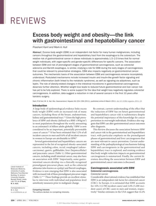
  • 6. REVIEWS cancer independently of the presence of gallstones. In the Chinese case–control study, the association between EBW and gallbladder cancer incidence was maintained even after adjustment for the presence of gallstones.69 Benign conditions linked to gallbladder cancer The gallbladder adenoma (or polyp) is recognized to have malignant potential76 and might be associated with EBW. In a large series of 264 gallbladder polyps, presence of the metabolic syndrome and increased waist circumference were independently associated with gallbladder polyp incidence.77 Other studies have suggested that BMI might also be a risk factor for gallbladder polyposis, particularly in males.78 There is a paucity of data linking EBW with other likely histological precursors of gallbladder cancer, such as chronic cholecystitis and gastric metaplasia, although these entities are common in obese patients. In a study of 199 morbidly obese Taiwanese patients undergoing cholecystectomy at the same time as bariatric surgery, all had chronic inflammatory cell infiltration and 27% had gastric metaplasia, although only 10% had gallstones.79 Cholangiocarcinoma EBW may also increase the risk of cholangiocarcinoma, which is a relatively rare cancer that has been increasing in incidence over recent decades.80 One large UK‑based case–control study of 372 cholangiocarcinoma cases estimated an OR of 1.5 (1.0–2.2) for obese patients versus those with a normal BMI.81 Another retrospective case– control study of patients with cholangio­ arcinoma c identified from the Surveillance, Epidemiology and End Results (SEER) database in the USA, reported that obesity was a significant risk factor for intrahepatic, but not extrahepatic, cholangiocarcinoma.82 However, there may be regional differences as a similar study in Denmark failed to show any significant increased risk of cholangiocarcinoma with increased BMI.83 Hepatic carcinogenesis There is strong evidence that obesity is associated with an increased risk of hepatocellular cancer (HCC). Changes in HCC incidence and obesity prevalence have both followed a similar upward pattern, suggesting that obesity may account for a significant proportion of HCC cases over and above better recognized risk factors, such as viral hepatitis and alcohol.84 Data from the UK Million Women Study showed that the risk of developing cirrhosis (a condition known to predispose to HCC) was linked to increasing BMI in a dose–response manner.85 Obesity is associated with nonalcoholic fatty liver disease (NAFLD), which is recognized as the hepatic manifestation of the metabolic syndrome. NAFLD encompasses a spectrum of histological states from isolated steatosis (fatty liver) and nonalcoholic steato­ hepatitis (NASH), through to fibrosis and cirrhosis. Population-based estimates for the prevalence of NAFLD range from 13% in nations where the prevalence of obesity is low, such as Japan,86 to approximately 30% in the USA.87 This figure rises to more than 90% in patients who are morbidly obese.88 The epidemiology of the link between EBW, NAFLD and HCC has been the subject of a recent systematic review and metaanalysis.89,90 In addition, current understanding of the mechanistic link between obesity, NAFLD and hepatic carcino­ enesis has been reviewed in detail elsewhere.84 g Therefore, only a brief summary of a large body of data is provided below. Hepatocellular cancer In a systematic review, seven out of 10 cohort studies reported significantly positive associations between BMI and HCC risk,89 with the risk being greater for men than women. In a separate meta-analysis, which included eight of the above cohort studies, plus two other cohort studies, obesity was associated with an RR of HCC of 1.9 (1.5–2.4) compared with a normal BMI; a similar gender difference was apparent (male RR 2.4 [1.8–3.2]; female RR 1.7 [1.4–2.0]).90 On the basis of prevalence data from the USA, it is estimated that 28% and 27% of cases of HCC in men and women, respectively, are attributable to EBW.90 EBW seems to be an independent risk factor for HCC separate from other causes of HCC. For example, in a study of 781,283 men from Korea, a region in which there is a high incidence of chronic HBV infection, obesityassociated HCC risk was maintained after controlling for HBV infection.91 In a separate study of 19,271 patients with cirrhosis who were listed for liver transplantation, obesity was found to be an independent predictor of HCC in patients with alcohol-related cirrhosis (OR 3.2), but not in patients with cirrhosis resulting from viral or autoimmune hepatitis.92 Another study of patients with HCV reported an HR of 1.9 (1.1–3.2) for overweight and 3.1 (1.4–6.8) for obese patients compared with their underweight counterparts.93 NASH NASH is considered to be the predisposing state of NAFLD for hepatic carcinogenesis. This is consistent with the close link between carcinogenesis and local inflammation throughout the gastrointestinal tract. Mechanistically, genetic and diet-induced obesity has been demonstrated to drive HCC development via chronic inflammation in mice.94 However, the relative contribution of NASH, as opposed to protumorigenic mechanisms linked to cirrhosis, during hepatic carcinogenesis requires further investigation. There are case reports of HCC in patients with noncirrhotic NASH,95,96 which implies that EBW can drive hepatic carcinogenesis via NASH independently of cirrhotic change. However, this may be a rare event as longitudinal studies have reported the incidence of HCC in patients with noncirrhotic NASH to be as low as 1–2%, whereas between 4–27% of cases of NASH transform to HCC following the development of cirrhosis.84 How does EBW drive carcinogenesis? How EBW promotes carcinogenesis in different tissues, including several parts of the gastrointestinal NATURE REVIEWS | GASTROENTEROLOGY HEPATOLOGY © 2011 Macmillan Publishers Limited. All rights reserved VOLUME 8  |  APRIL 2011  |  229
  • 7. REVIEWS Normal weight Innate and acquired immune activation in adipose tissue 1 Obesity 3 Gallstones GERD 4 Chronic inflammation ? 2 Mucosal inflammation TNF IL-6 MIF Insulin resistance Insulin IGF-1 Adipokines Modifiers: Physical activity Diet Glucose, FFAs Leptin Adiponectin 3 Gastrointestinal carcinogenesis Genotoxicity Mitogenic signaling to epithelial cells Apoptosis resistance Activated stromal–epithelial cell signaling Angiogenesis Host immunosurveillance Figure 1 | Mechanisms linking excess adiposity in overweight and obese states with gastrointestinal and hepatobiliary carcinogenesis. This schematic outlines the putative mechanisms that link adipose tissue in excess body weight with the cellular and molecular mechanisms that are critical for carcinogenesis. (1) Excess adiposity is associated with innate and acquired immune activation. (2) Some gastrointestinal cancers are linked to local mucosal inflammation and/or irritation secondary to obesity-related pathologies, such as GERD or gallstones. (3) Alternatively, obesity may drive gastrointestinal carcinogenesis directly via several interlinked mechanisms leading to exposure to protumorigenic cytokines, growth factors and adipokines, as well as increased substrate availability to neoplastic cells. (4) Diet composition and physical activity are closely linked to excess body weight and may modify the effect of excess adiposity on carcinogenesis at several levels, including reduced adipose tissue mass, modulation of chronic inflammation separate from changes in adipose tissue mass, and other direct effects on target cells, particularly genotoxicity by dietary carcinogens. Abbreviations: FFA, free fatty acid; IGF‑1, insulin-like growth factor 1; IL‑6, interleukin 6; MIF, macrophage migration inhibitory factor; TNF, tumor necrosis factor. and hepatobiliary tract, is still not understood but it is almost certainly a multifactorial process (Figure 1). A simple classification divides potential mechanisms into either indirect (for example EBW promotes GERD and gallstone formation, which then drive local inflammation-­driven carcinogenesis), or direct, in which pro­ umorigenic cell signaling occurs as a direct conset quence of excess adiposity. Proposed direct mechanisms probably exhibit complex inter-relationships (Figure 1); for example, chronic inflammation and insulin resistance co-exist as part of the metabolic syndrome, with proinflammatory signaling causally related to target cell insulin resistance.97 Increased estradiol production is believed to contribute to the increased risk of postmenopausal breast cancer and endometrial cancer that is associated with EBW.98 Moreover, therapeutic manipulation of sex hormone levels alters CRC risk in men and women.99 However, whether differences in sex hormone metabolism between males and females contribute to gender-specific differences in gastrointestinal and liver cancer risk associated with EBW or whether gender differences are explained by differences in the distribution and biology of VAT and SAT between the sexes is not known. For the purpose of this Review, we consider sex hormones most likely to modify EBW-related gastrointestinal and liver cancer risk rather than representing a direct mechanistic link 230  |  APRIL 2011  |  VOLUME 8 between excess adiposity and carcinogenesis in the gastro­intestinal tract. Great interest has been shown in the role of the intestinal microbiome in control of body weight.100 Therefore, the luminal microflora can be considered to be an etiological factor acting indirectly via modulation of body weight.101 In addition, luminal microflora could have a direct protumorigenic role during colorectal carcinogenesis. Putative molecular mechanisms linking obesity and carcinogenesis have previously been reviewed extensively.98,102,103 Therefore, only recent data relevant to the gastrointestinal tract is reviewed in detail here. Insulin and insulin-like growth factor signaling A well-established hypothesis linking EBW and gastrointestinal carcinogenesis, particularly CRC and pancreatic cancer, is that insulin resistance leads to direct mitogenic and antiapoptotic signaling by insulin and the insulin-like growth factor (IGF) axis (Figure 1).104 This hypothesis is supported by clinical observations, clinical biomarker data and mechanistic cell studies, but, as yet, few in vivo data have confirmed the rele­ vance of protumorigenic insulin and IGF signaling in humans. Increased CRC risk in patients with acromegaly, as well as increased CRC and pancreatic cancer risk www.nature.com/nrgastro © 2011 Macmillan Publishers Limited. All rights reserved
  • 8. REVIEWS in patients with T2DM and the metabolic syndrome, suggests that insulin and IGF signaling contributes to EBW-related cancer risk.60,105,106 Hyperinsulinemia in T2DM results in reduced hepatic synthesis of IGF binding protein (IGFBP) 1 and 2, which in turn leads to an increase in the circulating levels of bioactive, or ‘free’ IGF‑1. Insulin, IGF‑1 and IGF‑2 activate the insulin, IGF‑1 and IGF‑2 receptors, triggering an intracellular cascade, which stimulates the proliferation of CRC cells107 and promotion of tumor growth and metastasis.106,108,109 Downstream targets of these receptors include extracellular-signal-regulated kinase (ERK) and phosphatidylinositol 3 kinase (PI3K) pathways that mediate mitogenic cell signals.110 One critical downstream effector that has generated great interest is mammalian target of rapamycin (mTOR) and its downstream effector, S6 kinase 1, which both have a critical role in cellular nutrient sensing.111 Metformin, which is widely used in the treatment of T2DM, has shown potential for CRC chemoprevention in mouse models of colo­ ectal r carcinogenesis 112,113 through a mechanism believed to include direct inhibition of mTOR by adenosine monophosphate kinase (AMPK). The relationships between components of the insulin– IGF system and EBW are complex. In humans, although not all associations with BMI are linear, plasma levels of insulin and free IGF‑1 are higher in obesity, whilst IGFBP‑1 and IGFBP‑2 levels are reduced.104 Total IGF‑1 levels peak at a BMI of approximately 28 kg/m2 before reducing further in individuals with a BMI 28 kg/m2. A meta-analysis of five cohort studies revealed that the total IGF‑1 level predicts the RR of CRC.114 The associa­ tion between colorectal adenoma risk and IGF biomarker levels is inconsistent. One study demonstrated a positive association between levels of serum insulin and the IGF‑1:IGFBP‑3 ratio and colorectal adenoma risk,115 whilst another found a similar association only for ‘high-risk’ adenomas.116 Another study failed to detect any association between risk of colorectal adenoma and IGF biomarker levels.117 In addition to colorectal neoplasia, decreased IGFBP‑1 levels have been demonstrated to predict increased risk of pancreatic cancer. 118 IGF‑1 is over­ xpressed e in pancreatic cancer cells compared with normal pancreatic acini.119 Insulin and IGF signaling may also have a role in esophageal carcinogenesis. Genetic polymorphisms in the genes encoding the IGF‑1 receptor 120 and IGF‑2 receptor,121 which are known to increase expression of these receptors in tumor tissue compared with matched normal epithelium, are associ­ated with an increased risk of developing esophageal adenocarcinoma. Furthermore, IGF‑1 gene polymorphisms have also been shown to be associated with risk of Barrett esophagus.122 Alternatively, it has been proposed that insulin resistance may lead to increased energy substrate (glucose, triglycerides and free fatty acids) availability to mucosal cells in the gastrointestinal tract, promoting tumor growth directly and/or increasing oxidative stress and DNA mutagenesis (Figure 1). Chronic inflammation EBW can be considered to be a state of chronic low-grade inflammation, as demonstrated by increased systemic levels of proinflammatory markers and cytokines, such as C‑reactive protein (CRP) and interleukin (IL)‑6.123 Numerous proinflammatory cytokines, chemokines and other acute phase proteins, such as plasminogen activator inhibitor 1 and fibrinogen, are released from adipose tissue, which consists not only of adipocytes, but also immune cells, including macrophages (Figure 1).124 In the lean state, adipose tissue contains CD4+ T lympho­ ytes c with a T‑helper‑2 (TH2) profile and a proportionately higher population of regulatory CD4+ T lymphocytes (TREG) in relation to effector CD8 + T lympho­ ytes.125 c Obesity is associated with innate immune activation involving classic (M1) macrophage activation and a switch towards a TH1 profile and dominant effector CD8+ T lymphocytes,126 believed to be necessary for persistent adipose tissue inflammation.125 The precise mechanism(s) underlying immune activation associated with excess adiposity are not understood although some evidence supports the idea that Toll-like receptor (TLR) activation by free fatty acids and/or lipopolysaccharide (LPS) drives immune activation in adipose tissue.127 Many of the ‘classic’ cytokines and growth factors that are synthesized and released by adipose tissue, such as IL‑6, tumor necrosis factor (TNF) and macrophage migration inhibitory factor (MIF), are believed to have direct protumorigenic properties in the gastro­ ntestinal i and hepatobiliary tract (Figure 1). 128 However, it is not known whether increased systemic levels of pro­ tumorigenic factors act directly on gastrointestinal mucosal cells in an endocrine manner and/or whether a persistent systemic inflammatory state can drive induction of local tissue inflammation and subsequent production of the same factors, which then act in a paracrine manner. In an interesting study, Poullis et al.129 demonstrated a positive relationship between levels of fecal calprotectin (a marker of local intestinal inflamma­ tion) and obesity in healthy volunteers, independent of the serum CRP level, which suggests that local intestinal inflammation may occur in individuals with EBW independently of overt gastrointestinal disease. NASH is the paradigm for the causal relationship between local tissue inflammation and carcinogenesis associated with EBW.84 A study has demonstrated that 10% dietinduced weight loss is associ­ ted with a 25–50% reduca tion in pro­ nflammatory gene expression (including i TNF and IL‑8) in human colo­ ectal mucosa.130 Further r studies are required to confirm that weight loss reduces colorectal mucosal inflammation separate from the possible effects of increased physical exercise131 and dietary change per se. A close causal relationship is believed to exist between chronic inflammation and insulin resistance in the metabolic syndrome. One mechanism that has been proposed is nuclear factor κB (NFκB)-dependent inhibition of signaling downstream of the insulin receptor.132 There is some literature on the association between biomarkers of inflammation (particularly CRP) and NATURE REVIEWS | GASTROENTEROLOGY HEPATOLOGY © 2011 Macmillan Publishers Limited. All rights reserved VOLUME 8  |  APRIL 2011  |  231
  • 9. REVIEWS gastrointestinal cancer risk. In a meta-analysis of eight case–control studies, serum CRP level was demonstrated to be independently associated with an increased risk of CRC (RR 1.1 [1.0–1.3]). This relationship was only significant for men and for colon cancer, rather than for rectal cancer.133 However, one case–control study 134 and another cross-sectional study 135 have demonstrated no relationship between CRP levels and risk of colo­ rectal adenoma, although in the latter study a positive associa­ ion with risk of colorectal adenoma was evident t for serum IL‑6 and TNF levels.135 To date, no prospective study has investigated the association between increased levels of inflammatory biomarkers and future gastro­ ntestinal and liver cancer risk. i Adipokines Adipose tissue is an active endocrine organ that produces a range of hormones, collectively termed adipokines.126 Leptin and adiponectin are the adipokines that have been studied in the greatest depth (Figure 1). Similar to the insulin–IGF signaling hypothesis, there are convincing clinical biomarker data linking adipokine levels with increased gastrointestinal and liver cancer risk, and there are some functional preclinical data from animal models (see below), but there is a paucity of evidence confirming the precise role that these proteins play at differ­ nt stages of human gastrointestinal and e hepatobiliary carcinogenesis. Leptin Serum leptin levels are increased in obesity.136 The leptin receptor is overexpressed in CRC.137 Leptin has mitogenic activity in intestinal epithelial cells,138 reduces apoptosis in CRC cell lines139 and has also been shown to promote growth of azoxymethane-induced colorectal tumors in mice.140 Leptin is also implicated in esophageal carcinogenesis; mitogenic and antiapoptotic effects of leptin have been reported in esophageal adenocarcinoma cells in vitro.141 In a colonoscopy study in men, those with the highest tertile of circulating leptin levels had a BMI-adjusted 3.3fold greater risk of colorectal adenoma than those with lowest tertile leptin levels.142 The association between serum leptin levels and CRC is less clear although two prospective studies have shown positive associations with CRC in men.143,144 In a prospective study, there was a threefold increased risk of Barrett esophagus among men, but not women, in the highest quartile of serum leptin levels compared with the lowest, after adjustment for GERD symptoms.145 Leptin also has mitogenic activity in HCC.146 Adiponectin Adiponectin can be considered to be an insulins ­ ensitizing hormone, levels of which negatively correlate with BMI. 147 Adiponectin inhibits CRC cell growth, partly via modulation of AMPK activity and negative regulation of mTOR signaling.148 Exogenous administra­ ion of adiponectin reduces growth of t small intestinal polyps in the Apc Min/+ mouse model 232  |  APRIL 2011  |  VOLUME 8 of intestinal tumori­genesis,149 while an inverse associa­ tion between rectal dyplastic aberrant crypt focus number in humans and plasma adiponectin has been reported.150 A number of studies have reported plasma adiponectin levels to be inversely associated with the presence and multiplicity of colorectal adenomas151–153 and early CRC (defined as stage pT1 or below), following adjustment for BMI.154 Two case–control studies have demonstrated a negative relationship of adiponectin and adiponectin receptors with gastric cancer, including noncardia gastric cancers.155,156 Adiponectin may also have an inhibitory effect in esophageal carcinogenesis. In Barrett esophagus, expression of adiponectin receptors (Adipo‑R1 and Adipo‑R2) is downregulated. Adiponectin has been demon­ trated to have antiproliferative properties in s esophageal adenocarcinoma cell lines.157 There is evidence that adiponectin antagonizes leptin activity during hepatic carcinogenesis.158 The intestinal microbiome The intestinal microbiome in obese individuals is differ­ ent from that in normal weight individuals, and there is growing appreciation of the role of gut ecology in the promotion of obesity. 100,159,160 A lesser relative abundance of Bacteroides species is found in obese versus lean indivi­ uals and the proportion of Bacteroidetes d increases and Firmicutes decreases in association with diet-induced weight loss.100 This alteration in gut ecology is also a consistent feature of weight reduction after bari­ atric surgery.161 A case–control study determined that people with screening-detected colorectal adenoma had a signifi­ antly lower relative abundance of Bacteroidetes c than controls, although mean body weight data from the two groups were not presented.162 Emerging data demonstrate that gut bacteria may be involved in colorectal carcinogenesis. In a rat model of azoxymethane-induced tumors, germ-free rats developed smaller and fewer tumors than conventionally colonized littermates.163 There is also some evidence that levels of intramucosal and mucosal adherent bacteria are increased in patients with CRC.164,165 It is possible, therefore, that the altered intestinal microbiome in indivi­duals with EBW could drive carcinogenesis directly in the lower gastrointestinal tract. One postulated mechanism linking the intestinal microbiome and EBW-driven chronic inflammation is systemic exposure to bacterial LPS secondary to changes in bacterial load and/or changes in gut permeability. In animal models, circulating LPS levels increase under high-fat-diet conditions and also cause metabolic perturba­ ions resulting in insulin resistance.166 t Does weight loss decrease future cancer risk? A logical progression from the concept that EBW increases gastrointestinal and hepatobiliary cancer risk is that weight reduction should reduce subsequent cancer risk. Proof of the anticancer benefit of weight loss clearly has enormous public health consequences. However, to date, little evidence has accumulated to confirm or refute www.nature.com/nrgastro © 2011 Macmillan Publishers Limited. All rights reserved
  • 10. REVIEWS the hypothesis that weight reduction negatively regulates gastrointestinal and hepatobiliary carcinogenesis. The lack of data almost certainly reflects challenges in the design of observational studies in this field. Retrospective studies are subject to reverse causation bias, as weight reduction may be attributable to undiag­ nosed malignancy. Moreover, significant recall bias for self-reported weight is likely. Variation in individual weight over time is an important potential confounder in longitudinal cohort studies, with sustained weight loss rarely being achieved. The design of prospective studies of weight loss is also problematic. Investigation of CRC incidence requires long-term follow-up given the natural history of colorectal carcinogenesis over 10–15 years. Moreover, detection and removal of colo­ rectal adenomas during follow-up is a significant confounder in longitudinal studies. The low incidence of esophageal adenocarcinoma progression in Barrett esophagus means that the size and duration of a study using esophageal adenocarcinoma as an end point would be unfeasibly large and lengthy. Similar methodological problems are likely to blight studies of patients with NAFLD, combined with the relative inaccessibility of the liver for tissue analysis. Weight loss by behavioral changes The Iowa Women’s Health Study is the only major study that has prospectively investigated intentional dietary weight loss and CRC risk.167 In this study of more than 20,000 healthy postmenopausal women, individuals who self-reported an intentional weight loss of at least 20 lb (approximately 9 kg) or more, at some point in their lives, had a nonsignificant 18% decrease in CRC incidence compared with weight-stable controls.167 In a separate large cohort study of 65,000 Austrian adults with a mean follow-up of 8 years, neither weight loss nor weight gain was clearly associated with cancer incidence in the whole popula­ ion, although a reduction in BMI of more than t 0.1 kg/m2 per year was associated with a halving (HR 0.5 [0.3–0.9]) of the future risk of CRC in men when compared with men whose weight remained unchanged.168 Use of colorectal adenoma recurrence as a biomarker of CRC risk offers the opportunity to perform short-term studies. One Japanese study of screening colonoscopy demonstrated that individuals (who predominantly had normal BMI) who lost more than 5% of their initial body weight had lower rates of colorectal adenoma after 1 year compared with those whose weight was either unchanged or increased by 5%.24 There is evidence that behavioral change leading to weight loss reduces the severity of NASH.169 We are not aware of any reported investigation of the consequences of intentional weight loss for future HCC risk. Weight loss, physical activity and dietary change It is self-evident that body weight, diet and physical activity are inter-related factors. A simple line of reasoning is that reduction in calorific intake and increased physical activity could reduce future cancer risk via a reduction in EBW. However, there is evidence that dietary composition and physical exercise can modulate chronic inflammation independently of changes in the mass of adipose tissue.170,171 Moreover, calorie restriction has direct antineoplastic activity in preclinical models.172 Increased physical activity, independent of change in body weight, has been shown to reduce biomarkers of NAFLD in short-term clinical studies.173 Further study of the inter-relationship between EBW, diet and level of physical activity is required in order to inform future health education programs. A large prospec­tive cohort study from Denmark174 demonstrated that compliance with ‘healthy’ lifestyle factors, including maintenance of a waist circumference of 88 cm in men, was associated with a significant reduction (RR 0.8 [0.7–1.0]) in colon cancer in men during follow-up of almost 10 years. Weight loss following bariatric surgery The consistent, significant degree of weight loss after bariatric surgery provides an excellent opportunity to determine whether weight loss alters future gastrointestinal cancer risk, with the obvious caveats that physiological changes following surgery may confound weight-loss-associated outcomes and that lengthy follow-­ p is still required. Three reports have described u reduced cancer risks up to 10 years after bariatric surgery, with the largest risk reductions for hormoneresponsive endometrial and postmenopausal breast cancer, but also evident for CRC.175–177 By contrast, in a more-recent, population-based, case–control study of more than 13,000 patients undergoing bariatric surgery, there was no overall decrease in standardized incidence ratio for either breast or endometrial cancer, while the risk of CRC actually increased with time so that there was a twofold increase in risk after more than 10 years post-surgery.178 Short-term reductions in cancer risk after bari­ tric surgery may be unexpected given the a long natural history of gastrointestinal carcinogenesis, and could be explained by earlier diagnosis after bari­ atric surgery or a true biological effect of weight loss on existing tumor growth and/or progression. The possible increased risk of CRC beyond 10 years after surgery requires further investigation. Does EBW alter cancer outcomes? EBW also adversely affects short-term and long-term outcomes after diagnosis of gastrointestinal cancer. This is almost certainly multifactorial and is explained by effects of EBW on treatment efficacy and safety, as well as likely effects on disease progression, particularly metastatic potential. Colorectal cancer EBW adversely affects long-term survival following CRC diagnosis. The Iowa Women’s Health Study investi­ ated g the prognostic influence of baseline anthropometric measures on CRC outcomes during a 20-year follow-up period.179 After adjusting for age at diagnosis, smoking and stage of CRC at diagnosis, central adiposity, but not BMI, was a predictor of survival; women with the NATURE REVIEWS | GASTROENTEROLOGY HEPATOLOGY © 2011 Macmillan Publishers Limited. All rights reserved VOLUME 8  |  APRIL 2011  |  233
  • 11. REVIEWS highest tertile for waist–hip ratio had an HR of 1.37 (1.02–1.85) of CRC-related death compared with women with the lowest tertile waist–hip ratio.179 In a prospective Australian cohort study of 528 incident CRC cases, each 10 cm increase in waist circumference was associ­ ated with a 20% (5–37%) reduction in CRC-specific survival.180 Additionally, increasing body weight and a greater percentage of body fat, but not BMI, were significantly associated with increased CRC-related mortal­ ty i during a median follow-up of 5.5 years after CRC diagnosis.180 There is already preclinical evidence that obesity promotes liver tumor growth in mouse models of CRC liver metastasis.181,182 However, we are unaware of any human observational study that has investigated the association between pre-morbid weight, post-diagnosis weight change and CRC recurrence. In the short-term, a BMI 30 kg/m2 and increased waist–hip ratio are associated with worse CRC resection outcomes.179 Increased complication rates relate to rectal rather than colonic surgery and in men rather than women, and are explained by anastamotic leakage, increased blood transfusion requirements, longer surgery time and increased intra-abdominal collections in these patients.183 Further studies have shown reduced sphincter-sparing outcomes, increased permanent colo­ stomy rates following abdomino-perineal resection and a higher chance of local recurrence in men with obesity.184 These data are likely to be also partly explained by the less challenging anatomy of the wider female pelvis, but also by the hindrance of increased abdominal adipose tissue in men.185 There is also increasing evidence that efficacy of chemo­ herapy is modulated by body size. Randomized t controlled trials of adjuvant chemotherapy with 5‑flurouracil-­ ased regimens in patients with Dukes’ B b and C colon cancer have shown significantly worse overall survival and increased CRC recurrence rates in obese individuals compared with those who have a normal BMI.186,187 Further evidence of chemoresistance in obesity was demonstrated in a study by Guiu and colleagues,188 in which patients receiving treatment for metastatic CRC with bevacizumab who had increased VAT had reduced time to progression and overall survival, independent of BMI. Esophageal cancer The effect of obesity on esophageal adenocarcinoma outcomes has been harder to assess due to the negative energy balance and weight loss related to dysphagia prior to diagnosis. Consequently, the prevalence of obesity at diagnosis is lower than for CRC and there is a paucity of studies evaluating the impact of overweight and obesity on surgically-resectable esophageal adeno­ arcinoma. c Data from a large cohort study show that obesity increases the rate of anastomotic leakage and increases blood transfusion requirements, but does not result in significant differences in overall morbidity, in-hospital mortality, or in overall and disease-free 5‑year survival rates.189 The occurrence of other intraoperative complica­ tions, such as recurrent laryngeal nerve paralysis and 234  |  APRIL 2011  |  VOLUME 8 respiratory complications, is variable between studies and depends on whether the trans-hiatal or trans­thoracic approach is favored. 190,191 Other investigators have not shown any difference in the number of harvested lymph nodes or rates of negative resection margins in obese patients.192 Stomach cancer No studies have specifically investigated outcomes from gastric cardia cancer. However, one study did examine ‘proximal’ gastric cancer surgical outcomes in a Korean population; the results demonstrated similar short-term complications, as well as similar recurrence and survival rates in obese versus normal weight patients.193 In the largest study of its kind, including more than 7,900 Japanese patients with all forms of gastric cancer, overweight status was associated with an improved 5‑year survival.194 However, these data may not be generalizable to Western populations, in which obesity rates have been traditionally substantially higher than in Asia. Pancreatic cancer Evidence determining whether body size impacts on pancreatic cancer outcome is inconsistent. In one mouse model, obese mice that were injected with pancreatic cancer cells developed larger tumors, had more frequent metastasis and had decreased survival compared with lean mice.195 In patients with pancreatic cancer, two studies have shown that increasing BMI is associated with increased pancreatic fistula formation and prolonged hospital stay,196,197 although a similarly designed study previously showed no difference in fistula complica­ tions. 198 Increasing BMI also seems to increase the probability of lymph-node positivity from pancreatico­ duodenal resections.199 It has been suggested that BMI may be a poor discriminator of cancer outcome as one study showed that measures of VAT were superior in predicting higher wound and fistula complications. There are further inconsistencies in the effect of obesity on long-term survival from pancreatic cancer; studies have shown both improved200 and worsened59 5‑year survival following similar adjustments for disease stage. Conclusion EBW is a risk factor for multiple gastrointestinal and hepatobiliary cancers from the earliest, clinically apparent, premalignant phases of carcinogenesis and seems to also adversely affect cancer outcomes after diagnosis. As obesity prevalence continues on an upward trajectory globally, the cancer burden related to EBW will rise. The epidemiological link between EBW and increased cancer risk is established but further work is required to strengthen the argument that EBW is a preventable and/ or treatable factor for cancer prevention by weight loss. A recent model has highlighted the potential reduction in colon cancer incidence that is possible if ideal body weight is obtained at a population level.201 Further insights into the mechanistic basis of the link between EBW and carcinogenesis may lead to targeted chemoprevention strategies for obese individuals. In www.nature.com/nrgastro © 2011 Macmillan Publishers Limited. All rights reserved
  • 12. REVIEWS particular, the role of the obesity-associated intestinal microbiome in driving the systemic chronic inflammatory response in obesity and in mucosal protumorigenic signaling deserves further attention. Research into obesity-related adverse outcomes following gastrointestinal cancer diagnosis is in its infancy. In particular, an improved understanding of how EBW alters the biology and natural history of established cancer is required, alongside improved observational data on how EBW impacts on clinical care for patients with cancer. 1. 2. 3. 4. 5. 6. 7. 8. 9. 10. 11. 12. 13. 14. 15. 16. Moghaddam, A. A., Woodward, M. Huxley, R. Obesity and risk of colorectal cancer: a metaanalysis of 31 studies with 70,000 events. Cancer Epidemiol. Biomarkers Prev. 16, 2533–2547 (2007). Renehan, A. G., Tyson, M., Egger, M., Heller, R. F. Zwahlen, M. Body-mass index and incidence of cancer: a systematic review and meta-analysis of prospective observational studies. Lancet 371, 569–578 (2008). WHO. Obesity and overweight. Fact Sheet No 311. World Health Organization [online], http:// www.who.int/mediacentre/factsheets/fs311/ en/index.html (2006). World Cancer Research Fund. Food, Nutrition, Physical Activity, and the Prevention of Cancer: a Global Perspective 2nd edn (American Institute for Cancer Research, Washington, 2007). Vainoio, H. Bianchinin, F. (Eds) International Agency for Research in Cancer. Weight control and physical activity. (IARC Press, Lyon, 2002). Renehan, A. G. et al. Incident cancer burden attributable to excess body mass index in 30 European countries. Int. J. Cancer 126, 692–702 (2010). Wolk, A. et al. A prospective study of obesity and cancer risk (Sweden). Cancer Causes Control 12, 13–21 (2001). Siddiqui, A. et al. Obesity is associated with an increased prevalence of advanced adenomatous colon polyps in a male veteran population. Dig. Dis. Sci. 54, 1560–1564 (2009). Jacobson, B. C., Chan, A. T., Giovannucci, E. L. Fuchs, C. S. Body mass index and Barrett’s oesophagus in women. Gut 58, 1460–1466 (2009). Sato, Y., Nozaki, R., Yamada, K., Takano, M. Haruma, K. Relation between obesity and adenomatous polyps of the large bowel. Dig. Endosc. 21, 154–157 (2009). Cook, M. B., Greenwood, D. C., Hardie, L. J., Wild, C. R. Forman, D. A systematic review and meta-analysis of the risk of increasing adiposity on Barrett’s esophagus. Am. J. Gastroenterol. 103, 292–300 (2008). Thygesen, L. C. et al. Prospective weight change and colon cancer risk in male US health professionals. Int. J. Cancer 123, 1160–1165 (2008). National Institutes for Health. Clinical guidelines on the identification, evaluation, and treatment of overweight and obesity in adults. The evidence report. (NHLBI, 1998). Larsson, S. C. Wolk, A. Obesity and colon and rectal cancer risk: a meta-analysis of prospective studies. Am. J. Clin. Nutr. 86, 556–565 (2007). Dai, Z., Xu, Y. C. Niu, L. Obesity and colorectal cancer risk: a meta-analysis of cohort studies. World J. Gastroenterol. 13, 4199–4206 (2007). Moore, L. L. et al. BMI and waist circumference as predictors of lifetime colon cancer risk in 17. 18. 19. 20. 21. 22. 23. 24. 25. 26. 27. 28. 29. 30. 31. 32. Review criteria The PubMed database was searched using the following terms: “obesity”, “body weight”, “gastrointestinal”, “colorectum”, “oesophagus”, “gastric”, “pancreas”, “hepatocellular”, “gallbladder”, “cancer”, “adenoma”, “insulin”, “inflammation” and “adipokine”. Terms were searched alone and in combinations. Original articles, reviews, letters and editorials in the English language published between 1990 and January 2011 were considered. Reference lists were also hand searched to identify further relevant literature. Framingham Study adults. Int. J. Obes. 28, 559–567 (2004). Wise, L. A., Rosenberg, L., Palmer, J. R. Adams-Campbell, L. L. Anthropometric risk factors for colorectal polyps in African-American women. Obesity 16, 859–868 (2008). Sedjo, R. L. et al. Change in body size and the risk of colorectal adenomas. Cancer Epidemiol. Biomarkers Prev. 16, 526–531 (2007). Larsen, I. K., Grotmol, T., Almendingen, K. Hoff, G. Lifestyle as a predictor for colonic neoplasia in asymptomatic individuals. BMC Gastroenterol. 6, 5 (2006). Neugut, A. I. et al. Obesity and colorectal adenomatous polyps. J. Natl Cancer Inst. 83, 359–361 (1991). Morois, S., Mesrine, S., Josset, M., Clavel-Chapelon, F. Boutron-Ruault, M.‑C. Anthropometric factors in adulthood and risk of colorectal adenomas. Am. J. Epidemiol. 172, 1166–1180 (2010). Giovannucci, E. et al. Physical activity, obesity, and risk for colon cancer and adenoma in men. Ann. Intern. Med. 122, 327–334 (1995). Giovannucci, E., Colditz, G. A., Stampfer, M. J. Willett, W. C. Physical activity obesity and risk of colorectal adenoma in women (United States). Cancer Causes Control 7, 253–263 (1996). Yamaji, Y. et al. The effect of body weight reduction on the incidence of colorectal adenoma. Am. J. Gastroenterol. 103, 2061–2067 (2008). Lieberman, D. A., Prindiville, S., Weiss, D. G. Willett, W. Risk factors for advanced colonic neoplasia and hyperplastic polyps in asymptomatic individuals. JAMA 290, 2959–2967 (2003). Anderson, J. C. et al. Body mass index: a marker for significant colorectal neoplasia in a screening population. J. Clin. Gastroenterol. 41, 285–290 (2007). Omata, F. et al. Modifiable risk factors for colorectal neoplasms and hyperplastic polyps. Intern. Med. 48, 123–128 (2009). Kim, Y. Lee, S. An association between colonic adenoma and abdominal obesity: a crosssectional study. BMC Gastroenterol. 9, 4 (2009). Betes, M. et al. Use of colonoscopy as a primary screening test for colorectal cancer in average risk people. Am. J. Gastroenterol. 98, 2648–2654 (2003). Bird, C. L., Frankl, H. D., Lee, E. R. Haile, R. W. Obesity, weight gain, large weight changes, and adenomatous polyps of the left colon and rectum. Am. J. Epidemiol. 147, 670–680 (1998). Kono, S., Shinchi, K. Imanishi, K. Body mass index and adenomas of the sigmoid colon in Japanese men. Eur. J. Epidemiol. 12, 425–426 (1996). Boutron-Ruault, M.‑C., Senesse, P Méance, S., ., Belghiti, C. Faivre, J. Energy intake, body mass NATURE REVIEWS | GASTROENTEROLOGY HEPATOLOGY © 2011 Macmillan Publishers Limited. All rights reserved 33. 34. 35. 36. 37. 38. 39. 40. 41. 42. 43. 44. 45. 46. index, physical activity, and the colorectal adenoma-carcinoma sequence. Nutr. Cancer 39, 50–57 (2001). Wallace, K. et al. The association of physical activity and body mass index with the risk of large bowel polyps. Cancer Epidemiol. Biomarkers Prev. 14, 2082–2086 (2005). Terry, M. B. et al. Risk factors for advanced colorectal adenomas: a pooled analysis. Cancer Epidemiol. Biomarkers Prev. 11, 622–629 (2002). Wolf, L. A., Terry, P . D., Potter, J. D. Bostick, R. M. Do factors related to endogenous and exogenous estrogens modify the relationship between obesity and risk of colorectal adenomas in women? Cancer Epidemiol. Biomarkers Prev. 16, 676–683 (2007). Almendingen, K., Hofstad, B. Vatn, M. H. Does high body fatness increase the risk of presence and growth of colorectal adenomas followed up in situ for 3 years? Am. J. Gastroenterol. 96, 2238–2246 (2001). Bayerdörffer, E. et al. Increased risk of ‘high-risk’ colorectal adenomas in overweight men. Gastroenterology 104, 137–144 (1993). Kang, H. W. et al. Visceral obesity and insulin resistance as risk factors for colorectal adenoma: a cross-sectional, case-control study. Am. J. Gastroenterol. 105, 178–187 (2010). Kim, J. H. et al. Is metabolic syndrome a risk factor for colorectal adenoma? Cancer Epidemiol. Biomarkers Prev. 16, 1543–1546 (2007). Kim, K. S. et al. The frequency and risk factors of colorectal adenoma in health‑check‑up subjects in South Korea: relationship to abdominal obesity and age. Gut Liver 4, 36–42 (2010). Jacobs, E. T. et al. Association between body mass index and colorectal neoplasia at follow-up colonoscopy: a pooling study. Am. J. Epidemiol. 169, 657–666 (2009). Sainsbury, A. et al. Increased colorectal epithelial cell proliferation and crypt fission associated with obesity and roux‑en‑Y gastric bypass. Cancer Epidemiol. Biomarkers Prev. 17, 1401–1410 (2008). Ferrante, J. M. et al. Colorectal cancer screening among obese versus non-obese patients in primary care practices. Cancer Detect. Prev. 30, 459–465 (2006). Rosen, A. B. Schneider, E. C. Colorectal cancer screening disparities related to obesity and gender. J. Gen. Intern. Med. 19, 332–338 (2004). Borg, B. B., Gupta, N. K., Zuckerman, G. R., Banerjee, B. Gyawali, C. P Impact of obesity on . bowel preparation for colonoscopy. Clin. Gastroenterol. Hepatol. 7, 670–675 (2009). Pera, M., Manterola, C., Vidal, O. Grande, L. Epidemiology of esophageal adenocarcinoma. J. Surg. Onc. 92, 151–159 (2005). VOLUME 8  |  APRIL 2011  |  235
  • 13. REVIEWS 47. Lagergren, J., Bergstrom, R., Lindgren, A. Nyren, O. Symptomatic gastroesophageal reflux as a risk factor for esophageal adenocarcinoma. N. Engl. J. Med. 340, 825–831 (1999). 48. Hampel, H., Abraham, N. S. El-Serag, H. B. Meta-analysis: obesity and the risk for gastroesophageal reflux disease and its complications. Ann. Intern. Med. 143, 199–211 (2005). 49. Corley, D. A. Kubo, A. Body mass index and gastroesophageal reflux disease: a systematic review and meta-analysis. Am. J. Gastroenterol. 101, 2619–2628 (2006). 50. Corley, D. A., Kubo, A. Zhao, W. Abdominal obesity, ethnicity and gastro-oesophageal reflux symptoms. Gut 56, 756–762 (2007). 51. Whiteman, D. et al. The role of obesity, gastroesophageal reflux and smoking in adenocarcinomas of the esophagus and esophago-gastric junction [abstract]. Gastroenterology 132, A418 (2007). 52. Yousef, F. et al. The incidence of esophageal cancer and high-grade dysplasia in Barrett’s esophagus: a systematic review and metaanalysis. Am. J. Epidemiol. 168, 237–249 (2008). 53. Kamat, P Wen, S. J., Morris, J. ., Anandasabapathy, S. Exploring the association between elevated body mass index and Barrett’s esophagus: a systematic review and metaanalysis. Ann. Thorac. Surg. 87, 655–662 (2009). 54. Edelstein, Z. R., Farrow, D. C., Bronner, M. P ., Rosen, S. N. Vaughan, T. L. Central adiposity and risk of Barrett’s esophagus. Gastroenterology 133, 403–411 (2007). 55. Corley, D. A. et al. Abdominal obesity and body mass index as risk factors for Barrett’s esophagus. Gastroenterology 133, 34–41 (2007). 56. Oberg, S., Wenner, J., Johansson, J., Walther, B. Willen, R. Barrett esophagus: risk factors for progression to dysplasia and adenocarcinoma. Ann. Surg. 242, 49–54 (2005). 57. Arslan, A. A. et al. Anthropometric measures, body mass index, and pancreatic cancer: a pooled analysis from the Pancreatic Cancer Cohort Consortium (PanScan). Arch. Intern. Med. 170, 791–802 (2010). 58. Larsson, S. C., Orsini, N. Wolk, A. Body mass index and pancreatic cancer risk: a metaanalysis of prospective studies. Int. J. Cancer 120, 1993–1998 (2007). 59. Li, D. et al. Body mass index and risk, age of onset, and survival in patients with pancreatic cancer. JAMA 301, 2553–2562 (2009). 60. Huxley, R., Ansary-Moghaddam, A., de Gonzalez, A. B., Barzi, F. Woodward, M. Type-II diabetes and pancreatic cancer: a metaanalysis of 36 studies. Br. J. Cancer 92, 2076–2083 (2005). 61. Permert, J. et al. Is profound peripheral insulin resistance in patients with pancreatic cancer caused by a tumor-associated factor? Am. J. Surg. 165, 61–66 (1993). 62. Lowenfels, A. B. et al. Pancreatitis and the risk of pancreatic cancer. N. Engl. J. Med. 328, 1433–1437 (1993). 63. Yang, P et al. Overweight, obesity and gastric . cancer risk: results from a meta-analysis of cohort studies. Eur. J. Cancer 45, 2867–2873 (2009). 64. Leung, W. K. et al. Screening for gastric cancer in Asia: current evidence and practice. Lancet Oncol. 9, 279–287 (2008). 65. Goldblum, J. R. et al. Helicobacter pylori infection, not gastroesophageal reflux, is the major cause of inflammation and intestinal 236  |  APRIL 2011  |  VOLUME 8 66. 67. 68. 69. 70. 71. 72. 73. 74. 75. 76. 77. 78. 79. 80. 81. 82. 83. 84. metaplasia of gastric cardiac mucosa. Am. J. Gastroenterol. 97, 302–311 (2002). Kelley, J. R. Duggan, J. M. Gastric cancer epidemiology and risk factors. J. Clin. Epidemiol. 56, 1–9 (2003). Wistuba, I. I. Gazdar, A. F. Gallbladder cancer: Lessons from a rare tumour. Nat. Rev. Cancer 4, 695–706 (2004). Engeland, A., Tretli, S., Austad, G. Bjorge, T. Height and body mass index in relation to colorectal and gallbladder cancer in two million Norwegian men and women. Cancer Causes Control 16, 987–996 (2005). Hsing, A. W. et al. Body size and the risk of biliary tract cancer: a population-based study in China. Br. J. Cancer 99, 811–815 (2008). Stampfer, M. J., Maclure, K. M., Colditz, G. A., Manson, J. E. Willett, W. C. Risk of symptomatic gallstones in women with severe obesity. Am. J. Clin. Nutr. 55, 652–658 (1992). Lazcano-Ponce, E. C. et al. Epidemiology and molecular pathology of gallbladder cancer. CA Cancer J. 51, 349–364 (2001). Warren, B. L. Marais, A. W. Carcinoma of the gallbladder: a possible regional predisposition in the western Cape and the northern Cape. S. Afr. J. Surg. 33, 161–164 (1995). Shrikhande, S. V., Barreto, S. G., Singh, S., Udwadia, T. E. Agarwal, A. K. Cholelithiasis in gallbladder cancer: coincidence, cofactor, or cause! Eur. J. Surg. Oncol. 36, 514–519 (2010). Randi, G., Franceschi, S. La Vecchia, C. Gallbladder cancer worldwide: geographical distribution and risk factors. Int. J. Cancer 118, 1591–1602 (2006). Mohandas, K. M. Patil, P Cholecystectomy . S. for asymptomatic gallstones can reduce gall bladder cancer mortality in northern Indian women. Ind. J. Gastroenterol. 25, 147–151 (2006). Duarte, I., Llanos, O., Domke, H., Harz, C. Valdivieso, V. Metaplasia and precursor lesions of gallbladder carcinoma. Frequency, distribution, and probability of detection in routine histologic samples. Cancer 72, 1878–1884 (1993). Lim, S. H. et al. Is metabolic syndrome one of the risk factors for gallbladder polyps Found by ultrasonography during health screening? Gut Liver 1, 138–144 (2007). Segawa, K. et al. Prevalence of gallbladder polyps among apparently healthy Japanese: ultrasonographic study. Am. J. Gastroenterol. 87, 630–633 (1992). Liew, P et al. Gallbladder disease among . L. obese patients in Taiwan. Obes. Surg. 17, 383–390 (2007). Patel, T. Worldwide trends in mortality from biliary tract malignancies. BMC Cancer 2, 10 (2002). Grainge, M. J., West, J., Solaymani-Dodaran, M., Aithal, G. P Card, T. R. The antecedents of . biliary cancer: a primary care case-control study in the United Kingdom. Br. J. Cancer 100, 178–180 (2008). Welzel, T. M. et al. Risk factors for intrahepatic and extrahepatic cholangiocarcinoma in the United States: a population-based case-control study. Clin. Gastroenterol. Hepatol. 5, 1221–1228 (2007). Welzel, T. M. et al. Risk factors for intrahepatic cholangiocarcinoma in a low-risk population: a nationwide case-control study. Int. J. Cancer 120, 638–641 (2007). Starley, B. Q., Calcagno, C. J. Harrison, S. A. Nonalcoholic fatty liver disease and hepatocellular carcinoma: a weighty connection. Hepatology 51, 1820–1832 (2010). 85. Liu, B., Balkwill, A., Reeves, G. Beral, V. Body mass index and risk of liver cirrhosis in middle aged UK women: prospective study. BMJ 340, c912 (2010). 86. Omagari, K. et al. Fatty liver in non-alcoholic nonoverweight Japanese adults: incidence and clinical characteristics. J. Gastroenterol. Hepatol. 17, 1098–1105 (2002). 87. Browning, J. D. et al. Prevalence of hepatic steatosis in an urban population in the United States: impact of ethnicity. Hepatology 40, 1387–1395 (2004). 88. Machado, M., Marques-Vidal, P Cortez-Pinto, H. . Hepatic histology in obese patients undergoing bariatric surgery. J. Hepatol. 45, 600–606 (2006). 89. Saunders, D., Seidel, D., Allison, M. Lyratzopoulos, G. Systematic review: the association between obesity and hepatocellular carcinoma—epidemiological evidence. Aliment. Pharmacol. Ther. 31, 1051–1063 (2010). 90. Larsson, S. C. Wolk, A. Overweight, obesity and risk of liver cancer: a meta-analysis of cohort studies. Br. J. Cancer 97, 1005–1008 (2007). 91. Oh, S. W., Yoon, Y. S. Shin, S. A. Effects of excess weight on cancer incidences depending on cancer sites and histologic findings among men: Korea National Health Insurance Corporation Study. J. Clin. Oncol. 23, 4742–4754 (2005). 92. Nair, S., Mason, A., Eason, J., Loss, G. Perrillo, R. P Is obesity an independent risk factor . for hepatocellular carcinoma in cirrhosis? Hepatology 36, 150–155 (2002). 93. Ohki, T. et al. Obesity is an independent risk factor for hepatocellular carcinoma development in chronic hepatitis C patients. Clin. Gastroenterol. Hepatol. 6, 459–464 (2008). 94. Park, E. J. et al. Dietary and genetic obesity promote liver inflammation and tumorigenesis by enhancing IL‑6 and TNF expression. Cell 140, 197–208 (2010). 95. Ichikawa, T. et al. Two cases of non-alcoholic steatohepatitis with development of hepatocellular carcinoma without cirrhosis. J. Gastroenterol. Hepatol. 21, 1865–1866 (2006). 96. Kawada, N. et al. Hepatocellular carcinoma arising from non-cirrhotic nonalcoholic steatohepatitis. J. Gastroenterol. 44, 1190–1194 (2009). 97. Wellen, K. E. Hotamisligil, G. S. Inflammation, stress, and diabetes. J. Clin. Invest. 115, 1111–1119 (2005). 98. Roberts, D. L., Dive, C. Renehan, A. G. Biological mechanisms linking obesity and cancer risk: new perspectives. Ann. Rev. Med. 61, 301–316 (2010). 99. Lin, J. H. Giovannucci, E. Sex hormones and colorectal cancer: what have we learned so far? J. Natl Cancer Inst. 102, 1746–1747 (2010). 100. Ley, R. E., Turnbaugh, P Klein, S. . J., Gordon, J. I. Microbial ecology: human gut microbes associated with obesity. Nature 444, 1022–1023 (2006). 101. Caesar, R., Fåk, F. Bäckhed, F. Effects of gut microbiota on obesity and atherosclerosis via modulation of inflammation and lipid metabolism. J. Int. Med. 268, 320–328 (2010). 102. Cowey, S. Hardy, R. W. The metabolic syndrome: a high-risk state for cancer? Am. J. Pathol. 169, 1505–1522 (2006). 103. Donohoe, C. L., Pidgeon, G. P Lysaght, J. ., Reynolds, J. V. Obesity and gastrointestinal cancer. Br. J. Surg. 97, 628–642 (2010). 104. Renehan, A. G., Frystyk, J. Flyvbjerg, A. Obesity and cancer risk: the role of the insulin-IGF axis. Trends Endocrinol. Metab. 17, 328–336 (2006). www.nature.com/nrgastro © 2011 Macmillan Publishers Limited. All rights reserved
  • 14. REVIEWS 105. Larsson, S. C., Giovannucci, E. Wolk, A. Diabetes and colorectal cancer incidence in the cohort of Swedish men. Diabetes Care 28, 1805–1807 (2005). 106. Clayton, P . E., Banerjee, I., Murray, P . G. Renehan, A. G. Growth hormone, the insulin-like growth factor axis, insulin and cancer risk. Nat. Rev. Endocrinol. 7, 11–24 (2011). 107. Giovannucci, E. Insulin, insulin-like growth factors and colon cancer: a review of the evidence. J. Nutr. 131, 3109S–3120S (2001). 108. Corpet, D. E., Jacquinet, C., Peiffer, G. Tache, S. Insulin injections promote the growth of aberrant crypt foci in the colon of rats. Nutr. Cancer 27, 316–320 (1997). 109. Samani, A. A., Yakar, S., LeRoith, D. Brodt, P . The role of the IGF system in cancer growth and metastasis: overview and recent insights. Endocr. Rev. 28, 20–47 (2007). 110. Sears, R. et al. Multiple Ras-dependent phosphorylation pathways regulate Myc protein stability. Genes Dev. 14, 2501–2514 (2000). 111. Hursting, S. D. et al. Reducing the weight of cancer: mechanistic targets for breaking the obesity-carcinogenesis link. Best Pract. Res. Clin. Endocrinol. Metab. 22, 659–669 (2008). 112. Hosono, K. et al. Metformin suppresses colorectal aberrant crypt foci in a short-term clinical trial. Cancer Prev. Res. 3, 1077–1083 (2010). 113. Tomimoto, A. et al. Metformin suppresses intestinal polyp growth in ApcMin/+ mice. Cancer Sci. 99, 2136–2141 (2008). 114. Renehan, A. G. et al. Insulin-like growth factor (IGF)‑I, IGF binding protein‑3, and cancer risk: systematic review and meta-regression analysis. Lancet 363, 1346–1353 (2004). 115. Schoen, R. E. et al. Insulin-like growth factor‑I and insulin are associated with the presence and advancement of adenomatous polyps. Gastroenterology 129, 464–475 (2005). 116. Renehan, A. G. et al. High-risk colorectal adenomas and serum insulin-like growth factors. Br. J. Surg. 88, 107–113 (2001). 117. Flood, A. et al. Serum concentrations of insulinlike growth factor and insulin-like growth factor binding protein 3 and recurrent colorectal adenomas. Cancer Epidemiol. Biomarkers Prev. 17, 1493–1498 (2008). 118. Wolpin, B. M. et al. Circulating insulin-like growth factor binding protein‑1 and the risk of pancreatic cancer. Cancer Res. 67, 7923–7928 (2007). 119. Bergmann, U., Funatomi, H., Yokoyama, M., Beger, H. G. Korc, M. Insulin-like growth factor I overexpression in human pancreatic cancer: evidence for autocrine and paracrine roles. Cancer Res. 55, 2007–2011 (1995). 120. Zhao, R., Macdonald, K. Casson, A. G. Insulinlike growth factor type I receptor gene expression and obesity in esophageal adenocarcinoma. Mol. Carcinog. 48, 982–988 (2009). 121. Hoyo, C. et al. IGF2R polymorphisms and risk of esophageal and gastric adenocarcinomas. Int. J. Cancer 125, 2673–2678 (2009). 122. McElholm, A. R. et al. A population-based study of IGF axis polymorphisms and the esophageal inflammation, metaplasia, adenocarcinoma sequence. Gastroenterology 139, 204–212.e3 (2010). 123. Park, H. S., Park, J. Y. Yu, R. Relationship of obesity and visceral adiposity with serum concentrations of CRP TNF-[alpha] and IL‑6. Diab. , Res. Clin. Pract. 69, 29–35 (2005). 124. Nishimura, S. et al. CD8+ effector T cells contribute to macrophage recruitment and adipose tissue inflammation in obesity. Nat. Med. 15, 914–920 (2009). 125. Feuerer, M. et al. Lean, but not obese, fat is enriched for a unique population of regulatory T cells that affect metabolic parameters. Nat. Med. 15, 930–939 (2009). 126. Balistreri, C. R., Caruso, C. Candore, G. The role of adipose tissue and adipokines in obesityrelated inflammatory diseases. Mediators Inflamm. 2010, 802078 (2010). 127. Song, M. J., Kim, K. H., Yoon, J. M. Kim, J. B. Activation of Toll-like receptor 4 is associated with insulin resistance in adipocytes. Biochem. Biophys. Res. Commun. 346, 739–745 (2006). 128. Balkwill, F., Charles, K. A. Mantovani, A. Smoldering and polarized inflammation in the initiation and promotion of malignant disease. Cancer Cell 7, 211–217 (2005). 129. Poullis, A. bowel inflammation as measured by faecal calprotectin: a link between lifestyle factors and colorectal cancer risk. Cancer Epidemiol. Biomarkers Prev. 13, 279–284 (2004). 130. Pendyala, S., Neff, L. M., Suárez-Fariñas, M. Holt, P Diet-induced weight loss reduces . R. colorectal inflammation: implications for colorectal carcinogenesis. Am. J. Clin. Nutr. 93, 234–242 (2011). 131. Martínez, M. E. et al. Physical activity, body mass index, and prostaglandin E2 levels in rectal mucosa. J. Natl Cancer Inst. 91, 950–953 (1999). 132. Hotamisligil, G. S. Inflammation and metabolic disorders. Nature 444, 860–867 (2006). 133. Tsilidis, K. K. et al. C‑reactive protein and colorectal cancer risk: a systematic review of prospective studies. Int. J. Cancer 123, 1133–1140 (2008). 134. Tsilidis, K. K. et al. C‑reactive protein and colorectal adenoma in the CLUE II cohort. Cancer Causes Control 19, 559–567 (2008). 135. Kim, S. et al. Circulating levels of inflammatory cytokines and risk of colorectal adenomas. Cancer Res. 68, 323–328 (2008). 136. Considine, R. V. et al. Serum immunoreactive leptin concentrations in normal-weight and obese humans. N. Engl. J. Med. 334, 292–295 (1996). 137. Aloulou, N. et al. Involvement of the leptin receptor in the immune response in intestinal cancer. Cancer Res. 68, 9413–9422 (2008). 138. Liu, Z. H., Uesaka, T., Watanabe, H. Kato, N. High fat diet enhances colonic cell proliferation and carcinogenesis in rats by elevating serum leptin. Int. J. Oncol. 19, 1009–1014 (2001). 139. Rouet-Benzineb, P et al. Leptin counteracts . sodium butyrate-induced apoptosis in human colon cancer HT‑29 cells via NF‑kappaB signaling. J. Biol. Chem. 279, 16495–16502 (2004). 140. Hirose, Y. et al. Enhancement of development of azoxymethane-induced colonic premalignant lesions in C57BL/KsJ-db/db mice. Carcinogenesis 25, 821–825 (2004). 141. Ogunwobi, O., Mutungi, G. Beales, I. L. P . Leptin stimulates proliferation and inhibits apoptosis in Barrett’s esophageal adenocarcinoma cells by cyclooxygenase‑2‑dependent, prostaglandin‑E2‑mediated transactivation of the epidermal growth factor receptor and c‑Jun NH2-terminal kinase activation. Endocrinology 147, 4505–4516 (2006). 142. Chia, V. M. et al. Leptin concentrations, leptin receptor polymorphisms, and colorectal adenoma risk. Cancer Epidemiol. Biomarkers Prev. 16, 2697–2703 (2007). 143. Stattin, P et al. Plasma leptin and colorectal . cancer risk: a prospective study in Northern Sweden. Oncol. Rep. 10, 2015–2021 (2003). NATURE REVIEWS | GASTROENTEROLOGY HEPATOLOGY © 2011 Macmillan Publishers Limited. All rights reserved 144. Stattin, P et al. Obesity and colon cancer: . does leptin provide a link? Int. J. Cancer 109, 149–152 (2004). 145. Kendall, B. J. et al. Leptin and the risk of Barrett’s oesophagus. Gut 57, 448–454 (2008). 146. Wang, S. N., Lee, K. T. Ker, C. G. Leptin in hepatocellular carcinoma. World J. Gastroenterol. 16, 5801–5809 (2010). 147. Arita, Y. et al. Paradoxical decrease of an adipose-specific protein, adiponectin, in obesity. Biochem. Biophys. Res. Commun. 257, 79–83 (1999). 148. Sugiyama, M. et al. Adiponectin inhibits colorectal cancer cell growth through the AMPK/ mTOR pathway. Int. J. Oncol. 34, 339–344 (2009). 149. Otani, K. et al. Adiponectin suppresses tumorigenesis in Apc(Min/+) mice. Cancer Lett. 288, 177–182 (2010). 150. Takahashi, H. et al. Association of visceral fat accumulation and plasma adiponectin with rectal dysplastic aberrant crypt foci in a clinical population. Cancer Sci. 100, 29–32 (2009). 151. Wei, E. K., Giovannucci, E., Fuchs, C. S., Willett, W. C. Mantzoros, C. S. Low plasma adiponectin levels and risk of colorectal cancer in men: A prospective study. J. Natl Cancer Inst. 97, 1688–1694 (2005). 152. Nakajima, T. E. et al. Adipocytokines as new promising markers of colorectal tumors: adiponectin for colorectal adenoma, and resistin and visfatin for colorectal cancer. Cancer Sci. 101, 1286–1291 (2010). 153. Yamaji, T., Iwasaki, M., Sasazuki, S. Tsugane, S. Interaction between adiponectin and leptin Influences the risk of colorectal adenoma. Cancer Res. 70, 5430–5437 (2010). 154. Otake, S. et al. Decreased levels of plasma adiponectin associated with increased risk of colorectal cancer. World J. Gastroenterol. 16, 1252–1257 (2010). 155. Ishikawa, M. et al. Plasma adiponectin and gastric cancer. Clin. Cancer Res. 11, 466–472 (2005). 156. Otani, K. et al. Adiponectin receptors are downregulated in human gastric cancer. J. Gastroenterol. 45, 918–927 (2010). 157. Konturek, P . C., Burnat, G., Rau, T., Hahn, E. G. Konturek, S. Effect of adiponectin and ghrelin on apoptosis of barrett adenocarcinoma cell line. Dig. Dis. Sci. 53, 597–605 (2008). 157. Sharma, D. et al. Adiponectin antagonizes the oncogenic actions of leptin in hepatocellular carcinogenesis. Hepatology 52, 1713–1722 (2010). 159. Backhed, F. et al. The gut microbiota as an environmental factor that regulates fat storage. Proc. Natl Acad. Sci. USA 101, 15718–15723 (2004). 160. Backhed, F., Manchester, J. K., Semenkovich, C. F. Gordon, J. I. Mechanisms underlying the resistance to diet-induced obesity in germ-free mice. Proc. Natl Acad. Sci. USA 104, 979–984 (2007). 161. Furet, J. P et al. Differential adaptation of human . gut microbiota to bariatric surgery-induced weight loss: links with metabolic and low-grade inflammation markers. Diabetes 59, 3049–3057 (2010). 162. Shen, X. J. et al. Molecular characterization of mucosal adherent bacteria and associations with colorectal adenomas. Gut Microbes 1, 138–147 (2010). 163. Vannucci, L. et al. Colorectal carcinogenesis in germ-free and conventionally reared rats: different intestinal environments affect the systemic immunity. Int. J. Oncol. 32, 609–617 (2008). VOLUME 8  |  APRIL 2011  |  237
  • 15. REVIEWS 164. Martin, H. M. et al. Enhanced Escherichia coli adherence and invasion in Crohn’s disease and colon cancer. Gastroenterology 127, 80–93 (2004). 165. Swidsinski, A. et al. Association between intraepithelial Escherichia coli and colorectal cancer. Gastroenterology 115, 281–286 (1998). 166. Cani, P et al. Metabolic endotoxemia initiates . D. obesity and insulin resistance. Diabetes 56, 1761–1772 (2007). 167. Parker, E. D. Folsom, A. R. Intentional weight loss and incidence of obesity-related cancers: the Iowa Women’s Health Study. Int. J. Obes. Relat. Metab. Disord. 27, 1447–1452 (2003). 168. Rapp, K. et al. Weight change and cancer risk in a cohort of more than 65 000 adults in Austria. Ann. Oncol. 19, 641–648 (2008). 169. St. George, A. et al. Effect of a lifestyle intervention in patients with abnormal liver enzymes and metabolic risk factors. J. Gastroenterol. Hepatol. 24, 399–407 (2009). 170. Stewart, L. K. et al. The influence of exercise training on inflammatory cytokines and C‑reactive protein. Med. Sci. Sports Exerc. 39, 1714–1719 (2007). 171. Rankin, J. W. Turpyn, A. D. Low carbohydrate, high fat diet increases C‑reactive protein during weight loss. J. Am. Coll. Nutr. 26, 163–169 (2007). 172. Tammariello, A. E. Milner, J. A. Mouse models for unraveling the importance of diet in colon cancer prevention. J. Nutr. Biochem. 21, 77–88 (2010). 173. Johnson, N. A. et al. Aerobic exercise training reduces hepatic and visceral lipids in obese individuals without weight loss. Hepatology 50, 1105–1112 (2009). 174. Kirkegaard, H. et al. Association of adherence to lifestyle recommendations and risk of colorectal cancer: a prospective Danish cohort study. BMJ 341, c5504 (2010). 175. Sjöström, L. et al. Effects of bariatric surgery on cancer incidence in obese patients in Sweden (Swedish Obese Subjects Study): a prospective, controlled intervention trial. Lancet Oncol. 10, 653–662 (2009). 176. Adams, T. D. et al. Cancer incidence and mortality after gastric bypass surgery. Obesity 17, 796–802 (2009). 177. Christou, N. V., Lieberman, M., Sampalis, F. Sampalis, J. S. Bariatric surgery reduces cancer risk in morbidly obese patients. Surg. Obes. Relat. Dis. 4, 691–695 (2008). 178. Plecka-Ostlund, M. Risk of obesity-related cancer after obesity surgery in a population-based cohort study. Ann. Surg. 252, 972–976 (2010). 238  |  APRIL 2011  |  VOLUME 8 179. Prizment, A. E., Flood, A., Anderson, K. E. Folsom, A. R. Survival of women with colon cancer in relation to precancer anthropometric characteristics: the Iowa Women’s Health Study. Cancer Epidemiol. Biomarkers Prev. 19, 2229–2237 (2010). 180. Haydon, A. M. M., MacInnis, R. J., English, D. R. Giles, G. G. Effect of physical activity and body size on survival after diagnosis with colorectal cancer. Gut 55, 62–67 (2006). 181. Wu, Y. J. et al. Insulin-like growth factor‑I regulates the liver microenvironment in obese mice and promotes liver metastasis. Cancer Res. 70, 57–67 (2010). 182. Earl, T. M. et al. Silencing of TLR4 decreases liver tumor burden in a murine model of colorectal metastasis and hepatic steatosis. Ann. Surg. Oncol. 16, 1043–1050 (2009). 183. Benoist, S., Panis, Y., Alves, A. Valleur, P . Impact of obesity on surgical outcomes after colorectal resection. Am. J. Surg. 179, 275–281 (2000). 184. Meyerhardt, J. A. et al. Impact of body mass index on outcomes and treatment-related toxicity in patients with stage II and III rectal cancer: findings from intergroup trial 0114. J. Clin. Oncol. 22, 648–657 (2004). 185. Moon, H. G. et al. Visceral obesity may affect oncologic outcome in patients with colorectal cancer. Ann. Surg. Oncol. 15, 1918–1922 (2008). 186. Meyerhardt, J. A. et al. Influence of body mass index on outcomes and treatment-related toxicity in patients with colon carcinoma. Cancer 98, 484–495 (2003). 187. Dignam, J. J. et al. Body mass index and outcomes in patients who receive adjuvant chemotherapy for colon cancer. J. Natl Cancer Inst. 98, 1647–1654 (2006). 188. Guiu, B. et al. Visceral fat area is an independent predictive biomarker of outcome after first-line bevacizumab-based treatment in metastatic colorectal cancer. Gut 59, 341–347 (2010). 189. Grotenhuis, B. A. et al. Prognostic value of body mass index on short-term and long-term outcome after resection of esophageal cancer. World J. Surg. 34, 2621–2627 (2010). 190. Healy, L. A. et al. Impact of obesity on outcomes in the management of localized adenocarcinoma of the esophagus and esophagogastric junction. J. Thorac. Cardiovasc. Surg. 134, 1284–1291 (2007). 191. Scipione, C. N., Chang, A. C., Pickens, A., Lau, C. L. Orringer, M. B. Transhiatal esophagectomy in the profoundly obese: implications and experience. Ann. Thorac. Surg. 84, 376–383 (2007). 192. Melis, M. et al. An elevated body mass index does not reduce survival after esophagectomy for cancer. Ann. Surg. Oncol. doi:10.1245/ s10434-010-1336-1. 193. Oh, S. J. et al. Effect of being overweight on postoperative morbidity and long-term surgical outcomes in proximal gastric carcinoma. J. Gastroenterol. Hepatol. 24, 475–479 (2009). 194. Tokunaga, M. et al. Better 5‑year survival rate following curative gastrectomy in overweight patients. Ann. Surg. Oncol. 16, 3245–3251 (2009). 195. Zyromski, N. J. et al. Obesity potentiates the growth and dissemination of pancreatic cancer. Surgery 146, 258–263 (2009). 196. Noun, R. et al. The impact of obesity on surgical outcome after pancreaticoduodenectomy. JOP 9, 468–476 (2008). 197. Rosso, E. et al. The role of “fatty pancreas” and of BMI in the occurrence of pancreatic fistula after pancreaticoduodenectomy. J. Gastrointest. Surg. 13, 1845–1851 (2009). 198. Lermite, E. et al. Risk factors of pancreatic fistula and delayed gastric emptying after pancreaticoduodenectomy with pancreaticogastrostomy. J. Am. Coll. Surg. 204, 588–596 (2007). 199. Fleming, J. B. et al. Influence of obesity on cancer-related outcomes after pancreatectomy to treat pancreatic adenocarcinoma. Arch. Surg. 144, 216–221 (2009). 200. Tsai, S. et al. Impact of obesity on perioperative outcomes and survival following pancreaticoduodenectomy for pancreatic cancer: a large single-institution study. J. Gastrointest. Surg. 14, 1143–1150 (2010). 201. de Vries, E. et al. Lifestyle changes and reduction of colon cancer incidence in Europe: a scenario study of physical activity promotion and weight reduction. Eur. J. Cancer 46, 2605–2616 (2010). Acknowledgements Work on the link between obesity, weight loss and cancer risk in the authors’ laboratory is funded by the Medical Research Council (UK) Medical Research Foundation. Author contributions Both authors contributed equally to all aspects of this article. Supplementary information Supplementary information is linked to the online version of the paper at www.nature.com/nrgastro. www.nature.com/nrgastro © 2011 Macmillan Publishers Limited. All rights reserved