SlideShare uma empresa Scribd logo
1 de 88
vvvv
Consists of;
• the gluteal region (buttocks)
• the thigh
• the leg, and
• the foot.
The Lower Limb
Functions of the Lower Limb
Support the body weight
The ligaments at the hip and knee joints facilitate locking of these
joints therefore reducing the amount of muscular energy
required to maintain a standing position.
Locomotion
To move the body through space. This involves integration of
movements at all joints of the lower limb to place the foot on the
ground and move the body over it.
THE PELVIS BONE
The pelvis is made up of the sacrum and the coccyx bound to each
other by dense ligaments, Ilium, Ischium, and Pubis.
The ilium with its iliac crest running between the anterior and posterior
superior iliac spines; below each of these are the corresponding
inferior spines. Well-defined ridges on its lateral surface are the strong
muscle markings of the glutei. Its inner aspect bears the large auricular
surface which articulates with the sacrum. The iliopectineal line runs
forward from the apex of the auricular surface and demarcates the true
from the false pelvis. The pubis comprises the body and the superior
and inferior pubic rami.
The ischium has a vertically disposed body, bearing the ischial spine on
its posterior border which demarcates an upper (greater) and lower
(lesser), sciatic notch. The inferior pole of the body bears the ischial
tuberosity then projects forwards almost at right angles into the ischial
ramus to meet the inferior pubic ramus. The obturator foramen lies
bounded by the body and rami of the pubis and the body and ramus of
the ischium. All three bones fuse at the acetabulum which forms the
socket for the femoral head, for which it bears a wide crescentic
articular surface.
THE FEMUR
The femur is the largest bone in the body. It is 18 in (45 cm) in
length, a measurement it shares with the vas, the spinal cord and
the thoracic duct and which is also the distance from the teeth to
the cardia of the stomach. The femoral head is two-thirds of a
sphere and faces upwards, medially and forwards. It is covered with
cartilage except for its central fovea where the ligamentum teres is
attached. The neck is 2 in (5 cm) long and is set at an angle of 125°
to the shaft. In the female, with her wider pelvis, the angle is
smaller. The junction between the neck and the shaft is marked
anteriorly by the trochanteric line, laterally by the greater
trochanter, medially and somewhat posteriorly by the lesser
trochanter and posteriorly by the prominent trochanteric crest,
which unites the two trochanters.
THE FEMORAL SHAFT
The femoral shaft is roughly circular in section at its middle but is
flattened posteriorly at each extremity. Posteriorly also it is marked by
a strong crest, the linea aspera. Inferiorly, this crest splits into the
medial and lateral supracondylar lines, leaving a flat popliteal surface
between them. The medial supracondylar line ends distally in the
adductor tubercle. The lower end of the femur bears the prominent
condyles which are separated by a deep intercondylar notch posteriorly
but which blend anteriorly to form an articular surface for the patella.
The lateral condyle is the more prominent of the two and acts as a
buttress to assist in preventing lateral displacement of the patella
The posterior aspect of right femur
The sources of blood supply of
the femoral head—along the
ligamentum teres, through the
diaphysis and via the retinacula.
Through the
diaphysis
The blood supply to the femoral head is
derived from vessels travelling
up from the diaphysis along the cancellous
bone, from vessels in the hip
capsule, where this is reflected on to the
neck in longitudinal bands or
retinacula, and from the artery in the
ligamentum teres; this third source
is negligible in adults, but essential in
children, when the femoral head is
separated from the neck by the cartilage of
the epiphyseal line
CLINICAL FEATURES
1. The upper end of the femur is a common site for fracture in the elderly. The
neck may break immediately beneath the head (subcapital), near its midpoint
(cervical) or adjacent to the trochanters (basal), or the fracture line may pass
between, along or just below the trochanter. Fractures of the femoral neck will
interrupt completely the blood supply from the diaphysis and, should the
retinacula also be torn, avascular necrosis of the head will be inevitable. The
nearer the fracture to the femoral head, the more tenuous the retinacular
blood supply and the more likely it is to be disrupted. Avascular necrosis of the
femoral head in children is seen in Perthe’s disease and in severe slipped
femoral epiphysis; both resulting from thrombosis of the artery of the
ligamentum teres. In contrast, pertrochanteric fractures, being outside the joint
capsule, leave the retinacula undisturbed; avascular necrosis, therefore, never
follows such injuries. There is a curious age pattern of hip injuries; children may
sustain greenstick fractures of the femoral neck, school boys may displace the
epiphysis of the femoral head, in adult life the hip dislocates and, in old age,
fracture of the neck of the femur again becomes the usual lesion.
The head and
neck of the femur,
showing the
terminology
of the common
fracture
sites
(a) A pertrochanteric fracture does not damage the retinacular blood
supply—aseptic bone necrosis does not occur. (b) A subcapital fracture
cuts off most of the retinacular supply to the head—aseptic bone
necrosis is common.
a b
(2) Fractures of the femoral shaft are accompanied by considerable
shortening due to the longitudinal contraction of the extremely strong
surrounding muscles. The proximal segment is flexed by iliacus and psoas
and abducted by gluteus medius and minimus, whereas the distal segment is
pulled medially by the adductor muscles. Reduction requires powerful
traction, to overcome the shortening, and then manipulation of the distal
fragment to line with the proximal segment; the limb must therefore be
abducted and also pushed forwards by using a large pad behind the knee.
Fractures of the lower end of the shaft, immediately above the condyles, are
relatively rare; fortunately so, because they may be extremely difficult to
treat since the small distal fragment is tilted backwards by gastrocnemius,
the only muscle which is attached to it. The sharp proximal edge of this distal
fragment may also tear the popliteal artery, which lies directly behind it.
The deformities of femoral shaft fractures. (a) Fracture
of the proximal shaft—the proximal fragment is flexed
by iliacus and psoas and abducted by gluteus
medius and minimus. (b) Fracture of the mid-shaft—
flexion of the proximal fragment by iliacus and psoas. (c)
Fracture of the distal shaft—the distal fragment is
angulated backwards by gastrocnemius—the popliteal
artery may be torn in this injury. (In all these fractures
overriding of the bone ends is produced by muscle
spasm.)
(3) The angle subtended by the femoral neck to the shaft may be
decreased, producing a coxa vara deformity. This may result from
adduction fractures, slipped of the femoral epiphysis or bone-softening
diseases. Coxa valga, where the angle is increased, is much rarer but
occurs in impacted abduction fractures. Note, however, that in children
the normal angle between the neck and shaft is about 160°.
THE PATELLA
The patella is a sesamoid bone, the largest in the body, in the
expansion of the quadriceps tendon, which continues from the apex of
the bone as the ligamentum patellae. The posterior surface of the
patella is covered with cartilage and articulates with the two femoral
condyles by means of a larger lateral and smaller medial facet.
Factors in the stability of the patella:
(i) the medial pull of
vastus medialis and
(ii)the high patellar articular surface
of the lateral femoral condyle. These
resist the tendency for lateral
displacement of the patella which
results from the valgus angulation
between the femur and the tibia
CLINICAL FEATURES
Lateral dislocation of the patella is resisted by the prominent
articular surface of the lateral femoral condyle and by the medial pull
of the lowermost fibers of vastus medialis which insert almost
horizontally along the medial margin of the patella. If the lateral
condyle of the femur is underdeveloped, or if there is a considerable
genu valgum(knock-knee deformity), recurrent dislocations of the
patella may occur.
 A direct blow on the patella may split or shatter it but the fragments are
not avulsed because the quadriceps expansion remains intact. The patella
may also be fractured transversely by violent contraction of the
quadriceps—for example, in trying to stop a backward fall. In this case, the
tear extends outwards into the quadriceps expansion, allowing the upper
bone fragment to be pulled proximally; there may be a gap of over 2 in (5
cm) between the bone ends. Reduction is impossible by closed
manipulation and operative repair of the extensor expansion is imperative.
Occasionally this same mechanism of sudden forcible quadriceps
contraction tears the quadriceps expansion above the patella, ruptures the
ligamentum patellae or avulses the tibial tubercle. It is interesting that
following complete excision of the patella for a comminuted fracture, knee
function and movement may return to 100% efficiency; it is difficult, then,
to ascribe any particular function to this bone other than protection of the
soft tissues of the knee joint anteriorly.
TIBIA
The upper end of the tibia is expanded into the medial and lateral condyles, the former having the
greater surface area of the two. Between the condyles is the intercondylar area which bears, at its
waist, the intercondylar eminence, projecting upwards slightly on either side as the medial and
lateral intercondylar tubercles. The tuberosity of the tibia is at the upper end of the anterior border
of the shaft and gives attachment to the ligamentum patellae. The anterior aspect of this tuberosity
is subcutaneous, only excepting the infrapatellar bursa immediately in front of it. The shaft of the
tibia is triangular in cross-section, its anterior border and anteromedial surface being subcutaneous
throughout their whole extent. The posterior surface of the shaft bears a prominent oblique line at
its upper end termed the soleal line, which not only marks the tibial origin of the soleus but also
delimits an area above into which is inserted the popliteus. The lower end of the tibia is expanded
and quadrilateral in section, bearing an additional surface, the fibular notch, for the lower
tibiofibular joint The medial malleolus projects from the medial extremity of the bone and is
grooved posteriorly by the tendon of tibialis posterior. The inferior surface of the lower end of the
tibia is smooth, cartilage covered and forms, with the malleoli, the upper articular surface of the
ankle joint..
The tibia and fibula.
CLINICAL FEATURES
 The upper end of the tibial shaft is one of the most common sites for
acute osteomyelitis. Fortunately, the capsule of the knee joint is
attached closely around the articular surfaces so that the upper
extremity of the tibial diaphysis is extracapsular; involvement of the
knee joint therefore only occurs in the late and neglected case.
 The shaft of the tibia is subcutaneous and unprotected
anteromedially throughout its course and is particularly slender in its
lower third. It is not surprising that the tibia is the commonest long
bone to be fractured and to suffer compound injury.
 The extensive subcutaneous surface of the tibia makes it a
delightfully accessible donor site for bone-grafts.
THE FIBULA
The fibula serves three functions. It is:
• an origin for muscles;
• a part of the ankle joint;
• a pulley for the tendons of peroneus longus and brevis.
It comprises the head with a styloid process(into which is inserted the
tendon of biceps), the neck(around which passes the common peroneal
nerve; the shaft and the lower end or lateral malleolus. The latter bears a
medial roughened surface for the lower tibiofibular joint, below which is the
articular facet for the talus. A groove on the posterior aspect of the
malleolus lodges the tendons of peroneus longus and brevis.
NB, The epiphysis of the growing end of a long bone is the first to
appear and last to fuse with its diaphysis; the exception is the epiphysis
of the upper end of the fibula which, although at the growing end,
appears after the distal epiphysis and fuses after the latter has blended
with the shaft. The site of the growing end is of considerable practical
significance; for example, if a child has to undergo and above-elbow
amputation, the humeral upper epiphyseal line continues to grow and
the elongating bone may well push its way through the stump end,
requiring reamputation.
THE HIP JOINT
The hip is the largest joint in the body. It is a perfect example of a ball-and-socket
joint. Its articular surfaces are the femoral head and the horse-shoe shaped
articular surface of the acetabulum, which is deepened by the fibrocartilaginous
labrum acetabulare. The non-articular lower part of the acetabulum, the acetabular
notch, is closed off below by the transverse acetabular ligament. From this notch is
given off the ligamentum teres, passing to the fovea on the femoral head. The
capsule of the hip is attached proximally to the margins of the acetabulum and to
the transverse acetabular ligament. Distally, it is attached along the trochanteric
line, the bases of the greater and lesser trochanters and, posteriorly, to the femoral
neck about 0.5 in (12 mm) from the trochanteric crest. From this distal attachment,
capsular fibres are reflected on to the femoral neck as retinacula and provide one
pathway for the blood supply to the femoral head. Note that acute osteomyelitis of
the upper femoral metaphysis will involve the neck which is intracapsular and
which will therefore rapidly produce a secondary pyogenic arthritis of the hip joint.
HIP JOINT CONTD
Three ligaments reinforce the capsule:
1. the iliofemoral (Y-shaped ligament of Bigelow)—which arises from the
anterior inferior iliac spine, bifurcates, and is inserted at each end of the
trochanteric line.
2. the pubofemoral—arising from the iliopubic junction to blend with the
medial aspect of the capsule;
3. the ischiofemoral—arising from the ischium to be inserted into the base
of the greater trochanter.
Of these, the iliofemoral is by far the strongest and resists hyperextension
strains on the hip. In posterior dislocation it usually remains intact. The
synovium of the hip covers the non-articular surfaces of the joint and
occasionally bulges out anteriorly to form a bursa beneath the psoas tendon
where this crosses the front of the joint.
Movements
The hip is capable of a wide range of movements—flexion, extension, abduction,
adduction, medial and lateral rotation and circumduction. The principal muscles
acting on the joint are:
 flexors—iliacus and psoas major assisted by rectus femoris, sartorius, pectineus;
extensors—gluteus maximus, the hamstrings;
 adductors—adductor longus, brevis and magnus assisted by gracilis and
pectineus;
 abductors—gluteus medius and minimus, tensor fasciae latae;
lateral rotators—principally gluteus maximus assisted by the obturators, gemelli
and quadratus femoris;
medial rotators—tensor fasciae latae and anterior fibres of gluteus medius and
minimus.
Relations
The hip joint is surrounded by muscles:
anteriorly—iliacus, psoas and pectineus, together with the femoral artery
and vein;
laterally—tensor fasciae latae, gluteus medius and minimus;
posteriorly—the tendon of obturator internus with the gemelli, quadratus
femoris, the sciatic nerve and, more superficially, gluteus maximus;
superiorly—the reflected head of rectus femoris lying in contact with the
joint capsule;
inferiorly—the obturator externus, passing back to be inserted into the
trochanteric fossa.
Surgical exposure of the hip joint therefore inevitably involves considerable and
deep dissection. The lateral approach comprises splitting down through the fibres
of tensor fasciae latae, gluteus medius and minimus on to the femoral neck.
Further access may be obtained by detaching the greater trochanter with the
gluteal insertions. The anterior approach passes between gluteus medius and
minimus laterally and sartorius medially, then dividing the reflected head of rectus
femoris to expose the anterior aspect of the hip joint. More room may be obtained
by detaching these glutei from the external aspect of the ilium. The posterior
approach is through an angled incision commencing at the posterior superior iliac
spine, passing to the greater trochanter and then dropping vertically downwards
from this point. Gluteus maximus is split in the line of its fibres and then incised
along its tendinous insertion. Gluteus medius and minimus are detached from their
insertions into the greater trochanter (or the trochanter is detached and
subsequently wired back in place.) and an excellent view of the hip joint is thus
obtained.
NERVE SUPPLY
The hip receives fibres from the femoral, sciatic and obturator nerves.
It is important to note that these nerves also supply the knee joint and,
for this reason, it is not uncommon for a patient, particularly a child, to
complain bitterly of pain in the knee.
CLINICAL FEATURES
Trendelenburg’s test
The stability of the hip in the standing position depends on two factors, the
strength of the surrounding muscles and the integrity of the lever system of the
femoral neck and head within the intact hip joint. When standing on one leg, the
abductors of the hip on this side (gluteus medius and minimus and tensor fasciae
latae) come into powerful action to maintain fixation at the hip joint, so much so
that the pelvis actually rises slightly on the opposite side. If, however, there is any
defect in these muscles or lever mechanism of the hip joint, the weight of the body
in these circumstances forces the pelvis to tilt downwards on the opposite side.
This positive Trendelenburg test is seen if the hip abductors are paralysed (e.g.
poliomyelitis), if there is an old unreduced or congenital dislocation of the hip, if
the head of the femur has been destroyed by disease or removed operatively
(pseudarthrosis), if there is an un-united fracture of the femoral neck. The test may
be said to indicate ‘a defect in the osseo-muscular stability of the hip joint’. A
patient with any of the conditions enumerated above walks with a characteristic
‘dipping gait’.
DISLOCATION OF THE HIP JOINT
The hip is usually dislocated backwards and this is produced by a force
applied along the femoral shaft with the hip in the flexed position (e.g.
the knee striking against the opposite seat when a train runs into the
buffers). If the hip is also in the adducted position, the head of the
femur is unsupported posteriorly by the acetabulum and dislocation
can occur without an associated acetabular fracture. If the hip is
abducted, dislocation must be accompanied by a fracture of the
posterior acetabular lip. The sciatic nerve, a close posterior relation of
the hip, is in danger of damage in these injuries.
Dislocation of the hip. If the hip is forced into posterior
dislocation while adducted (a), there is no associated fracture
of the posterior acetabular lip (b). Dislocation in the abducted
position (c) can only occur with a concomitant acetabular
fracture
(The inset figure indicates the plane of these diagrams
Reduction of a dislocated hip is quite simple providing that a deep
anaesthetic is used to relax the surrounding muscles; the hip is flexed,
rotated into the neutral position and lifted back into the acetabulum.
Occasionally, forcible abduction of the hip will dislocate the hip
forwards. Violent force along the shaft (e.g. a fall from a height) may
thrust the femoral head through the floor of the acetabulum,
producing a central dislocation of the hip.
THE KNEE JOINT
The knee is a hinge joint made up of the articulations between the
femoral and tibial condyles and between the patella and the patellar
surface of the femur.
(a) The knee—anterior view; the
knee is flexed and the patella has
been
turned downwards. (b) The right
knee in transverse section
The capsule is attached to the margins of these articular surfaces but
communicates above with the suprapatellar bursa (between the lower
femoral shaft and the quadriceps), posteriorly with the bursa under the
medial head of gastrocnemius and often, through it, with the bursa under
semimembranosus. It may also communicate with the bursa under the
lateral head of gastrocnemius. The capsule is also perforated posteriorly by
popliteus, which emerges from it in much the same way that the long head
of biceps bursts out of the shoulder joint. The capsule of the knee joint is
reinforced on each side by the medial and lateral collateral ligaments, the
latter passing to the head of the fibula and lying free from the capsule.
Anteriorly, the capsule is considerably strengthened by the ligamentum
patellae, and, on each side of the patella, by the medial and lateral patellar
retinacula, which are expansions from vastus medialis and lateralis.
Posteriorly, the tough oblique ligament arises as an expansion from the
insertion of semimembranosus and blends with the joint capsule.
MOVEMENT OF THE KNEE JOINT
The principal knee movements are flexion and extension, but note on yourself that
some degree of rotation of the knee is possible when this joint is in the flexed
position. In full extension, i.e. in the standing position, the knee is quite rigid
because the medial condyle of the tibia, being rather larger than the lateral
condyle, rides forward on the medial femoral condyle, thus ‘screwing’ the joint
firmly together. The first step in flexion of the fully extended knee is ‘unscrewing’ or
internal rotation. This is brought about by popliteus, which arises from the lateral
side of the lateral condyle of the femur, emerges from the joint capsule posteriorly
and is inserted into the back of the upper end of the tibia.
The principal muscles acting on the knee are:
I. extensor—quadriceps femoris;
II. flexors—hamstrings assisted by gracilis, gastrocnemius and sartorius;
III. medial rotator—popliteus (‘unscrews the knee’).
CLINICAL FEATURES
1. The stability of the knee depends upon the strength of its
surrounding muscles and of its ligaments. Of the two, the muscles are
by far the more important. Providing quadriceps femoris is powerfully
developed, the knee will function satisfactorily even in the face of
considerable ligamentous damage. Conversely, the most skilful surgical
repair of torn ligaments is doomed to failure unless the muscles are
functioning strongly; without their support, reconstructed ligaments
will merely stretch once more.
2. When considering soft tissue injuries of the knee joint, think of three
Cs that may be damaged—the Collateral ligaments, the Cruciates and
the Cartilages. The collateral ligaments are taut in full extension of the
knee and are, therefore, only liable to injury in this position. The medial
ligament may be partly or completely torn when a violent abduction
strain is applied, whereas an adduction force may damage the lateral
ligament.
The cruciate ligaments may both be torn (along with the collateral
ligaments) in severe abduction or adduction injuries. The anterior
cruciate, which is taut in extension, may be torn by violent
hyperextension of the knee or in anterior dislocation of the tibia on the
femur. Since it resists rotation, it may also be torn in a violent twisting
injury to the knee. The posterior cruciate tears in a posterior
dislocation. If both the cruciate ligaments are torn, unnatural
anteroposterior mobility of the knee can be demonstrated. If there is
only increased forward mobility, the anterior cruciate ligament has
been divided or is lax. Increased backward mobility implies a lesion of
the posterior cruciate. The semilunar cartilages can only tear when the
knee is flexed and is thus able to rotate.
THETIBIOFIBULA JOINT
• The tibia and fibula are connected by:
I. The superior tibiofibular joint, a synovial joint between the head of
the fibula and the lateral condyle of the tibia;
II. The interosseous membrane, which is crossed by the anterior tibial
vessels above and pierced by the perforating branch of the
peroneal artery below;
III. The inferior tibiofibular joint, a fibrous joint, the only one in the
limbs, between the triangular areas of each bone immediately
above the ankle joint.
THE ANKLE JOINT
The ankle is a hinge joint formed by the malleoli and lower end of the
tibia and the body of the talus. The capsule of the joint fits closely
around its articular surfaces, and, as in every hinge joint, it is weak
anteriorly and posteriorly but reinforced laterally and medially by
collateral ligaments.
The ankle in coronal section.
MOVEMENT OF THE ANKLE
The ankle joint is capable of being flexed and extended (plantar- and
dorsiflexion). The body of the talus is slightly wider anteriorly and, in
full extension, becomes firmly wedged between the malleoli.
Conversely, in flexion, there is slight laxity at the joint and some degree
of side to side tilting is possible.
The principal muscles acting on the ankle are:
 dorsiflexors—tibialis anterior assisted by extensor digitorum longus,
extensor hallucis longus and peroneus tertius;
 plantarflexors—gastrocnemius and soleus assisted by tibialis
posterior, flexor hallucis longus and flexor digitorum longus.
1. The collateral ligaments of the ankle can be sprained or completely torn by
forcible abduction or adduction, the lateral ligament being far the more
frequently affected. If the ligament is completely disrupted the talus can be
tilted in its mortice; this is difficult to demonstrate clinically and is best
confirmed by taking an anteroposterior radiograph of the ankle while forcibly
inverting the foot.
2. The most usual ankle fracture is that produced by an abduction-external
rotation injury; the patient catches his foot in a rabbit hole, his body and his
tibia internally rotate while the foot is rigidly held. First there is a torsional
spinal fracture of the lateral malleolus, then avulsion of the medial collateral
ligament, with or without avulsion of a flake of the medial malleolus and,
finally, as the tibia is carried forwards, the posterior margin of the lower end of
the tibia shears off against the talus. These stages are termed 1st, 2nd and 3rd
degree Pott’s fractures. Notice that, with widening of the joint, there is forward
dislocation of the tibia on the talus, producing characteristic prominence of the
heel in this injury.
Inversion and eversion of the foot take place at the talocalcaneal
articulations and at the mid-tarsal joints between the calcaneum and
the cuboid and between the talus and the navicular. Of these, the
talocalcaneal joint is the more important. Hold your calcaneus between
your finger and thumb; inversion and eversion are prevented. Loss of
these rotatory movements of the foot, e.g. after injury or because of
arthritis, results in quite severe disability because the foot cannot
adapt itself to walking on rough or sloping ground. Inversion is brought
about by tibialis anterior and posterior assisted by the long extensor
and flexor tendons of the hallux; eversion is the duty of peroneus
longus and brevis, (peroneus tertius forms part of the extensor
muscles). The other tarsal joints allow slight gliding movements only.
THE ARCHES OF THE FOOT
The longitudinal arches of the right foot.
(a) Medial view. (b) Lateral view.
The bones of the foot are arranged in the form of two longitudinal
arches. The medial arch comprises calcaneus, talus, navicular, the three
cuneiforms and the three medial metatarsals; the apex of this arch is the
talus. The lateral arch, which is lower, comprises the calcaneus, cuboid
and the lateral two metatarsals. The foot plays a double role; it functions
as a rigid support for the weight of the body in the standing position, and
as a mobile springboard during walking and running..
The arches are maintained by:
1.The shape of the interlocking bones;
2.The ligaments of the foot;
3.muscle action.
The ligaments concerned are:
1. The dorsal, plantar and interosseous ligaments between the small bones of the forefoot
2. The spring ligament, which passes from the sustentaculum tali of the calcaneus forward to the
tuberosity of the navicular and which supports the inferior aspect of the head of the talus; the
short plantar ligament which stretches from the plantar surface of the calcaneus to the
cuboid;
3. The long plantar ligament which arises from the posterior tuberosity on the plantar surface of
the calcaneus, covers the short plantar ligament, forms a tunnel for peroneus longus tendon
with the cuboid, and is inserted into the bases of the 2nd, 3rd and 4th metatarsals. These
ligaments are reinforced in their action by the plantar aponeurosis which is the condensed
deep fascia of the sole of the foot.
Plantar aspect of the left foot to show
the attachments of the important
ligaments and long tendons. (The head
of the talus is hidden, deep to the spring
ligament).
Three important zones of the
lower limb
1.The femoral triangle
This triangle is bounded:
superiorly—by the inguinal ligament;
medially—by the medial border of adductor longus;
laterally—by the medial border of sartorius.
Its floor consists of iliacus, the tendon of psoas, pectineus and adductor longus.
The roof is formed by the skin, superficial fascia, containing the superficial inguinal
lymph nodes and the great saphenous vein with its tributaries and the deep fascia
(fascia lata), which is pierced by the saphenous vein at the saphenous opening.
The contents of the triangle are the femoral vein, artery and nerve together with
the deep inguinal nodes.
The femoral triangle and its content
The fascia lata
The deep fascia of the thigh, or fascia lata, extends downwards to ensheath
the whole lower limb except over the subcutaneous surface of the tibia (to
whose margins it adheres), and at the saphenous opening. Above, it is
attached all around to the root of the lower limb—that is to say, to the
inguinal ligament, pubis, ischium, sacrotuberous ligament, sacrum and
coccyx and the iliac crest. The fascia of the thigh is particularly dense
laterally (the iliotibial tract), where it receives tensor fasciae latae, and
posteriorly, where the greater part of gluteus maximus is inserted into it. The
iliotibial tract, when tensed by its attached muscles, assists in the
stabilization of the hip and the extended knee when standing. The tough
lateral fascia of the thigh is an excellent source of this material for hernia and
dural repairs.
The femoral sheath and femoral canal
The femoral artery and vein enter the femoral triangle from beneath
the inguinal ligament within a fascial tube termed the femoral sheath.
This is derived from the extraperitoneal intra-abdominal fascia, its
anterior wall arising from the transversalis fascia and its posterior wall
from the fascia covering the iliacus. The medial part of the femoral
sheath contains a small, almost vertically placed gap, the femoral canal,
which is about 0.5 in (12 mm) in length and which just admits the tip of
the little finger. The greater width of the female pelvis means the canal
is somewhat larger in the female and femoral hernia are, consequently,
commoner in this sex.
The boundaries of the femoral canal are:
anteriorly—the inguinal ligament;
medially—the sharp free edge of the pectineal part of the inguinal ligament,
termed the lacunar ligament(Gimbernat’s ligament);
laterally—the femoral vein;
posteriorly—the pectineal ligament (of Astley Cooper), which is the
thickened periosteum along the pectineal border of the superior pubic
ramus and which continues medially with the pectineal part of the inguinal
ligament. The canal contains a plug of fat and a constant lymph node—the
node of the femoral canal or Cloquet’s gland. The canal has two functions:
first, as a dead space for expansion of the distended femoral vein and,
second, as a lymphatic pathway from the lower limb to the external iliac
nodes.
The femoral canal and its surrounds.
NB, The femoral nerve lies outside
the femoral sheath
Femoral hernia
The great importance of the femoral canal is, of course, that it is a
potential point of weakness in the abdominal wall through which may
develop a femoral hernia. Unlike the indirect inguinal hernia, this is
never due to a congenital sac and, although cases do occur rarely in
children, it is never found in the newborn.
The relationship of an indirect inguinal and a femoral hernia
to the pubic tubercle; the inguinal hernia emerges above and
medial to the tubercle, the femoral hernia lies below and
lateral to it.
The adductor canal (of Hunter) or
subsartorial canal
This canal leads on from the apex of the femoral triangle. Its boundaries are:
posteriorly—adductor longus and magnus;
anterolaterally—vastus medialis;
anteromedially—the sartorius, which lies on a fascial sheet, forming the roof
of the canal.
The contents of the canal are the femoral artery, the femoral vein (which lies
behind the artery), the saphenous nerve and, in its upper part, the nerve to
vastus medialis from the femoral nerve. John Hunter described the exposure
and ligation of the femoral artery in this canal for aneurysm of the popliteal
artery; this method has the advantage that the artery at this site is healthy
and will not tear when tied, as may happen if ligation is attempted
immediately above the aneurysm.
The popliteal fossa
The popliteal fossa is the distal continuation of the adductor canal. This ‘fossa’ is, in
fact, a closely packed compartment which only becomes the rhomboid-shaped
space of anatomical diagrams when opened up at operation or by dissection.
Its boundaries are:
superolaterally—biceps tendon;
superomedially—semimembranosus reinforced by semitendinosus;
inferomedially and inferolaterally—the medial and lateral heads of gastrocnemius.
The roof of the fossa is deep fascia which is pierced by the small saphenous vein as
this enters the popliteal vein, three cutaneous nerves
Its floor, from above down, is formed by: the popliteal surface of the femur; the
posterior aspect of the knee joint; the popliteus muscle covering the upper
posterior surface of the tibia. From without in, the popliteal fossa contains nerves,
vein and artery
The popliteal fossa. (a) Superficial dissection. (b) Deep.
(c)Floor.
Clinical features
The popliteal fossa is another good example of the value of thinking anatomically
when considering the differential diagnosis of a mass situated in a particular
anatomical area. When examining a lump in the popliteal region, let these
possibilities pass through your mind:
• skin and soft tissues—sebaceous cyst, lipoma, sarcoma;
• vein—varicosities of the short saphenous vein in the roof of the fossa;
• artery—popliteal aneurysm;
• lymph nodes—infection secondary to suppuration in the foot;
• knee joint—joint effusion;
• tendons—enlarged bursae, especially those beneath semimembranosus and the
heads of gastrocnemius;
• bones—a tumour of the lower end of femur or upper end of tibia.
The arteries of the lower limb
Femoral artery
The femoral artery is the distal continuation of the external iliac artery
beyond the inguinal ligament. It traverses the femoral triangle and the
adductor canal of Hunter, then terminates a hand’s breadth above the
adductor tubercle by passing through the hiatus in adductor magnus to
become the popliteal artery. Throughout its course, the femoral artery
is accompanied by its vein, which lies first on the medial side of the
artery and then passes posteriorly to it at the apex of the femoral
triangle.
Branches
In the groin, the femoral artery gives off:
• the superficial circumflex iliac artery;
• the superficial epigastric artery;
• the superficial external pudendal artery.
These three vessels are encountered in the groin incision for repair for an inguinal
hernia. Their corresponding veins drain into the great saphenous vein. The
profunda femoris arises posterolaterally from the femoral artery 2 in (5 cm) distal
to the inguinal ligament. It is conventional to call the femoral artery above this
branch the common femoral, and below it, the superficial femoral artery. The
profunda passes deep to adductor longus and gives off medial and lateral
circumflex branches and four perforating branches. These are important both as
the source of blood supply to the great muscles of the thigh and as collateral
channels which link the rich arterial anastomoses around the hip and the knee.
CLINICAL ANATOMY
1. The femoral artery in the upper 4 in (10 cm) of its course lies in the femoral
triangle where it is quite superficial and, in consequence, easily injured. A
laceration of the femoral artery at this site is an occupational hazard of
butchers and bullfighters.
2. The femoral artery at the groin is readily punctured by a hypodermic needle
and is the most convenient site from which to obtain arterial blood samples.
Arteriography of the peripheral leg vessels is also easily performed at this point
3. Arteriosclerotic changes, with consequent thrombotic arterial occlusion,
frequently commence at the lower end of the femoral artery, perhaps as a
result of compression of the diseased vessel by the margins of the hiatus in
adductor magnus. Collateral circulation is maintained via anastomoses
between the branches of profunda femoris and the popliteal artery. If
arteriography demonstrates a patent arterial tree distal to the block, it is
possible to bypass the occluded segment by means of a graft between the
common femoral and popliteal arteries.
Popliteal artery
The popliteal artery continues on from the femoral artery at the
adductor hiatus and terminates at the lower border of the popliteus
muscle. It lies deep within the popliteal fossa, being covered
superficially by the popliteal vein and, more superficially still, crossed
by the tibial nerve. The popliteal artery gives off muscular branches,
geniculate branches (to the knee joint) and terminal branches, the
anterior and posterior tibial arteries.
Clinical features
1. Aneurysm of the popliteal artery, once common, is now rare. Its
frequency in former days was associated with the repeated
traumata of horse-riding and the wearing of high riding-boots.
Pressure of the aneurysm on the adjacent vein may cause venous
thrombosis and peripheral oedema; pressure on the tibial nerve
may cause severe pain in the leg.
2. The popliteal artery is exposed by deep dissection in the midline
within the popliteal fossa, care being taken not to injure the more
superficial vein and nerve. It can also be exposed by a medial
approach, which divides the insertion of adductor magnus and
detaches the origin of the medial head of gastrocnemius from the
tibia.
Posterior tibial artery
The posterior tibial artery is the larger of the terminal branches of the popliteal
artery. It descends deep to soleus, where it can be exposed by splitting
gastrocnemius and soleus in the midline, then becomes superficial in the lower
third of the leg and passes behind the medial malleolus between the tendons of
flexor digitorum longus and flexor hallucis longus. It is accompanied by its
corresponding vein and by the tibial nerve. Below the ankle, the posterior tibial
artery divides into the medial and lateral plantar arteries which constitute the
principal blood supply to the foot. As well as branches to muscles and skin and a
large nutrient branch to the tibia, the posterior tibial artery gives off the peroneal
artery about 1.5 in (4 cm) from it origin. The peroneal artery runs down the
posterior aspect of the fibula, close to the medial margin of the bone, supplying
adjacent muscles and giving a nutrient branch to the fibula. Above the ankle it gives
off its perforating branch which pierces the interosseous membrane, descends over
the lateral malleolus and anastomoses with the arteries of dorsum of the foot.
Anterior tibial artery
The anterior tibial artery arises at the bifurcation of the popliteal artery. It
passes forwards between the tibia and fibula under the lower border of
popliteus over the upper margin of the interosseous membrane and
descends on this structure in the anterior compartment of the leg. At first
deeply buried, it becomes superficial just above the ankle between the
tendons of extensor hallucis longus and tibialis anterior, being crossed
superficially by the former immediately proximal to the line of the ankle
joint. The artery continues over the dorsum of the foot as the dorsalis pedis
(where its pulse may be palpated); this gives off the arcuate which, in turn,
supplies cutaneous branches to the backs of the toes. Dorsalis pedis itself
plunges between the 1st and 2nd metatarsals to join the lateral plantar
artery in the formation of the plantar arch, from which branches run
forwards to supply the plantar aspects of the toes.
The relations of the posterior tibial artery as it passes behind the
medial malleolus
The veins of the lower limb
The veins of the lower limb are divided into the deep and superficial groups
according to their relationship to the investing deep fascia of the leg. The
deep veins accompany the corresponding major arteries. The superficial
veins are the great and small(or long and short) saphenous veins and their
tributaries. The small (short) saphenous vein commences at the ankle behind
the lateral malleolus where it drains the lateral side of the dorsal venous
plexus of the foot. It courses over the back of the calf, perforates the deep
fascia over the popliteal fossa and terminates in the popliteal vein. One or
more branches run upwards and medially from it to join the great saphenous
vein. The small saphenous vein is accompanied by the sural nerve—a
sensory branch of the tibial nerve, which may be damaged in operating on
varices of this vein.
The great (long) saphenous vein drains the medial part of the venous plexus on the
dorsum of the foot and passes upwards immediately in front of the medial
malleolus; here branches of the saphenous nerve lie in front of and behind the
vein. The vein then ascends over the posterior parts of the medial condyles of the
tibia and femur to the groin where it pierces the deep fascia at the saphenous
opening 1in(2.5 cm) below the inguinal ligament, to enter the femoral vein
immediately medial to the femoral pulse. The great saphenous vein is joined by
one or more branches from the small saphenous, and by the lateral accessory vein
which usually enters the main vein at the mid-thigh, although it may not do so until
the saphenous opening is reached. At the groin a number of tributaries from the
lower abdominal wall, thigh and scrotum enter the great saphenous vein; these
tributaries are variable in number and arrangement but usually comprise
1. the superficial epigastric vein;
2. the superficial circumflex iliac vein;
3. the superficial external pudendal vein.
The superficial vains of the lower limb
Clinical features
1. Knowledge of the constant position of the great saphenous vein lying immediately in front of
the medial malleolus is of great importance. Its presence at this site, even if not visible in an
obese or collapsed patient, may be life-saving when urgent transfusion is required.
Occasionally, the immediately adjacent saphenous nerve is caught up by a ligature during this
procedure—the patient, if conscious, will complain bitterly of pain if this is done.
2. The saphenous veins frequently become dilated, incompetent and varicose. Usually this is
idiopathic but may result from the increased venous pressure caused by more proximal venous
obstruction (a pelvic tumour or the pregnant uterus, for example) or may be secondary to
obstruction of the deep venous pathway of the leg by thrombosis.
3. Stagnation of blood in the skin of the lower limb may result from venous thrombosis or valve
incompetence; the skin, in consequence, is poorly nourished and easily breaks down into a
varicose ulcerif subjected to even minor trauma. This is especially liable to occur over the
subcutaneous anteromedial surface of the tibia where the cutaneous blood supply is least
generous.
4. In operating upon varicose veins it is important that all tributaries of the groin are ligated as
well as the main saphenous trunk; if one tributary escapes, it in turn becomes dilated and
produces recurrence of the varices.
The course and distribution of the
principal nerves of the lower limb
The nerves of the lower limb are derived from the lumbar and sacral plexuses:
The lumbar plexus:
The lumbar plexus originates from the anterior primary rami of L1–4. The trunks of the plexus traverse psoas
major and emerge from its lateral border. There are two exceptions: the obturator nerve appears at the medial
border of psoas tendon, and the genitofemoral nerve emerges on the anterior aspect of the muscle. The
principal branches of the plexus are the femoral nerve and the obturator nerve. The femoral nerve (L2– 4)
passes through the substance of psoas then under the inguinal ligament a finger’s breadth lateral to the
femoral artery, to break up into its terminal branches after a course in lower limb of only some 2 in (5 cm).Its
branches are:
1. muscular—to the anterior compartment of the thigh (quadriceps, sartorius and pectineus);
2. cutaneous—the medial and intermediate cutaneous nerves of the thigh and the saphenous nerve, which
traverses the adductor canal to supply the skin of the medial side of the leg, ankle and foot to the great
toe;
3. articular—to the hip and knee joints.
The femoral nerve supplies the skin of the medial and anterior aspects of the thigh via its medial and
intermediate cutaneous branches, but the lateral aspect is supplied by the lateral cutaneous nerve of the thigh
(L2–3). This arises directly from the lumbar plexus and enters the thigh usually by passing deep to the inguinal
ligament.
The obturator nerve(L2–4) emerges from the medial aspect of the
psoas and runs downwards and forwards, deep to the internal iliac
vessels, to reach the superior part of the obturator foramen. This nerve
traverses, in company with the obturator vessels, to enter the thigh. Its
branches are:
1. muscular—to obturator externus, the adductor muscles and
gracilis;
2. cutaneous—to an area of skin over the medial aspect of the thigh;
3. articular—to the hip and knee joints.
Plan of the lumbar plexus (muscular branches have been omitted
for clarity)
Clinical features
1. Spasm of the adductor muscles of the thigh in spastic paraplegia can be
relieved by division of the obturator nerve (obturator neurectomy)
2. Rarely, an obturator hernia develops through the canal where the
obturator nerve and vessels traverse the membrane covering the
obturator foramen. Pressure of a strangulated obturator hernia upon the
nerve causes referred pain in its area of cutaneous distribution, so that
intestinal obstruction associated with pain along the medial side of the
thigh is suggestive of this diagnosis.
3. The femoral and obturator nerves, as well as the sciatic nerve and its
branches, supply sensory fibres to both the hip and the knee; it is not
uncommon for hip disease to present, disguised as pain in the knee.
The sacral plexus
This plexus originates from the anterior primary rami of L4–5, S1– 4. Note
that L4 is shared by both plexuses, a branch from it joining L5 to form the
lumbosacral trunk which carries its contribution to the sacral plexus. The
sacral nerves emerge from the anterior sacral foramina and unite in front of
piriformis where they are joined by the lumbosacral trunk. Branches from
the plexus supply:
1. the pelvic muscles;
2. the muscles of the hip;
3. the skin of the buttock and the back of the thigh.
The plexus itself terminates as the pudendal nerve and the sciatic nerve. The
pudendal nerve supply the skin of the external genitalia.
Plan of the sacral plexus.
Pudendal nerve contd
It arises as the lower main division of the sacral plexus although it is
dwarfed by the giant sciatic nerve. It leaves the pelvis through the
greater foramen below the piriformis muscle. Within the canal it first
gives off the inferior rectal nerve, which crosses the fossa to innervate
the external anal sphincter and the perianal skin, and then divides into
the perineal nerve and the dorsal nerve of the penis (or clitoris). The
perineal nerve is the larger of the two.
Clinical features
The pudendal nerve can be blocked with local anaesthetic prior to
forceps delivery by inserting a long needle through the vaginal wall and
guided by a finger to the ischial spine, which can be palpated per
vaginam. Alternatively, the needle can be introduced just medial to the
ischial tuberosity to a depth of 1 in (2.5 cm). When the procedure is
carried out bilaterally there is loss of the anal reflex (which is a useful
test that a successful block has been achieved), relaxation of the pelvic
floor muscles and loss of sensation to the vulva and lower one third of
the vagina.
The sciatic nerve
The sciatic nerve (L4, 5, S1–3) is the largest nerve in the body. It is
broad and flat at its origin, although peripherally it becomes rounded.
The nerve emerges from the greater sciatic foramen distal to piriformis
and under cover of gluteus maximus, crosses the posterior surface of
the ischium, crosses obturator internus, with its gemelli, quadratus
femoris and descends on adductor magnus. Here it lies deep to the
hamstrings and is crossed only by the long head of biceps. The sciatic
nerve terminates by dividing into the tibial and common peroneal
nerves. The level of this division is variable—usually it is at the mid-
thigh, but the two nerves may be separate even at their origins from
the sacral plexus.
Branches
The trunk of the sciatic nerve supplies the hamstring muscles (biceps,
semimembranosus, semitendinosus) and also the adductor magnus,
the latter being innervated also by the obturator nerve. All the muscle
branches apart from the one to the short head of biceps arise on the
medial side of the nerve; its lateral border is therefore the side of
relative safety in its operative exposure. The tibial nerve(L4, 5, S1–3) is
the larger of the two terminal branches of the sciatic nerve; it traverses
the popliteal fossa superficial to the popliteal vein and artery, which it
crosses from the lateral to the medial side.
Branches
(a)in popliteal fossa
muscular—to gastrocnemius, soleus and popliteus;
cutaneous—the sural nerve, which descends over the back of the calf, behind the lateral
malleolus to the 5th toe; it receives a communicating branch from the common peroneal
nerve and supplies the lateral side of the leg, foot and 5th toe; articular—to the knee joint.
It then descends deep to soleus, in company with the posterior tibial vessels, passes on
their lateral side behind the medial malleolus to end by dividing into the media land lateral
plantar nerves.
(b)in the leg
The tibial nerve supplies flexor halluces longus, flexor digitorum longus and tibialis
posterior. Its terminal plantar branches supply the intrinsic muscles and skin of the sole of
the foot, the medial plantar nerve having an equivalent distribution to that of the median
nerve in the hand, the lateral plantar nerve being comparable to the ulnar nerve.
The common peroneal (fibular) nerve
The common peroneal nerve (L4, 5, S1, 2) is the smaller of the terminal
branches of the sciatic nerve. It enters the upper part of the popliteal
fossa, passes along the medial border of the biceps tendon, then
curves around the neck of the fibula where it lies in the substance of
peroneus longus and divides into its terminal branches, the deep
peroneal and superficial peroneal nerves.
Dissection of the sciatic nerve in the thigh and popliteal fossa
“This Day, We shall seal up their mouths,
and their hands will speak to Us, and their
legs will bear witness to what they used to
earn.”
(Q 36:35)

Mais conteúdo relacionado

Mais procurados

An easy way to learn upper limb muscles
An easy way to learn upper limb musclesAn easy way to learn upper limb muscles
An easy way to learn upper limb musclesChristiane Riedinger
 
Axillary artery and axillary vein
Axillary artery and axillary veinAxillary artery and axillary vein
Axillary artery and axillary veinkamil khan
 
Anatomy of anterior compartment of arm
Anatomy of anterior compartment of armAnatomy of anterior compartment of arm
Anatomy of anterior compartment of armDr. Mohammad Mahmoud
 
Anatomy of anterior compartment of forearm
Anatomy  of anterior compartment of forearmAnatomy  of anterior compartment of forearm
Anatomy of anterior compartment of forearmDr. Mohammad Mahmoud
 
Anterior,medial compartment of thigh,adductor canal &obturator nerve Dr.N.Mug...
Anterior,medial compartment of thigh,adductor canal &obturator nerve Dr.N.Mug...Anterior,medial compartment of thigh,adductor canal &obturator nerve Dr.N.Mug...
Anterior,medial compartment of thigh,adductor canal &obturator nerve Dr.N.Mug...MUGUNTHAN Dr.Mugunthan
 
Surface Markings of Thorax.pptx
Surface Markings of Thorax.pptxSurface Markings of Thorax.pptx
Surface Markings of Thorax.pptxMathew Joseph
 
Muscles and nerves in the thoracic wall
Muscles and nerves in the thoracic wallMuscles and nerves in the thoracic wall
Muscles and nerves in the thoracic wallDr. Mohammad Mahmoud
 
2. shoulder joint & its applied anatomy 07[1]
2. shoulder joint & its applied anatomy   07[1]2. shoulder joint & its applied anatomy   07[1]
2. shoulder joint & its applied anatomy 07[1]MBBS IMS MSU
 
5.posterior compartment of the thigh
5.posterior compartment of the thigh5.posterior compartment of the thigh
5.posterior compartment of the thighDr. Mohammad Mahmoud
 

Mais procurados (20)

Muscles of the back
Muscles of the back Muscles of the back
Muscles of the back
 
Slideshow: Anterior Thigh
Slideshow: Anterior ThighSlideshow: Anterior Thigh
Slideshow: Anterior Thigh
 
An easy way to learn upper limb muscles
An easy way to learn upper limb musclesAn easy way to learn upper limb muscles
An easy way to learn upper limb muscles
 
Upper limb muscles
Upper limb musclesUpper limb muscles
Upper limb muscles
 
4.gluteal region
4.gluteal region4.gluteal region
4.gluteal region
 
Thoracic wall
Thoracic wallThoracic wall
Thoracic wall
 
Axillary artery and axillary vein
Axillary artery and axillary veinAxillary artery and axillary vein
Axillary artery and axillary vein
 
The fibula
The fibulaThe fibula
The fibula
 
Cervical vertebra
Cervical vertebra Cervical vertebra
Cervical vertebra
 
3.medial side of the thigh
3.medial side of the thigh3.medial side of the thigh
3.medial side of the thigh
 
Anatomy of anterior compartment of arm
Anatomy of anterior compartment of armAnatomy of anterior compartment of arm
Anatomy of anterior compartment of arm
 
Anatomy of anterior compartment of forearm
Anatomy  of anterior compartment of forearmAnatomy  of anterior compartment of forearm
Anatomy of anterior compartment of forearm
 
Anterior,medial compartment of thigh,adductor canal &obturator nerve Dr.N.Mug...
Anterior,medial compartment of thigh,adductor canal &obturator nerve Dr.N.Mug...Anterior,medial compartment of thigh,adductor canal &obturator nerve Dr.N.Mug...
Anterior,medial compartment of thigh,adductor canal &obturator nerve Dr.N.Mug...
 
Popliteal fossa
Popliteal fossa   Popliteal fossa
Popliteal fossa
 
Surface Markings of Thorax.pptx
Surface Markings of Thorax.pptxSurface Markings of Thorax.pptx
Surface Markings of Thorax.pptx
 
Muscles and nerves in the thoracic wall
Muscles and nerves in the thoracic wallMuscles and nerves in the thoracic wall
Muscles and nerves in the thoracic wall
 
Sciatic nerve
Sciatic nerveSciatic nerve
Sciatic nerve
 
2. shoulder joint & its applied anatomy 07[1]
2. shoulder joint & its applied anatomy   07[1]2. shoulder joint & its applied anatomy   07[1]
2. shoulder joint & its applied anatomy 07[1]
 
Axillary artey ppt
Axillary artey pptAxillary artey ppt
Axillary artey ppt
 
5.posterior compartment of the thigh
5.posterior compartment of the thigh5.posterior compartment of the thigh
5.posterior compartment of the thigh
 

Semelhante a Applied and clinical anatomy of lower limb

Osteology upper limb by Dr G Kamau
Osteology upper limb by Dr G KamauOsteology upper limb by Dr G Kamau
Osteology upper limb by Dr G KamauMathewJude
 
Clavicle anatomy.pdf
Clavicle anatomy.pdfClavicle anatomy.pdf
Clavicle anatomy.pdfmhmad farooq
 
Hip joint - proximal and distal articular surface ppt
Hip joint - proximal and distal articular surface pptHip joint - proximal and distal articular surface ppt
Hip joint - proximal and distal articular surface pptChrislynLues
 
Atlantoaxial and occipital joint
Atlantoaxial and occipital jointAtlantoaxial and occipital joint
Atlantoaxial and occipital jointntkhab24
 
Proximal humerus fractures anatomy and classification
Proximal humerus fractures anatomy and classificationProximal humerus fractures anatomy and classification
Proximal humerus fractures anatomy and classificationSai Prasanth Grandhi
 
The skeleton of the lower limb
The skeleton of the lower limbThe skeleton of the lower limb
The skeleton of the lower limbKamal Deen
 
KNEE JOINT GK.pptx
KNEE JOINT GK.pptxKNEE JOINT GK.pptx
KNEE JOINT GK.pptxMrSHAH18
 
hip joint anatomy physiology and injuries.pptx
hip joint anatomy physiology and injuries.pptxhip joint anatomy physiology and injuries.pptx
hip joint anatomy physiology and injuries.pptx9459654457
 
01 Bones of upper limb.ppt111111111111111
01 Bones of upper limb.ppt11111111111111101 Bones of upper limb.ppt111111111111111
01 Bones of upper limb.ppt111111111111111JamesAmaduKamara
 
clavicle Anatomy.pptx
clavicle Anatomy.pptxclavicle Anatomy.pptx
clavicle Anatomy.pptxmhmad farooq
 
09 Articulations Selected Articulations In Depth
09 Articulations   Selected Articulations In Depth09 Articulations   Selected Articulations In Depth
09 Articulations Selected Articulations In Depthguest334add
 

Semelhante a Applied and clinical anatomy of lower limb (20)

Osteology upper limb by Dr G Kamau
Osteology upper limb by Dr G KamauOsteology upper limb by Dr G Kamau
Osteology upper limb by Dr G Kamau
 
Joints of lower limb
Joints of lower limbJoints of lower limb
Joints of lower limb
 
hipjointanatomy
hipjointanatomyhipjointanatomy
hipjointanatomy
 
Hip joint anatomy
Hip joint anatomyHip joint anatomy
Hip joint anatomy
 
Hip joint biomecanics
Hip joint biomecanicsHip joint biomecanics
Hip joint biomecanics
 
Clavicle anatomy.pdf
Clavicle anatomy.pdfClavicle anatomy.pdf
Clavicle anatomy.pdf
 
Hip joint - proximal and distal articular surface ppt
Hip joint - proximal and distal articular surface pptHip joint - proximal and distal articular surface ppt
Hip joint - proximal and distal articular surface ppt
 
Atlantoaxial and occipital joint
Atlantoaxial and occipital jointAtlantoaxial and occipital joint
Atlantoaxial and occipital joint
 
04 ankle fractures ppt
04 ankle fractures ppt04 ankle fractures ppt
04 ankle fractures ppt
 
Proximal humerus fractures anatomy and classification
Proximal humerus fractures anatomy and classificationProximal humerus fractures anatomy and classification
Proximal humerus fractures anatomy and classification
 
The skeleton of the lower limb
The skeleton of the lower limbThe skeleton of the lower limb
The skeleton of the lower limb
 
The patella
The patellaThe patella
The patella
 
KNEE JOINT GK.pptx
KNEE JOINT GK.pptxKNEE JOINT GK.pptx
KNEE JOINT GK.pptx
 
hip joint anatomy physiology and injuries.pptx
hip joint anatomy physiology and injuries.pptxhip joint anatomy physiology and injuries.pptx
hip joint anatomy physiology and injuries.pptx
 
01 Bones of upper limb.ppt111111111111111
01 Bones of upper limb.ppt11111111111111101 Bones of upper limb.ppt111111111111111
01 Bones of upper limb.ppt111111111111111
 
KNEE JOINT GK.pptx
KNEE JOINT GK.pptxKNEE JOINT GK.pptx
KNEE JOINT GK.pptx
 
Fracture of neck of femur
Fracture of neck of femurFracture of neck of femur
Fracture of neck of femur
 
LIGAMENTS BPT.pdf
LIGAMENTS BPT.pdfLIGAMENTS BPT.pdf
LIGAMENTS BPT.pdf
 
clavicle Anatomy.pptx
clavicle Anatomy.pptxclavicle Anatomy.pptx
clavicle Anatomy.pptx
 
09 Articulations Selected Articulations In Depth
09 Articulations   Selected Articulations In Depth09 Articulations   Selected Articulations In Depth
09 Articulations Selected Articulations In Depth
 

Último

Top Quality Call Girl Service Kalyanpur 6378878445 Available Call Girls Any Time
Top Quality Call Girl Service Kalyanpur 6378878445 Available Call Girls Any TimeTop Quality Call Girl Service Kalyanpur 6378878445 Available Call Girls Any Time
Top Quality Call Girl Service Kalyanpur 6378878445 Available Call Girls Any TimeCall Girls Delhi
 
Call Girls Dehradun Just Call 9907093804 Top Class Call Girl Service Available
Call Girls Dehradun Just Call 9907093804 Top Class Call Girl Service AvailableCall Girls Dehradun Just Call 9907093804 Top Class Call Girl Service Available
Call Girls Dehradun Just Call 9907093804 Top Class Call Girl Service AvailableDipal Arora
 
Call Girls Faridabad Just Call 9907093804 Top Class Call Girl Service Available
Call Girls Faridabad Just Call 9907093804 Top Class Call Girl Service AvailableCall Girls Faridabad Just Call 9907093804 Top Class Call Girl Service Available
Call Girls Faridabad Just Call 9907093804 Top Class Call Girl Service AvailableDipal Arora
 
Call Girls Bangalore Just Call 9907093804 Top Class Call Girl Service Available
Call Girls Bangalore Just Call 9907093804 Top Class Call Girl Service AvailableCall Girls Bangalore Just Call 9907093804 Top Class Call Girl Service Available
Call Girls Bangalore Just Call 9907093804 Top Class Call Girl Service AvailableDipal Arora
 
Call Girls Visakhapatnam Just Call 9907093804 Top Class Call Girl Service Ava...
Call Girls Visakhapatnam Just Call 9907093804 Top Class Call Girl Service Ava...Call Girls Visakhapatnam Just Call 9907093804 Top Class Call Girl Service Ava...
Call Girls Visakhapatnam Just Call 9907093804 Top Class Call Girl Service Ava...Dipal Arora
 
Premium Call Girls Cottonpet Whatsapp 7001035870 Independent Escort Service
Premium Call Girls Cottonpet Whatsapp 7001035870 Independent Escort ServicePremium Call Girls Cottonpet Whatsapp 7001035870 Independent Escort Service
Premium Call Girls Cottonpet Whatsapp 7001035870 Independent Escort Servicevidya singh
 
Call Girls Cuttack Just Call 9907093804 Top Class Call Girl Service Available
Call Girls Cuttack Just Call 9907093804 Top Class Call Girl Service AvailableCall Girls Cuttack Just Call 9907093804 Top Class Call Girl Service Available
Call Girls Cuttack Just Call 9907093804 Top Class Call Girl Service AvailableDipal Arora
 
Lucknow Call girls - 8800925952 - 24x7 service with hotel room
Lucknow Call girls - 8800925952 - 24x7 service with hotel roomLucknow Call girls - 8800925952 - 24x7 service with hotel room
Lucknow Call girls - 8800925952 - 24x7 service with hotel roomdiscovermytutordmt
 
VIP Call Girls Indore Kirti 💚😋 9256729539 🚀 Indore Escorts
VIP Call Girls Indore Kirti 💚😋  9256729539 🚀 Indore EscortsVIP Call Girls Indore Kirti 💚😋  9256729539 🚀 Indore Escorts
VIP Call Girls Indore Kirti 💚😋 9256729539 🚀 Indore Escortsaditipandeya
 
Night 7k to 12k Navi Mumbai Call Girl Photo 👉 BOOK NOW 9833363713 👈 ♀️ night ...
Night 7k to 12k Navi Mumbai Call Girl Photo 👉 BOOK NOW 9833363713 👈 ♀️ night ...Night 7k to 12k Navi Mumbai Call Girl Photo 👉 BOOK NOW 9833363713 👈 ♀️ night ...
Night 7k to 12k Navi Mumbai Call Girl Photo 👉 BOOK NOW 9833363713 👈 ♀️ night ...aartirawatdelhi
 
Call Girls Ooty Just Call 9907093804 Top Class Call Girl Service Available
Call Girls Ooty Just Call 9907093804 Top Class Call Girl Service AvailableCall Girls Ooty Just Call 9907093804 Top Class Call Girl Service Available
Call Girls Ooty Just Call 9907093804 Top Class Call Girl Service AvailableDipal Arora
 
Call Girls Aurangabad Just Call 9907093804 Top Class Call Girl Service Available
Call Girls Aurangabad Just Call 9907093804 Top Class Call Girl Service AvailableCall Girls Aurangabad Just Call 9907093804 Top Class Call Girl Service Available
Call Girls Aurangabad Just Call 9907093804 Top Class Call Girl Service AvailableDipal Arora
 
Call Girls Service Jaipur Grishma WhatsApp ❤8445551418 VIP Call Girls Jaipur
Call Girls Service Jaipur Grishma WhatsApp ❤8445551418 VIP Call Girls JaipurCall Girls Service Jaipur Grishma WhatsApp ❤8445551418 VIP Call Girls Jaipur
Call Girls Service Jaipur Grishma WhatsApp ❤8445551418 VIP Call Girls Jaipurparulsinha
 
Russian Call Girls in Jaipur Riya WhatsApp ❤8445551418 VIP Call Girls Jaipur
Russian Call Girls in Jaipur Riya WhatsApp ❤8445551418 VIP Call Girls JaipurRussian Call Girls in Jaipur Riya WhatsApp ❤8445551418 VIP Call Girls Jaipur
Russian Call Girls in Jaipur Riya WhatsApp ❤8445551418 VIP Call Girls Jaipurparulsinha
 
Best Rate (Guwahati ) Call Girls Guwahati ⟟ 8617370543 ⟟ High Class Call Girl...
Best Rate (Guwahati ) Call Girls Guwahati ⟟ 8617370543 ⟟ High Class Call Girl...Best Rate (Guwahati ) Call Girls Guwahati ⟟ 8617370543 ⟟ High Class Call Girl...
Best Rate (Guwahati ) Call Girls Guwahati ⟟ 8617370543 ⟟ High Class Call Girl...Dipal Arora
 
Night 7k to 12k Chennai City Center Call Girls 👉👉 7427069034⭐⭐ 100% Genuine E...
Night 7k to 12k Chennai City Center Call Girls 👉👉 7427069034⭐⭐ 100% Genuine E...Night 7k to 12k Chennai City Center Call Girls 👉👉 7427069034⭐⭐ 100% Genuine E...
Night 7k to 12k Chennai City Center Call Girls 👉👉 7427069034⭐⭐ 100% Genuine E...hotbabesbook
 
💎VVIP Kolkata Call Girls Parganas🩱7001035870🩱Independent Girl ( Ac Rooms Avai...
💎VVIP Kolkata Call Girls Parganas🩱7001035870🩱Independent Girl ( Ac Rooms Avai...💎VVIP Kolkata Call Girls Parganas🩱7001035870🩱Independent Girl ( Ac Rooms Avai...
💎VVIP Kolkata Call Girls Parganas🩱7001035870🩱Independent Girl ( Ac Rooms Avai...Taniya Sharma
 
All Time Service Available Call Girls Marine Drive 📳 9820252231 For 18+ VIP C...
All Time Service Available Call Girls Marine Drive 📳 9820252231 For 18+ VIP C...All Time Service Available Call Girls Marine Drive 📳 9820252231 For 18+ VIP C...
All Time Service Available Call Girls Marine Drive 📳 9820252231 For 18+ VIP C...Arohi Goyal
 
VIP Hyderabad Call Girls Bahadurpally 7877925207 ₹5000 To 25K With AC Room 💚😋
VIP Hyderabad Call Girls Bahadurpally 7877925207 ₹5000 To 25K With AC Room 💚😋VIP Hyderabad Call Girls Bahadurpally 7877925207 ₹5000 To 25K With AC Room 💚😋
VIP Hyderabad Call Girls Bahadurpally 7877925207 ₹5000 To 25K With AC Room 💚😋TANUJA PANDEY
 
(Rocky) Jaipur Call Girl - 09521753030 Escorts Service 50% Off with Cash ON D...
(Rocky) Jaipur Call Girl - 09521753030 Escorts Service 50% Off with Cash ON D...(Rocky) Jaipur Call Girl - 09521753030 Escorts Service 50% Off with Cash ON D...
(Rocky) Jaipur Call Girl - 09521753030 Escorts Service 50% Off with Cash ON D...indiancallgirl4rent
 

Último (20)

Top Quality Call Girl Service Kalyanpur 6378878445 Available Call Girls Any Time
Top Quality Call Girl Service Kalyanpur 6378878445 Available Call Girls Any TimeTop Quality Call Girl Service Kalyanpur 6378878445 Available Call Girls Any Time
Top Quality Call Girl Service Kalyanpur 6378878445 Available Call Girls Any Time
 
Call Girls Dehradun Just Call 9907093804 Top Class Call Girl Service Available
Call Girls Dehradun Just Call 9907093804 Top Class Call Girl Service AvailableCall Girls Dehradun Just Call 9907093804 Top Class Call Girl Service Available
Call Girls Dehradun Just Call 9907093804 Top Class Call Girl Service Available
 
Call Girls Faridabad Just Call 9907093804 Top Class Call Girl Service Available
Call Girls Faridabad Just Call 9907093804 Top Class Call Girl Service AvailableCall Girls Faridabad Just Call 9907093804 Top Class Call Girl Service Available
Call Girls Faridabad Just Call 9907093804 Top Class Call Girl Service Available
 
Call Girls Bangalore Just Call 9907093804 Top Class Call Girl Service Available
Call Girls Bangalore Just Call 9907093804 Top Class Call Girl Service AvailableCall Girls Bangalore Just Call 9907093804 Top Class Call Girl Service Available
Call Girls Bangalore Just Call 9907093804 Top Class Call Girl Service Available
 
Call Girls Visakhapatnam Just Call 9907093804 Top Class Call Girl Service Ava...
Call Girls Visakhapatnam Just Call 9907093804 Top Class Call Girl Service Ava...Call Girls Visakhapatnam Just Call 9907093804 Top Class Call Girl Service Ava...
Call Girls Visakhapatnam Just Call 9907093804 Top Class Call Girl Service Ava...
 
Premium Call Girls Cottonpet Whatsapp 7001035870 Independent Escort Service
Premium Call Girls Cottonpet Whatsapp 7001035870 Independent Escort ServicePremium Call Girls Cottonpet Whatsapp 7001035870 Independent Escort Service
Premium Call Girls Cottonpet Whatsapp 7001035870 Independent Escort Service
 
Call Girls Cuttack Just Call 9907093804 Top Class Call Girl Service Available
Call Girls Cuttack Just Call 9907093804 Top Class Call Girl Service AvailableCall Girls Cuttack Just Call 9907093804 Top Class Call Girl Service Available
Call Girls Cuttack Just Call 9907093804 Top Class Call Girl Service Available
 
Lucknow Call girls - 8800925952 - 24x7 service with hotel room
Lucknow Call girls - 8800925952 - 24x7 service with hotel roomLucknow Call girls - 8800925952 - 24x7 service with hotel room
Lucknow Call girls - 8800925952 - 24x7 service with hotel room
 
VIP Call Girls Indore Kirti 💚😋 9256729539 🚀 Indore Escorts
VIP Call Girls Indore Kirti 💚😋  9256729539 🚀 Indore EscortsVIP Call Girls Indore Kirti 💚😋  9256729539 🚀 Indore Escorts
VIP Call Girls Indore Kirti 💚😋 9256729539 🚀 Indore Escorts
 
Night 7k to 12k Navi Mumbai Call Girl Photo 👉 BOOK NOW 9833363713 👈 ♀️ night ...
Night 7k to 12k Navi Mumbai Call Girl Photo 👉 BOOK NOW 9833363713 👈 ♀️ night ...Night 7k to 12k Navi Mumbai Call Girl Photo 👉 BOOK NOW 9833363713 👈 ♀️ night ...
Night 7k to 12k Navi Mumbai Call Girl Photo 👉 BOOK NOW 9833363713 👈 ♀️ night ...
 
Call Girls Ooty Just Call 9907093804 Top Class Call Girl Service Available
Call Girls Ooty Just Call 9907093804 Top Class Call Girl Service AvailableCall Girls Ooty Just Call 9907093804 Top Class Call Girl Service Available
Call Girls Ooty Just Call 9907093804 Top Class Call Girl Service Available
 
Call Girls Aurangabad Just Call 9907093804 Top Class Call Girl Service Available
Call Girls Aurangabad Just Call 9907093804 Top Class Call Girl Service AvailableCall Girls Aurangabad Just Call 9907093804 Top Class Call Girl Service Available
Call Girls Aurangabad Just Call 9907093804 Top Class Call Girl Service Available
 
Call Girls Service Jaipur Grishma WhatsApp ❤8445551418 VIP Call Girls Jaipur
Call Girls Service Jaipur Grishma WhatsApp ❤8445551418 VIP Call Girls JaipurCall Girls Service Jaipur Grishma WhatsApp ❤8445551418 VIP Call Girls Jaipur
Call Girls Service Jaipur Grishma WhatsApp ❤8445551418 VIP Call Girls Jaipur
 
Russian Call Girls in Jaipur Riya WhatsApp ❤8445551418 VIP Call Girls Jaipur
Russian Call Girls in Jaipur Riya WhatsApp ❤8445551418 VIP Call Girls JaipurRussian Call Girls in Jaipur Riya WhatsApp ❤8445551418 VIP Call Girls Jaipur
Russian Call Girls in Jaipur Riya WhatsApp ❤8445551418 VIP Call Girls Jaipur
 
Best Rate (Guwahati ) Call Girls Guwahati ⟟ 8617370543 ⟟ High Class Call Girl...
Best Rate (Guwahati ) Call Girls Guwahati ⟟ 8617370543 ⟟ High Class Call Girl...Best Rate (Guwahati ) Call Girls Guwahati ⟟ 8617370543 ⟟ High Class Call Girl...
Best Rate (Guwahati ) Call Girls Guwahati ⟟ 8617370543 ⟟ High Class Call Girl...
 
Night 7k to 12k Chennai City Center Call Girls 👉👉 7427069034⭐⭐ 100% Genuine E...
Night 7k to 12k Chennai City Center Call Girls 👉👉 7427069034⭐⭐ 100% Genuine E...Night 7k to 12k Chennai City Center Call Girls 👉👉 7427069034⭐⭐ 100% Genuine E...
Night 7k to 12k Chennai City Center Call Girls 👉👉 7427069034⭐⭐ 100% Genuine E...
 
💎VVIP Kolkata Call Girls Parganas🩱7001035870🩱Independent Girl ( Ac Rooms Avai...
💎VVIP Kolkata Call Girls Parganas🩱7001035870🩱Independent Girl ( Ac Rooms Avai...💎VVIP Kolkata Call Girls Parganas🩱7001035870🩱Independent Girl ( Ac Rooms Avai...
💎VVIP Kolkata Call Girls Parganas🩱7001035870🩱Independent Girl ( Ac Rooms Avai...
 
All Time Service Available Call Girls Marine Drive 📳 9820252231 For 18+ VIP C...
All Time Service Available Call Girls Marine Drive 📳 9820252231 For 18+ VIP C...All Time Service Available Call Girls Marine Drive 📳 9820252231 For 18+ VIP C...
All Time Service Available Call Girls Marine Drive 📳 9820252231 For 18+ VIP C...
 
VIP Hyderabad Call Girls Bahadurpally 7877925207 ₹5000 To 25K With AC Room 💚😋
VIP Hyderabad Call Girls Bahadurpally 7877925207 ₹5000 To 25K With AC Room 💚😋VIP Hyderabad Call Girls Bahadurpally 7877925207 ₹5000 To 25K With AC Room 💚😋
VIP Hyderabad Call Girls Bahadurpally 7877925207 ₹5000 To 25K With AC Room 💚😋
 
(Rocky) Jaipur Call Girl - 09521753030 Escorts Service 50% Off with Cash ON D...
(Rocky) Jaipur Call Girl - 09521753030 Escorts Service 50% Off with Cash ON D...(Rocky) Jaipur Call Girl - 09521753030 Escorts Service 50% Off with Cash ON D...
(Rocky) Jaipur Call Girl - 09521753030 Escorts Service 50% Off with Cash ON D...
 

Applied and clinical anatomy of lower limb

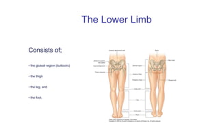
  • 1. vvvv Consists of; • the gluteal region (buttocks) • the thigh • the leg, and • the foot. The Lower Limb
  • 2. Functions of the Lower Limb Support the body weight The ligaments at the hip and knee joints facilitate locking of these joints therefore reducing the amount of muscular energy required to maintain a standing position. Locomotion To move the body through space. This involves integration of movements at all joints of the lower limb to place the foot on the ground and move the body over it.
  • 3. THE PELVIS BONE The pelvis is made up of the sacrum and the coccyx bound to each other by dense ligaments, Ilium, Ischium, and Pubis. The ilium with its iliac crest running between the anterior and posterior superior iliac spines; below each of these are the corresponding inferior spines. Well-defined ridges on its lateral surface are the strong muscle markings of the glutei. Its inner aspect bears the large auricular surface which articulates with the sacrum. The iliopectineal line runs forward from the apex of the auricular surface and demarcates the true from the false pelvis. The pubis comprises the body and the superior and inferior pubic rami.
  • 4.
  • 5. The ischium has a vertically disposed body, bearing the ischial spine on its posterior border which demarcates an upper (greater) and lower (lesser), sciatic notch. The inferior pole of the body bears the ischial tuberosity then projects forwards almost at right angles into the ischial ramus to meet the inferior pubic ramus. The obturator foramen lies bounded by the body and rami of the pubis and the body and ramus of the ischium. All three bones fuse at the acetabulum which forms the socket for the femoral head, for which it bears a wide crescentic articular surface.
  • 6. THE FEMUR The femur is the largest bone in the body. It is 18 in (45 cm) in length, a measurement it shares with the vas, the spinal cord and the thoracic duct and which is also the distance from the teeth to the cardia of the stomach. The femoral head is two-thirds of a sphere and faces upwards, medially and forwards. It is covered with cartilage except for its central fovea where the ligamentum teres is attached. The neck is 2 in (5 cm) long and is set at an angle of 125° to the shaft. In the female, with her wider pelvis, the angle is smaller. The junction between the neck and the shaft is marked anteriorly by the trochanteric line, laterally by the greater trochanter, medially and somewhat posteriorly by the lesser trochanter and posteriorly by the prominent trochanteric crest, which unites the two trochanters.
  • 7. THE FEMORAL SHAFT The femoral shaft is roughly circular in section at its middle but is flattened posteriorly at each extremity. Posteriorly also it is marked by a strong crest, the linea aspera. Inferiorly, this crest splits into the medial and lateral supracondylar lines, leaving a flat popliteal surface between them. The medial supracondylar line ends distally in the adductor tubercle. The lower end of the femur bears the prominent condyles which are separated by a deep intercondylar notch posteriorly but which blend anteriorly to form an articular surface for the patella. The lateral condyle is the more prominent of the two and acts as a buttress to assist in preventing lateral displacement of the patella
  • 8. The posterior aspect of right femur
  • 9. The sources of blood supply of the femoral head—along the ligamentum teres, through the diaphysis and via the retinacula. Through the diaphysis The blood supply to the femoral head is derived from vessels travelling up from the diaphysis along the cancellous bone, from vessels in the hip capsule, where this is reflected on to the neck in longitudinal bands or retinacula, and from the artery in the ligamentum teres; this third source is negligible in adults, but essential in children, when the femoral head is separated from the neck by the cartilage of the epiphyseal line
  • 10. CLINICAL FEATURES 1. The upper end of the femur is a common site for fracture in the elderly. The neck may break immediately beneath the head (subcapital), near its midpoint (cervical) or adjacent to the trochanters (basal), or the fracture line may pass between, along or just below the trochanter. Fractures of the femoral neck will interrupt completely the blood supply from the diaphysis and, should the retinacula also be torn, avascular necrosis of the head will be inevitable. The nearer the fracture to the femoral head, the more tenuous the retinacular blood supply and the more likely it is to be disrupted. Avascular necrosis of the femoral head in children is seen in Perthe’s disease and in severe slipped femoral epiphysis; both resulting from thrombosis of the artery of the ligamentum teres. In contrast, pertrochanteric fractures, being outside the joint capsule, leave the retinacula undisturbed; avascular necrosis, therefore, never follows such injuries. There is a curious age pattern of hip injuries; children may sustain greenstick fractures of the femoral neck, school boys may displace the epiphysis of the femoral head, in adult life the hip dislocates and, in old age, fracture of the neck of the femur again becomes the usual lesion.
  • 11. The head and neck of the femur, showing the terminology of the common fracture sites (a) A pertrochanteric fracture does not damage the retinacular blood supply—aseptic bone necrosis does not occur. (b) A subcapital fracture cuts off most of the retinacular supply to the head—aseptic bone necrosis is common. a b
  • 12. (2) Fractures of the femoral shaft are accompanied by considerable shortening due to the longitudinal contraction of the extremely strong surrounding muscles. The proximal segment is flexed by iliacus and psoas and abducted by gluteus medius and minimus, whereas the distal segment is pulled medially by the adductor muscles. Reduction requires powerful traction, to overcome the shortening, and then manipulation of the distal fragment to line with the proximal segment; the limb must therefore be abducted and also pushed forwards by using a large pad behind the knee. Fractures of the lower end of the shaft, immediately above the condyles, are relatively rare; fortunately so, because they may be extremely difficult to treat since the small distal fragment is tilted backwards by gastrocnemius, the only muscle which is attached to it. The sharp proximal edge of this distal fragment may also tear the popliteal artery, which lies directly behind it.
  • 13. The deformities of femoral shaft fractures. (a) Fracture of the proximal shaft—the proximal fragment is flexed by iliacus and psoas and abducted by gluteus medius and minimus. (b) Fracture of the mid-shaft— flexion of the proximal fragment by iliacus and psoas. (c) Fracture of the distal shaft—the distal fragment is angulated backwards by gastrocnemius—the popliteal artery may be torn in this injury. (In all these fractures overriding of the bone ends is produced by muscle spasm.)
  • 14. (3) The angle subtended by the femoral neck to the shaft may be decreased, producing a coxa vara deformity. This may result from adduction fractures, slipped of the femoral epiphysis or bone-softening diseases. Coxa valga, where the angle is increased, is much rarer but occurs in impacted abduction fractures. Note, however, that in children the normal angle between the neck and shaft is about 160°.
  • 15. THE PATELLA The patella is a sesamoid bone, the largest in the body, in the expansion of the quadriceps tendon, which continues from the apex of the bone as the ligamentum patellae. The posterior surface of the patella is covered with cartilage and articulates with the two femoral condyles by means of a larger lateral and smaller medial facet.
  • 16. Factors in the stability of the patella: (i) the medial pull of vastus medialis and (ii)the high patellar articular surface of the lateral femoral condyle. These resist the tendency for lateral displacement of the patella which results from the valgus angulation between the femur and the tibia
  • 17. CLINICAL FEATURES Lateral dislocation of the patella is resisted by the prominent articular surface of the lateral femoral condyle and by the medial pull of the lowermost fibers of vastus medialis which insert almost horizontally along the medial margin of the patella. If the lateral condyle of the femur is underdeveloped, or if there is a considerable genu valgum(knock-knee deformity), recurrent dislocations of the patella may occur.
  • 18.  A direct blow on the patella may split or shatter it but the fragments are not avulsed because the quadriceps expansion remains intact. The patella may also be fractured transversely by violent contraction of the quadriceps—for example, in trying to stop a backward fall. In this case, the tear extends outwards into the quadriceps expansion, allowing the upper bone fragment to be pulled proximally; there may be a gap of over 2 in (5 cm) between the bone ends. Reduction is impossible by closed manipulation and operative repair of the extensor expansion is imperative. Occasionally this same mechanism of sudden forcible quadriceps contraction tears the quadriceps expansion above the patella, ruptures the ligamentum patellae or avulses the tibial tubercle. It is interesting that following complete excision of the patella for a comminuted fracture, knee function and movement may return to 100% efficiency; it is difficult, then, to ascribe any particular function to this bone other than protection of the soft tissues of the knee joint anteriorly.
  • 19. TIBIA The upper end of the tibia is expanded into the medial and lateral condyles, the former having the greater surface area of the two. Between the condyles is the intercondylar area which bears, at its waist, the intercondylar eminence, projecting upwards slightly on either side as the medial and lateral intercondylar tubercles. The tuberosity of the tibia is at the upper end of the anterior border of the shaft and gives attachment to the ligamentum patellae. The anterior aspect of this tuberosity is subcutaneous, only excepting the infrapatellar bursa immediately in front of it. The shaft of the tibia is triangular in cross-section, its anterior border and anteromedial surface being subcutaneous throughout their whole extent. The posterior surface of the shaft bears a prominent oblique line at its upper end termed the soleal line, which not only marks the tibial origin of the soleus but also delimits an area above into which is inserted the popliteus. The lower end of the tibia is expanded and quadrilateral in section, bearing an additional surface, the fibular notch, for the lower tibiofibular joint The medial malleolus projects from the medial extremity of the bone and is grooved posteriorly by the tendon of tibialis posterior. The inferior surface of the lower end of the tibia is smooth, cartilage covered and forms, with the malleoli, the upper articular surface of the ankle joint..
  • 20. The tibia and fibula.
  • 21. CLINICAL FEATURES  The upper end of the tibial shaft is one of the most common sites for acute osteomyelitis. Fortunately, the capsule of the knee joint is attached closely around the articular surfaces so that the upper extremity of the tibial diaphysis is extracapsular; involvement of the knee joint therefore only occurs in the late and neglected case.  The shaft of the tibia is subcutaneous and unprotected anteromedially throughout its course and is particularly slender in its lower third. It is not surprising that the tibia is the commonest long bone to be fractured and to suffer compound injury.  The extensive subcutaneous surface of the tibia makes it a delightfully accessible donor site for bone-grafts.
  • 22. THE FIBULA The fibula serves three functions. It is: • an origin for muscles; • a part of the ankle joint; • a pulley for the tendons of peroneus longus and brevis. It comprises the head with a styloid process(into which is inserted the tendon of biceps), the neck(around which passes the common peroneal nerve; the shaft and the lower end or lateral malleolus. The latter bears a medial roughened surface for the lower tibiofibular joint, below which is the articular facet for the talus. A groove on the posterior aspect of the malleolus lodges the tendons of peroneus longus and brevis.
  • 23. NB, The epiphysis of the growing end of a long bone is the first to appear and last to fuse with its diaphysis; the exception is the epiphysis of the upper end of the fibula which, although at the growing end, appears after the distal epiphysis and fuses after the latter has blended with the shaft. The site of the growing end is of considerable practical significance; for example, if a child has to undergo and above-elbow amputation, the humeral upper epiphyseal line continues to grow and the elongating bone may well push its way through the stump end, requiring reamputation.
  • 24. THE HIP JOINT The hip is the largest joint in the body. It is a perfect example of a ball-and-socket joint. Its articular surfaces are the femoral head and the horse-shoe shaped articular surface of the acetabulum, which is deepened by the fibrocartilaginous labrum acetabulare. The non-articular lower part of the acetabulum, the acetabular notch, is closed off below by the transverse acetabular ligament. From this notch is given off the ligamentum teres, passing to the fovea on the femoral head. The capsule of the hip is attached proximally to the margins of the acetabulum and to the transverse acetabular ligament. Distally, it is attached along the trochanteric line, the bases of the greater and lesser trochanters and, posteriorly, to the femoral neck about 0.5 in (12 mm) from the trochanteric crest. From this distal attachment, capsular fibres are reflected on to the femoral neck as retinacula and provide one pathway for the blood supply to the femoral head. Note that acute osteomyelitis of the upper femoral metaphysis will involve the neck which is intracapsular and which will therefore rapidly produce a secondary pyogenic arthritis of the hip joint.
  • 25. HIP JOINT CONTD Three ligaments reinforce the capsule: 1. the iliofemoral (Y-shaped ligament of Bigelow)—which arises from the anterior inferior iliac spine, bifurcates, and is inserted at each end of the trochanteric line. 2. the pubofemoral—arising from the iliopubic junction to blend with the medial aspect of the capsule; 3. the ischiofemoral—arising from the ischium to be inserted into the base of the greater trochanter. Of these, the iliofemoral is by far the strongest and resists hyperextension strains on the hip. In posterior dislocation it usually remains intact. The synovium of the hip covers the non-articular surfaces of the joint and occasionally bulges out anteriorly to form a bursa beneath the psoas tendon where this crosses the front of the joint.
  • 26.
  • 27. Movements The hip is capable of a wide range of movements—flexion, extension, abduction, adduction, medial and lateral rotation and circumduction. The principal muscles acting on the joint are:  flexors—iliacus and psoas major assisted by rectus femoris, sartorius, pectineus; extensors—gluteus maximus, the hamstrings;  adductors—adductor longus, brevis and magnus assisted by gracilis and pectineus;  abductors—gluteus medius and minimus, tensor fasciae latae; lateral rotators—principally gluteus maximus assisted by the obturators, gemelli and quadratus femoris; medial rotators—tensor fasciae latae and anterior fibres of gluteus medius and minimus.
  • 28. Relations The hip joint is surrounded by muscles: anteriorly—iliacus, psoas and pectineus, together with the femoral artery and vein; laterally—tensor fasciae latae, gluteus medius and minimus; posteriorly—the tendon of obturator internus with the gemelli, quadratus femoris, the sciatic nerve and, more superficially, gluteus maximus; superiorly—the reflected head of rectus femoris lying in contact with the joint capsule; inferiorly—the obturator externus, passing back to be inserted into the trochanteric fossa.
  • 29. Surgical exposure of the hip joint therefore inevitably involves considerable and deep dissection. The lateral approach comprises splitting down through the fibres of tensor fasciae latae, gluteus medius and minimus on to the femoral neck. Further access may be obtained by detaching the greater trochanter with the gluteal insertions. The anterior approach passes between gluteus medius and minimus laterally and sartorius medially, then dividing the reflected head of rectus femoris to expose the anterior aspect of the hip joint. More room may be obtained by detaching these glutei from the external aspect of the ilium. The posterior approach is through an angled incision commencing at the posterior superior iliac spine, passing to the greater trochanter and then dropping vertically downwards from this point. Gluteus maximus is split in the line of its fibres and then incised along its tendinous insertion. Gluteus medius and minimus are detached from their insertions into the greater trochanter (or the trochanter is detached and subsequently wired back in place.) and an excellent view of the hip joint is thus obtained.
  • 30. NERVE SUPPLY The hip receives fibres from the femoral, sciatic and obturator nerves. It is important to note that these nerves also supply the knee joint and, for this reason, it is not uncommon for a patient, particularly a child, to complain bitterly of pain in the knee.
  • 31. CLINICAL FEATURES Trendelenburg’s test The stability of the hip in the standing position depends on two factors, the strength of the surrounding muscles and the integrity of the lever system of the femoral neck and head within the intact hip joint. When standing on one leg, the abductors of the hip on this side (gluteus medius and minimus and tensor fasciae latae) come into powerful action to maintain fixation at the hip joint, so much so that the pelvis actually rises slightly on the opposite side. If, however, there is any defect in these muscles or lever mechanism of the hip joint, the weight of the body in these circumstances forces the pelvis to tilt downwards on the opposite side. This positive Trendelenburg test is seen if the hip abductors are paralysed (e.g. poliomyelitis), if there is an old unreduced or congenital dislocation of the hip, if the head of the femur has been destroyed by disease or removed operatively (pseudarthrosis), if there is an un-united fracture of the femoral neck. The test may be said to indicate ‘a defect in the osseo-muscular stability of the hip joint’. A patient with any of the conditions enumerated above walks with a characteristic ‘dipping gait’.
  • 32. DISLOCATION OF THE HIP JOINT The hip is usually dislocated backwards and this is produced by a force applied along the femoral shaft with the hip in the flexed position (e.g. the knee striking against the opposite seat when a train runs into the buffers). If the hip is also in the adducted position, the head of the femur is unsupported posteriorly by the acetabulum and dislocation can occur without an associated acetabular fracture. If the hip is abducted, dislocation must be accompanied by a fracture of the posterior acetabular lip. The sciatic nerve, a close posterior relation of the hip, is in danger of damage in these injuries.
  • 33. Dislocation of the hip. If the hip is forced into posterior dislocation while adducted (a), there is no associated fracture of the posterior acetabular lip (b). Dislocation in the abducted position (c) can only occur with a concomitant acetabular fracture (The inset figure indicates the plane of these diagrams
  • 34. Reduction of a dislocated hip is quite simple providing that a deep anaesthetic is used to relax the surrounding muscles; the hip is flexed, rotated into the neutral position and lifted back into the acetabulum. Occasionally, forcible abduction of the hip will dislocate the hip forwards. Violent force along the shaft (e.g. a fall from a height) may thrust the femoral head through the floor of the acetabulum, producing a central dislocation of the hip.
  • 35. THE KNEE JOINT The knee is a hinge joint made up of the articulations between the femoral and tibial condyles and between the patella and the patellar surface of the femur.
  • 36. (a) The knee—anterior view; the knee is flexed and the patella has been turned downwards. (b) The right knee in transverse section
  • 37. The capsule is attached to the margins of these articular surfaces but communicates above with the suprapatellar bursa (between the lower femoral shaft and the quadriceps), posteriorly with the bursa under the medial head of gastrocnemius and often, through it, with the bursa under semimembranosus. It may also communicate with the bursa under the lateral head of gastrocnemius. The capsule is also perforated posteriorly by popliteus, which emerges from it in much the same way that the long head of biceps bursts out of the shoulder joint. The capsule of the knee joint is reinforced on each side by the medial and lateral collateral ligaments, the latter passing to the head of the fibula and lying free from the capsule. Anteriorly, the capsule is considerably strengthened by the ligamentum patellae, and, on each side of the patella, by the medial and lateral patellar retinacula, which are expansions from vastus medialis and lateralis. Posteriorly, the tough oblique ligament arises as an expansion from the insertion of semimembranosus and blends with the joint capsule.
  • 38. MOVEMENT OF THE KNEE JOINT The principal knee movements are flexion and extension, but note on yourself that some degree of rotation of the knee is possible when this joint is in the flexed position. In full extension, i.e. in the standing position, the knee is quite rigid because the medial condyle of the tibia, being rather larger than the lateral condyle, rides forward on the medial femoral condyle, thus ‘screwing’ the joint firmly together. The first step in flexion of the fully extended knee is ‘unscrewing’ or internal rotation. This is brought about by popliteus, which arises from the lateral side of the lateral condyle of the femur, emerges from the joint capsule posteriorly and is inserted into the back of the upper end of the tibia. The principal muscles acting on the knee are: I. extensor—quadriceps femoris; II. flexors—hamstrings assisted by gracilis, gastrocnemius and sartorius; III. medial rotator—popliteus (‘unscrews the knee’).
  • 39. CLINICAL FEATURES 1. The stability of the knee depends upon the strength of its surrounding muscles and of its ligaments. Of the two, the muscles are by far the more important. Providing quadriceps femoris is powerfully developed, the knee will function satisfactorily even in the face of considerable ligamentous damage. Conversely, the most skilful surgical repair of torn ligaments is doomed to failure unless the muscles are functioning strongly; without their support, reconstructed ligaments will merely stretch once more.
  • 40. 2. When considering soft tissue injuries of the knee joint, think of three Cs that may be damaged—the Collateral ligaments, the Cruciates and the Cartilages. The collateral ligaments are taut in full extension of the knee and are, therefore, only liable to injury in this position. The medial ligament may be partly or completely torn when a violent abduction strain is applied, whereas an adduction force may damage the lateral ligament.
  • 41. The cruciate ligaments may both be torn (along with the collateral ligaments) in severe abduction or adduction injuries. The anterior cruciate, which is taut in extension, may be torn by violent hyperextension of the knee or in anterior dislocation of the tibia on the femur. Since it resists rotation, it may also be torn in a violent twisting injury to the knee. The posterior cruciate tears in a posterior dislocation. If both the cruciate ligaments are torn, unnatural anteroposterior mobility of the knee can be demonstrated. If there is only increased forward mobility, the anterior cruciate ligament has been divided or is lax. Increased backward mobility implies a lesion of the posterior cruciate. The semilunar cartilages can only tear when the knee is flexed and is thus able to rotate.
  • 42. THETIBIOFIBULA JOINT • The tibia and fibula are connected by: I. The superior tibiofibular joint, a synovial joint between the head of the fibula and the lateral condyle of the tibia; II. The interosseous membrane, which is crossed by the anterior tibial vessels above and pierced by the perforating branch of the peroneal artery below; III. The inferior tibiofibular joint, a fibrous joint, the only one in the limbs, between the triangular areas of each bone immediately above the ankle joint.
  • 43. THE ANKLE JOINT The ankle is a hinge joint formed by the malleoli and lower end of the tibia and the body of the talus. The capsule of the joint fits closely around its articular surfaces, and, as in every hinge joint, it is weak anteriorly and posteriorly but reinforced laterally and medially by collateral ligaments.
  • 44. The ankle in coronal section.
  • 45. MOVEMENT OF THE ANKLE The ankle joint is capable of being flexed and extended (plantar- and dorsiflexion). The body of the talus is slightly wider anteriorly and, in full extension, becomes firmly wedged between the malleoli. Conversely, in flexion, there is slight laxity at the joint and some degree of side to side tilting is possible. The principal muscles acting on the ankle are:  dorsiflexors—tibialis anterior assisted by extensor digitorum longus, extensor hallucis longus and peroneus tertius;  plantarflexors—gastrocnemius and soleus assisted by tibialis posterior, flexor hallucis longus and flexor digitorum longus.
  • 46. 1. The collateral ligaments of the ankle can be sprained or completely torn by forcible abduction or adduction, the lateral ligament being far the more frequently affected. If the ligament is completely disrupted the talus can be tilted in its mortice; this is difficult to demonstrate clinically and is best confirmed by taking an anteroposterior radiograph of the ankle while forcibly inverting the foot. 2. The most usual ankle fracture is that produced by an abduction-external rotation injury; the patient catches his foot in a rabbit hole, his body and his tibia internally rotate while the foot is rigidly held. First there is a torsional spinal fracture of the lateral malleolus, then avulsion of the medial collateral ligament, with or without avulsion of a flake of the medial malleolus and, finally, as the tibia is carried forwards, the posterior margin of the lower end of the tibia shears off against the talus. These stages are termed 1st, 2nd and 3rd degree Pott’s fractures. Notice that, with widening of the joint, there is forward dislocation of the tibia on the talus, producing characteristic prominence of the heel in this injury.
  • 47. Inversion and eversion of the foot take place at the talocalcaneal articulations and at the mid-tarsal joints between the calcaneum and the cuboid and between the talus and the navicular. Of these, the talocalcaneal joint is the more important. Hold your calcaneus between your finger and thumb; inversion and eversion are prevented. Loss of these rotatory movements of the foot, e.g. after injury or because of arthritis, results in quite severe disability because the foot cannot adapt itself to walking on rough or sloping ground. Inversion is brought about by tibialis anterior and posterior assisted by the long extensor and flexor tendons of the hallux; eversion is the duty of peroneus longus and brevis, (peroneus tertius forms part of the extensor muscles). The other tarsal joints allow slight gliding movements only.
  • 48. THE ARCHES OF THE FOOT The longitudinal arches of the right foot. (a) Medial view. (b) Lateral view. The bones of the foot are arranged in the form of two longitudinal arches. The medial arch comprises calcaneus, talus, navicular, the three cuneiforms and the three medial metatarsals; the apex of this arch is the talus. The lateral arch, which is lower, comprises the calcaneus, cuboid and the lateral two metatarsals. The foot plays a double role; it functions as a rigid support for the weight of the body in the standing position, and as a mobile springboard during walking and running..
  • 49. The arches are maintained by: 1.The shape of the interlocking bones; 2.The ligaments of the foot; 3.muscle action. The ligaments concerned are: 1. The dorsal, plantar and interosseous ligaments between the small bones of the forefoot 2. The spring ligament, which passes from the sustentaculum tali of the calcaneus forward to the tuberosity of the navicular and which supports the inferior aspect of the head of the talus; the short plantar ligament which stretches from the plantar surface of the calcaneus to the cuboid; 3. The long plantar ligament which arises from the posterior tuberosity on the plantar surface of the calcaneus, covers the short plantar ligament, forms a tunnel for peroneus longus tendon with the cuboid, and is inserted into the bases of the 2nd, 3rd and 4th metatarsals. These ligaments are reinforced in their action by the plantar aponeurosis which is the condensed deep fascia of the sole of the foot.
  • 50. Plantar aspect of the left foot to show the attachments of the important ligaments and long tendons. (The head of the talus is hidden, deep to the spring ligament).
  • 51. Three important zones of the lower limb 1.The femoral triangle This triangle is bounded: superiorly—by the inguinal ligament; medially—by the medial border of adductor longus; laterally—by the medial border of sartorius. Its floor consists of iliacus, the tendon of psoas, pectineus and adductor longus. The roof is formed by the skin, superficial fascia, containing the superficial inguinal lymph nodes and the great saphenous vein with its tributaries and the deep fascia (fascia lata), which is pierced by the saphenous vein at the saphenous opening. The contents of the triangle are the femoral vein, artery and nerve together with the deep inguinal nodes.
  • 52. The femoral triangle and its content
  • 53. The fascia lata The deep fascia of the thigh, or fascia lata, extends downwards to ensheath the whole lower limb except over the subcutaneous surface of the tibia (to whose margins it adheres), and at the saphenous opening. Above, it is attached all around to the root of the lower limb—that is to say, to the inguinal ligament, pubis, ischium, sacrotuberous ligament, sacrum and coccyx and the iliac crest. The fascia of the thigh is particularly dense laterally (the iliotibial tract), where it receives tensor fasciae latae, and posteriorly, where the greater part of gluteus maximus is inserted into it. The iliotibial tract, when tensed by its attached muscles, assists in the stabilization of the hip and the extended knee when standing. The tough lateral fascia of the thigh is an excellent source of this material for hernia and dural repairs.
  • 54. The femoral sheath and femoral canal The femoral artery and vein enter the femoral triangle from beneath the inguinal ligament within a fascial tube termed the femoral sheath. This is derived from the extraperitoneal intra-abdominal fascia, its anterior wall arising from the transversalis fascia and its posterior wall from the fascia covering the iliacus. The medial part of the femoral sheath contains a small, almost vertically placed gap, the femoral canal, which is about 0.5 in (12 mm) in length and which just admits the tip of the little finger. The greater width of the female pelvis means the canal is somewhat larger in the female and femoral hernia are, consequently, commoner in this sex.
  • 55. The boundaries of the femoral canal are: anteriorly—the inguinal ligament; medially—the sharp free edge of the pectineal part of the inguinal ligament, termed the lacunar ligament(Gimbernat’s ligament); laterally—the femoral vein; posteriorly—the pectineal ligament (of Astley Cooper), which is the thickened periosteum along the pectineal border of the superior pubic ramus and which continues medially with the pectineal part of the inguinal ligament. The canal contains a plug of fat and a constant lymph node—the node of the femoral canal or Cloquet’s gland. The canal has two functions: first, as a dead space for expansion of the distended femoral vein and, second, as a lymphatic pathway from the lower limb to the external iliac nodes.
  • 56. The femoral canal and its surrounds. NB, The femoral nerve lies outside the femoral sheath
  • 57. Femoral hernia The great importance of the femoral canal is, of course, that it is a potential point of weakness in the abdominal wall through which may develop a femoral hernia. Unlike the indirect inguinal hernia, this is never due to a congenital sac and, although cases do occur rarely in children, it is never found in the newborn.
  • 58. The relationship of an indirect inguinal and a femoral hernia to the pubic tubercle; the inguinal hernia emerges above and medial to the tubercle, the femoral hernia lies below and lateral to it.
  • 59. The adductor canal (of Hunter) or subsartorial canal This canal leads on from the apex of the femoral triangle. Its boundaries are: posteriorly—adductor longus and magnus; anterolaterally—vastus medialis; anteromedially—the sartorius, which lies on a fascial sheet, forming the roof of the canal. The contents of the canal are the femoral artery, the femoral vein (which lies behind the artery), the saphenous nerve and, in its upper part, the nerve to vastus medialis from the femoral nerve. John Hunter described the exposure and ligation of the femoral artery in this canal for aneurysm of the popliteal artery; this method has the advantage that the artery at this site is healthy and will not tear when tied, as may happen if ligation is attempted immediately above the aneurysm.
  • 60. The popliteal fossa The popliteal fossa is the distal continuation of the adductor canal. This ‘fossa’ is, in fact, a closely packed compartment which only becomes the rhomboid-shaped space of anatomical diagrams when opened up at operation or by dissection. Its boundaries are: superolaterally—biceps tendon; superomedially—semimembranosus reinforced by semitendinosus; inferomedially and inferolaterally—the medial and lateral heads of gastrocnemius. The roof of the fossa is deep fascia which is pierced by the small saphenous vein as this enters the popliteal vein, three cutaneous nerves Its floor, from above down, is formed by: the popliteal surface of the femur; the posterior aspect of the knee joint; the popliteus muscle covering the upper posterior surface of the tibia. From without in, the popliteal fossa contains nerves, vein and artery
  • 61. The popliteal fossa. (a) Superficial dissection. (b) Deep. (c)Floor.
  • 62. Clinical features The popliteal fossa is another good example of the value of thinking anatomically when considering the differential diagnosis of a mass situated in a particular anatomical area. When examining a lump in the popliteal region, let these possibilities pass through your mind: • skin and soft tissues—sebaceous cyst, lipoma, sarcoma; • vein—varicosities of the short saphenous vein in the roof of the fossa; • artery—popliteal aneurysm; • lymph nodes—infection secondary to suppuration in the foot; • knee joint—joint effusion; • tendons—enlarged bursae, especially those beneath semimembranosus and the heads of gastrocnemius; • bones—a tumour of the lower end of femur or upper end of tibia.
  • 63. The arteries of the lower limb Femoral artery The femoral artery is the distal continuation of the external iliac artery beyond the inguinal ligament. It traverses the femoral triangle and the adductor canal of Hunter, then terminates a hand’s breadth above the adductor tubercle by passing through the hiatus in adductor magnus to become the popliteal artery. Throughout its course, the femoral artery is accompanied by its vein, which lies first on the medial side of the artery and then passes posteriorly to it at the apex of the femoral triangle.
  • 64. Branches In the groin, the femoral artery gives off: • the superficial circumflex iliac artery; • the superficial epigastric artery; • the superficial external pudendal artery. These three vessels are encountered in the groin incision for repair for an inguinal hernia. Their corresponding veins drain into the great saphenous vein. The profunda femoris arises posterolaterally from the femoral artery 2 in (5 cm) distal to the inguinal ligament. It is conventional to call the femoral artery above this branch the common femoral, and below it, the superficial femoral artery. The profunda passes deep to adductor longus and gives off medial and lateral circumflex branches and four perforating branches. These are important both as the source of blood supply to the great muscles of the thigh and as collateral channels which link the rich arterial anastomoses around the hip and the knee.
  • 65. CLINICAL ANATOMY 1. The femoral artery in the upper 4 in (10 cm) of its course lies in the femoral triangle where it is quite superficial and, in consequence, easily injured. A laceration of the femoral artery at this site is an occupational hazard of butchers and bullfighters. 2. The femoral artery at the groin is readily punctured by a hypodermic needle and is the most convenient site from which to obtain arterial blood samples. Arteriography of the peripheral leg vessels is also easily performed at this point 3. Arteriosclerotic changes, with consequent thrombotic arterial occlusion, frequently commence at the lower end of the femoral artery, perhaps as a result of compression of the diseased vessel by the margins of the hiatus in adductor magnus. Collateral circulation is maintained via anastomoses between the branches of profunda femoris and the popliteal artery. If arteriography demonstrates a patent arterial tree distal to the block, it is possible to bypass the occluded segment by means of a graft between the common femoral and popliteal arteries.
  • 66. Popliteal artery The popliteal artery continues on from the femoral artery at the adductor hiatus and terminates at the lower border of the popliteus muscle. It lies deep within the popliteal fossa, being covered superficially by the popliteal vein and, more superficially still, crossed by the tibial nerve. The popliteal artery gives off muscular branches, geniculate branches (to the knee joint) and terminal branches, the anterior and posterior tibial arteries.
  • 67. Clinical features 1. Aneurysm of the popliteal artery, once common, is now rare. Its frequency in former days was associated with the repeated traumata of horse-riding and the wearing of high riding-boots. Pressure of the aneurysm on the adjacent vein may cause venous thrombosis and peripheral oedema; pressure on the tibial nerve may cause severe pain in the leg. 2. The popliteal artery is exposed by deep dissection in the midline within the popliteal fossa, care being taken not to injure the more superficial vein and nerve. It can also be exposed by a medial approach, which divides the insertion of adductor magnus and detaches the origin of the medial head of gastrocnemius from the tibia.
  • 68. Posterior tibial artery The posterior tibial artery is the larger of the terminal branches of the popliteal artery. It descends deep to soleus, where it can be exposed by splitting gastrocnemius and soleus in the midline, then becomes superficial in the lower third of the leg and passes behind the medial malleolus between the tendons of flexor digitorum longus and flexor hallucis longus. It is accompanied by its corresponding vein and by the tibial nerve. Below the ankle, the posterior tibial artery divides into the medial and lateral plantar arteries which constitute the principal blood supply to the foot. As well as branches to muscles and skin and a large nutrient branch to the tibia, the posterior tibial artery gives off the peroneal artery about 1.5 in (4 cm) from it origin. The peroneal artery runs down the posterior aspect of the fibula, close to the medial margin of the bone, supplying adjacent muscles and giving a nutrient branch to the fibula. Above the ankle it gives off its perforating branch which pierces the interosseous membrane, descends over the lateral malleolus and anastomoses with the arteries of dorsum of the foot.
  • 69. Anterior tibial artery The anterior tibial artery arises at the bifurcation of the popliteal artery. It passes forwards between the tibia and fibula under the lower border of popliteus over the upper margin of the interosseous membrane and descends on this structure in the anterior compartment of the leg. At first deeply buried, it becomes superficial just above the ankle between the tendons of extensor hallucis longus and tibialis anterior, being crossed superficially by the former immediately proximal to the line of the ankle joint. The artery continues over the dorsum of the foot as the dorsalis pedis (where its pulse may be palpated); this gives off the arcuate which, in turn, supplies cutaneous branches to the backs of the toes. Dorsalis pedis itself plunges between the 1st and 2nd metatarsals to join the lateral plantar artery in the formation of the plantar arch, from which branches run forwards to supply the plantar aspects of the toes.
  • 70. The relations of the posterior tibial artery as it passes behind the medial malleolus
  • 71. The veins of the lower limb The veins of the lower limb are divided into the deep and superficial groups according to their relationship to the investing deep fascia of the leg. The deep veins accompany the corresponding major arteries. The superficial veins are the great and small(or long and short) saphenous veins and their tributaries. The small (short) saphenous vein commences at the ankle behind the lateral malleolus where it drains the lateral side of the dorsal venous plexus of the foot. It courses over the back of the calf, perforates the deep fascia over the popliteal fossa and terminates in the popliteal vein. One or more branches run upwards and medially from it to join the great saphenous vein. The small saphenous vein is accompanied by the sural nerve—a sensory branch of the tibial nerve, which may be damaged in operating on varices of this vein.
  • 72. The great (long) saphenous vein drains the medial part of the venous plexus on the dorsum of the foot and passes upwards immediately in front of the medial malleolus; here branches of the saphenous nerve lie in front of and behind the vein. The vein then ascends over the posterior parts of the medial condyles of the tibia and femur to the groin where it pierces the deep fascia at the saphenous opening 1in(2.5 cm) below the inguinal ligament, to enter the femoral vein immediately medial to the femoral pulse. The great saphenous vein is joined by one or more branches from the small saphenous, and by the lateral accessory vein which usually enters the main vein at the mid-thigh, although it may not do so until the saphenous opening is reached. At the groin a number of tributaries from the lower abdominal wall, thigh and scrotum enter the great saphenous vein; these tributaries are variable in number and arrangement but usually comprise 1. the superficial epigastric vein; 2. the superficial circumflex iliac vein; 3. the superficial external pudendal vein.
  • 73. The superficial vains of the lower limb
  • 74. Clinical features 1. Knowledge of the constant position of the great saphenous vein lying immediately in front of the medial malleolus is of great importance. Its presence at this site, even if not visible in an obese or collapsed patient, may be life-saving when urgent transfusion is required. Occasionally, the immediately adjacent saphenous nerve is caught up by a ligature during this procedure—the patient, if conscious, will complain bitterly of pain if this is done. 2. The saphenous veins frequently become dilated, incompetent and varicose. Usually this is idiopathic but may result from the increased venous pressure caused by more proximal venous obstruction (a pelvic tumour or the pregnant uterus, for example) or may be secondary to obstruction of the deep venous pathway of the leg by thrombosis. 3. Stagnation of blood in the skin of the lower limb may result from venous thrombosis or valve incompetence; the skin, in consequence, is poorly nourished and easily breaks down into a varicose ulcerif subjected to even minor trauma. This is especially liable to occur over the subcutaneous anteromedial surface of the tibia where the cutaneous blood supply is least generous. 4. In operating upon varicose veins it is important that all tributaries of the groin are ligated as well as the main saphenous trunk; if one tributary escapes, it in turn becomes dilated and produces recurrence of the varices.
  • 75. The course and distribution of the principal nerves of the lower limb The nerves of the lower limb are derived from the lumbar and sacral plexuses: The lumbar plexus: The lumbar plexus originates from the anterior primary rami of L1–4. The trunks of the plexus traverse psoas major and emerge from its lateral border. There are two exceptions: the obturator nerve appears at the medial border of psoas tendon, and the genitofemoral nerve emerges on the anterior aspect of the muscle. The principal branches of the plexus are the femoral nerve and the obturator nerve. The femoral nerve (L2– 4) passes through the substance of psoas then under the inguinal ligament a finger’s breadth lateral to the femoral artery, to break up into its terminal branches after a course in lower limb of only some 2 in (5 cm).Its branches are: 1. muscular—to the anterior compartment of the thigh (quadriceps, sartorius and pectineus); 2. cutaneous—the medial and intermediate cutaneous nerves of the thigh and the saphenous nerve, which traverses the adductor canal to supply the skin of the medial side of the leg, ankle and foot to the great toe; 3. articular—to the hip and knee joints. The femoral nerve supplies the skin of the medial and anterior aspects of the thigh via its medial and intermediate cutaneous branches, but the lateral aspect is supplied by the lateral cutaneous nerve of the thigh (L2–3). This arises directly from the lumbar plexus and enters the thigh usually by passing deep to the inguinal ligament.
  • 76. The obturator nerve(L2–4) emerges from the medial aspect of the psoas and runs downwards and forwards, deep to the internal iliac vessels, to reach the superior part of the obturator foramen. This nerve traverses, in company with the obturator vessels, to enter the thigh. Its branches are: 1. muscular—to obturator externus, the adductor muscles and gracilis; 2. cutaneous—to an area of skin over the medial aspect of the thigh; 3. articular—to the hip and knee joints.
  • 77. Plan of the lumbar plexus (muscular branches have been omitted for clarity)
  • 78. Clinical features 1. Spasm of the adductor muscles of the thigh in spastic paraplegia can be relieved by division of the obturator nerve (obturator neurectomy) 2. Rarely, an obturator hernia develops through the canal where the obturator nerve and vessels traverse the membrane covering the obturator foramen. Pressure of a strangulated obturator hernia upon the nerve causes referred pain in its area of cutaneous distribution, so that intestinal obstruction associated with pain along the medial side of the thigh is suggestive of this diagnosis. 3. The femoral and obturator nerves, as well as the sciatic nerve and its branches, supply sensory fibres to both the hip and the knee; it is not uncommon for hip disease to present, disguised as pain in the knee.
  • 79. The sacral plexus This plexus originates from the anterior primary rami of L4–5, S1– 4. Note that L4 is shared by both plexuses, a branch from it joining L5 to form the lumbosacral trunk which carries its contribution to the sacral plexus. The sacral nerves emerge from the anterior sacral foramina and unite in front of piriformis where they are joined by the lumbosacral trunk. Branches from the plexus supply: 1. the pelvic muscles; 2. the muscles of the hip; 3. the skin of the buttock and the back of the thigh. The plexus itself terminates as the pudendal nerve and the sciatic nerve. The pudendal nerve supply the skin of the external genitalia.
  • 80. Plan of the sacral plexus.
  • 81. Pudendal nerve contd It arises as the lower main division of the sacral plexus although it is dwarfed by the giant sciatic nerve. It leaves the pelvis through the greater foramen below the piriformis muscle. Within the canal it first gives off the inferior rectal nerve, which crosses the fossa to innervate the external anal sphincter and the perianal skin, and then divides into the perineal nerve and the dorsal nerve of the penis (or clitoris). The perineal nerve is the larger of the two.
  • 82. Clinical features The pudendal nerve can be blocked with local anaesthetic prior to forceps delivery by inserting a long needle through the vaginal wall and guided by a finger to the ischial spine, which can be palpated per vaginam. Alternatively, the needle can be introduced just medial to the ischial tuberosity to a depth of 1 in (2.5 cm). When the procedure is carried out bilaterally there is loss of the anal reflex (which is a useful test that a successful block has been achieved), relaxation of the pelvic floor muscles and loss of sensation to the vulva and lower one third of the vagina.
  • 83. The sciatic nerve The sciatic nerve (L4, 5, S1–3) is the largest nerve in the body. It is broad and flat at its origin, although peripherally it becomes rounded. The nerve emerges from the greater sciatic foramen distal to piriformis and under cover of gluteus maximus, crosses the posterior surface of the ischium, crosses obturator internus, with its gemelli, quadratus femoris and descends on adductor magnus. Here it lies deep to the hamstrings and is crossed only by the long head of biceps. The sciatic nerve terminates by dividing into the tibial and common peroneal nerves. The level of this division is variable—usually it is at the mid- thigh, but the two nerves may be separate even at their origins from the sacral plexus.
  • 84. Branches The trunk of the sciatic nerve supplies the hamstring muscles (biceps, semimembranosus, semitendinosus) and also the adductor magnus, the latter being innervated also by the obturator nerve. All the muscle branches apart from the one to the short head of biceps arise on the medial side of the nerve; its lateral border is therefore the side of relative safety in its operative exposure. The tibial nerve(L4, 5, S1–3) is the larger of the two terminal branches of the sciatic nerve; it traverses the popliteal fossa superficial to the popliteal vein and artery, which it crosses from the lateral to the medial side.
  • 85. Branches (a)in popliteal fossa muscular—to gastrocnemius, soleus and popliteus; cutaneous—the sural nerve, which descends over the back of the calf, behind the lateral malleolus to the 5th toe; it receives a communicating branch from the common peroneal nerve and supplies the lateral side of the leg, foot and 5th toe; articular—to the knee joint. It then descends deep to soleus, in company with the posterior tibial vessels, passes on their lateral side behind the medial malleolus to end by dividing into the media land lateral plantar nerves. (b)in the leg The tibial nerve supplies flexor halluces longus, flexor digitorum longus and tibialis posterior. Its terminal plantar branches supply the intrinsic muscles and skin of the sole of the foot, the medial plantar nerve having an equivalent distribution to that of the median nerve in the hand, the lateral plantar nerve being comparable to the ulnar nerve.
  • 86. The common peroneal (fibular) nerve The common peroneal nerve (L4, 5, S1, 2) is the smaller of the terminal branches of the sciatic nerve. It enters the upper part of the popliteal fossa, passes along the medial border of the biceps tendon, then curves around the neck of the fibula where it lies in the substance of peroneus longus and divides into its terminal branches, the deep peroneal and superficial peroneal nerves.
  • 87. Dissection of the sciatic nerve in the thigh and popliteal fossa
  • 88. “This Day, We shall seal up their mouths, and their hands will speak to Us, and their legs will bear witness to what they used to earn.” (Q 36:35)