SlideShare uma empresa Scribd logo
1 de 4
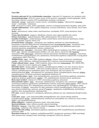
Neuro DDx EC
Persistent embyronic ICA to vertebrobasilar connections: trigeminal (#1), hypoglossal, otic, proatlantic artery
Intraaxial hemorrhage: HTN (#1), tumor, trauma, AVM, aneurysm, coagulopathy, amyloid angiopathy, emboli,
hemorrhagic infarction, vasculitis, HTN encephalopathy (eclampsia, cyclosporine)
Aneurysm: saccular – degenerative, trauma, mycotic, vasculopathies; fusiform – atherosclerotic; dissecting –
trauma > vasculopathy > spontaneous
SAH: aneurysm (#1), trauma, AVM, coagulopathy, extension of intraparenchymal bleed, idiopathic, spinal AVM
Vascular malformations: AVM, capillary telangiectasia, cavernous malformation, venous anomaly, vein of Galen
AVM or varix
Stroke: atherosclerosis, cardiac emboli, arterial dissection, vasculopathy, IVDA, venous thrombosis, blood
dyscrasias
Venous sinus thrombosis: pregnancy, dehydration, infection, tumor, hypercoagulable state, trauma
Primary brain injury: epidural hematoma, SDH, subdural hygroma, DAI, cortical contusion
Secondary brain injury: cerebral herniation, diffuse cerebral edema, arterial dissection (spontaneous, trauma,
HTN, vasculopathy, migraine, IVDA), CCF
Extraaxial masses: hemisphere – meningioma, mets, lymphoma, arachnoid cyst, dermoid/epidermoid,
hemorrhagic or infectious fluid collections, hemangiopericytoma; clivus/prepontine cistern – meningioma, mets,
chordoma, chondrosarcoma; CPA mass – acoustic neuroma, meningioma, mets, epidermoid, petrous apex
cholesterol granuloma, paraganglioma, aneurysm, arachnoid cyst
Intraaxial masses: hemisphere – astrocytoma, GBM, oligodendroglioma, lymphoma, mets, PNET, ganglioglioma,
DNET; sella – adenoma, craniopharyngioma, Rathke’s cyst, mets, abscess, sarcoid, EG stalk; brainstem –
astrocytoma, GBM; pineal region – germinoma, teratoma, pineocytoma, pineoblastoma; cerebellum – astrocytoma,
medulloblastoma, hemangioblastoma, mets, ependymoma, choroid plexus papilloma; temporal lobe in young patient
– ganglioglioma, DNET, JPA, PXA
Multiple lesions: tumor – mets, GBM, lymphoma; infection – abscess, fungus, cysticercosis, toxoplasmosis;
vascular – embolic infarctions, multifocal hemorrhage, DAI, contusions, cavernous hemangiomas, vasculitis
Corpus callosum lesions: tumors – GBM, lymphoma, lipoma, mets (rare); demyelinating – MS, ADEM, PML;
infarct – always also involves cingulate gyrus; trauma - DAI
Intrasellar masses: pituitary adenoma, apoplexy, craniopharyngioma, Rathke’s cyst, mets, aneurysm, abscess
Suprasellar masses: SATCHMOE – sellar lesion extending superiorly, sarcoid, aneurysm, arachnoid cyst,
teratoma, craniopharyngioma, hypothalamic glioma, mets, meningioma, optic nerve glioma, EG,
epidermoid/dermoid; adults – macroadenoma (#1), meningioma, glioma, craniopharyngioma, aneurysm; children –
craniopharyngioma (#1), glioma, germinoma, hypothalamic hamartoma, EG
Posterior fossa tumors: adults – mets, hemangioblastoma, astrocytoma, choroid plexus papilloma, meningioma,
epidermoid, dysplastic gangliocytoma; children -–JPA, medulloblastoma, brainstem glioma, ependymoma
Brain tumor in infant: teratoma (#1), PNET, choroid plexus papilloma, astrocytoma
Intraventricular tumors: adults – astrocytoma, subependymoma, meningioma, mets, cysticercosis; children –
choroid plexus papilloma, ependymoma, medulloblastoma, teratoma, astrocytoma; lateral ventricle – PNET, choroid
plexus papilloma, glioma, JPA, subependymoma, astrocytoma, mets, oligodendroglioma, meningioma, central
neurocytoma; 3rd
ventricle – astrocytoma, EG stalk, germinoma, extrinsic craniopharyngioma, colloid cyst, glioma,
mets, pituitary or pineal mass, aneurysm, sarcoid; 4th
ventricle – ependymoma, medulloblastoma, choroid plexus
papilloma, mets, hemangioblastoma, subependymoma
CPA mass: acoustic neuroma (#1), meningioma, epidermoid, arachnoid cyst, mets, ependymoma through Luschka,
lipoma
Cystic mass: tumor – cystic astrocytoma/GBM, hemangioblastoma, mets, necrotic pituitary adenoma,
craniopharyngioma; benign – epidermoid/dermoid, arachnoid cyst, porencephalic cyst, colloid cyst, cavum variants,
Rathke’s cyst, thrombosed aneurysm
Diffuse infiltrative: lymphoma, gliomatosis cerebri, white matter disease
Restricted diffusion: infarct, infection, possibly subacute hemorrhage
Metastasis: intraaxial – lung, breast, colon, melanoma; extraaxial – breast, lymphoma, prostate, neuroblastoma;
hemorrhagic – melanoma, renal, chorioCA, thyroid
Tumors with CSF seeding: choroid plexus papilloma, ependymoma, PNET (medulloblastoma), piineoblastoma,
germinoma, GBM
Hyperdense lesion on CT: lymphoma, medulloblastoma/PNET, ependymoma, germinoma, GBM, hemorrhagic
mets, mucinous mets, osteogenic tumor, hemorrhage, meningioma, colloid cyst, aneurysm
Calcified intraparenchymal lesions: oligodendroglioma, ependymoma, mucinous adenoCA, osteogenic sarcoma,
toxoplasmosis, CMV, cysticercosis, TB, AVM, aneurysm, TS, Sturge-Weber, hematoma; sellar lesions –
meningioma, craniopharyngioma, germ cell tumor, aneurysm
T2 hypointense lesions: ferritin, hemosiderin, deoxyhemoglobin, intracellular methemoglobin, melanin,
calcification, lymphoma, myeloma, neuroblastoma, fibrous tissue (meningioma), high protein concentration, flow
void
T1 hyperintense lesions: Gd, methemoglobin, melanin, certain states of calcium, fat (dermoid), high protein
concentration (colloid cyst), slow flow
Lesions with no enhancement: cysts, tumors with intact BBB (low-grade gliomas)
Lesions with strong enhancement: meningioma, medulloblastoma/PNET, AVM, paraganglioma, aneurysm, HIV-
associated lymphoma, GBM
Ring enhancement: mets, abscess, GBM, infarct, contusion, AIDS, lymphoma, demyelinating, resolving
hematoma, radiation
Diffuse meningeal enhancement: meningitis, carcinomatosis (lymphoma, mets), post-op, SAH, intracranial
hypotension, CSF leak
Basilar meningeal enhancement: infection - TB (#1), fungal, pyogenic (more common on convexity),
cysticercosis; tumor – lymphoma, leukemia, carcinomatosis; inflammatory – sarcoid, rheumatoid pachymeningitis,
drugs, pantopaque, ruptured dermoid
Ependymal enhancement: tumor – lymphoma, mets, CSF seeding (PNET, GBM); infection – spread of
meningitis, CMV (rare); inflammatory ventriculitis – postshunt or after instrumentation, posthemorrhage
T2 hypointense basal ganglia lesions: old age, any chronic degenerative disease (MS, Parkinson’s), childhood
hypoxia
T2 hyperintense basal ganglia lesions: tumor – lymphoma, NF; ischemia – hypoxic encephalopathy, venous
infarction; neurodegenerative diseases (uncommon), Leigh’s dz; toxin – CO, CN, H2S poisoning, hypoglycemia,
methanol; infection – Cryptococcus, parasites
T1 hyperintense basal ganglia lesions: dystrophic calcifications (any cause), hepatic failure, NF, manganese
Basal ganglia calcification: physiologic (#1), hypoparathyroid, HPT, TORCH, AIDS, TB, toxoplasmosis,
cysticercosis (common), lead, CO, radiation, chemotherapy, Fahr’s disease, mitochondrial (common), ischemic-
hypoxic injury
White matter disease: demyelinating (MS, ADEM, CPM), dysmyelinating (leukodystrophies), tumor (lymphoma,
mets) vasculopathies (small vessel ischemic dz, vasculitis, HTN, eclampsia, migraines, radiation, chemotherapy,
cyclosporine, IVDA), inflammatory (Lyme, sarcoid, HIV, PML, CMV)
Wallerian degeneration: infarction, trauma, demyelinating, radiation, neurodegenerative, tumor
Neurodegenerative disorders: WM – demyelinating, dysmyelinating; GM – Alzheimer’s, Pick’s, multiinfarct
dementia, Parkinson’s, lysosomal storage disorders, Wernicke’s, Creutzfeldt-Jakob, mesial temporal sclerosis; BG –
Huntington’s, Wilson’s, Fahr’s, Leigh’s, ALS
Cerebellar atrophy: oligopontocerebellar degeneration, alcohol, dilantin, hemosiderin deposition
Noncommunicating hydrocephalus: Foramen of Monro obstruction – 3rd
ventricle tumors, colloid cyst,
oligodendroglioma, central neurocytoma, giant cell astrocytoma in TS, ependymoma, suprasellar tumors; aqueduct
obstruction – congenital aqueductal stenosis, ventriculitis, IVH, tumor (mesencephalic, pineal, posterior 3rd
ventricle
region); 4th
ventricle obstruction – DW malformation, IVH, infection, subependymoma, exophytic brainstem glioma,
posterior fossa tumors
Communicating hydrocephalus: meningitis (infectious, carcinomatous), SAH, surgery, venous thrombosis; NPH
Cystic supratentorial congenital anomalies: holoprosencephaly, hydrancephaly, aqueductal stenosis, callosal
dysgenesis, porencephaly, arachnoid cyst, cystic teratoma, epidermoid/dermoid, vein of Galen AVM
Posterior fossa cystic abnormalities: DW malformation (vermian hypoplasia/aplasia and large posterior fossa),
DW variant (normal size posterior fossa and vermian hypoplasia), megacisterna magna (normal vermis),
retrocerebellar arachnoid cyst (must show mass effect), Chiari 4 (near complete absence of cerebellum),
epidermoid/dermoid, cystic tumor, Joubert’s syndrome (superior vermian hypoplasia/aplasia),
rhomboencephalosynapsis (vermian hypoplasia/aplasia + fusion)
Absent septum pellucidum: holoprosencephaly, ACC, septooptic dysplasia, Chiari 2
Migration and sulcation anomalies: lissencephaly, schizencephaly, polymicrogyria, pachygyria, cortical
heterotopia (focal, diffuse, subependymal), hemimegalencephaly
Phakomatoses: NF, TS, VHL, Sturge-Weber
Diffuse marrow involvement: mets, myeloma, lymphoma, leukemia, anemia, Paget’s, FD
Spine DDx
Spinal cord compression: criteria – no CSF seen around cord, narrowed AP diameter of cord (<7mm), deformity
of cord; causes – infection (TB, pyogenic), compression fracture (CA, trauma), spondylosis and disk disease
(herniated nucleus, hypertrophy of ligaments, osteophyte, facet hypertrophy), primary bone disorders (Paget’s),
epidural hematoma
Intramedullary lesions: astrocytoma (#1), ependymoma (#2), hemangioblastoma, lymphoma, mets (rare),
demyelinating disease/myelitis, syrinx, AVM, trauma (contusion), radiation, sarcoid, infection (rare), infarction
Intradural extramedullary lesions: nerve sheath tumor (#1), meningioma, drop mets, lipoma, teratomatous lesion,
arachnoid cyst, arachnoiditis/meningitis, AVM/AVF, ependymoma, sequestered disc fragment, lymphoma, sarcoid,
pantopaque
Extradural lesions: disc disease, mets, lymphoma, epidural abscess, epidural hematoma, lipomatosis (thoracic),
synovial cyst, extramedullary hematopoiesis, Tarlov cyst, discitis/osteomyelitis, spondylolysis, RA
Syrinx: primary – Chiari malformations, spinal dysraphism, DW, diastematomyelia; acquired – tumor
(astrocytoma, ependymoma), trauma (spinal cord injury, vascular insult), inflammatory (arachnoiditis/meningitis,
SAH)
Head and Neck DDx
External auditory canal: exostoses, malignant otitis externa, atresia
Clivus mass: chordoma, chondrosarcoma, plasmacytoma, mets, lymphoma, FD, EG
Petrous apex mass: cholesterol granuloma, mucocele, petrous apicitis, epidermoid, mets, myeloma,
chondrosarcoma, meningioma, aneurysm
Soft tissue mass in middle ear: cholesteatoma, cholesterol granuloma, glomus tympanicum tumor, aberrant ICA,
high or dehiscent jugular bulb
Intracanalicular IAC masses: exclusively intracanalicular – acoustic neuroma, facial neuroma, hemangioma,
lipoma; not primarily intracanalicular – meningioma, epidermoid
Hearing loss: conductive – otitis media, cholesteatoma, otosclerosis, trauma (longitudinal fracture); sensorineural –
idiopathic hereditary, acoustic neuroma, trauma (transverse fracture)
Pulsatile tinnitus: aberrant ICA, jugular bulb anomalies, glomus jugulare, glomus tympanicum, AVM, ICA
aneurysm at petrous apex
Jugular fossa mass: glomus jugulare (#1), NF (#2), schwannoma, chondrosarcoma, mets
Orbital masses by etiology: tumors – hemangioma (adults: cavernous; children: capillary), lymphoma, mets
(neuroblastoma, breast), lymphangioma, rhabdomyosarcoma, hemangiopericytoma, neurofibroma; inflammatory –
pseudotumor, thyroid ophthalmopathy, cellulitis, abscess, Wegener’s; vascular – carotid-cavernous fistula, venous
varix, thrombosis of superior ophthalmic vein; trauma – hematoma, FB, lens dislocation
Extraconal disease: nasal disease – infection, neoplasm; orbital bone disease – subperiosteal abscess,
osteomyelitis, FD, tumors, trauma; sinus disease – mucocele, invasive infections, neoplasm; lacrimal gland disease –
adenitis, lymphoma, pseudotumor, tumor
Intraconal disease: well-defined margins – hemangioma, schwannoma, orbital varix, meningioma; ill-defined
margins – pseudotumor, infection, lymphoma, mets; muscle enlargement – pseudotumor, Graves’, myositis, carotid-
cavernous fistula
Vascular orbital lesions: tumor – hemangioma, lymphangioma, hemangioendothelioma, hemangiopericytoma,
meningioma, hypervascular mets; vascular (with enlarged superior ophthalmic vein) – carotid cavernous fistula,
cavernous thrombosis, orbital varix, ophthalmic artery aneurysm
Optic neuritis: abnormal T2 signal and enhancement but not enlarged – MS, sarcoid, infection
Optic neuropathy: abnormal T2 signal only – compression, ischemia, pharmacologic, toxins, trauma
Optic nerve tumor: abnormal T2 signal and enhancement and nerve enlarged – glioma, meningioma
Optic nerve sheath enlargement: tumor – optic nerve glioma, meningioma, meningeal carcinomatosis, mets,
lymphoma, leukemia; inflammatory – optic neuritis, pseudotumor, sarcoid; increased intracranial pressure; trauma –
hematoma
Tramtrack enhancement of orbital nerve: optic nerve meningioma, optic neuritis, idiopathic, pseudotumor,
sarcoid, lymphoma, leukemia, perioptic hemorrhage, mets, normal variant
Ocular muscle enlargement: thyroid ophthalmopathy (#1, painless), pseudotumor (painful), infection from
adjacent sinus, TB, sarcoid, carotid cavernous fistula, hemorrhage, tumor
Childhood orbital masses: retinoblastoma, rhabdomyosarcoma, optic nerve glioma, lymphoma, leukemia,
hemangioma, lymphangioma, dermoid, neuroblastoma
Adult orbital masses: hemangioma, schwannoma, melanoma, meningioma, lymphoma, pseudotumor, trauma
Cystic orbital lesions: dermoid, epidermoid, teratoma, ABC, cholesterol granuloma, colobomatous cyst
T1 hyperintense orbital masses: tumor – melanoma, retinoblastoma, choroidal mets, hemangioma; detachment –
Coat’s disease, persistent hyperplastic primary vitreous, trauma; other – hemorrhage, phthisis bulbi
Globe calcifications: tumor – retinoblastoma (95%), astrocytic hamartoma (TS, NF), choroidal osteoma; infection
(chorioretinitis) – toxoplasmosis, herpes, CMV, rubella; other – phthisis bulbi (calcification in endstage disease,
shrunken bulb), optic nerve drusen (most common cause of calcifications in adults, bilateral)
Micropthalmia: persistent hyperplastic primary vitreous, retinopathy of prematurity, congenital rubella, phthisis
bulbi
Sudden onset proptosis: orbital varix, hemorrhage into cavernous hemangioma or lymphangioma, CCF,
thrombosis of superior orbital vein
Lacrimal gland enlargement: benign lymphoid hyperplasia, pseudotumor, sarcoid, Sjogren syndrome,
pleomorphic adenoma, adenoid cystic CA, lymphoma, leukemia, dacryoadenitis
Diffuse bone abnormality: FD, Paget’s, thalassemia, osteopetrosis, craniometaphyseal dysplasia, mets
Radioopaque sinus: normal variant – hypoplasia, unilateral thick bone; sinusitis (acute: AFL; chronic: mucosal
thickening, retention cysts) – allergic, aspergillosus, mucor, sarcoid, Wegener’s; solid masses – SCC, polyp,
inverted papilloma, lymphoma, juvenile angiofibroma (most common tumor in children), mucocele (expansile,
associated with CF in children), esthesioneuroblastoma, mets, osteoma, FD; postsurgical – Caldwell-Luc
Mucosal space mass: SCC, lymphoma, rhabdomyosarcoma, melanoma, adenoids, juvenile angiofibroma,
Thornwald’s cyst
Parapharyngeal and carotid space masses: salivary gland tumors (80% benign), vagal schwannoma, cervical
sympathetic plexus schwannoma, glomus vagale, nasopharyngeal CA, lymphadenopathy, abscess, cellulitis
Prevertebral mass: mets, chordoma, osteomyelitis, abscess, hematoma
Sublingual space mass: lymphangioma, ranula, hemangioma, lingual thyroid, inflammatory
Simultaneous sublingual and submandibular space mass: diving ranula, lymphangioma, abscess
Post-styloid parapharyngeal mass: salivary tissue, nerves, nodes, glomus tumor
Prestyloid parapharyngeal mass: pleomorphic adenoma, Warthin’s, mucoepidermoid, adenoid cystic, branchial
cleft cyst, neurogenic tumor, hemangioma, node
Bilateral parotid low attenuation lesions: HIV lymphoepithelial cysts, Sjogren’s, Warthin’s tumor, infection
Enlarged parotids: obesity, DM, alcohol, cirrhosis, malnutrition, drugs
Sialoliths: sarcoid, Sjogren’s, HPT
Cystic extrathyroid lesions: neck – branchial cleft cyst (lat to carotid), thyroglossal duct cyst (midline mass),
ranula (retention cyst of sublingual glands), retention cysts of mucous glands (parotid), cystic hygroma
(lymphangioma, most common < 2y/o); nasooropharnyx – Thornwald’s cyst, mucus retention cyst, necrotic SCC;
larynx, paralaryngeal space – laryngocele, mucus retention cyst
Cystic thyroid lesions: colloid cyst, cystic degeneration, cystic papillary tumor, cystic mets
Bilateral thyroid masses: lymphoma, mets (RCC, lung), multiple primary tumors, MNG, thyroiditis, cysts
Neck lymphadenopathy: enlarged Waldeyer’s ring – lymphoma, mononucleosis, HIV; skin lesions – KS, sarcoid,
lymphoma, CA, cat-scratch, TB, Actinomycosis; enlarged nodular salivary glands – HIV, Sjogren, sarcoid,
lymphoma, cat-scratch; calcified – thyroid CA, treated lymphoma, sarcoid, silicosis, TB
Solid neck mass: SCC of larynx or nasooropharynx, lymphadenopathy, parotid tumor, neurofibroma, glomus
tumor, dermoid, teratoma, infection, granulomatous inflammation, ectopic thyroid
Vascular head and neck mass: glomus tumor – carotid body, vagale, jugulare, tympanicum; hemangioma; AVM;
aneurysm (often ICA) – pseudoaneurysm, posttraumatic
Vocal cord paralysis: tumor, post-op, iatrogenic, idiopathic
AIDS: ENT complications in 50%; parotid – multiple intraparotid cystic masses (benign lymphoepithelial lesion),
lymphadenopathy; sinonasal – sinusitis, KS; oral cavity – Candida, periodontal an gingival infections;
pharynx/larynx – opportunistic infections, epiglottitis, lymphoma; temporal bone (rare) – otitis media, otitis externa
Odontogenic: cysts, ameloblastoma, odontogenic carcinoma or sarcoma; nonodontogenic – osteosarcoma,
chondrosarcoma, Ewing’s, myeloma

Mais conteúdo relacionado

Mais procurados

Haemangiomas And Vascular Malformations
Haemangiomas And Vascular MalformationsHaemangiomas And Vascular Malformations
Haemangiomas And Vascular Malformations
plasticclinic
 
tumor and tumor like condition of blood vessel ,Dr sanjay bhardwaj ,jnmc amu ...
tumor and tumor like condition of blood vessel ,Dr sanjay bhardwaj ,jnmc amu ...tumor and tumor like condition of blood vessel ,Dr sanjay bhardwaj ,jnmc amu ...
tumor and tumor like condition of blood vessel ,Dr sanjay bhardwaj ,jnmc amu ...
9953028663
 
Vascular Anomalies (therapy) - HAWKINS
Vascular Anomalies (therapy) - HAWKINSVascular Anomalies (therapy) - HAWKINS
Vascular Anomalies (therapy) - HAWKINS
Matt Hawkins, MD
 

Mais procurados (20)

Haemangiomas And Vascular Malformations
Haemangiomas And Vascular MalformationsHaemangiomas And Vascular Malformations
Haemangiomas And Vascular Malformations
 
Hemangiomas and vascular malformations/ oral surgery courses  
Hemangiomas and vascular malformations/ oral surgery courses  Hemangiomas and vascular malformations/ oral surgery courses  
Hemangiomas and vascular malformations/ oral surgery courses  
 
Vascular malformations
Vascular malformationsVascular malformations
Vascular malformations
 
tumor and tumor like condition of blood vessel ,Dr sanjay bhardwaj ,jnmc amu ...
tumor and tumor like condition of blood vessel ,Dr sanjay bhardwaj ,jnmc amu ...tumor and tumor like condition of blood vessel ,Dr sanjay bhardwaj ,jnmc amu ...
tumor and tumor like condition of blood vessel ,Dr sanjay bhardwaj ,jnmc amu ...
 
Vascular lesions in the head and neck regions
Vascular lesions in the head and neck regionsVascular lesions in the head and neck regions
Vascular lesions in the head and neck regions
 
Vascular tumors
Vascular tumorsVascular tumors
Vascular tumors
 
Vascular anomalies
Vascular anomaliesVascular anomalies
Vascular anomalies
 
Vascular malformations of the head and neck
Vascular malformations of the head and neckVascular malformations of the head and neck
Vascular malformations of the head and neck
 
Vascular malformation
Vascular malformationVascular malformation
Vascular malformation
 
Hemangiomas and Vascular Malformations /certified fixed orthodontic courses b...
Hemangiomas and Vascular Malformations /certified fixed orthodontic courses b...Hemangiomas and Vascular Malformations /certified fixed orthodontic courses b...
Hemangiomas and Vascular Malformations /certified fixed orthodontic courses b...
 
Vascular tumors
Vascular tumorsVascular tumors
Vascular tumors
 
Vascular Anomalies (therapy) - HAWKINS
Vascular Anomalies (therapy) - HAWKINSVascular Anomalies (therapy) - HAWKINS
Vascular Anomalies (therapy) - HAWKINS
 
Topic vascular anomalies
Topic vascular anomaliesTopic vascular anomalies
Topic vascular anomalies
 
Recent trends in management of vascular malformation
 Recent trends in management of vascular malformation Recent trends in management of vascular malformation
Recent trends in management of vascular malformation
 
MENINGIOMA DDs
MENINGIOMA DDsMENINGIOMA DDs
MENINGIOMA DDs
 
Vascular anomalies
Vascular anomaliesVascular anomalies
Vascular anomalies
 
Vascular Anomalies: Vascular Tumours and Vascular malformations
Vascular Anomalies: Vascular Tumours and Vascular malformationsVascular Anomalies: Vascular Tumours and Vascular malformations
Vascular Anomalies: Vascular Tumours and Vascular malformations
 
Vascular anomalies of head and neck
Vascular anomalies of head and neckVascular anomalies of head and neck
Vascular anomalies of head and neck
 
Haemangioma and vascular anomelies
Haemangioma and vascular anomeliesHaemangioma and vascular anomelies
Haemangioma and vascular anomelies
 
HEMANGIOMA / oral surgery courses
HEMANGIOMA / oral surgery courses HEMANGIOMA / oral surgery courses
HEMANGIOMA / oral surgery courses
 

Destaque

Aventura Na 3 Idade
Aventura Na 3 IdadeAventura Na 3 Idade
Aventura Na 3 Idade
Armin Caldas
 
Articulo 20 minutos dia 15 noviembre 2012
Articulo 20 minutos dia 15 noviembre 2012Articulo 20 minutos dia 15 noviembre 2012
Articulo 20 minutos dia 15 noviembre 2012
soypublica
 
Entrada general al Club Natació Granollers
Entrada general al Club Natació GranollersEntrada general al Club Natació Granollers
Entrada general al Club Natació Granollers
Club Natació Granollers
 

Destaque (17)

Mesmo Que Doa
Mesmo Que DoaMesmo Que Doa
Mesmo Que Doa
 
Banco di collaudo per dispositivi pneumatici del sistema frenante ferroviario...
Banco di collaudo per dispositivi pneumatici del sistema frenante ferroviario...Banco di collaudo per dispositivi pneumatici del sistema frenante ferroviario...
Banco di collaudo per dispositivi pneumatici del sistema frenante ferroviario...
 
Aventura Na 3 Idade
Aventura Na 3 IdadeAventura Na 3 Idade
Aventura Na 3 Idade
 
04
0404
04
 
Music video analysis
Music video analysisMusic video analysis
Music video analysis
 
FINAL NEWSLETTER
FINAL NEWSLETTERFINAL NEWSLETTER
FINAL NEWSLETTER
 
Microsoft Project
Microsoft ProjectMicrosoft Project
Microsoft Project
 
Plansatsing og parkering - parkering som sentralt virkemiddel i byutviklingen...
Plansatsing og parkering - parkering som sentralt virkemiddel i byutviklingen...Plansatsing og parkering - parkering som sentralt virkemiddel i byutviklingen...
Plansatsing og parkering - parkering som sentralt virkemiddel i byutviklingen...
 
MSM 1
MSM 1MSM 1
MSM 1
 
La televisión educativa
La televisión educativaLa televisión educativa
La televisión educativa
 
Peliculas
PeliculasPeliculas
Peliculas
 
Articulo 20 minutos dia 15 noviembre 2012
Articulo 20 minutos dia 15 noviembre 2012Articulo 20 minutos dia 15 noviembre 2012
Articulo 20 minutos dia 15 noviembre 2012
 
ALbum de fotografías
ALbum de fotografíasALbum de fotografías
ALbum de fotografías
 
Khkh
KhkhKhkh
Khkh
 
Entrada general al Club Natació Granollers
Entrada general al Club Natació GranollersEntrada general al Club Natació Granollers
Entrada general al Club Natació Granollers
 
Power point drag_and_drop
Power point drag_and_dropPower point drag_and_drop
Power point drag_and_drop
 
Guia Planeación
Guia PlaneaciónGuia Planeación
Guia Planeación
 

Semelhante a Neuro d dx

L75- Pathology of CNS Tumors.ppt
L75- Pathology of CNS Tumors.pptL75- Pathology of CNS Tumors.ppt
L75- Pathology of CNS Tumors.ppt
Gowthun
 
Cns tumors and pns
Cns tumors and pnsCns tumors and pns
Cns tumors and pns
MUBOSScz
 
Anatomy of mediastinum and its disorders
Anatomy of mediastinum and its disordersAnatomy of mediastinum and its disorders
Anatomy of mediastinum and its disorders
GIREESH G
 
Lymfomyobrne
LymfomyobrneLymfomyobrne
Lymfomyobrne
MUBOSScz
 
Presentation1.pptx, supratentorial brain tumour
Presentation1.pptx, supratentorial brain tumourPresentation1.pptx, supratentorial brain tumour
Presentation1.pptx, supratentorial brain tumour
Abdellah Nazeer
 

Semelhante a Neuro d dx (20)

Presentation1, Short Cases Quiz..pptx
Presentation1, Short Cases Quiz..pptxPresentation1, Short Cases Quiz..pptx
Presentation1, Short Cases Quiz..pptx
 
intracerebral calcification
intracerebral calcificationintracerebral calcification
intracerebral calcification
 
Phakomatoses
PhakomatosesPhakomatoses
Phakomatoses
 
Phakomatoses ppt
Phakomatoses pptPhakomatoses ppt
Phakomatoses ppt
 
L75- Pathology of CNS Tumors.ppt
L75- Pathology of CNS Tumors.pptL75- Pathology of CNS Tumors.ppt
L75- Pathology of CNS Tumors.ppt
 
Cns tumors and pns
Cns tumors and pnsCns tumors and pns
Cns tumors and pns
 
NON MELANOTIC 2.pdf
NON MELANOTIC 2.pdfNON MELANOTIC 2.pdf
NON MELANOTIC 2.pdf
 
Anatomy of mediastinum and its disorders
Anatomy of mediastinum and its disordersAnatomy of mediastinum and its disorders
Anatomy of mediastinum and its disorders
 
Intracranial vascular disease
Intracranial vascular diseaseIntracranial vascular disease
Intracranial vascular disease
 
Neuro signs
Neuro signsNeuro signs
Neuro signs
 
Normal & abnormal radiology of brain part iv
Normal & abnormal radiology of brain part ivNormal & abnormal radiology of brain part iv
Normal & abnormal radiology of brain part iv
 
Lymfomyobrne
LymfomyobrneLymfomyobrne
Lymfomyobrne
 
Imaging in multiple ring enhancing brain lesions
Imaging in multiple ring enhancing brain lesionsImaging in multiple ring enhancing brain lesions
Imaging in multiple ring enhancing brain lesions
 
Presentation1.pptx, supratentorial brain tumour
Presentation1.pptx, supratentorial brain tumourPresentation1.pptx, supratentorial brain tumour
Presentation1.pptx, supratentorial brain tumour
 
neurocut S (1).pptx
neurocut S (1).pptxneurocut S (1).pptx
neurocut S (1).pptx
 
imagingofintracranialinfectionsincludingcovid19pk2-200605190339 (1).pptx
imagingofintracranialinfectionsincludingcovid19pk2-200605190339 (1).pptximagingofintracranialinfectionsincludingcovid19pk2-200605190339 (1).pptx
imagingofintracranialinfectionsincludingcovid19pk2-200605190339 (1).pptx
 
vascular anomalies.pptx
vascular anomalies.pptxvascular anomalies.pptx
vascular anomalies.pptx
 
1. pediatric tumors dr. sinhasan, mdzah
1. pediatric tumors  dr. sinhasan, mdzah1. pediatric tumors  dr. sinhasan, mdzah
1. pediatric tumors dr. sinhasan, mdzah
 
Approach to CNS tumors Dr. Muhammad Bin Zulfiqar
Approach to CNS tumors Dr. Muhammad Bin ZulfiqarApproach to CNS tumors Dr. Muhammad Bin Zulfiqar
Approach to CNS tumors Dr. Muhammad Bin Zulfiqar
 
Problem oriented approach in pediatric radiology
Problem oriented approach in pediatric radiologyProblem oriented approach in pediatric radiology
Problem oriented approach in pediatric radiology
 

Mais de Dr. Himadri Sikhor Das

cord tuberculoma & spondylodiskitis at same level
cord tuberculoma & spondylodiskitis at same levelcord tuberculoma & spondylodiskitis at same level
cord tuberculoma & spondylodiskitis at same level
Dr. Himadri Sikhor Das
 
Imaging in neuro ophthalmology & revisting orbital imaging.2012 (1) (1)
Imaging in neuro ophthalmology & revisting orbital imaging.2012 (1) (1)Imaging in neuro ophthalmology & revisting orbital imaging.2012 (1) (1)
Imaging in neuro ophthalmology & revisting orbital imaging.2012 (1) (1)
Dr. Himadri Sikhor Das
 
Imaging the cv junction.part 2. himadri s das
Imaging the cv junction.part 2. himadri s dasImaging the cv junction.part 2. himadri s das
Imaging the cv junction.part 2. himadri s das
Dr. Himadri Sikhor Das
 
Imaging the cv junction.part 1. himadri s das
Imaging the cv junction.part 1. himadri s dasImaging the cv junction.part 1. himadri s das
Imaging the cv junction.part 1. himadri s das
Dr. Himadri Sikhor Das
 
MR Morphology Of Intracranial Tuberculomas
MR Morphology Of Intracranial TuberculomasMR Morphology Of Intracranial Tuberculomas
MR Morphology Of Intracranial Tuberculomas
Dr. Himadri Sikhor Das
 
Cerebral Venous Sinus Anatomy.Paper Iria 2009.Ghy
Cerebral Venous Sinus Anatomy.Paper Iria 2009.GhyCerebral Venous Sinus Anatomy.Paper Iria 2009.Ghy
Cerebral Venous Sinus Anatomy.Paper Iria 2009.Ghy
Dr. Himadri Sikhor Das
 
Imaging In Obstructive Biliopathy.Aizawl
Imaging In Obstructive Biliopathy.AizawlImaging In Obstructive Biliopathy.Aizawl
Imaging In Obstructive Biliopathy.Aizawl
Dr. Himadri Sikhor Das
 

Mais de Dr. Himadri Sikhor Das (20)

member. ESR2016
member. ESR2016member. ESR2016
member. ESR2016
 
cord tuberculoma & spondylodiskitis at same level
cord tuberculoma & spondylodiskitis at same levelcord tuberculoma & spondylodiskitis at same level
cord tuberculoma & spondylodiskitis at same level
 
Dr Himadri Sikhor Das
Dr Himadri Sikhor DasDr Himadri Sikhor Das
Dr Himadri Sikhor Das
 
Healthcare Express
Healthcare ExpressHealthcare Express
Healthcare Express
 
Imaging in neuro ophthalmology & revisting orbital imaging.2012 (1) (1)
Imaging in neuro ophthalmology & revisting orbital imaging.2012 (1) (1)Imaging in neuro ophthalmology & revisting orbital imaging.2012 (1) (1)
Imaging in neuro ophthalmology & revisting orbital imaging.2012 (1) (1)
 
Overview & role of imaging of ms
Overview & role of  imaging of msOverview & role of  imaging of ms
Overview & role of imaging of ms
 
Imaging the cv junction.part 2. himadri s das
Imaging the cv junction.part 2. himadri s dasImaging the cv junction.part 2. himadri s das
Imaging the cv junction.part 2. himadri s das
 
Imaging the cv junction.part 1. himadri s das
Imaging the cv junction.part 1. himadri s dasImaging the cv junction.part 1. himadri s das
Imaging the cv junction.part 1. himadri s das
 
Overview & role of imaging of ms
Overview & role of  imaging of msOverview & role of  imaging of ms
Overview & role of imaging of ms
 
Overview Of Hepatocellular Cas
Overview Of Hepatocellular CasOverview Of Hepatocellular Cas
Overview Of Hepatocellular Cas
 
Unusual Cord And Disc Tb
Unusual Cord And Disc TbUnusual Cord And Disc Tb
Unusual Cord And Disc Tb
 
MR maging In Orthopaedics
MR maging In OrthopaedicsMR maging In Orthopaedics
MR maging In Orthopaedics
 
Functional MRI in Neuroradio
Functional  MRI in NeuroradioFunctional  MRI in Neuroradio
Functional MRI in Neuroradio
 
MR Morphology Of Intracranial Tuberculomas
MR Morphology Of Intracranial TuberculomasMR Morphology Of Intracranial Tuberculomas
MR Morphology Of Intracranial Tuberculomas
 
Cerebral Venous Sinus Anatomy.Paper Iria 2009.Ghy
Cerebral Venous Sinus Anatomy.Paper Iria 2009.GhyCerebral Venous Sinus Anatomy.Paper Iria 2009.Ghy
Cerebral Venous Sinus Anatomy.Paper Iria 2009.Ghy
 
Clinical Imaging Of The Bp
Clinical Imaging Of The BpClinical Imaging Of The Bp
Clinical Imaging Of The Bp
 
Nf 1
Nf 1Nf 1
Nf 1
 
Imaging In Trauma
Imaging In TraumaImaging In Trauma
Imaging In Trauma
 
Unusual Cord And Disc Tb
Unusual Cord And Disc TbUnusual Cord And Disc Tb
Unusual Cord And Disc Tb
 
Imaging In Obstructive Biliopathy.Aizawl
Imaging In Obstructive Biliopathy.AizawlImaging In Obstructive Biliopathy.Aizawl
Imaging In Obstructive Biliopathy.Aizawl
 

Último

Último (20)

Night 7k to 12k Chennai City Center Call Girls 👉👉 7427069034⭐⭐ 100% Genuine E...
Night 7k to 12k Chennai City Center Call Girls 👉👉 7427069034⭐⭐ 100% Genuine E...Night 7k to 12k Chennai City Center Call Girls 👉👉 7427069034⭐⭐ 100% Genuine E...
Night 7k to 12k Chennai City Center Call Girls 👉👉 7427069034⭐⭐ 100% Genuine E...
 
Call Girls Gwalior Just Call 8617370543 Top Class Call Girl Service Available
Call Girls Gwalior Just Call 8617370543 Top Class Call Girl Service AvailableCall Girls Gwalior Just Call 8617370543 Top Class Call Girl Service Available
Call Girls Gwalior Just Call 8617370543 Top Class Call Girl Service Available
 
Top Rated Bangalore Call Girls Richmond Circle ⟟ 9332606886 ⟟ Call Me For Ge...
Top Rated Bangalore Call Girls Richmond Circle ⟟  9332606886 ⟟ Call Me For Ge...Top Rated Bangalore Call Girls Richmond Circle ⟟  9332606886 ⟟ Call Me For Ge...
Top Rated Bangalore Call Girls Richmond Circle ⟟ 9332606886 ⟟ Call Me For Ge...
 
VIP Hyderabad Call Girls Bahadurpally 7877925207 ₹5000 To 25K With AC Room 💚😋
VIP Hyderabad Call Girls Bahadurpally 7877925207 ₹5000 To 25K With AC Room 💚😋VIP Hyderabad Call Girls Bahadurpally 7877925207 ₹5000 To 25K With AC Room 💚😋
VIP Hyderabad Call Girls Bahadurpally 7877925207 ₹5000 To 25K With AC Room 💚😋
 
Call Girls Service Jaipur {8445551418} ❤️VVIP BHAWNA Call Girl in Jaipur Raja...
Call Girls Service Jaipur {8445551418} ❤️VVIP BHAWNA Call Girl in Jaipur Raja...Call Girls Service Jaipur {8445551418} ❤️VVIP BHAWNA Call Girl in Jaipur Raja...
Call Girls Service Jaipur {8445551418} ❤️VVIP BHAWNA Call Girl in Jaipur Raja...
 
(Low Rate RASHMI ) Rate Of Call Girls Jaipur ❣ 8445551418 ❣ Elite Models & Ce...
(Low Rate RASHMI ) Rate Of Call Girls Jaipur ❣ 8445551418 ❣ Elite Models & Ce...(Low Rate RASHMI ) Rate Of Call Girls Jaipur ❣ 8445551418 ❣ Elite Models & Ce...
(Low Rate RASHMI ) Rate Of Call Girls Jaipur ❣ 8445551418 ❣ Elite Models & Ce...
 
Call Girls Vasai Virar Just Call 9630942363 Top Class Call Girl Service Avail...
Call Girls Vasai Virar Just Call 9630942363 Top Class Call Girl Service Avail...Call Girls Vasai Virar Just Call 9630942363 Top Class Call Girl Service Avail...
Call Girls Vasai Virar Just Call 9630942363 Top Class Call Girl Service Avail...
 
Pondicherry Call Girls Book Now 9630942363 Top Class Pondicherry Escort Servi...
Pondicherry Call Girls Book Now 9630942363 Top Class Pondicherry Escort Servi...Pondicherry Call Girls Book Now 9630942363 Top Class Pondicherry Escort Servi...
Pondicherry Call Girls Book Now 9630942363 Top Class Pondicherry Escort Servi...
 
All Time Service Available Call Girls Marine Drive 📳 9820252231 For 18+ VIP C...
All Time Service Available Call Girls Marine Drive 📳 9820252231 For 18+ VIP C...All Time Service Available Call Girls Marine Drive 📳 9820252231 For 18+ VIP C...
All Time Service Available Call Girls Marine Drive 📳 9820252231 For 18+ VIP C...
 
Trichy Call Girls Book Now 9630942363 Top Class Trichy Escort Service Available
Trichy Call Girls Book Now 9630942363 Top Class Trichy Escort Service AvailableTrichy Call Girls Book Now 9630942363 Top Class Trichy Escort Service Available
Trichy Call Girls Book Now 9630942363 Top Class Trichy Escort Service Available
 
Call Girls Service Jaipur {9521753030} ❤️VVIP RIDDHI Call Girl in Jaipur Raja...
Call Girls Service Jaipur {9521753030} ❤️VVIP RIDDHI Call Girl in Jaipur Raja...Call Girls Service Jaipur {9521753030} ❤️VVIP RIDDHI Call Girl in Jaipur Raja...
Call Girls Service Jaipur {9521753030} ❤️VVIP RIDDHI Call Girl in Jaipur Raja...
 
8980367676 Call Girls In Ahmedabad Escort Service Available 24×7 In Ahmedabad
8980367676 Call Girls In Ahmedabad Escort Service Available 24×7 In Ahmedabad8980367676 Call Girls In Ahmedabad Escort Service Available 24×7 In Ahmedabad
8980367676 Call Girls In Ahmedabad Escort Service Available 24×7 In Ahmedabad
 
Call Girls Rishikesh Just Call 8250077686 Top Class Call Girl Service Available
Call Girls Rishikesh Just Call 8250077686 Top Class Call Girl Service AvailableCall Girls Rishikesh Just Call 8250077686 Top Class Call Girl Service Available
Call Girls Rishikesh Just Call 8250077686 Top Class Call Girl Service Available
 
Jogeshwari ! Call Girls Service Mumbai - 450+ Call Girl Cash Payment 90042684...
Jogeshwari ! Call Girls Service Mumbai - 450+ Call Girl Cash Payment 90042684...Jogeshwari ! Call Girls Service Mumbai - 450+ Call Girl Cash Payment 90042684...
Jogeshwari ! Call Girls Service Mumbai - 450+ Call Girl Cash Payment 90042684...
 
Call Girls Guntur Just Call 8250077686 Top Class Call Girl Service Available
Call Girls Guntur  Just Call 8250077686 Top Class Call Girl Service AvailableCall Girls Guntur  Just Call 8250077686 Top Class Call Girl Service Available
Call Girls Guntur Just Call 8250077686 Top Class Call Girl Service Available
 
Top Rated Hyderabad Call Girls Chintal ⟟ 9332606886 ⟟ Call Me For Genuine Se...
Top Rated  Hyderabad Call Girls Chintal ⟟ 9332606886 ⟟ Call Me For Genuine Se...Top Rated  Hyderabad Call Girls Chintal ⟟ 9332606886 ⟟ Call Me For Genuine Se...
Top Rated Hyderabad Call Girls Chintal ⟟ 9332606886 ⟟ Call Me For Genuine Se...
 
Call Girls Visakhapatnam Just Call 8250077686 Top Class Call Girl Service Ava...
Call Girls Visakhapatnam Just Call 8250077686 Top Class Call Girl Service Ava...Call Girls Visakhapatnam Just Call 8250077686 Top Class Call Girl Service Ava...
Call Girls Visakhapatnam Just Call 8250077686 Top Class Call Girl Service Ava...
 
Independent Call Girls In Jaipur { 8445551418 } ✔ ANIKA MEHTA ✔ Get High Prof...
Independent Call Girls In Jaipur { 8445551418 } ✔ ANIKA MEHTA ✔ Get High Prof...Independent Call Girls In Jaipur { 8445551418 } ✔ ANIKA MEHTA ✔ Get High Prof...
Independent Call Girls In Jaipur { 8445551418 } ✔ ANIKA MEHTA ✔ Get High Prof...
 
Call Girls in Delhi Triveni Complex Escort Service(🔝))/WhatsApp 97111⇛47426
Call Girls in Delhi Triveni Complex Escort Service(🔝))/WhatsApp 97111⇛47426Call Girls in Delhi Triveni Complex Escort Service(🔝))/WhatsApp 97111⇛47426
Call Girls in Delhi Triveni Complex Escort Service(🔝))/WhatsApp 97111⇛47426
 
Call Girls Ahmedabad Just Call 9630942363 Top Class Call Girl Service Available
Call Girls Ahmedabad Just Call 9630942363 Top Class Call Girl Service AvailableCall Girls Ahmedabad Just Call 9630942363 Top Class Call Girl Service Available
Call Girls Ahmedabad Just Call 9630942363 Top Class Call Girl Service Available
 

Neuro d dx

  • 1. Neuro DDx EC Persistent embyronic ICA to vertebrobasilar connections: trigeminal (#1), hypoglossal, otic, proatlantic artery Intraaxial hemorrhage: HTN (#1), tumor, trauma, AVM, aneurysm, coagulopathy, amyloid angiopathy, emboli, hemorrhagic infarction, vasculitis, HTN encephalopathy (eclampsia, cyclosporine) Aneurysm: saccular – degenerative, trauma, mycotic, vasculopathies; fusiform – atherosclerotic; dissecting – trauma > vasculopathy > spontaneous SAH: aneurysm (#1), trauma, AVM, coagulopathy, extension of intraparenchymal bleed, idiopathic, spinal AVM Vascular malformations: AVM, capillary telangiectasia, cavernous malformation, venous anomaly, vein of Galen AVM or varix Stroke: atherosclerosis, cardiac emboli, arterial dissection, vasculopathy, IVDA, venous thrombosis, blood dyscrasias Venous sinus thrombosis: pregnancy, dehydration, infection, tumor, hypercoagulable state, trauma Primary brain injury: epidural hematoma, SDH, subdural hygroma, DAI, cortical contusion Secondary brain injury: cerebral herniation, diffuse cerebral edema, arterial dissection (spontaneous, trauma, HTN, vasculopathy, migraine, IVDA), CCF Extraaxial masses: hemisphere – meningioma, mets, lymphoma, arachnoid cyst, dermoid/epidermoid, hemorrhagic or infectious fluid collections, hemangiopericytoma; clivus/prepontine cistern – meningioma, mets, chordoma, chondrosarcoma; CPA mass – acoustic neuroma, meningioma, mets, epidermoid, petrous apex cholesterol granuloma, paraganglioma, aneurysm, arachnoid cyst Intraaxial masses: hemisphere – astrocytoma, GBM, oligodendroglioma, lymphoma, mets, PNET, ganglioglioma, DNET; sella – adenoma, craniopharyngioma, Rathke’s cyst, mets, abscess, sarcoid, EG stalk; brainstem – astrocytoma, GBM; pineal region – germinoma, teratoma, pineocytoma, pineoblastoma; cerebellum – astrocytoma, medulloblastoma, hemangioblastoma, mets, ependymoma, choroid plexus papilloma; temporal lobe in young patient – ganglioglioma, DNET, JPA, PXA Multiple lesions: tumor – mets, GBM, lymphoma; infection – abscess, fungus, cysticercosis, toxoplasmosis; vascular – embolic infarctions, multifocal hemorrhage, DAI, contusions, cavernous hemangiomas, vasculitis Corpus callosum lesions: tumors – GBM, lymphoma, lipoma, mets (rare); demyelinating – MS, ADEM, PML; infarct – always also involves cingulate gyrus; trauma - DAI Intrasellar masses: pituitary adenoma, apoplexy, craniopharyngioma, Rathke’s cyst, mets, aneurysm, abscess Suprasellar masses: SATCHMOE – sellar lesion extending superiorly, sarcoid, aneurysm, arachnoid cyst, teratoma, craniopharyngioma, hypothalamic glioma, mets, meningioma, optic nerve glioma, EG, epidermoid/dermoid; adults – macroadenoma (#1), meningioma, glioma, craniopharyngioma, aneurysm; children – craniopharyngioma (#1), glioma, germinoma, hypothalamic hamartoma, EG Posterior fossa tumors: adults – mets, hemangioblastoma, astrocytoma, choroid plexus papilloma, meningioma, epidermoid, dysplastic gangliocytoma; children -–JPA, medulloblastoma, brainstem glioma, ependymoma Brain tumor in infant: teratoma (#1), PNET, choroid plexus papilloma, astrocytoma Intraventricular tumors: adults – astrocytoma, subependymoma, meningioma, mets, cysticercosis; children – choroid plexus papilloma, ependymoma, medulloblastoma, teratoma, astrocytoma; lateral ventricle – PNET, choroid plexus papilloma, glioma, JPA, subependymoma, astrocytoma, mets, oligodendroglioma, meningioma, central neurocytoma; 3rd ventricle – astrocytoma, EG stalk, germinoma, extrinsic craniopharyngioma, colloid cyst, glioma, mets, pituitary or pineal mass, aneurysm, sarcoid; 4th ventricle – ependymoma, medulloblastoma, choroid plexus papilloma, mets, hemangioblastoma, subependymoma CPA mass: acoustic neuroma (#1), meningioma, epidermoid, arachnoid cyst, mets, ependymoma through Luschka, lipoma Cystic mass: tumor – cystic astrocytoma/GBM, hemangioblastoma, mets, necrotic pituitary adenoma, craniopharyngioma; benign – epidermoid/dermoid, arachnoid cyst, porencephalic cyst, colloid cyst, cavum variants, Rathke’s cyst, thrombosed aneurysm Diffuse infiltrative: lymphoma, gliomatosis cerebri, white matter disease Restricted diffusion: infarct, infection, possibly subacute hemorrhage Metastasis: intraaxial – lung, breast, colon, melanoma; extraaxial – breast, lymphoma, prostate, neuroblastoma; hemorrhagic – melanoma, renal, chorioCA, thyroid Tumors with CSF seeding: choroid plexus papilloma, ependymoma, PNET (medulloblastoma), piineoblastoma, germinoma, GBM Hyperdense lesion on CT: lymphoma, medulloblastoma/PNET, ependymoma, germinoma, GBM, hemorrhagic mets, mucinous mets, osteogenic tumor, hemorrhage, meningioma, colloid cyst, aneurysm
  • 2. Calcified intraparenchymal lesions: oligodendroglioma, ependymoma, mucinous adenoCA, osteogenic sarcoma, toxoplasmosis, CMV, cysticercosis, TB, AVM, aneurysm, TS, Sturge-Weber, hematoma; sellar lesions – meningioma, craniopharyngioma, germ cell tumor, aneurysm T2 hypointense lesions: ferritin, hemosiderin, deoxyhemoglobin, intracellular methemoglobin, melanin, calcification, lymphoma, myeloma, neuroblastoma, fibrous tissue (meningioma), high protein concentration, flow void T1 hyperintense lesions: Gd, methemoglobin, melanin, certain states of calcium, fat (dermoid), high protein concentration (colloid cyst), slow flow Lesions with no enhancement: cysts, tumors with intact BBB (low-grade gliomas) Lesions with strong enhancement: meningioma, medulloblastoma/PNET, AVM, paraganglioma, aneurysm, HIV- associated lymphoma, GBM Ring enhancement: mets, abscess, GBM, infarct, contusion, AIDS, lymphoma, demyelinating, resolving hematoma, radiation Diffuse meningeal enhancement: meningitis, carcinomatosis (lymphoma, mets), post-op, SAH, intracranial hypotension, CSF leak Basilar meningeal enhancement: infection - TB (#1), fungal, pyogenic (more common on convexity), cysticercosis; tumor – lymphoma, leukemia, carcinomatosis; inflammatory – sarcoid, rheumatoid pachymeningitis, drugs, pantopaque, ruptured dermoid Ependymal enhancement: tumor – lymphoma, mets, CSF seeding (PNET, GBM); infection – spread of meningitis, CMV (rare); inflammatory ventriculitis – postshunt or after instrumentation, posthemorrhage T2 hypointense basal ganglia lesions: old age, any chronic degenerative disease (MS, Parkinson’s), childhood hypoxia T2 hyperintense basal ganglia lesions: tumor – lymphoma, NF; ischemia – hypoxic encephalopathy, venous infarction; neurodegenerative diseases (uncommon), Leigh’s dz; toxin – CO, CN, H2S poisoning, hypoglycemia, methanol; infection – Cryptococcus, parasites T1 hyperintense basal ganglia lesions: dystrophic calcifications (any cause), hepatic failure, NF, manganese Basal ganglia calcification: physiologic (#1), hypoparathyroid, HPT, TORCH, AIDS, TB, toxoplasmosis, cysticercosis (common), lead, CO, radiation, chemotherapy, Fahr’s disease, mitochondrial (common), ischemic- hypoxic injury White matter disease: demyelinating (MS, ADEM, CPM), dysmyelinating (leukodystrophies), tumor (lymphoma, mets) vasculopathies (small vessel ischemic dz, vasculitis, HTN, eclampsia, migraines, radiation, chemotherapy, cyclosporine, IVDA), inflammatory (Lyme, sarcoid, HIV, PML, CMV) Wallerian degeneration: infarction, trauma, demyelinating, radiation, neurodegenerative, tumor Neurodegenerative disorders: WM – demyelinating, dysmyelinating; GM – Alzheimer’s, Pick’s, multiinfarct dementia, Parkinson’s, lysosomal storage disorders, Wernicke’s, Creutzfeldt-Jakob, mesial temporal sclerosis; BG – Huntington’s, Wilson’s, Fahr’s, Leigh’s, ALS Cerebellar atrophy: oligopontocerebellar degeneration, alcohol, dilantin, hemosiderin deposition Noncommunicating hydrocephalus: Foramen of Monro obstruction – 3rd ventricle tumors, colloid cyst, oligodendroglioma, central neurocytoma, giant cell astrocytoma in TS, ependymoma, suprasellar tumors; aqueduct obstruction – congenital aqueductal stenosis, ventriculitis, IVH, tumor (mesencephalic, pineal, posterior 3rd ventricle region); 4th ventricle obstruction – DW malformation, IVH, infection, subependymoma, exophytic brainstem glioma, posterior fossa tumors Communicating hydrocephalus: meningitis (infectious, carcinomatous), SAH, surgery, venous thrombosis; NPH Cystic supratentorial congenital anomalies: holoprosencephaly, hydrancephaly, aqueductal stenosis, callosal dysgenesis, porencephaly, arachnoid cyst, cystic teratoma, epidermoid/dermoid, vein of Galen AVM Posterior fossa cystic abnormalities: DW malformation (vermian hypoplasia/aplasia and large posterior fossa), DW variant (normal size posterior fossa and vermian hypoplasia), megacisterna magna (normal vermis), retrocerebellar arachnoid cyst (must show mass effect), Chiari 4 (near complete absence of cerebellum), epidermoid/dermoid, cystic tumor, Joubert’s syndrome (superior vermian hypoplasia/aplasia), rhomboencephalosynapsis (vermian hypoplasia/aplasia + fusion) Absent septum pellucidum: holoprosencephaly, ACC, septooptic dysplasia, Chiari 2 Migration and sulcation anomalies: lissencephaly, schizencephaly, polymicrogyria, pachygyria, cortical heterotopia (focal, diffuse, subependymal), hemimegalencephaly Phakomatoses: NF, TS, VHL, Sturge-Weber Diffuse marrow involvement: mets, myeloma, lymphoma, leukemia, anemia, Paget’s, FD
  • 3. Spine DDx Spinal cord compression: criteria – no CSF seen around cord, narrowed AP diameter of cord (<7mm), deformity of cord; causes – infection (TB, pyogenic), compression fracture (CA, trauma), spondylosis and disk disease (herniated nucleus, hypertrophy of ligaments, osteophyte, facet hypertrophy), primary bone disorders (Paget’s), epidural hematoma Intramedullary lesions: astrocytoma (#1), ependymoma (#2), hemangioblastoma, lymphoma, mets (rare), demyelinating disease/myelitis, syrinx, AVM, trauma (contusion), radiation, sarcoid, infection (rare), infarction Intradural extramedullary lesions: nerve sheath tumor (#1), meningioma, drop mets, lipoma, teratomatous lesion, arachnoid cyst, arachnoiditis/meningitis, AVM/AVF, ependymoma, sequestered disc fragment, lymphoma, sarcoid, pantopaque Extradural lesions: disc disease, mets, lymphoma, epidural abscess, epidural hematoma, lipomatosis (thoracic), synovial cyst, extramedullary hematopoiesis, Tarlov cyst, discitis/osteomyelitis, spondylolysis, RA Syrinx: primary – Chiari malformations, spinal dysraphism, DW, diastematomyelia; acquired – tumor (astrocytoma, ependymoma), trauma (spinal cord injury, vascular insult), inflammatory (arachnoiditis/meningitis, SAH) Head and Neck DDx External auditory canal: exostoses, malignant otitis externa, atresia Clivus mass: chordoma, chondrosarcoma, plasmacytoma, mets, lymphoma, FD, EG Petrous apex mass: cholesterol granuloma, mucocele, petrous apicitis, epidermoid, mets, myeloma, chondrosarcoma, meningioma, aneurysm Soft tissue mass in middle ear: cholesteatoma, cholesterol granuloma, glomus tympanicum tumor, aberrant ICA, high or dehiscent jugular bulb Intracanalicular IAC masses: exclusively intracanalicular – acoustic neuroma, facial neuroma, hemangioma, lipoma; not primarily intracanalicular – meningioma, epidermoid Hearing loss: conductive – otitis media, cholesteatoma, otosclerosis, trauma (longitudinal fracture); sensorineural – idiopathic hereditary, acoustic neuroma, trauma (transverse fracture) Pulsatile tinnitus: aberrant ICA, jugular bulb anomalies, glomus jugulare, glomus tympanicum, AVM, ICA aneurysm at petrous apex Jugular fossa mass: glomus jugulare (#1), NF (#2), schwannoma, chondrosarcoma, mets Orbital masses by etiology: tumors – hemangioma (adults: cavernous; children: capillary), lymphoma, mets (neuroblastoma, breast), lymphangioma, rhabdomyosarcoma, hemangiopericytoma, neurofibroma; inflammatory – pseudotumor, thyroid ophthalmopathy, cellulitis, abscess, Wegener’s; vascular – carotid-cavernous fistula, venous varix, thrombosis of superior ophthalmic vein; trauma – hematoma, FB, lens dislocation Extraconal disease: nasal disease – infection, neoplasm; orbital bone disease – subperiosteal abscess, osteomyelitis, FD, tumors, trauma; sinus disease – mucocele, invasive infections, neoplasm; lacrimal gland disease – adenitis, lymphoma, pseudotumor, tumor Intraconal disease: well-defined margins – hemangioma, schwannoma, orbital varix, meningioma; ill-defined margins – pseudotumor, infection, lymphoma, mets; muscle enlargement – pseudotumor, Graves’, myositis, carotid- cavernous fistula Vascular orbital lesions: tumor – hemangioma, lymphangioma, hemangioendothelioma, hemangiopericytoma, meningioma, hypervascular mets; vascular (with enlarged superior ophthalmic vein) – carotid cavernous fistula, cavernous thrombosis, orbital varix, ophthalmic artery aneurysm Optic neuritis: abnormal T2 signal and enhancement but not enlarged – MS, sarcoid, infection Optic neuropathy: abnormal T2 signal only – compression, ischemia, pharmacologic, toxins, trauma Optic nerve tumor: abnormal T2 signal and enhancement and nerve enlarged – glioma, meningioma Optic nerve sheath enlargement: tumor – optic nerve glioma, meningioma, meningeal carcinomatosis, mets, lymphoma, leukemia; inflammatory – optic neuritis, pseudotumor, sarcoid; increased intracranial pressure; trauma – hematoma Tramtrack enhancement of orbital nerve: optic nerve meningioma, optic neuritis, idiopathic, pseudotumor, sarcoid, lymphoma, leukemia, perioptic hemorrhage, mets, normal variant Ocular muscle enlargement: thyroid ophthalmopathy (#1, painless), pseudotumor (painful), infection from adjacent sinus, TB, sarcoid, carotid cavernous fistula, hemorrhage, tumor
  • 4. Childhood orbital masses: retinoblastoma, rhabdomyosarcoma, optic nerve glioma, lymphoma, leukemia, hemangioma, lymphangioma, dermoid, neuroblastoma Adult orbital masses: hemangioma, schwannoma, melanoma, meningioma, lymphoma, pseudotumor, trauma Cystic orbital lesions: dermoid, epidermoid, teratoma, ABC, cholesterol granuloma, colobomatous cyst T1 hyperintense orbital masses: tumor – melanoma, retinoblastoma, choroidal mets, hemangioma; detachment – Coat’s disease, persistent hyperplastic primary vitreous, trauma; other – hemorrhage, phthisis bulbi Globe calcifications: tumor – retinoblastoma (95%), astrocytic hamartoma (TS, NF), choroidal osteoma; infection (chorioretinitis) – toxoplasmosis, herpes, CMV, rubella; other – phthisis bulbi (calcification in endstage disease, shrunken bulb), optic nerve drusen (most common cause of calcifications in adults, bilateral) Micropthalmia: persistent hyperplastic primary vitreous, retinopathy of prematurity, congenital rubella, phthisis bulbi Sudden onset proptosis: orbital varix, hemorrhage into cavernous hemangioma or lymphangioma, CCF, thrombosis of superior orbital vein Lacrimal gland enlargement: benign lymphoid hyperplasia, pseudotumor, sarcoid, Sjogren syndrome, pleomorphic adenoma, adenoid cystic CA, lymphoma, leukemia, dacryoadenitis Diffuse bone abnormality: FD, Paget’s, thalassemia, osteopetrosis, craniometaphyseal dysplasia, mets Radioopaque sinus: normal variant – hypoplasia, unilateral thick bone; sinusitis (acute: AFL; chronic: mucosal thickening, retention cysts) – allergic, aspergillosus, mucor, sarcoid, Wegener’s; solid masses – SCC, polyp, inverted papilloma, lymphoma, juvenile angiofibroma (most common tumor in children), mucocele (expansile, associated with CF in children), esthesioneuroblastoma, mets, osteoma, FD; postsurgical – Caldwell-Luc Mucosal space mass: SCC, lymphoma, rhabdomyosarcoma, melanoma, adenoids, juvenile angiofibroma, Thornwald’s cyst Parapharyngeal and carotid space masses: salivary gland tumors (80% benign), vagal schwannoma, cervical sympathetic plexus schwannoma, glomus vagale, nasopharyngeal CA, lymphadenopathy, abscess, cellulitis Prevertebral mass: mets, chordoma, osteomyelitis, abscess, hematoma Sublingual space mass: lymphangioma, ranula, hemangioma, lingual thyroid, inflammatory Simultaneous sublingual and submandibular space mass: diving ranula, lymphangioma, abscess Post-styloid parapharyngeal mass: salivary tissue, nerves, nodes, glomus tumor Prestyloid parapharyngeal mass: pleomorphic adenoma, Warthin’s, mucoepidermoid, adenoid cystic, branchial cleft cyst, neurogenic tumor, hemangioma, node Bilateral parotid low attenuation lesions: HIV lymphoepithelial cysts, Sjogren’s, Warthin’s tumor, infection Enlarged parotids: obesity, DM, alcohol, cirrhosis, malnutrition, drugs Sialoliths: sarcoid, Sjogren’s, HPT Cystic extrathyroid lesions: neck – branchial cleft cyst (lat to carotid), thyroglossal duct cyst (midline mass), ranula (retention cyst of sublingual glands), retention cysts of mucous glands (parotid), cystic hygroma (lymphangioma, most common < 2y/o); nasooropharnyx – Thornwald’s cyst, mucus retention cyst, necrotic SCC; larynx, paralaryngeal space – laryngocele, mucus retention cyst Cystic thyroid lesions: colloid cyst, cystic degeneration, cystic papillary tumor, cystic mets Bilateral thyroid masses: lymphoma, mets (RCC, lung), multiple primary tumors, MNG, thyroiditis, cysts Neck lymphadenopathy: enlarged Waldeyer’s ring – lymphoma, mononucleosis, HIV; skin lesions – KS, sarcoid, lymphoma, CA, cat-scratch, TB, Actinomycosis; enlarged nodular salivary glands – HIV, Sjogren, sarcoid, lymphoma, cat-scratch; calcified – thyroid CA, treated lymphoma, sarcoid, silicosis, TB Solid neck mass: SCC of larynx or nasooropharynx, lymphadenopathy, parotid tumor, neurofibroma, glomus tumor, dermoid, teratoma, infection, granulomatous inflammation, ectopic thyroid Vascular head and neck mass: glomus tumor – carotid body, vagale, jugulare, tympanicum; hemangioma; AVM; aneurysm (often ICA) – pseudoaneurysm, posttraumatic Vocal cord paralysis: tumor, post-op, iatrogenic, idiopathic AIDS: ENT complications in 50%; parotid – multiple intraparotid cystic masses (benign lymphoepithelial lesion), lymphadenopathy; sinonasal – sinusitis, KS; oral cavity – Candida, periodontal an gingival infections; pharynx/larynx – opportunistic infections, epiglottitis, lymphoma; temporal bone (rare) – otitis media, otitis externa Odontogenic: cysts, ameloblastoma, odontogenic carcinoma or sarcoma; nonodontogenic – osteosarcoma, chondrosarcoma, Ewing’s, myeloma