2. 220 Lung cancer with neuroendocrine features
Downloaded from http://jjco.oxfordjournals.org/ by guest on March 28, 2013
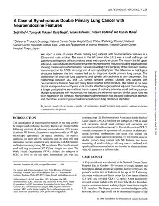
Figure 1. (a) Macroscopic findings for LLL tumors. A yellow- white well definedmass measuring 14 x 10.5 x 8 em in the LLL was compressing the surrounding
normal lung parenchyma with formation of a capsule-like structure. (bHe) Microscopic findings for LLL tumor. Small cell component consists of smallcells with
largeN/Cratio,scantycytoplasm andfinely granular nuclearchromatin withhighmitoticactivity (b). Cellsin smallcell/large cellcomponent have eosinophilic larger
cytoplasmand vesicular nucleuswithdistinctnucleolus (c). Organoid components form welldemarcated roundto ovoidclusters resembling atypical carcinoidin the
background of smallcell/large cells or spindlecells(d).Sarcomatous components havespindle-shaped cellsof VaI;OUS sizes(e).Hematoxylin andeosinstain;original
magnifications, x333 (b, c), x l67 (d), x130 (e).
small size of the specimen. Computed tomography of the chest of the lung was also resected because another nodule was found
showed no enlargement of the mediastinal or hilar lymph nodes. in the LUL during the operation. Radiation therapy to the brain
Magnetic resonance imaging (MRI) of the brain showed a 6 x 6 was performed when the brain mass was found to have grown to
mm solitary mass in the right cerebellar hemisphere. The LLL 14 x 14 mm on MRI in January 1995, suggesting a brain
was resected in December 1994. Part of the left upper lobe (LUL) metastasis. After 50 Gy of radiation to the brain, the nodule could
3. Jpn J Clin OncoI1999;29(4) 221
Downloaded from http://jjco.oxfordjournals.org/ by guest on March 28, 2013
Figure 2. (a) Macroscopicfindings for LLL and LUL tumors. A white round nodulemeasuring2.1 x 1.7 x 1.5 em was presentin the LUL(S1+2) with a clear margin.
(b), (c) Microscopicfindings for LUL tumor. The tumor consists of glandularstructure lined by tall columnar cells with hyperchromatic nucleiand coarse-granular
chromatin (b). There were organoid nests showing occasional rosette formation (c). Hematoxylin and eosin stain; original magnifications, x83 (b), x4 10 (c).
no longer be detected on MRI of the brain. No chemotherapy was protein (Dako), Factor VIII (Dako), p53 (Nichirei, Tokyo, Japan)
given. Left leg pain occurred in February 1997 due to bone and Rb (MK-15-1 , MBL, Nagoya, Japan).
metastasis. Computed tomography of the chest showed multiple A yellow-white, well defined mass measuring 14 x 10.5 x 8 cm
pulmonary metastases. Dyspnea developed and the patient died in S10 of the LLL was compressing the surrounding normal lung
in February 1997, 2 years and 4 months after the operation. An parenchyma with formation of a capsule-like structure; the center
autopsy was not performed. of the mass revealed extensive hemorrhage and necrosis . (Fig.
We performed an immunohistochemical analysis to the for- 1a). The LLL showed poor aeration and moderate anthracosis and
malin-fixed, paraffin-embedded sections by the avidin-biotin fibrosis , especially beneath the pleura, but no emphysema.
complex (ABC) method (6). Biotinylated secondary antibody A round, white nodule measuring 2.1 x 1.7 x 1.5 em and having
and ABC reagents were purchased from Dako Japan (Kyoto, well defined margins was found in Sl+2 of the LUL . The tumor
Japan). Primary antibodies used were against keratin (AE1/AE3, was solid and there was no evidence of hemorrhage or necrosis.
Dako, Dakopatts, Glostrup, Denmark; CAM5.2, Becton Dickin- A sharp pleural indentation was detected (Fig. 2a).
son, San Jose, CA, USA), surfactant apoprotien A (Sp-A) (PE-lO, The LLL tumor was largely necrotic, but there were viable
Dako), surfactant apoprotein D (Sp-D) (6B2, Yamasa, Chiba, tumor cells in the periphery. It consisted of four components:
Japan), CD56 (Lu243, Nippon Kayaku, Tokyo, Japan), chromo- small cell, large cell, organoid and sarcomatous . The small cell
granin A (Dako) , synaptophysin (Dako), CDS7 (Leu7, Becton component was composed of oval to spindle-shaped cells slightly
Dickinson), gastrin-releasing peptide (GRP) (Dako), serotonin larger than lymphocytes and having a large N/C ratio, scanty
(SHT-H209, Dako), calcitonin (Dako) , CEA (011, Mochida, cytoplasm and oval nuclei with fmely granular chromatin but no
Tokyo, Japan), vimentin (V9, Dako), myoglobin (Dako), desmin nucleoli (Fig. 1b). The large cell component consisted of
(033, Dako) , alpha-smooth muscle actin (lA4, Dako), S-lOO polygonal to round cells that were larger than the cells in the small
4. 222 Lung cancer with neuroendocrine features
cell component and they contained abundant eosinophilic cyto- columnar cells. The tumor cells contained relatively abundant
plasm and a vesicular nucleus with one or two prominent nucleoli eosinophilic cytoplasm and hyperchromatic nuclei with coarse-
(Fig. 1c). Small cells and large cells were frequently intermingled. granular chromatin and distinct eosinophilic nucleoli (Fig. 2b).
Both types of cells had high mitotic activity [22/10 high power Mitotic activity was high (25/10 HPF). The stroma was scant. The
field (HPF)] and clusters of pure small cell carcinoma cells were tumor had a focal solid growth area with organoid nests showing
also observed focally. In the organoid component, the neoplastic occasional rosette formation (Fig. 2c). Nuclear palisading was also
cells formed well demarcated round to ovoid clusters resembling seen in the periphery of the nests. No lymphatic permeation or
atypical carcinoid in the background with small cell/large cells or vascular invasion was observed. Hilar lymph nodes contained no
spindle cells (Fig. 1d). The cells forming the organoid structure metastatic tumors. Mediastinal lymph nodes were not explored.
were polygonal to spindle shaped and had a fmely granular nucleus The results of the immunohistochemical analysis are shown in
with one or two prominent nucleoli. Mitotic activity was high Table 1. The small cell/large cell component. of the LLL was
(24/10 HPF). The sarcomatous cells were spindle shaped and their diffusely positive for CD56 (Fig. 3a) and chromogranin A and
nuclei were variable in size and contained coarse chromatin with partially positive for keratin.. The organoid component was
one or two prominent nucleoli (Fig. Ie). The small cell/large cell positive for CD56, chromogranin A, synaptophysin (Fig. 3b),
carcinoma, spindle cell sarcomatous carcinoma and organoid GRP and CEA. The sarcomatous component was positive for
components were highly intermingled with areas of transition vimentin (Fig. 3c) and some spindle-shaped also cells showed an
between them; the proportions of these components were about 65, immunoreaction for keratin (Fig. 3d). The LUL tumor was
Downloaded from http://jjco.oxfordjournals.org/ by guest on March 28, 2013
30 and 5%, respectively. No lymphatic permeation or vascular positive for CD56, chromogranin A (Fig. 4), GRP, calcitonin,
invasion was observed. The remaining non-neoplastic pulmonary CEA and keratin. The positive rates for p53 were 79% of the cells
parenchyma showed no evidence of carcinoid tumorlets or in the small cell/large cell component, 13% in the organoid
neuroendocrine cell hyperplasia. component, 80% in sarcomatous component and 8% in the LUL
The LUL tumor had a clear margin and showed expansive tumor. Rb was positive only in the LUL tumor (30%) and
growth. It consisted of irregular-shaped glands lined by tall negative in all components of the LLL tumor.
Table 1. Immunohistochemical profiles of left lower lobe and left upper lobe tumors
Antigens LLL tumor LUL tumor
S/L Organoid Sarcomatous
Keratin (AEI/AE3) + (partly) ± +
Keratin (CAM5.2) ± + + +++
Sp-A + (partly)
Sp-D
CD56 + + +
Chromogranin A + +
Synaptophysin + +++
CD57
GRP + + (partly)
Serotonin
Calcitonin +
CEA + +
Vimentin +
Myoglobin
Desmin
Alpha-smooth muscle actin
S-IOO protein
Factor VIII
p53* 79% 13% 80% 8%
Rb* 30%
LLL, left lower lobe; LUL, left upper lobe; S/L, small cell/large cell carcinoma; Sp-A, surfactant apoprotein A; Sp-D, surfactant apoprotein D; GRP, gastrin-releasing
peptide. *The percentage of immunoreactive cells counted in 500 cells.
5. Jpn J Clin OncoI1999;29(4) 223
I
Downloaded from http://jjco.oxfordjournals.org/ by guest on March 28, 2013
I
/
- l,.:
"
..
. ; "/
,~
"
I
" .:' . '~1.J:
; ~
If.
';r" .,-
,"
, / .,
• >
." " J;
-
'/
,-' - .' /{.
,
.
{.
I ." ( .' .
. .
.~
" 0#
, ~ I
'(
'.
I .
Figure 3. Immunohistochemical findings for LLL tumor. Small cell/large cell component was diffusely positive for CD56 (a), Organoid component was positive for
synaptophysin (b). Sarcomatous component was positive for vimentin (c) but some spindle-shaped cells showed immunoreaction for keratin (d). Original
magnification, x4 1O.
DISCUSSION
Our differential diagnosis of the LLL tumor was (i) small cell
carcinoma with spindle cell component, (ii) large cell NE
carcinoma and (iii) atypical carcinoid. The high mitotic activity
(22/10 HPF) ruled out atypical carcinoid, although some parts of
the LLL tumor showed features resembling atypical carcinoid,
such as an organoid growth pattern, tumor cells containing
moderately eosinophilic, finely granular cytoplasm and nuclei
possessing finely granular chromatin. Small cell carcinoma was
preferred over large cell NE cancer because the LLL tumor
consisted mainly of two kinds of cells, small-sized tumor cells
with a high N/C ratio and large-sized polygonal to round tumor
cells with one or two prominent nucleoli . In addition, the small
cells and large cells were highly intermingled and clusters of pure
small cell carcinoma cells were also observed focally, We
therefore concluded that the LLL tumor was a mixed small Figure 4. Immunohistochemical findings for LUL tumor. It was positive for
chromogranin A. Original magnification, x41O.
cell/large cell carcinoma, one of the subtypes of small cell lung
carcinoma in the IASLC classification (3),
Spindle cell carcinoma has been found most often in associ- and infrequently with small cell carcinoma (7). Tsubota et al. (4)
ation with giant cell carcinoma and adenocarcinoma, less reported a case of combined small cell (pure type) and spindle cell
frequently with large cell carcinoma or squamous cell carcinoma carcinoma of the lung. The spindle cell carcinoma was predomi-
6. 224 Lung cancer with. neuroendocrine features
nant and immunoreactive for smooth-muscle actin, but not for NE The survival time in our case was 2 years and 4 months without
markers, in their case, whereas the small cell/large cell compo- chemotherapy, longer than in ordinary small cell lung cancer (10).
nent in our case occupied more of the tumor than the spindle cells. The median survival for limited stage small cell lung cancer treated
Moreover, the spindle cell sarcomatous area in our case did not by surgery alone has been reported to be about 6 months (11).
show clear NE features or differentiation to mesenchyme but Tsubota et al. did not comment on the outcome of their case of a
exhibited epithelial differentiation, indicating poorly differen- combined small cell and spindle cell carcinoma of the lung. Small
tiated carcinoma or sarcomatoid change of carcinoma. The cell lung cancer has a poor prognosis although it is sensitive to
spindle cell sarcomatous component was immunohistochemi- chemotherapy and radiation. In contrast to small cell lung cancer,
cally positive for p53 and the frequency of positive cells was spindle cell carcinoma is also generally considered to have a poor
almost same as in the small cell/large cell component, suggesting prognosis and to be resistant to irradiation and chemotherapy
that although their phenotypes were different, a similar genetic (12,13). The metastatic brain tumor in our case was as sensitive to
abnormality may have occurred in both. radiation therapy as small cell lung cancer; however, it recurred 2
We considered three possible diagnoses for the LUL mass: years after surgery, a much longer latent period than in ordinary
(i) moderately differentiated adenocarcinoma, tubular type, with NE small cell lung cancer. No lymph node metastasis was observed in
features, (ii) combined adenocarcinomaand large cell NE carcinoma our case despite the large tumor, a finding also different from usual
small cell lung cancer. The reason for the comparatively good
and (iii) metastatic carcinoma from the LLL tumor. The LUL tumor
outcome in our case remains unclear. Two-year disease-free
exhibited glandular structures lined by tall columnar cells and
Downloaded from http://jjco.oxfordjournals.org/ by guest on March 28, 2013
survivors represented 13% of patients presenting with limited
organoid nests of large pleomorphic cells with rosette formation and
small cell lung cancer but only 2% of those with extensive disease
nuclear palisading. The latter pattern may be seen in large cell NE
(10). More than 80% of 2-year survivors of small lung cell lung
carcinoma (1); however, the clear glandular differentiation favored
cancer have been found to have received chest irradiation and
adenocarcinoma rather than large cell NE carcinoma.
almost all extensive disease patients had metastases confmed to a
Immunohistochemical testing yielded different characteristics
single organ system (11). In our case, however, the brain was the
in the LUL and LLL tumor. Calcitonin and Rb were positive only
only metastatic site at the time of presentation. Hardly any
in the LUL tumor. Furthermore, the LLL tumor exhibited no
long-term survivors of resected small cell lung cancer have had
evidence of glandular differentiation, indicating that the LUL
lymph node metastases or distant metastases (14,15). The present
tumor was not a metastasis of the LLL tumor. Immunohisto- case was exceptional, because the patient survived for a long time
chemical study showed that both the organoid and the glandular in spite of the brain metastasis and having been treated only by
component of the LUL tumor had NE features, indicating that it resection of the lung tumors and radiotherapy to the brain. Slow
was an adenocarcinoma with NE features rather than a combina- growth may have been one of the characteristics of the tumors in
tion of adenocarcinoma and NE carcinoma. our case, despite their high mitotic activity.
The relationship between LLL and LUL tumors is a matter of In conclusion, we have reported a unique case of synchronous
controversy. The LUL tumor was immunohistochemically posi- double primary lung cancers with a combination of small
tive for p53 the same as the organoid area in the LLL tumor, but cell/large cell carcinoma and a spindle sarcomatous component
positive cells were less frequent than in the small cell/large cell in the LLL and adenocarcinoma with NE features in the LUL. At
or spindle cell sarcomatous lesions. The NE marker study showed presentation the tumor had metastasized to the brain, but not to
that the LUL tumor and the organoid area in the LLL might lymph nodes. Radiation to the brain after resection of the lung
differentiate with NE feature better than small cell/large cell or tumors was very effective. The survival time in our patient, who
spindle lesions. LUL tumor. resembled the organoid component did not receive chemotherapy, was 2 years and 4 months. Multiple
in the LLL in the immunohistochemical pattern of the NE lung cancers with NE features are extremely rare and similar
markers (CD56, chromogranin A, synaptophysin, GRP). The cases have not been reported in the literature.
same genetic change may have occurred in the areas of these two
tumors exhibiting NE features, even though their morphology
Acknowledgments
was different. Genetic analysis might resolve this issue.
The only case of multiple lung cancer with NE features reported This work was supported in part by a Grant from the Ministry of
previously was a case of bronchial carcinoid, small cell carcinoma Health and Welfare for the 2nd term Comprehensive Strategy for
and adenocarcinoma of the right lung described by Jung-Legg et Cancer Control and a Grant-in Aid for Cancer Research from the
al. (8) The series of synchronous double primary lung cancers Ministry of Health and Welfare, Japan.
reported by Carey et al. (9) included cases of combined non-small
cell lung cancer and small cell lung cancer or carcinoid, but there
were no cases of double cancer with NE differentiation demon- References
strated by immunohistochemistry. Since these cases were not
1. Travis WD, Linnoila RI, Tsokos MG, Hitchcock CL, Cutler GB Ir, Nieman L,
immunohistochemically tested for NE markers, some of them may et al. Neuroendocrine tumors of the lung with proposed criteria for large-cell
have been double cancer, both of which had NE differentiation. In neuroendocrine carcinoma. An ultrastructural, immunohistochemical and
flow cytometric study of 35 cases. Am J Surg PathoI1991;15:529-33.
any event, the occurrence of synchronous double cancers with NE 2. Histological typing of lung tumors. International Histological Classification
features in both appears to be a rare event. of Tumors. Geneva: World Health Organization 1981.
7. Jpn J Clin OncoI1999;29(4) 225
3. HirschFR, MatthewsMJ,Aisner S, Campobasso 0, ElemaJD, GazdarAF,et 9. Carey FA,DonnellySC, WalkerWS, Cameron EWJ, Lamb D. Synchronous
al. Histopathologic classification of small cell lung cancer. Changing primary lung cancers: prevalence in surgical material and clinical implica-
concepts and terminology. Cancer 1988;62:973-7. tions. Thorax 1993;48:344-6.
4. TsubotaYT,KawaguchiT,HosoT, NishinoE, TravisWD. A combinedsmall 10. Ihde DC, Pass HI, Glatstein E: Small cell lung cancer. In: Devita VT Jr,
cell and spindle cell carcinoma of the lung. Report of a unique case with Hellman S, Rosenberg S, editors. Cancer. Principles and Practice of
immunohistochemical and ultrastructural studies. Am J SUiX Pathol Oncology,5th ed. Philadelphia: Lippincott-Raven 1997;911-49.
1992;16:1108-15. 11. Morstyn G, Ihde DC, Lichter AS, Bunn PA, Carney DN, Glastein E, et al.
5. Colby TV, Koss MN, Travis WD. Small cell carcinoma and large cell Small cell lung cancer 1973-1983: early progress and recent obstacles. lnt J
neuroendocrinecarcinoma.In: Tumorsof the Lower RespiratoryTract.Atlas Radiat Oncol Bioi Phys 1984;10:515-39.
of TumorPathology,Third Series, Fasc. 13.Washington, DC: ArmedForces 12. Nappi 0, Glasner SD, Swanson PE, Wick MR. Biphasic and monophasic
Institute of Pathology 1995;235-57. sarcomatoid carcinomas of the lung: a reappraisal of 'carcinosarcomas' and
6. Hsu S, RaineL, Franger H. Use of avidin-biotin-peroxidasecomplex(ABC) 'spindle cell carcinomas.' Am J Clin Patho/1994;102:331-40.
in immunoperoxidase techniques:a comparisonbetweenABCand unlabeled 13. Ro JY, Chen JL, Lee JS, Sahin AA, Ordonez NG, Ayala AG. Sarcomatoid
antibody (PAP) procedures. J Histochem Cytochem 1981 ;29:577-80. carcinomaof the lung: immunohistochemical and ultrastructural studiesof 14
7. Fishback NF, Travis WD, Moran CA, Guinee DG Jr, McCarthy WF, Koss cases. Cancer 1992;69:376-86.
MN. Pleomorphic (spindle/giant cell) carcinoma of the lung. Cancer 14. Coolen L, Eeckhout AVD, DeneffeG, DemedtsM, Vansteenkiste 1. Surgical
1994;73:2936-45. treatmentof small cell lung cancer. Eur J Cardiothorac Surg 1995;9:59-64.
8. Jung-Legg Y, McGowan SE, Sweeney KG, Zitzman JL, Pugatch RD. 15. AngelettiCA, MacchiariniP,Mussi A, Basolo F. InfluenceofT and N stages
Synchronoustriple malignanttumors of the lung. A case report of bronchial on long-term survival in resecteablesmall cell lung cancer. Eur J SUiR Oncol
carcinoid,small cell carcinoma and adenocarcinomaof the right lung. Am J 1989;15:337-40.
Clin Patho/1986;85:96-101.
Downloaded from http://jjco.oxfordjournals.org/ by guest on March 28, 2013