2. Humans – definite male or females – hermaphroditism –
sexual reproduction by mating
Sex determination - genetic differentiation – genotype –
chromosomal sex differentiation.
Human chromosomes – 44 autosomes + 2 sex
chromosomes – karyotype
Female: 44 + XX, Male: 44 + XY
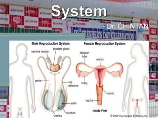
Gonads (Testis - testes & Ovary - ovaries) - Mature male
gamete – sperm (sperms), mature female gamete –
ovum (ova)
Oogonia (22+X), Spermatogonia (22+X or 22+Y)
3.
4. Genetic sex determination
Y is smaller than X, lighter, swim faster –
significance
Inactive X in female – condensed mass - Barr
body – sex chromatin – significance
Sex differentiation
- Gonadal
- Genital
7. Phenotypic
Internal genitalia – Male Wolffian duct &
Female Mullerian duct
Male - Leydig cells – testosterone –
epididymis, vas, seminal vesicles
MIS – regression of Mullerian duct by apoptosis
Female - No MIS – oviduct, uterus, vagina
No testosterone – Wolffian duct degenerate
11. Trisomy (47) – non disjunction of sex
chromosomes
–
abnormal
growth,
development & mental retardation
XXY – Klinefelter syndrome – seminiferous
dysgenesis – positive sex chromatin test
- Poor development of testis – sterility
- N int. ext. genitalia
- Tall & obese
- Gynaecomastia
- Poor development of sec. sex characters
12.
13. XXX – superfemale
- Poor sexual development
- Scanty menses
- Mental retardation
- High FSH, LH, estradiol
- positive sex chromatin test
Down syndrome
- mongolism
- Autosomal trisomy of 21 chromosome
14. Monosomy (45)
Turner’s syndrome – 44 + XO – female
phenotype – gonadal dysgenesis
- Ovarian dysgenesis
- Delayed puberty
- Scanty or no menses (amenorrhea)
- Infertility, amastia
- Mental retardation
- Dwarfism: webbed neck, low hair
line, ptosis, epicanthus, micrognathia, Co
15.
16.
17. Early diagnosis – in utero
Amniocentesis – Amniotic fluid is
collected by inserting a needle into the
amniotic cavity through ant. Abdo. Wall
CVS (chorionic villus sampling)
needle biopsy of chorionic villi
–
19. The reproductive functions of the male can
be divided into three major subdivisions:
(1)spermatogenesis, which means simply
the formation of sperm;
(2)performance of the male sexual act; and
(3)regulation of male reproductive functions
by the various hormones.
20.
21. During formation of the embryo, the
primordial germ cells migrate into the
testes,
and become immature germ cells called
spermatogonia which lie in 2 or 3 layers of
the inner surfaces of the seminiferous
tubules.
The spermatogonia begin to undergo mitotic
division, beginning at puberty, and
continually proliferate and differentiate
22.
23.
24. 1. Testosterone, secreted by the Leydig cells
located in the interstitium of the testis, is
essential for growth and division of the
testicular germinal cells, which is the first stage
in forming sperm.
2. LH, secreted by the anterior pituitary gland,
stimulates the Leydig cells to secrete
testosterone.
3. FSH, also secreted by the anterior pituitary
25. 4. Estrogens, formed from testosterone by
the Sertoli cells when they are stimulated by
FSH, are probably also essential for
spermiogenesis.
5. GH is necessary for controlling
background metabolic functions of the
testes. GH hormone specifically promotes
early division of the spermatogonia
themselves; in its absence, as in pituitary
dwarfs,
spermatogenesis
is
severely
26. After formation in the seminiferous tubules, the
sperm require several days to pass through the
6-meter-long tubule of the epididymis.
Sperm removed from the seminiferous tubules
and from the early portions of the epididymis
are nonmotile, and they cannot fertilize an
ovum.
However, after the sperm have been in the
epididymis for some 18 to 24 hours, they
develop the capability of motility, even though
several inhibitory proteins in the epididymal
fluid still prevent final motility until after
27. 120 million sperm each day - A small quantity
stored in the epididymis - most are stored in the
vas deferens.
They can remain stored, maintaining their fertility,
for at least a month. During this time, they are kept
in a deeply suppressed inactive state by multiple
inhibitory substances in the secretions of the
ducts.
The Sertoli cells and the epithelium of the
epididymis secrete a special fluid that is ejaculated
28. Motile – fertile
1 to 4 mm/min
Neutral and slightly alkaline medium
Life expectancy of ejaculated sperm in the
female genital tract is only 1 to 2 days
Temperature – sperm bank (- 100 degree)
29. Each seminal vesicle is a tortuous, loculated tube
lined with a secretory epithelium that secretes a
mucoid material containing an abundance of
fructose,
citric
acid,
nutrient
substances, prostaglandins and fibrinogen.
Empties shortly after the vas deferens.
Prostaglandins:
(1) React with the female cervical mucus to make it
more receptive to sperm movement
(2) Cause backward, reverse peristaltic contractions
in the uterus and fallopian tubes to move the
ejaculated sperm toward the ovaries
30. Thin, milky fluid that contains calcium, citrate
ion, phosphate ion, a clotting enzyme, and a
profibrinolysin.
A slightly alkaline characteristic of the prostatic
fluid is quite important for successful
fertilization of the ovum, because the fluid of
the vas deferens is relatively acidic owing to the
presence of citric acid and metabolic end
products of the sperm.
The vaginal secretions of the female are acidic
(pH of 3.5 to 4.0). Sperm do not become
optimally motile until the pH of the surrounding
31. The fluid and sperm from the vas deferens
(about 10 per cent of the total),
fluid from the seminal vesicles (almost 60
per cent),
fluid from the prostate gland (about 30 per
cent),
and
small
amounts
from
the
mucous
32. Average pH – 7.5
The prostatic fluid gives the semen a milky
appearance, and fluid from the seminal vesicles and
mucous glands gives the semen a mucoid
consistency.
Clotting enzyme from the prostatic fluid - a weak
fibrin coagulum that holds the semen in the deeper
regions of the vagina. The coagulum then dissolves
during the next 15 to 30 minutes because of lysis by
fibrinolysin
formed
from
the
prostatic
profibrinolysin.
In the early minutes after ejaculation, the sperm
remain relatively immobile, possibly because of the
33. On coming in contact with the fluids of the
female genital tract, multiple changes occur
that activate the sperm for the final
processes of fertilization.
These collective changes are called
capacitation of the spermatozoa – 1 to 10
hours.
1. The uterine and fallopian tube fluids wash
34. 2. While the spermatozoa remain in the fluid of the male
genital ducts, they are continually exposed to many
floating vesicles from the seminiferous tubules containing
large amounts of cholesterol.
This cholesterol is continually added to the cellular
membrane covering the sperm acrosome, toughening
this membrane and preventing release of its enzymes.
After ejaculation, the sperm deposited in the vagina swim
away from the cholesterol vesicles upward into the
uterine cavity, and they gradually lose much of their other
excess cholesterol over the next few hours. In so doing,
the membrane at the head of the sperm (the acrosome)
becomes much weaker.
35. 3. The membrane of the sperm also becomes
much more permeable to calcium ions, so that
calcium now enters the sperm in abundance
and changes the activity of the flagellum,
giving it a powerful whiplash motion in contrast
to its previously weak undulating motion.
In addition, the calcium ions causes release of
acrosomal enzymes rapidly and easily as the
sperm penetrates the granulosa cell mass
surrounding the ovum.
36. Stored in the acrosome of the sperm are large
quantities of hyaluronidase and proteolytic enzymes.
Hyaluronidase depolymerizes the hyaluronic acid
polymers in the intercellular cement that hold the
ovarian granulosa cells together.
The proteolytic enzymes digest proteins in the
structural elements of tissue cells that still adhere to
the ovum.
When the sperm reaches the zona pellucida of the
ovum, the anterior membrane of the sperm itself
37. The entire acrosome dissolves,
acrosomal enzymes are released.
and
all
the
Within minutes, these enzymes open a penetrating
pathway for passage of the sperm head through the
zona pellucida to the inside of the ovum.
Within another 30 minutes, the cell membranes of
the sperm head and of the oocyte fuse with each
other to form a single cell.
At the same time, the genetic material of the sperm
and the oocyte combine to form a completely new
cell genome, containing equal numbers of
chromosomes and genes from mother and father.
38.
39. Within a few minutes after the first sperm penetrates
the zona pellucida of the ovum, calcium ions diffuse
inward through the oocyte membrane
and cause multiple cortical granules to be released
by exocytosis from the oocyte into the perivitelline
space.
These granules contain substances that permeate all
portions of the zona pellucida and prevent binding of
additional sperm,
and they even cause any sperm that have already
40.
41. Bilateral orchitis – mumps – sterility
(Infertility)
Congenital stricture of genital duct
Effect of temperature
Cryptorchidism – undescended testis &
Rx – role of testosterone
42. Volume: 3.5 ml, Color – white, SG – 1.028, pH
– 7.35 to 7.50
Count: 35 – 200 (120) million sperm / ml
< 20 million – infertile
Sperm
Morphology
physically, having two heads,
shaped heads, or abnormal Tails
abnormal
abnormally
Sperm Motility - entirely nonmotile or relatively
nonmotile
43.
44. Neuronal Stimulus - The most important source of
sensory nerve signals for initiating the male sexual
act is the glans penis.
The glans contains an especially sensitive sensory
end organ system that transmits into the CNS that
special modality of sensation called sexual
sensation.
The slippery massaging action of intercourse on
the glans stimulates the sensory end-organs, and
the sexual signals in turn pass through the
45. Impulses may also enter the spinal cord from
areas adjacent to the penis - stimulation of the
anal epithelium, the scrotum, and perineal
structures in general can send signals into the cord
that add to the sexual sensation.
Sexual
sensations
the
urethra, bladder, prostate, seminal vesicles, testes
and vas deferens.
Mild infection and inflammation of these sexual
organs sometimes cause almost continual sexual
desire, and some “aphrodisiac” drugs, such as
cantharidin, increase sexual desire by irritating the
46. Psychic Element - nocturnal emissions
Role of spinal cord
Penile Erection — Role of the Parasympathetic
Nerves.
Erection is caused by parasympathetic impulses that
pass from the sacral portion of the spinal cord through
the pelvic nerves to the penis.
Ach, NO, VIP
NO especially relaxes the arteries of the penis, as well
as relaxes the trabecular meshwork of smooth muscle
fibers in the erectile tissue of the corpora cavernosa
and corpus spongiosum in the shaft of the penis.
47.
48. Lubrication, a Parasympathetic Function
During sexual stimulation, the parasympathetic impulses,
in addition to promoting erection, cause the urethral
glands and the bulbourethral glands to secrete mucus.
This mucus flows through the urethra during intercourse
to aid in the lubrication during coitus.
However, most of the lubrication of coitus is provided by
the female sexual organs rather than by the male.
Without satisfactory lubrication, the male sexual act is
seldom successful because unlubricated intercourse
causes grating, painful sensations that inhibit rather than
excite sexual sensations.
49. Emission and Ejaculation—Function of
the Sympathetic Nerves
When the sexual stimulus becomes
extremely intense, spinal cord - sympathetic
impulses – T 12 to L 2 - the hypogastric and
pelvic sympathetic nerve plexuses to initiate
emission
Contraction
of
vas,
prostate,
seminal
50. Filling of int. urethra – pudendal nerves - feeling
of sudden fullness - rhythmical contraction of
the internal genital organs and cause
contraction of the ischiocavernosus and
bulbocavernosus muscles.
Rhythmical, wavelike increases in pressure in
both the erectile tissue of the penis and the
genital ducts and urethra, which “ejaculate” the
semen from the urethra to the exterior. This
final process is called ejaculation.
51. Androgens - testosterone, dihydrotestosterone, and
Androstenedione
Testosterone is formed by the interstitial cells of
Leydig, which lie in the interstices between the
seminiferous tubules and constitute about 20 per
cent of the mass of the adult testes.
Leydig cells are almost nonexistent in the testes
during childhood when the testes secrete almost no
testosterone, but they are numerous in the newborn
male infant for the first few months of life and in the
adult male any time after puberty.
Resistant to X – ray & heat, tumor
52. Secretion of Androgens Elsewhere in the
Body – adrenal gland – steroid compound
from cholesterol
Masculinizing effect
Role in female
Adrenal tumor – adrenogenital syndrome
arrhenoblastoma
53.
54. 97 % loosely bound with plasma albumin or
more tightly bound with a beta globulin called
sex hormone–binding globulin and circulates in
the blood in these states for 30 minutes to
several hours.
In tissue to DHT by 5 alpha reductase
Rapidly converted, mainly by the liver, into
androsterone and dehydroepiandrosterone and
simultaneously
conjugated
as
either
glucuronides or sulfates - excreted either into
the gut by way of the liver bile or into the urine
55. Small amounts of estrogens are formed in the
male (about 1/5th the amount in the
nonpregnant female), and a reasonable
quantity of estrogens can be recovered from a
man’s urine.
(1) The concentration of estrogens in the fluid
of the seminiferous tubules is quite high and
probably plays an important role in
spermiogenesis. This estrogen is believed to be
formed by the Sertoli cells by converting
testosterone to estradiol.
(2) Much larger amounts (80%) of estrogens
56.
57. Fetal development – 7th week of embryonic
life by genital ridge – injection & removal
Fetal testis - formation of a penis and a
scrotum - formation of the prostate gland,
seminal vesicles, and male genital ducts.
Descent of testes
Undescended testis & role of testosterone in
58. Development of Adult Primary
Secondary Sexual Characteristics
and
After puberty – enlargement of penis,
scrotum & testes
Distribution of body hair
Baldness
Voice change – enlargement of larynx
59. Skin thickness, secretion by sebaceous glands
Male adolescence – acne
Protein formation & muscle development –
synthetic androgens
Bone matrix & calcium retention – pelvic bone –
osteoporosis Rx
The epiphyses of the long bones to unite with
the shafts of the bones – castration in eunuch
60. Effect on BMR
Effect on RBC
Effect on
Balance
Electrolyte
MOA – intracellular
receptor protein - nucleus
and
Water
cytoplasmic
61.
62.
63. GnRH - the arcuate nuclei of the
hypothalamus
- hypothalamic-hypophysial portal vascular
system
- anterior pituitary gland
- stimulates the release of the two
gonadotropins, LH and FSH.
GnRH is secreted intermittently a few
minutes at a time once every 1 to 3 hours –
64. Ant. Pituitary – gonadotropes – gonadotropic
hormones
Negative feedback of testosterone – more
on hypothalamus – less on ant. Pituitary
FSH binds with specific FSH receptors
attached to the Sertoli cells in the
seminiferous tubules.
To initiate spermatogenesis, both FSH and
65. When the seminiferous tubules fail to produce
sperm, secretion of FSH by the anterior
pituitary gland increases markedly.
When spermatogenesis proceeds too rapidly,
pituitary secretion of FSH diminishes. The
cause of this negative feedback effect on the
anterior pituitary is believed to be secretion by
the Sertoli cells of still another hormone called
inhibin.
Strong direct effect on the anterior pituitary
gland - slight effect on the hypothalamus
66.
67. During childhood the hypothalamus simply
does not secrete significant amounts of
GnRH.
The slightest secretion of any sex steroid
hormones exerts a strong inhibitory effect on
hypothalamic secretion of GnRH.
At the time of puberty, the secretion of
hypothalamic GnRH breaks through the
childhood inhibition, and adult sexual life
68. • Body changes at puberty in boys (male secondary sex
characteristics).
• External genitalia: Penis increases in length and width.
Scrotum becomes pigmented and rugose.
• Internal genitalia: Seminal vesicles enlarge and secrete and
begin to form fructose. Prostate and bulbourethral glands
enlarge and secrete.
• Voice: Larynx enlarges, vocal cords increase in length and
thickness, and voice becomes deeper.
• Hair growth: Beard appears. Hairline on scalp recedes
anterolaterally. Pubic hair grows with male (triangle with apex
up) pattern. Hair appears in axillas, on chest, and around
anus; general body hair increases.
• Mental: More aggressive, active attitude. Interest in opposite
sex develops.
• Body conformation: Shoulders broaden, muscles enlarge.
• Skin: Sebaceous gland secretion thickens and increases
69. After puberty, spermatogenesis usually continues
until death.
Most men, however, begin to exhibit slowly
decreasing sexual functions in their late 40s or 50s,
and one study showed that the average age for
terminating intersexual relations
was 68.
This decline in sexual function is related to decrease
in testosterone secretion. The decrease in male
sexual function is called the male climacteric.
Hot flashes, suffocation, and psychic disorders
70. Prostate – BHP
The prostate gland remains relatively small throughout
childhood and begins to grow at puberty under the
stimulus of testosterone.
This gland reaches an almost stationary size by the age
of 20 years and remains at this size up to the age of
about 50 years. At that time, in some men it begins to
involute, along with decreased production of testosterone
by the testes.
A benign prostatic fibroadenoma frequently develops in
the prostate in many older men and can cause urinary
obstruction.
This hypertrophy is caused not by testosterone but
71. Prostate cancer - death
Once cancer of the prostate gland does occur,
the cancerous cells are usually stimulated to
more rapid growth by testosterone and are
inhibited by removal of both testes so that
testosterone cannot be formed.
Prostatic cancer usually can be inhibited by
administration of estrogens.
72. When the testes of a male fetus are
nonfunctional during fetal life, none of the male
sexual characteristics develop in the fetus.
Instead, female organs are formed. The reason
for this is that the basic genetic characteristic of
the fetus, whether male or female, is to form
female sexual organs if there are no sex
hormones.
But in the presence of testosterone, formation
of female sexual organs is suppressed, and
instead, male organs are induced.
73. When a boy loses his testes before puberty, a
state of eunuchism ensues in which he
continues to have infantile sex organs and
other infantile sexual characteristics throughout
life.
The height of an adult eunuch is slightly greater
than that of a normal man because the bone
epiphyses are slow to unite, although the bones
are quite thin and the muscles are considerably
weaker.
The voice is childlike, there is no loss of hair on
the head, and the normal adult masculine hair
74.
75. When a man is castrated after puberty, some of
his male secondary sexual characteristics
revert to those of a child and others remain of
adult masculine character.
The sexual organs regress slightly in size but
not to a
childlike state, and the voice regresses from the
bass
quality only slightly.