SlideShare uma empresa Scribd logo
1 de 3
Baixar para ler offline
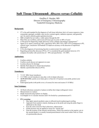
Soft Tissue Ultrasound: Abscess versus Cellulitis
                                    Geoffrey E. Hayden, MD
                             Director of Emergency Ultrasonography
                                Vanderbilt Emergency Medicine

Background:

    •   CT is the gold standard for the diagnosis of soft tissue infections, but is of course expensive, time-
        consuming, sparingly available, may involve contrast agents, radiation exposure, and generally
        can’t be used to directly guide aspiration/incision
    •   MRI is helpful but not really a viable option through the ED
    •   Plain films are worthless, and reveal soft tissue gas in as few as 40% of cases
    •   U/S is great for diagnosing simple abscesses versus cellulitis and changes management1-5
    •   Squire et al. report a sensitivity=98%, specificity=88%, PPV=93%, NPV=97% (comparison to PE,
        clinical exam; conclusion: ED bedside US improves accuracy in the detection of superficial
        abscesses)6
    •   U/S in the diagnosis of necrotizing fasciitis is controversial; few studies exist7
    •   Regarding peritonsillar abscesses (PTA), CT is considered the gold standard; however, U/S
        compares favorably with sensitivities of ~ 90% and specificities ~ 83-100%

Applications:

    •   Confirm cellulitis
    •   Confirm occult abscess not apparent on exam
    •   Localize abscess for drainage
    •   Identify fluid adjacent to deeper fascial planes
    •   Identify a peritonsillar abscess (PTA)

Transducers:

    •   7.5-10+ MHz linear transducers
    •   3.5-5 MHz curved transducers may play a role in deeper abscesses
    •   ALWAYS USE a probe cover (latex glove is fine) for U/S (protect your probe, protect your
        patients)
    •   Endovaginal probe (with probe cover, of course) for intra-oral exams (5-10 MHz)

Sono Technique:

    •   All fluid collections scanned in 2 planes to define their shape (orthogonal views)
    •   Measurements in 2 planes
    •   Depth measured using markers on display
    •   Use Doppler to r/o vascular structure (don’t be fooled by a pseudoaneurysm!!)

    •   PTA scanning:
           • May apply topical anesthetic spray to affected tonsil/oropharyngeal swelling
           • Approach from medial to lateral, choking up on the probe and using the pinky finger to
               stabilized on the cheek
           • Scan the entire tonsil for fluid collections, enhancement
           • Gentle pressure may be applied to assess for “squish sign”
           • Note the location of the internal carotid artery, which runs anterior to the jugular vein in
               the carotid sheath; it is usually located posterolateral to the tonsil within 5mm to 25mm
               of a peritonsillar abscess
Normal sono findings/anatomy:

    •       Subcutaneous tissue generally appears hypoechoic with randomly distributed hyperechoic strands
            that represent connective tissue
    •       Fascial planes are hyperechoic
    •       Muscle has a characteristic striated appearance in a longitudinal plane
    •       Vascular structures: anechoic
    •       Nerves: stippled appearance
    •       Lymph nodes: classic circular to oval shape, echogenic centers with hypoechoic rims
    •       Normal tonsil has the appearance of a typical lymph node, with a hypoechoic rim and generally
            echogenic center; may be isoechoic throughout

Abnormal sono findings:

    •       Cellulitis:
                • Diffuse thickening of subcutaneous layer due to edema amidst the fat and connective
                      tissue
                • Edema evolves as well defined hypoechoic septae between the fat and connective tissue;
                      characteristic “cobble-stone” appearance

    •       Abscess:
                • Sonographic appearance is quite variable
                • Ranges from anechoic to irregularly hyperechoic, internal echoes; may find hyperechoic
                     sediment, septae, or even gas
                • Ranges from round and generally well-defined to irregular, lobulated
                • Posterior acoustic enhancement may be your only sonographic finding
                • “Squish sign” with compression: ability to induce motion in the material with
                     palpation/pressure

    •       Necrotizing fasciitis:
                • Marked thickening of the subcutaneous layer (i.e. cellulitis)
                • Layer of anechoic fluid measuring >4mm, adjacent to the deep fascia
                • Subcutaneous gas (acoustic shadowing and reverberation artifact) may be present

    •       Peritonsillar abscess:
                • Usually appears as a hypoechoic, heterogeneous mass, though appearance may be
                     variable
                • Commonly see posterior enhancement

Pitfalls:

    •       Sometimes difficult to differentiate between interconnected bands of edema fluid and an irregular
            abscess/pus collection
    •       May miss abscess if isoechoic to surrounding tissue and no posterior enhancement or “squish
            sign” appreciated

    •       PTA exams may be limited by trismus or gag reflex
    •       Inferiorly located abscesses can be missed by failure to scan in the longitudinal plane

Pearls:

    •       Use contralateral side to delineate pathology
    •       Look for areas of echogenicity (suggestive of occult abscess) or a “squish sign” even if no obvious
            fluid collection
•   Use plenty of U/S gel so you limit direct pressure (allows visualization without “hurting” your
       patient)
   •   Use water bath for hand or foot infections (the water provides the interface, even better than U/S
       gel!!)


1.      Bureau NJ, Chhem RK, Cardinal E. Musculoskeletal infections: US
manifestations. Radiographics 1999;19(6):1585-92.
2.      Chao HC, Lin SJ, Huang YC, Lin TY. Sonographic evaluation of cellulitis in
children. J Ultrasound Med 2000;19(11):743-9.
3.      Swartz MN. Clinical practice. Cellulitis. N Engl J Med 2004;350(9):904-12.
4.      Tayal VS, Hasan N, Norton HJ, Tomaszewski CA. The effect of soft-tissue
ultrasound on the management of cellulitis in the emergency department. Acad Emerg
Med 2006;13(4):384-8.
5.      Vincent LM. Ultrasound of soft tissue abnormalities of the extremities. Radiol
Clin North Am 1988;26(1):131-44.
6.      Squire BT, Fox JC, Anderson C. ABSCESS: applied bedside sonography for
convenient evaluation of superficial soft tissue infections. Acad Emerg Med
2005;12(7):601-6.
7.      Yen ZS, Wang HP, Ma HM, Chen SC, Chen WJ. Ultrasonographic screening of
clinically-suspected necrotizing fasciitis. Acad Emerg Med 2002;9(12):1448-51.

Mais conteúdo relacionado

Mais procurados

Presentation1.pptx, ultrasound examination of the wrist joint.
Presentation1.pptx, ultrasound examination of the wrist joint.Presentation1.pptx, ultrasound examination of the wrist joint.
Presentation1.pptx, ultrasound examination of the wrist joint.Abdellah Nazeer
 
Presentation1, radiological imaging of undescended testis.
Presentation1, radiological imaging of undescended testis.Presentation1, radiological imaging of undescended testis.
Presentation1, radiological imaging of undescended testis.Abdellah Nazeer
 
Presentation1.pptx, radiological anatomy of the naso, oro and hypopharynx.
Presentation1.pptx, radiological anatomy of the naso, oro and hypopharynx.Presentation1.pptx, radiological anatomy of the naso, oro and hypopharynx.
Presentation1.pptx, radiological anatomy of the naso, oro and hypopharynx.Abdellah Nazeer
 
Presentation1.pptx, radiological imaging of adult neck masses.
Presentation1.pptx, radiological imaging of adult neck masses.Presentation1.pptx, radiological imaging of adult neck masses.
Presentation1.pptx, radiological imaging of adult neck masses.Abdellah Nazeer
 
Imaging of the neck part i
Imaging of the neck part iImaging of the neck part i
Imaging of the neck part iWafik Ebrahim
 
Temporal bone Radiologic anatomy.. In depth
Temporal bone Radiologic anatomy.. In depthTemporal bone Radiologic anatomy.. In depth
Temporal bone Radiologic anatomy.. In depthAhmed Hamdy Hamdy
 
Contrast media used with ct
Contrast media used with ctContrast media used with ct
Contrast media used with ctDR Laith
 
ascending urethrogram
ascending urethrogramascending urethrogram
ascending urethrogramNasin Usman
 
Temporal bone radiology
Temporal bone radiologyTemporal bone radiology
Temporal bone radiologySatish Naga
 
Diagnostic Imaging of Intraspinal Masses
Diagnostic Imaging of Intraspinal MassesDiagnostic Imaging of Intraspinal Masses
Diagnostic Imaging of Intraspinal MassesMohamed M.A. Zaitoun
 
RADIO LOGICAL ANATOMY OF HEAD AND NECK CANCERS
RADIO LOGICAL ANATOMY OF HEAD AND NECK CANCERSRADIO LOGICAL ANATOMY OF HEAD AND NECK CANCERS
RADIO LOGICAL ANATOMY OF HEAD AND NECK CANCERSIsha Jaiswal
 
Presentation1.pptx, radiological anatomy of the shoulder joint.
Presentation1.pptx, radiological anatomy of the shoulder joint.Presentation1.pptx, radiological anatomy of the shoulder joint.
Presentation1.pptx, radiological anatomy of the shoulder joint.Abdellah Nazeer
 
Imaging Acute Abdomen (Part 1)
Imaging Acute Abdomen (Part 1)Imaging Acute Abdomen (Part 1)
Imaging Acute Abdomen (Part 1)Rathachai Kaewlai
 
Peri anal fistula mri
Peri anal fistula mriPeri anal fistula mri
Peri anal fistula mriAli Jiwani
 
Anatomy of neck spaces
Anatomy of neck spacesAnatomy of neck spaces
Anatomy of neck spacesWalid Rezk
 
Anatomy of neck spaces and levels of cervical
Anatomy of neck spaces and levels of cervicalAnatomy of neck spaces and levels of cervical
Anatomy of neck spaces and levels of cervicalairwave12
 
Emergency Ultrasound Course -Lecture 01 -Introduction to acute abdomen
Emergency Ultrasound Course -Lecture 01 -Introduction to acute abdomenEmergency Ultrasound Course -Lecture 01 -Introduction to acute abdomen
Emergency Ultrasound Course -Lecture 01 -Introduction to acute abdomenDr.Ismail Sayed Ismail
 

Mais procurados (20)

Presentation1.pptx, ultrasound examination of the wrist joint.
Presentation1.pptx, ultrasound examination of the wrist joint.Presentation1.pptx, ultrasound examination of the wrist joint.
Presentation1.pptx, ultrasound examination of the wrist joint.
 
Presentation1, radiological imaging of undescended testis.
Presentation1, radiological imaging of undescended testis.Presentation1, radiological imaging of undescended testis.
Presentation1, radiological imaging of undescended testis.
 
Presentation1.pptx, radiological anatomy of the naso, oro and hypopharynx.
Presentation1.pptx, radiological anatomy of the naso, oro and hypopharynx.Presentation1.pptx, radiological anatomy of the naso, oro and hypopharynx.
Presentation1.pptx, radiological anatomy of the naso, oro and hypopharynx.
 
Presentation1.pptx, radiological imaging of adult neck masses.
Presentation1.pptx, radiological imaging of adult neck masses.Presentation1.pptx, radiological imaging of adult neck masses.
Presentation1.pptx, radiological imaging of adult neck masses.
 
Imaging of the neck part i
Imaging of the neck part iImaging of the neck part i
Imaging of the neck part i
 
Temporal bone Radiologic anatomy.. In depth
Temporal bone Radiologic anatomy.. In depthTemporal bone Radiologic anatomy.. In depth
Temporal bone Radiologic anatomy.. In depth
 
Contrast media used with ct
Contrast media used with ctContrast media used with ct
Contrast media used with ct
 
ascending urethrogram
ascending urethrogramascending urethrogram
ascending urethrogram
 
Temporal bone radiology
Temporal bone radiologyTemporal bone radiology
Temporal bone radiology
 
Diagnostic Imaging of Intraspinal Masses
Diagnostic Imaging of Intraspinal MassesDiagnostic Imaging of Intraspinal Masses
Diagnostic Imaging of Intraspinal Masses
 
RADIO LOGICAL ANATOMY OF HEAD AND NECK CANCERS
RADIO LOGICAL ANATOMY OF HEAD AND NECK CANCERSRADIO LOGICAL ANATOMY OF HEAD AND NECK CANCERS
RADIO LOGICAL ANATOMY OF HEAD AND NECK CANCERS
 
Abdominal x ray
Abdominal x rayAbdominal x ray
Abdominal x ray
 
Presentation1.pptx, radiological anatomy of the shoulder joint.
Presentation1.pptx, radiological anatomy of the shoulder joint.Presentation1.pptx, radiological anatomy of the shoulder joint.
Presentation1.pptx, radiological anatomy of the shoulder joint.
 
Imaging Acute Abdomen (Part 1)
Imaging Acute Abdomen (Part 1)Imaging Acute Abdomen (Part 1)
Imaging Acute Abdomen (Part 1)
 
Peri anal fistula mri
Peri anal fistula mriPeri anal fistula mri
Peri anal fistula mri
 
Usg neck
Usg neckUsg neck
Usg neck
 
Anatomy of neck spaces
Anatomy of neck spacesAnatomy of neck spaces
Anatomy of neck spaces
 
Anatomy of neck spaces and levels of cervical
Anatomy of neck spaces and levels of cervicalAnatomy of neck spaces and levels of cervical
Anatomy of neck spaces and levels of cervical
 
RADIOLOGY PRESENTATION
RADIOLOGY PRESENTATIONRADIOLOGY PRESENTATION
RADIOLOGY PRESENTATION
 
Emergency Ultrasound Course -Lecture 01 -Introduction to acute abdomen
Emergency Ultrasound Course -Lecture 01 -Introduction to acute abdomenEmergency Ultrasound Course -Lecture 01 -Introduction to acute abdomen
Emergency Ultrasound Course -Lecture 01 -Introduction to acute abdomen
 

Semelhante a Soft tissue ultrasound

Ultrasonography - Head and Neck applications
Ultrasonography - Head and Neck applicationsUltrasonography - Head and Neck applications
Ultrasonography - Head and Neck applicationsaparna666
 
Thyroid Ultrasound and TIRADS
Thyroid Ultrasound and TIRADSThyroid Ultrasound and TIRADS
Thyroid Ultrasound and TIRADSNomanKhan297
 
acute appendicitis final.pptx
acute appendicitis final.pptxacute appendicitis final.pptx
acute appendicitis final.pptxNeeraj Ojha
 
Soft tissue calcification orofacial region
Soft tissue calcification orofacial regionSoft tissue calcification orofacial region
Soft tissue calcification orofacial regionDr. Monali Prajapati
 
Otosclerosis and its management.pdf
Otosclerosis and its management.pdfOtosclerosis and its management.pdf
Otosclerosis and its management.pdfVirginia Pame
 
VESTIBULAR SCHWANNOMA.pptx
VESTIBULAR SCHWANNOMA.pptxVESTIBULAR SCHWANNOMA.pptx
VESTIBULAR SCHWANNOMA.pptxKarishmaMishra13
 
Case of the week : Thyroglossal cyst
Case of the week : Thyroglossal cystCase of the week : Thyroglossal cyst
Case of the week : Thyroglossal cystDr Abdalla M. Gamal
 
Chest X-ray & Interpretation.pptx
Chest X-ray & Interpretation.pptxChest X-ray & Interpretation.pptx
Chest X-ray & Interpretation.pptxAshish yadav
 
thyroid_usg.dos (1).pptx
thyroid_usg.dos (1).pptxthyroid_usg.dos (1).pptx
thyroid_usg.dos (1).pptxNeerajOjha17
 
Nasopalatine duct cyst
Nasopalatine duct cystNasopalatine duct cyst
Nasopalatine duct cystDr AJINS CB
 
Ultrasound of salivary glands
Ultrasound of salivary glandsUltrasound of salivary glands
Ultrasound of salivary glandsMylapally Rohit
 
Intussusception in Adults-Submucosal Lipoma at Transverse colon-A rare cause
 Intussusception in Adults-Submucosal Lipoma at Transverse colon-A rare cause Intussusception in Adults-Submucosal Lipoma at Transverse colon-A rare cause
Intussusception in Adults-Submucosal Lipoma at Transverse colon-A rare causeDr.Santosh Atreya
 
Testicular Torsion
Testicular TorsionTesticular Torsion
Testicular TorsionAmeen Rageh
 

Semelhante a Soft tissue ultrasound (20)

Ultrasonography - Head and Neck applications
Ultrasonography - Head and Neck applicationsUltrasonography - Head and Neck applications
Ultrasonography - Head and Neck applications
 
Thyroid Ultrasound and TIRADS
Thyroid Ultrasound and TIRADSThyroid Ultrasound and TIRADS
Thyroid Ultrasound and TIRADS
 
acute appendicitis final.pptx
acute appendicitis final.pptxacute appendicitis final.pptx
acute appendicitis final.pptx
 
Soft tissue calcification orofacial region
Soft tissue calcification orofacial regionSoft tissue calcification orofacial region
Soft tissue calcification orofacial region
 
Role of ultrasound in ovarian lesions
Role of ultrasound in ovarian lesionsRole of ultrasound in ovarian lesions
Role of ultrasound in ovarian lesions
 
Acute appendicitis
Acute appendicitisAcute appendicitis
Acute appendicitis
 
Otosclerosis and its management.pdf
Otosclerosis and its management.pdfOtosclerosis and its management.pdf
Otosclerosis and its management.pdf
 
Nasolabial cyst
Nasolabial cystNasolabial cyst
Nasolabial cyst
 
VESTIBULAR SCHWANNOMA.pptx
VESTIBULAR SCHWANNOMA.pptxVESTIBULAR SCHWANNOMA.pptx
VESTIBULAR SCHWANNOMA.pptx
 
10 neck masses - copy
10   neck masses - copy10   neck masses - copy
10 neck masses - copy
 
Case of the week : Thyroglossal cyst
Case of the week : Thyroglossal cystCase of the week : Thyroglossal cyst
Case of the week : Thyroglossal cyst
 
Chest X-ray & Interpretation.pptx
Chest X-ray & Interpretation.pptxChest X-ray & Interpretation.pptx
Chest X-ray & Interpretation.pptx
 
Fistula in ano
Fistula in anoFistula in ano
Fistula in ano
 
thyroid_usg.dos (1).pptx
thyroid_usg.dos (1).pptxthyroid_usg.dos (1).pptx
thyroid_usg.dos (1).pptx
 
Nasopalatine duct cyst
Nasopalatine duct cystNasopalatine duct cyst
Nasopalatine duct cyst
 
USG Guided Thoracentesis
USG Guided ThoracentesisUSG Guided Thoracentesis
USG Guided Thoracentesis
 
Spleen Radiology
Spleen RadiologySpleen Radiology
Spleen Radiology
 
Ultrasound of salivary glands
Ultrasound of salivary glandsUltrasound of salivary glands
Ultrasound of salivary glands
 
Intussusception in Adults-Submucosal Lipoma at Transverse colon-A rare cause
 Intussusception in Adults-Submucosal Lipoma at Transverse colon-A rare cause Intussusception in Adults-Submucosal Lipoma at Transverse colon-A rare cause
Intussusception in Adults-Submucosal Lipoma at Transverse colon-A rare cause
 
Testicular Torsion
Testicular TorsionTesticular Torsion
Testicular Torsion
 

Mais de Rashidi Ahmad

Mais de Rashidi Ahmad (20)

Humanitarian charter
Humanitarian charterHumanitarian charter
Humanitarian charter
 
Terrorism in msia
Terrorism in msiaTerrorism in msia
Terrorism in msia
 
Fracture care
Fracture careFracture care
Fracture care
 
Kenali ular (snake)
Kenali ular (snake) Kenali ular (snake)
Kenali ular (snake)
 
Acute poisoning
Acute poisoningAcute poisoning
Acute poisoning
 
Pain score in ed
Pain score in edPain score in ed
Pain score in ed
 
Pseudotumour
PseudotumourPseudotumour
Pseudotumour
 
Nasal fb
Nasal fbNasal fb
Nasal fb
 
Post resuscitation care
Post resuscitation carePost resuscitation care
Post resuscitation care
 
Trauma scan
Trauma scanTrauma scan
Trauma scan
 
Mild traumatic brain injury
Mild traumatic brain injuryMild traumatic brain injury
Mild traumatic brain injury
 
Mannitol Vs Hypertonic Saline
Mannitol Vs Hypertonic SalineMannitol Vs Hypertonic Saline
Mannitol Vs Hypertonic Saline
 
Sedation With Ketamine
Sedation With KetamineSedation With Ketamine
Sedation With Ketamine
 
Shock Trial
Shock TrialShock Trial
Shock Trial
 
Is The Mediastinum Wide
Is The Mediastinum WideIs The Mediastinum Wide
Is The Mediastinum Wide
 
Emergency Fluid Therapy
Emergency Fluid TherapyEmergency Fluid Therapy
Emergency Fluid Therapy
 
Humanitarian Mercy Conference
Humanitarian Mercy ConferenceHumanitarian Mercy Conference
Humanitarian Mercy Conference
 
Burr Hole
Burr HoleBurr Hole
Burr Hole
 
Burr Hole
Burr HoleBurr Hole
Burr Hole
 
Mercury Poisoning
Mercury PoisoningMercury Poisoning
Mercury Poisoning
 

Soft tissue ultrasound

  • 1. Soft Tissue Ultrasound: Abscess versus Cellulitis Geoffrey E. Hayden, MD Director of Emergency Ultrasonography Vanderbilt Emergency Medicine Background: • CT is the gold standard for the diagnosis of soft tissue infections, but is of course expensive, time- consuming, sparingly available, may involve contrast agents, radiation exposure, and generally can’t be used to directly guide aspiration/incision • MRI is helpful but not really a viable option through the ED • Plain films are worthless, and reveal soft tissue gas in as few as 40% of cases • U/S is great for diagnosing simple abscesses versus cellulitis and changes management1-5 • Squire et al. report a sensitivity=98%, specificity=88%, PPV=93%, NPV=97% (comparison to PE, clinical exam; conclusion: ED bedside US improves accuracy in the detection of superficial abscesses)6 • U/S in the diagnosis of necrotizing fasciitis is controversial; few studies exist7 • Regarding peritonsillar abscesses (PTA), CT is considered the gold standard; however, U/S compares favorably with sensitivities of ~ 90% and specificities ~ 83-100% Applications: • Confirm cellulitis • Confirm occult abscess not apparent on exam • Localize abscess for drainage • Identify fluid adjacent to deeper fascial planes • Identify a peritonsillar abscess (PTA) Transducers: • 7.5-10+ MHz linear transducers • 3.5-5 MHz curved transducers may play a role in deeper abscesses • ALWAYS USE a probe cover (latex glove is fine) for U/S (protect your probe, protect your patients) • Endovaginal probe (with probe cover, of course) for intra-oral exams (5-10 MHz) Sono Technique: • All fluid collections scanned in 2 planes to define their shape (orthogonal views) • Measurements in 2 planes • Depth measured using markers on display • Use Doppler to r/o vascular structure (don’t be fooled by a pseudoaneurysm!!) • PTA scanning: • May apply topical anesthetic spray to affected tonsil/oropharyngeal swelling • Approach from medial to lateral, choking up on the probe and using the pinky finger to stabilized on the cheek • Scan the entire tonsil for fluid collections, enhancement • Gentle pressure may be applied to assess for “squish sign” • Note the location of the internal carotid artery, which runs anterior to the jugular vein in the carotid sheath; it is usually located posterolateral to the tonsil within 5mm to 25mm of a peritonsillar abscess
  • 2. Normal sono findings/anatomy: • Subcutaneous tissue generally appears hypoechoic with randomly distributed hyperechoic strands that represent connective tissue • Fascial planes are hyperechoic • Muscle has a characteristic striated appearance in a longitudinal plane • Vascular structures: anechoic • Nerves: stippled appearance • Lymph nodes: classic circular to oval shape, echogenic centers with hypoechoic rims • Normal tonsil has the appearance of a typical lymph node, with a hypoechoic rim and generally echogenic center; may be isoechoic throughout Abnormal sono findings: • Cellulitis: • Diffuse thickening of subcutaneous layer due to edema amidst the fat and connective tissue • Edema evolves as well defined hypoechoic septae between the fat and connective tissue; characteristic “cobble-stone” appearance • Abscess: • Sonographic appearance is quite variable • Ranges from anechoic to irregularly hyperechoic, internal echoes; may find hyperechoic sediment, septae, or even gas • Ranges from round and generally well-defined to irregular, lobulated • Posterior acoustic enhancement may be your only sonographic finding • “Squish sign” with compression: ability to induce motion in the material with palpation/pressure • Necrotizing fasciitis: • Marked thickening of the subcutaneous layer (i.e. cellulitis) • Layer of anechoic fluid measuring >4mm, adjacent to the deep fascia • Subcutaneous gas (acoustic shadowing and reverberation artifact) may be present • Peritonsillar abscess: • Usually appears as a hypoechoic, heterogeneous mass, though appearance may be variable • Commonly see posterior enhancement Pitfalls: • Sometimes difficult to differentiate between interconnected bands of edema fluid and an irregular abscess/pus collection • May miss abscess if isoechoic to surrounding tissue and no posterior enhancement or “squish sign” appreciated • PTA exams may be limited by trismus or gag reflex • Inferiorly located abscesses can be missed by failure to scan in the longitudinal plane Pearls: • Use contralateral side to delineate pathology • Look for areas of echogenicity (suggestive of occult abscess) or a “squish sign” even if no obvious fluid collection
  • 3. Use plenty of U/S gel so you limit direct pressure (allows visualization without “hurting” your patient) • Use water bath for hand or foot infections (the water provides the interface, even better than U/S gel!!) 1. Bureau NJ, Chhem RK, Cardinal E. Musculoskeletal infections: US manifestations. Radiographics 1999;19(6):1585-92. 2. Chao HC, Lin SJ, Huang YC, Lin TY. Sonographic evaluation of cellulitis in children. J Ultrasound Med 2000;19(11):743-9. 3. Swartz MN. Clinical practice. Cellulitis. N Engl J Med 2004;350(9):904-12. 4. Tayal VS, Hasan N, Norton HJ, Tomaszewski CA. The effect of soft-tissue ultrasound on the management of cellulitis in the emergency department. Acad Emerg Med 2006;13(4):384-8. 5. Vincent LM. Ultrasound of soft tissue abnormalities of the extremities. Radiol Clin North Am 1988;26(1):131-44. 6. Squire BT, Fox JC, Anderson C. ABSCESS: applied bedside sonography for convenient evaluation of superficial soft tissue infections. Acad Emerg Med 2005;12(7):601-6. 7. Yen ZS, Wang HP, Ma HM, Chen SC, Chen WJ. Ultrasonographic screening of clinically-suspected necrotizing fasciitis. Acad Emerg Med 2002;9(12):1448-51.