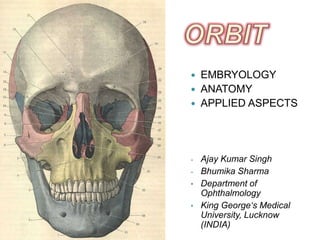
1. EMBRYOLOGY
ANATOMY
APPLIED ASPECTS
- Ajay Kumar Singh
- Bhumika Sharma
• Department of
Ophthalmology
• King George‘s Medical
University, Lucknow
(INDIA)
2. INTRODUCTION
Orbit is the anatomical space bounded:
Superiorly – Anterior cranial fossa
Medially - Nasal cavity & Ethmoidal air sinuses
Inferiorly - Maxillary sinus
Laterally - Middle cranial fossa & Temporal fossa
3.
4. EMBRYOLOGY
Orbital walls- derived from cranial neural crest
cells which expand to form:
Frontonasal process
Maxillary process
Lateral nasal process + Maxillary process =
medial, inferior and lateral orbital walls
Capsule of forebrain forms orbital roof
5. EMBRYOLOG
Y
Early in the human
development eyes point
almost in the opposite
direction.
As the facial growth occurs,
the angle between the optic
stalks decreases and is ~68˚
in an adult.
7. EMBRYOLOG
Frontal, Zygomatic, Maxillary and Palatine bones-
Intramembranous origin
First bone- Maxillary (at 6 wks of intrauterine life)
- develops from elements in the region of the canine tooth
- secondary ossification centres in the orbitonasal and
premaxillary regions
Other bones develop at around 7 wks of intrauterine
life
8. EMBRYOLOG
Sphenoid bone- both enchondral and
intramembranous origins
Lesser wing of the sphenoid- 7 wks (Enchondral)
Greater wing of the sphenoid- 10 wks
(Intramembranous)
Both wings join- 16 wks
Ossification is complete at birth (except orbital apex)
9. CLINICAL SIGNIFICANCE
DERMOID CYSTS:
Most common orbital
cystic lesions
Origin:
◦ Pouches of ectoderm
trapped into bony
sutures
◦ Most common site
frontozygomatic suture
10. EMBRYOLOG
CEPHALOCOELES:
Reflect orbital
entrapment of
neuroectoderm
Most commonly-
◦ At the junction of frontal
& ethmoid
Pathology:
◦ Herniation of brain
parenchyma into the
orbit
11. EMBRYOLOG
FIBROUS DYSPLASIA:
Benign, developmental
fibro-osseous lesion
Origin:
◦ Arrest in maturation at
woven bone stage
Pathology:
◦ Bone replaced by
fibrous tissue
12.
13. DIMENSIONS
Quadrilateral pyramid
Base - forwards, laterally, downwards
Apex - optic foramen
Volume of orbital cavity ≈ 30 cc in adults
14. DIMENSIONS
Rim:
- Horizontally ≈ 40 mm
- Vertically ≈ 35 mm
Interorbital width
≈ 25 mm
Extraorbital width
≈ 100 mm
Depth
◦ Medially ≈ 42 mm
◦ Laterally ≈ 50 mm
18. ROOF
Underlies Frontal sinus and
Anterior cranial fossa
Formed by-
◦ 1. Frontal bone (Orbital
plate)
◦ 2. Lesser wing of Sphenoid
Triangular
Faces downwards, and Left orbit
slightly forwards
19. ROOF
Concave anteriorly, almost flat posteriorly
The anterior concavity is greatest about 1.5 cm from
the orbital margin & corresponds to the equator of
the globe.
Thin, transluscent and fragile (except the lesser
wing of the sphenoid)
20. ROOF
LANDMARKS
• 1. FOSSA FOR THE LACRIMAL GLAND-
LOCATION:
behind the zygomatic process of the frontal bone
CONTENTS:
lacrimal gland
some orbital fat (accessory fossa of Rochon-
Duvigneaud)
21. ROOF
2. TROCHLEAR FOSSA (FOVEA)
LOCATION:
4 mm from the orbital margin
CONTENTS:
insertion of tendinous pulley of Superior Oblique
o sometimes (≈10%) surmounted by a spicule of bone
(Spina trochlearis)
o Extremely rarely trochlea completely ossified
cracks easily
SURFACE ANATOMY:
Palpable just within the supero-medial angle
22. ROOF
3. SUPRAORBITAL
NOTCH:
LOCATION:
≈15 mm lateral to the
superomedial angle
TRANSMITS:
- Supraorbital nerve
- Supraorbital vessels
SURFACE ANATOMY: Right orbit
- At the junction of lateral
2/3rd and medial 1/3rd
- About two finger breadth
23. ROOF
4. OPTIC FORAMEN:
LOCATION:
- Lies medial to superior
orbital fissure
- at the apex
- Present in the lesser wing
of sphenoid
TRANSMITS:
- Optic nerve with its
meninges
Left orbit
- Ophthalmic artery
24. ROOF
Cribra orbitalia:
- apertures apparent on the medial side of anterior
portion of the lacrimal fossa
- for veins from diploë to the orbit
- Best marked in the fetus and infant
Frontosphenoidal suture:
- between frontal and the lesser wing of the sphenoid
- usually obliterated in the adults
25. ROOF
CLINICAL SIGNIFICANCE
Thin and fragile
Easily fractured by direct
violence (penetrating orbital
injuries)
Frontal lobe injury
26. ROOF
Reinforced
- Laterally- greater wing of sphenoid
- Anteriorly- superior orbital margin
So, fractures tend to pass towards medial side
At junction of the roof and medial wall, the suture line lies
in proximity to cribriform plate of ethmoid
rupture of dura mater
CSF escapes into orbit/nose/both
27. ROOF
Since the roof is perforated neither by major
nerves nor by blood vessels, so it can be easily
nibbled away in transfrontal orbitotomy.
28. MEDIAL WALL
Thinnest orbital wall
Formed(Antero-posteriorly)
1. Frontal process of
Maxilla
2. Lacrimal bone
3. Orbital plate of Ethmoid
4. Body of the sphenoid
Almost parallel to each other Left orbit
29. LANDMARKS
LACRIMAL FOSSA:
- Formed by:
- frontal process of
maxilla
- lacrimal bone
- Boundaries:
- Anterior- anterior
lacrimal crest
Right orbit - Posterior- posterior
lacrimal crest
30. MEDIAL
WALL
- Dimensions-
- Length≈ 14 mm
- Depth≈ 5 mm
- Continuous below with bony nasolacrimal canal
- Content-
- Lacrimal sac
31. MEDIAL WALL
ANTERIOR LACRIMAL CREST*-
- upward continuation of the inferior orbital margin
- Ill defined above but well marked below
- Surface anatomy-
- Palpable along the medial orbital margin (anteriorly)
POSTERIOR LACRIMAL CREST*-
- downward extension of the superior orbital margin
- Surface anatomy-
- Palpable along the medial orbital margin, posterior to
the lacrimal fossa
*significant landmarks in lacrimal sac surgery
32. MEDIAL WALL
FRONTO ETHMOIDAL SUTURE LINE
- Marks the approximate level of ethmoidal sinus
roof
- Breach of this suture may open the frontal sinus,
or the cranial cavity
- Anterior and posterior ethmoidal foramina are
present in the suture line
33. MEDIAL WAL
Anterior ethmoidal foramen
- 20-25 mm posterior from the anterior lacrimal crest
- Opens in the anterior cranial fossa at the side of the
cribriform plate of ethmoid
- Transmits-
- anterior ethmoidal nerve & vessels
34. MEDIAL
WALL
Posterior ethmoidal
foramen
- 32-35 mm posterior from
anterior lacrimal crest
- 7 mm anterior to the
anterior rim of optic
canal
- Transmits
Left orbit
- posterior ethmoidal
nerve & vessels
35. MEDIAL WAL
Weber’s suture
Lies anterior to lacrimal fossa
Also known as sutura longitudinalis imperfecta
Runs parallel to anterior lacrimal crest
Branches of infraorbital artery pass through this
groove to supply the nasal mucosa
Bleeding may occur from these vessels during
DCR surgeries
36. MEDIAL
WALL
CLINICAL SIGNIFICANCE
Anteriorly located suture indicates predominance
of lacrimal bone
Posteriorly located suture indicates the
predominance of maxillary bone*
*If maxillary component is predominant, it
becomes difficult to perform osteotomy to reach
the sac during DCR, because the maxillary bone
is very thick.
37. MEDIAL WALL
Medial wall extremely fragile (presence of
ethmoidal air cells and nasal cavity)
Accidental lateral displacement of medial wall-
traumatic hypertelorism
Medial wall provides alternate access route to
the orbit through the sinus
38. MEDIAL WAL
Ethmoid
- Thinnest bone of the orbit
- Vascular connections with ethmoid sinus through foramina
- Inflammation in the ethmoid sinus spreads readily to the
orbit
Tumours of the nasal cavity can breach the lamina
papyracea to involve the orbit
Lacrimal bone can be easily penetrated during
endoscopic DCR
During surgery, hemorrhage is most troublesome due to
injury to ethmoidal vessels.
39. FLOOR
• Shortest orbital wall
• Roughly triangular
• Formed by-
• Orbital plate of maxilla
(major)
• Orbital surface of
Zygomatic bone
(anterolateral)
• Orbital plate of Palatine Right orbit
bone
40. FLOOR
Bordered laterally by inferior orbital fissure and
medially by maxilloethmoidal suture
Overlies maxillary sinus
41. FLOOR
LANDMARKS
Infraorbital Infraorbital Infraorbital
groove canal foramen
≈4 mm inferior to the inferior orbital margin
Transmits
- Infraorbital nerve
- Infraorbital vessels
42. FLOOR
CLINICAL SIGNIFICANCE
BLOW OUT FRACTURES:
◦ Fractures of the orbital floor
◦ Infraorbital nerves and
vessels are almost invariably
involved
◦ Patient presents with
Diplopia
Restricted
movements(upgaze)
Paresthesia
43. LATERAL WALL
Formed by-
◦ 1. Zygomatic bone
◦ 2. Greater wing of
sphenoid
Thickest orbital wall
Separates orbit from-
◦ Middle cranial fossa
◦ Temporal fossa
At an angle of about 90°
Right orbit
with each other
44. LATERAL
WALL
LANDMARKS
LATERAL ORBITAL
TUBERCLE OF
WHITNALL:
- 4-5 mm behind the
lateral orbital rim
- 11 mm inferior to the
frontozygomatic
suture line
Right orbit
45. LATERAL
WALL
- Gives attachment to:
- Check ligament of lateral rectus
- Lockwood’s ligament
- Lateral canthal tendon
- The aponeurosis of the levator palpebrae
superioris
- Orbital septum
- Lacrimal fascia
46. LATERAL
WALL
CLINICAL SIGNIFICANCE
In resection of maxilla, the Whitnall’s tubercle is
spared, otherwise
Damage to Lockwood’s ligament
Inferior dystopia of eye ball
Diplopia
47. LATERAL WAL
SPINA RECTI LATERALIS:
- at the junction of wide & narrow portions of the
superior orbital fissure
- Produced by a groove lodging superior ophthalmic
vein
- Gives origin to a part of Lateral Rectus
48. LATERAL WAL
ZYGOMATIC GROOVE:
- EXTENT:
- From the anterior end of the inferior orbital fissure to a
foramen in the zygomatic bone
- CONTENTS:
- Zygomatic nerve
- Zygomatic vessels
49. LATERAL WAL
CLINICAL SIGNIFICANCE
Lateral wall protects only the posterior half of the
eyeball, hence palpation of retrobulbar tumours is
easier.
Frontal process of zygoma & zygomatic process of
frontal bone protect the globe from lateral trauma-
known as facial buttress area.
Just behind the facial buttress area, is the
zygomaticosphenoid suture, which is the preferred
site for lateral orbitotomy.
50. LATERAL WAL
Anteriorly, superior margin of inferior
Orbital fissure joins suture between
zygomatic and greater wing of sphenoid
(line of relative weakness)
extends to frontozygomatic suture
Frequently involved in zygomatic bone
fracture
52. SUPERIOR ORBITAL MARGIN
- formed by- Frontal bone
- concave downwards, convex forwards
- sharp in lateral 2/3rd ,rounded in medial 1/3rd
- at the junction- supraorbital notch (sometimes
foramen)*
- *Site for nerve block.
53. SUPERIOR ORBITAL
MARGIN
Sometimes-
o Arnold’s notch/foramen
Present medial to supraorbital notch
Transmits
medial branches of supraorbital nerve & vessels
o Supraciliary canal
Near the supraorbital notch
Transmits
nutrient artery
a branch of supraorbital nerve to frontal air sinus
54. SUPERIOR ORBITAL
MARGIN
SURFACE ANATOMY:
- Well marked prominence
- More prominent laterally than medially
- Eyebrow corresponds to the margin only in a part
- Head- under the margin
- Body- along the margin
- Tail- above the margin
55. LATERAL ORBITAL MARGIN:
- formed by
- zygomatic process of frontal
- the zygomatic bone
- strongest portion of margin
56. LATERAL ORBITAL
MARGIN
CLINICAL SIGNIFICANCE
Lateral orbital rim is recessed on its deep aspect ≈
0.75 cm above the rim margin to accommodate the
lacrimal gland
Prone to fracture
57. LATERAL ORBITAL MAR
Narrowest and weakest part- frontozygomatic
suture
Prone for separation following blunt trauma
58. INFERIOR ORBITAL MARGIN:
Formed by-
- Zygomatic
- Maxilla
- suture between the two is sometimes marked by a
tubercle- felt 4-5 mm above the infraorbital foramen
SURFACE ANATOMY:
- Palpable as a sharp ridge, beyond which the finger can
pass into the orbit
59. INFERIOR ORBITAL MAR
CLINICAL SIGNIFICANCE
At the junction of lateral 2/3rd & medial 1/3rd just within
the rim- small depression- origin of Inferior oblique
Prone to fracture
Disruption of Inferior oblique
Diplopia
Penetrating injuries may severe lacrimal passages
60. MEDIAL ORBITAL MARGIN:
- Formed by
- Frontal process of maxilla (anterior lacrimal crest)
- Lacrimal bone (posterior lacrimal crest)
63. OPTIC CANAL
Leads from the middle cranial fossa to the apex of
the orbit
Orbital opening- vertically oval
In the middle- circular (≈5mm)
Intracranial- horizontally oval
Length ≈ 8-12 mm
- Attained at 4-5 years of age
Boundaries-
- Medially- Body of the sphenoid
Right orbit
- Laterally- Lesser wing of the sphenoid
64. OPTIC
CANAL
Directed- forwards, laterally and downwards
Distance between
◦ Intracranial openings≈ 25mm
◦ Orbital openings≈ 30mm
Transmits-
◦ Optic nerve & its meninges
◦ Ophthalmic artery
65. OPTIC CANA
Processus falciformis: The roof of the canal
reaches farther forwards than the floor
anteriorly, while posteriorly, the floor projects
beyond the roof. Fold of dura mater filling the
gap in the roof is called Processus falciformis.
66. OPTIC CANA
CLINICAL SIGNIFICANCE
Optic nerve glioma or Meningioma may lead to
unilateral enlargement of Optic canal
CT-Scan showing lesion in Left Strut view of Optic
optic nerve Canal
(Normal)
67. SUPERIOR ORBITAL FISSURE
Also known as Sphenoidal
fissure
Lateral to the optic foramen
at the orbital apex
comma-shaped gap between the
roof and the lateral wall
Left orbit
Bounded by- Lesser and greater
wings of the sphenoid
69. SUPERIOR ORBITAL
FISSURE
22 mm long
Largest communication between the orbit and
the middle cranial fossa
Its tip lies 30-40 mm from the frontozygomatic
suture
70. SUPERIOR ORBITAL
FISSURE
Lateral superior part of the fissure is narrower
than the medial inferior part.
- At the junction of the two lies spina recti
lateralis
71. SUPERIOR ORBITAL
FISSURE
LANDMARK
Annulus of Zinn
- Spans both superior orbital fissure & the optic
canal
- Gives origin to the four recti muscles
72. SUPERIOR ORBITAL
FISSURE
CLINICAL SIGNIFANCE
Inflammation of the superior orbital fissure and
apex may result in a multitude of signs
including ophthalmoplegia and venous outflow
obstruction
TOLOSA HUNT SYNDROME
73. SUPERIOR ORBITAL
FISSURE
Fracture at superior orbital fissure
Involvement of cranial nerves
Diplopia, Ophthalmoplegia,
Exophthalmos, Ptosis,
SUPERIOR ORBITAL SYNDROME
(Rochon-Duvigneaud syndrome)
74. SUPERIOR ORBITAL
FISSURE
Manner of involvement of nerves may be helpful in
predicting the site and extent of the lesion.
Divisions of III’rd nerve ± VI’th nerve
Annulus of Zinn (Purely intraconal lesion)
III’rd, IV’th and VI’th nerve
Entire length of the fissure involved
75. INFERIOR ORBITAL FISSURE
Also known as sphenomaxillary
fissure
Between floor and the lateral wall
Bounded by-
o Medially- Maxilla and orbital
process of palatine
o Laterally- Greater wing of the
sphenoid
o Anterior aspect- closed by
Zygomatic bone Left orbit
76. INFERIOR ORBITAL
FISSURE
Transmits-
- Venous drainage from the inferior part of the
orbit to the pterygoid plexus
- neural branches from the pterygopalatine
ganglion
- the zygomatic nerve
- the infraorbital nerve
Closed in the living by the periorbita & the
Muller’s muscle
Serves as the posterior limit of surgical
subperiosteal dissection along the orbital floor
78. PERIORBITA (Orbital periosteum)
Loosely adherent to the bones
Sensory innervation by branches of V’th nerve
Fixed firmly at
- Orbital margins (Arcus marginale)
- Suture lines
- Various fissures & foramina
- Lacrimal fossa
79. PERIORBITA
CLINICAL SIGNIFICANCE
Surgery in the orbital roof in the areas of
fissures and suture lines may be complicated
by cerebrospinal fluid leakage .
80. ORBITAL SEPTAL SYSTEM
Includes the connective tissue septa which are
suspended from the periorbita to form a
complex radial and circumferential
interconnecting slings.
These septa surround Extraocular muscles,
Optic nerve, neuro-vascular elements and the
fat lobules.
81. TENON’S CAPSULE
Also known as Fascia bulbi or bulbar sheath.
Dense, elastic and vascular connective tissue that
surrounds the globe (except over the cornea).
Begins anteriorly at the perilimbal sclera, extends around
the globe to the optic nerve, and fuses with the dural
sheath and the sclera.
Separated from the sclera by periscleral lymph space,
which is in continuation with subdural and subarachnoid
spaces.
82. CONTENTS OF THE ORBIT
Eye ball
Muscles
◦ 4 Recti
◦ 2 obliques
◦ Levator palpebrae superioris
◦ Muller’s muscle (Musculus orbitalis)
Left orbit
Nerves
◦ Sensory- branches of V’th Nerve
◦ Motor- III’rd, IV’th & VI’th Nerve
◦ Autonomic- Nerves to the Lacrimal gland
◦ Ciliary ganglion
83. CONTENTS OF THE
ORBIT
Vessels
◦ Arteries-
Internal carotid system- branches of ophthalmic artery
External carotid system- a branch of internal maxillary
artery
◦ Veins-
Superior ophthalmic vein
Inferior ophthalmic vein
◦ Lymphatics-
none
Lacrimal gland
Lacrimal sac
Orbital fat, reticular tissue & orbital fascia
84. NERVES
CILIARY GANGLION
- Peripheral parasympathetic
ganglion
- Lies between Optic nerve and
Lateral Rectus muscle
- ≈1cm anterior to the optic
foramen
- 3 posterior roots
- Sensory root
- Nasociliary Nerve
- Motor root
- Nerve to inferior oblique
- Sympathetic root
- Branches from internal
85. SURGICAL SPACES
SUBPERIOSTEAL SPACE:
◦ Between orbital bones and the periorbita
◦ Limited anteriorly by strong adhesions of periorbita to
the orbital rim
86. SURGICAL
SPACES
PERIPHERAL ORBITAL SPACE (ORBITAL SPACE)
- Bounded:
- peripherally by periorbita
- internally by the four recti with their intermuscular
septa
- anteriorly by the septum orbitale
- Posteriorly, it merges with the central space
88. SURGICAL
SPACES
CENTRAL SPACE
- Also known as muscular cone or retrobulbar space
- Bounded:
- Anteriorly by Tenon’s capsule
- Peripherally by four recti with their intermuscular septa
- In the posterior part, continuous with the peripheral orbital
space
89. SURGICAL
CONTENTS: SPACES
Central orbital fat
Nerves
◦ Optic nerve (with its meninges)
◦ Oculomotor
Superior and inferior divisions
◦ Abducent
◦ Nasociliary
◦ Ciliary ganglion
Vessels
◦ Ophthalmic artery
◦ Superior ophthalmic vein
90. SURGICAL
SUBTENON’S SPACE* SPACES
- Between the sclera and the Tenon’s capsule
- *Pus collected in this space is drained by incision of
Tenon’s capsule through the conjunctiva
- *Site for drug instillation
91.
92. AGE RELATED VARIATIONS
Infantile orbits are more divergent (≈115°) than
those of adults (≈40-45°)
Orbital axes
- Lie in horizontal plane in infants
- slope downwards (≈15-20°) in adults
93. AGE RELATED
VARIATIONS
Orbital fissures are relatively larger in childhood than
in adults (owing to the narrowness of the greater
wing of sphenoid)
Orbital index- higher in children than in adults
(transverse diameter increases relatively more in
the later life)
Interorbital distance is smaller in children- may give
false impression of squint
94. AGE RELATED
VARIATIONS
Roof much larger than floor in infancy
Optic canal has no length at birth- a foramen
- at 1 year of age≈ 4 mm
Periorbita much thicker and stronger at birth than in
adults
95. AGE RELATED
VARIATIONS
SENILE CHANGES-
Holes, particularly in the roof due to absorption of
the bony wall
Orbital fissures become wider
96. GENDER RELATED VARIATIONS
MALES FEMALES
• Glabella & • Larger
supraciliary ridges • More elongated
more marked • Rounder
• Upper margins
sharper
• Frontal eminences
more marked
97. TAKE HOME
MESSAGE…………………...
Knowledge of orbital anatomy and its variations
helps to determine the pathology as well as the
site, direction and extent of the incision during
elective exploration of the orbit.
It is also must for understanding the clinical
course and planning the management in cases
of accidental incisions/explorations.