

Recomendados
Mais conteúdo relacionado
Mais procurados
Mais procurados (20)
Destaque
Semelhante a Ugibllding
Semelhante a Ugibllding (20)
Mais de Ajay Agade
Último
Último (20)
Ugibllding
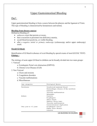
- 1. 1 Upper Gastrointestinal Bleeding Def : Upper gastrointestinal bleeding is from a source between the pharynx and the ligament of Trietz. This type of bleeding is characterised by hematemesis and melena. Bleeding from obscure sources: Defined as bleeding of : • unknown origin that persists or recurs, • that is recurrent or persistent iron deficiency anemia, • occult blood test positivity, or visible bleeding, • after a negative initial or primary endoscopy (colonoscopy and/or upper endoscopy) result. Occult GI bleed: Identification of GI bleed in absence of overt bleeding by special exams of stool (GUIAC TEST) Etiology : The etiology of acute upper GI bleed in children can be broadly divided into two main groups: 1. Variceal a. Extrahepatic Portal vein obstruction (EHPVO) b. Chronic Liver Disease (CLD) 2. Non Variceal a. Ulcers and erosions b. Coagulation disorders c. Vascular malformations d. Miscellaneous UGI BLEEDING IN CHILDREN
- 2. 2 Grading : Bleeding : SHOCK –ve: MILD SHOCK +ve : rapid BT >400ml or 20ml/kg Recovered from shock MODERATE shock persist, bleeding persist ,more BT required SEVERE SEVERE bleeding criteria: 1. 2. 3. 4. Shock 20% of blood volume required immediately (or) ≥40% of blood required in 24hrs to stabilize hemodynamically Fall of Hb < 8gm/dl PSEUDO / SPURIOUS : Newborns who have swallowed maternal blood can present with significant melena or hematemesis while appearing stable clinically. The Apt Downey test performed on the emesis identifies the source of bleeding, conclusively number of substances such as food coloring agents, vegetables such as beetroot, drugs like ampicillin and phenobarbital may mimic hematochezia. Similarly, melena may be mimicked by iron preparations, spinach, chocolate, blueberries, grape juice, or licorice. History and Physical Examination: Most GI bleeding is not life threatening, it is necessary to determine: the source degree possible cause of the bleeding. A complete and thorough history and physical examination is therefore vital. Four questions need to be answered by taking history and physical examination: 1. spurious Vs True bleeding UGI BLEEDING IN CHILDREN
- 3. 3 2. source : Is the child actually bleeding from the gastrointestinal tract? Hemoptysis Vs Hematemisis , rule out Munchausen syndrome 3. What is the site of bleeding? a. Melena is indicative of a significant blood loss (over 2% of blood volume), most likely taking place proximal to the ligament of Trietz. b. Lesions proximal to the ligament of Trietz presents usually as vomiting of bright red or coffee ground blood. c. Streaking of blood on the stools is indicative of lesion in the rectum or anal canal. d. Bright red or dark red blood in the stool is indicative of lesions associated in the ileum or colon. e. In the event of massive bleeding from the upper tract, bright red rectal bleeding may occur in about 10%of cases . f. The presence of blood in the gastric aspirate is very suggestive of bleeding above the ligament of Trietz 4. How much blood has been lost? a. If bleeding is slow, as much as 13% of blood can be lost without any hemodynamic change. b. The loss of palmar crease erythema may be seen when the hand is hyperextended, as a sign of 50% or more blood volume loss Diagnosis: Diagnosis is based on direct observation of blood in the stool or vomitus. Microscopic blood loss in the stool can be confirmed with a faecal occult blood test. For upper GI bleeding, a nasogastric tube is placed to confirm the presence of fresh blood and to evaluate the degree of active bleeding Endoscopy : Esophagogastroduodenoscopy (EGD) and colonoscopy re currently considered the first-line diagnostic procedures of choice for upper and lower GI bleeding, respectively. The site and the cause of bleeding can be identified in 85 to 90% of the patients. Endoscopy also provides immediate access to the bleeding site for any possible intervention. UGI BLEEDING IN CHILDREN
- 4. 4 There are definite advantages of performing an endoscopy within the first 24 h of bleed. These include higher diagnostic accuracy, achieving hemostasis quickly, possibly preventing complications, decreasing transfusions and length of hospital stay for these patients . However, there is no advantage in performing an endoscopy within the first 6 h as compared to 6–24 h. Additionally, complication rate for emergency endoscopy is four times higher than an elective endoscopy Radionuclide Studies : Radionuclide imaging techniques allow identification of those patients who are actively bleeding. (99m) Tc-labeled erythrocytes and (99m)Tc sulfur colloid are two commonly used techniques to detect active bleeding diagnostic sensitivity of the scans 39.1% scintigraphy may be helpful in risk-stratifying patients and planning radiological and surgical interventions. Conventional Angiography: useful in the evaluation of difficult to diagnose cases of recurrent UGI bleeding . Arteriography is best performed in patients in whom bleeding is severe and active. Even 0.5 ml/min of the bleeding rate can be demonstrated angiographically accurate angiographic diagnosis is more likely in acute GI bleeding than in chronic GI bleeding, (71% vs 55%) . Angiography is also effective in demonstrating lesions not bleeding currently as in vascular malformation, telangiectasias and hemangiomas CT Angiography FAL) CT angiography is an excellent tool for fast and accurate diagnosis and localization of acute GI bleeding. (FAL) Advantages include widespread availability, speed, reproducibility, minimal invasiveness and detection of bleeding in the endoscopically inaccessible small bowel. UGI BLEEDING IN CHILDREN
- 5. 5 Limitations of CT angiography are the lack of therapeutic options in comparison to those that are available with endoscopy, colonoscopy and conventional angiography, radiation dose, and risks affiliated with usage of contrast material Role of BUN: BUN increases in view of increased protein load presented to the small intestine following a GI bleeding episode or a decreased excretion from the kidneys of urea nitrogen due to hypovolemia. A direct relationship exists between BUN/ Cr ratio and Delta Hb in upper GI bleeding ∆Hb = 0.08 (BUN/Cr) + 0.8 Ratio of BUN/Cr of ≥30 has 98% specificity and sensitivity of 68.8% of upper GI bleeding. Those who have a BUN/Cr ratio >30 have a high probability that Hb will fall later . When BUN/Cr ratio returns to normal: active bleeding has stopped Management of Gastrointestinal Bleeding: The goals of therapy in a child with GI bleeding should include: 1. Hemodynamic resuscitation 2. cessation of bleeding source 3. Etiological identification 4. prevention of future episodes of GI bleeding UGI BLEEDING IN CHILDREN
- 6. 6 Initial assessment and classification of illness Immediate steps be followed for managing upper GI bleed: UGI BLEEDING IN CHILDREN
- 7. 7 Pharmacologic Management of Mucosal Bleeding: Mucosal bleeding is most common type of upper GI bleed in critically ill children. Therapy in these groups of patients is directed at neutralizing and/or preventing the release of acid. Antacids: In children with gastric and duodenal ulcers, antacids need to be given for a minimum of 48 h after bleeding has subsided. In children more than 5 years of age, magnesium and aluminum hydroxide in doses of the 30 ml/h for the first 48 h followed by same dose at 1 and 3 h after meals throughout the remainder of hospitalization H2 Receptor Antagonists : The H2 receptor antagonists are a class of drugs used to block the action of histamine on parietal cells in the stomach, decreasing the production of acid by these cells. H2 antagonists are used in the treatment of gastritis, peptic ulcers and superficial mucosal erosions. UGI BLEEDING IN CHILDREN
- 8. 8 They have been surpassed in popularity by the more effective proton pump inhibitors. Proton Pump Inhibitors (PPIs): Proton pump inhibitors act by irreversibly blocking the hydrogen/potassium adenosine triphosphatase enzyme system (the H+/K+ ATPase, or, more common, gastric proton pump) of the gastric parietal cell. The proton pump is the terminal stage in gastric acid secretion, being directly responsible for secreting H+ ions into the gastric lumen. Targeting the terminal step in acid production, as well as the irreversible nature of the inhibition, results in their ability to reduce gastric acid secretion by up to 99%. Sucralfate : Sucralfate is a sucrose sulfate-aluminum complex that acts locally in an acidic environment (pH<4). It reacts with acid in the stomach to form a cross-linking, viscous, paste-like material which has an acid buffering effect for as long as 6 to 8 h. It also attaches to proteins (such as albumin and fibrinogen) on the surface of erosions and ulcers to form stable insoluble complexes. These complexes serve as protective barriers at the ulcer surface, preventing further damage from acid, pepsin, and bile. Endoscopic Management of Mucosal Bleeding : various techniques like thermal therapy, sclerosant therapy, clips, and thrombin/fibrin glue . it was recommended that epinephrine injection alone should not be used. UGI BLEEDING IN CHILDREN
- 9. 9 Pharmacologic Management of Variceal Bleeding: Primary objective of therapy is immediate cessation of the bleeding. Start all patients on H2 receptor blocker drugs or Proton pump inhibitors. A vasoactive drug should also be started to decrease the splanchnic pressures. Choose between octreotide and somatostatin except that the latter is costlier. The indications for use of these drugs are: o Lesions that are not amenable to or in which endoscopic therapy has failed; o Child is too unstable for intervention; o children who have arterial bleeding (Dieulafoy’s lesion), o children who have known varices undergoing stabilization before endoscopic therapy. o Growing evidence in studies is accumulating over the superiority of terlipressin over other vasoactive drug Endoscopic Management: Endoscopy should be performed when the patient has been stabilized and preferably within 24 h of admission or onset of hemorrhage . The modalities available for controlling acute variceal bleeding are either variceal ligation or sclerotherapy. Sclerotherapy can control acute variceal bleeding in 70–100% of cases. In more experienced hands, hemostasis can be achieved in more than 90% of cases. endoscopic variceal ligation therapy significantly reduced rebleeding, mortality, frequency of esophageal strictures and the number of sessions required to achieve variceal eradication when compared with injection sclerotherapy . Sclerosants are effective in controlling active variceal bleeding but are less favored than variceal ligation because of associated complications UGI BLEEDING IN CHILDREN
- 10. 10 Surgical Management: In cases where conservative management fails with combined pharmacotherapy and endoscopic treatments, shunt and nonshunt surgeries are the definitive treatment. For primary UGI bleeding in children it is recommend that when more than 85 mL/kg of blood is transfused within 1 1/2 h and bleeding has not subsided, surgery should be considered For intrahepatic portal hypertension, transjugular intrahepatic portosystemic shunting (TIPS) provides temporary decompression of the intrahepatic portal vein into the hepatic veins. Surgical portosystemic or portoportal shunts for GI bleeding are now reserved for refractory cases and/or when liver transplantation is not an option. Prognosis: Prognostic factors associated with increased mortality: The coexistence of another severe medical disorder Coagulation disorder Failure to identify the bleeding site Hemoglobin level <7 g/dL, and/or a hematocrit value of <20% at presentation >85 ml/kg blood loss without surgical intervention UGI BLEEDING IN CHILDREN