Evolution of radiation 2012
•Transferir como PPT, PDF•
13 gostaram•4,591 visualizações

This document discusses the evolution of radiation therapy from its discovery in the late 19th century to modern techniques. It traces developments such as the discovery of x-rays and radioactivity, early radium and x-ray therapies, and the introduction of cobalt-60 and linear accelerators to improve targeting ability. Modern advances discussed include intensity-modulated radiation therapy (IMRT), image-guided radiation therapy (IGRT), proton beam therapy, and radiosurgery techniques like Gamma Knife and Cyberknife which allow extremely precise high dose radiation treatments.

Recomendados
Recomendados
Mais conteúdo relacionado
Mais procurados
Mais procurados (20)
Destaque
Destaque (20)
Semelhante a Evolution of radiation 2012
Semelhante a Evolution of radiation 2012 (20)
Mais de Robert J Miller MD
Mais de Robert J Miller MD (20)
Último
Último (20)
Evolution of radiation 2012
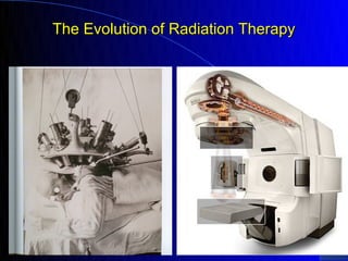
- 1. The Evolution of Radiation Therapy
- 4. Thomas Hodgkin in 1832 Boy with Hodgkin’s in 1920 Until the 1960s, among patients with an advanced stage of Hodgkin's disease the median survival was 2 years, and only 5% of patients lived beyond 4 years. Currently it is curable in 85 to 95% of cases, depending on its stage
- 5. History of Radiotherapy 1895 Roentgen discovers Xrays 1896 Becquerel discovers radioactivity 1898 Curie discovers radium late 1890’s therapeutic use 1920’s reliable Xray tubes (150-300Kv) 1950’s Cobalt (1Mv or million volt) 1960’s Linear Accelerator (4 - 25 million) 1970’s computers and CT scans 1980’s 3-D radiotherapy 1990’s 3D conformal therapy 2002 IMRT (intensity modulated radiotherapy) 2002+ IGRT (image guided and radiosurgery)
- 6. Wilhelm Conrad Röntgen On the evening of November 8, 1895, he found that, if the discharge tube is enclosed in a sealed, thick black carton to exclude all light, and if he worked in a dark room, a paper plate covered on one side with barium platinocyanide placed in the path of the rays became fluorescent even when it was as far as two meters from the discharge tube. ( 27 March 1845 – 10 February 1923) was a German physicist, an achievement that earned him the first Nobel Prize in Physics in 1901
- 7. Emil Grubbe Medical Student in Chicago March 29, 1896 in an X-ray Tube factory in Chicago he began to bombard Rose Lee an elderly woman with recurrent breast cancer and had the first documented response to radiation
- 8. Antoine Henri Becquerel In 1896, decided to investigate whether there was any connection between X-rays and naturally occurring phosphorescence. He had inherited from his father a supply of uranium salts, which phosphoresce on exposure to light. When the salts were placed near to a photographic plate covered with opaque paper, the plate was discovered to be fogged. (15 December 1852 – 25 August 1908) was a French physicist, Nobel laureate, and the discoverer of radioactivity along with Marie Skłodowska- Curie and Pierre Curie, for which all three won the 1903 Nobel Prize in Physics.
- 9. Marie Curie The discovery of radioactivity by Henri Becquerel led to in 1898 the Curies to the isolation of polonium, named after the country of Marie's birth, and radium. Mme. Curie developed methods for the separation of radium from radioactive residues in sufficient quantities to allow for its characterization and the careful study of its properties, therapeutic properties in particular. (7 November 1867 – 4 July 1934) was a French-Polish physicist and chemist, She was the first person honored with two Nobel Prizes—in physics and chemistry. She was the first female professor at the University of Paris, and in 1995 became the first woman to be entombed on her own merits in the Pantheon in Paris.
- 10. What is polonium-210, how can it kill you? Saturday, November 25, 2006 LONDON - Polonium-210 - the radioactive substance that killed a former Russian spy in London - is one of the world's rarest elements, first discovered in the 19th century by scientists Marie and Pierre Curie. Alexander Litvenko
- 12. Pre- 1904 X-ray Machine
- 13. Radium Pack
- 15. 1902 Cervix Contact X-ray Tube
- 16. 1930 Radium Tele-Curie Machine
- 17. Radiation in the early 20th Century “Weak Discouraged Men! Now Bubble Over with Joyous Vitality Through the Use of Glands and Radium” Vita Radium Suppositories (ca.1930)
- 18. Any Risks of Harm from Radiation?
- 19. Undark and the Radium Girls
- 20. Undark and the Radium Girls
- 21. Undark and the Radium Girls In 1902, inventor William J. Hammer left Paris with a curious souvenir. The famous scientists Pierre and Marie Curie had provided him with some samples of their radium salt crystals. Radioactivity was somewhat new to science, so its properties and dangers were not well understood; but the radium’s slight blue-green glow and natural warmth indicated that it was clearly a fascinating material. Hammer went on to combine his radium salt with glue and a compound called zinc sulfide which glowed in the presence of radiation. The result was glow- in-the-dark paint. US Radium employed hundreds of women at their factory in Orange, New Jersey, After a few strokes a brush tended to lose its shape, so the women’s managers encouraged them to use their lips and tongues to keep the tips of the camel hair brushes sharp and clean. The glowing paint was completely flavorless, and the supervisors assured them that rosy cheeks would be the only physical side effect
- 22. Undark and the Radium Girls At their first appearance in court in January 1928, two were bedridden, and none could raise their arms to take the oath. Grace Fryer, still described by reporters as “pretty,” was unable to walk, required a back brace to sit up, and had lost all of her teeth. The “Radium Girls” began appearing in headlines nationwide, and the grim descriptions of their hopeless condition reached Marie Curie in Paris. “I would be only too happy to give any aid that I could,” she said, adding, “there is absolutely no means of destroying the substance once it enters the human body.” The last of the famous Radium Girls died in the 1930s, the dial painters had ingested anywhere from a few hundred to a few thousand microcuries of radium per year. One tenth of a microcurie is now considered to be the maximum safe exposure. Marie Curie herself died of radiation-related ailments in 1934. Because radium has a half-life of 1,600 years, her lab notebooks are said to be too highly contaminated to be safely handled even today. Radium continued to be used to illuminate watches until about 1968, but under much safer conditions.
- 23. At the recommendation of his doctor, Byers began drinking Radithor, and he continued to do so long averaged three bottles a day for two years. Byers stopped consuming Radithor in 1930 when his teeth started falling out and holes appeared in his skull. Perhaps more than anything else, his death in 1932 alerted the public, and much of the medical Radithor (ca. 1928) profession, of the harmful effects of "mild" radium therapy.
- 24. Shoe-Fitting X-Ray Machine The shoe fitting fluoroscope x-ray machine was a common fixture in shoe stores during the 1930s, 1940s and 1950s. A typical unit consisted of a vertical wooden cabinet with an opening near the bottom into which the feet were placed. When you looked through one of the three viewing ports on the top of the cabinet (e.g., one for the child being fitted, one for the child's parent, and the third for the shoe salesman or saleswoman), you would see a fluorescent image of the bones of the feet and the outline of the shoes.
- 25. The Gra-Maze Uranium Comforter (ca. 1965) "This is your personal radioactive uranium comforter. Actually your own health mine in miniature, If you followed the suggestion on the pad and checked it with a Geiger counter, you would find it to be measurably radioactive.
- 28. Skull Film Roentgenogram circa 1923
- 29. Electron Charged Rays for Cancer, Vienna, 1938
- 30. Radium Bomb London 1934 4 Radium sources close to the patient’s skin. Each source focused on the cancer from a different angle, maximizing the dose to the localized area. Protective devices for the medical practitioner were non-existent at this time
- 31. COBALT The use of 60Co sources for teletherapy was begun in the 1950s as a replacement for the 250 kVp x-ray treatment machines that were then in common use. The skin-sparing effects of 60Co treatment were immediately recognized and such effects were no longer limitations on treatment.
- 32. Henry Kaplan "In the 1950s, I began to hear cocktail party conversations about an interesting new atom smasher being developed on the campus," An atom smasher, otherwise known as a particle accelerator, uses electromagnetic fields to propel charged particles to great energies. By slamming accelerated electrons into a target made of heavy metal, high-energy X-ray beams result. Kaplan thought this machine could be harnessed to deliver X- ray therapy that was superior to the unreliable, weak and unfocused radiation therapy approaches of his day.
- 33. First Medical Linear Accelerator at Stanford in 1956
- 34. In January 1956 the machine was ready to be used on their first patient, a boy with retinoblastoma in his one remaining eye after surgeons had removed the tumor in the other eye. Destroying the tumor while sparing the eye would have been impossible with earlier, less-focused radiation sources.
- 36. Use of Cobalt in Cervix Cancer Patterns of Care Data 100 80 60 Cobalt 40 LinAc 20 0 1978 1983 1988 Montana IJROBP 1995;32:14381
- 38. Penetration of Radiation by Energy Level
- 39. Skin Sparing and the Energy of the Beam
- 40. Selecting the Proper Dose of Radiation “It may be a bit over-exposed”
- 41. Prostate Cancer Outcome based on Dose of Radiation Biochemical relapse-free results by treatment modality. The study population comprised 2991 consecutive patients treated at the Cleveland Clinic Foundation or Memorial Sloan Kettering at Mercy Medical Center
- 42. How Much is Safe ?
- 43. Strategies to Improve the Effectiveness of Radiation Use chemical or chemotherapy to make the cancer cells more sensitive to radiation Design ways to focus the radiation more precisely and allow the use of higher doses (conformal 3D, IMRT or radiosurgery) Implant the radiation directly into the tumor (brachytherapy, with wires or seeds) or isotopes or monoclonal antibodies
- 44. Esophagus Cancer Survival - Radiation plus Chemotherapy versus Radiation Alone 5 Year Survival Radiation = 0% Radiation plus Chemotherapy = 26%
- 45. Erbitux + Radiation ERBITUX binds specifically to epidermal growth factor receptor on both normal and tumor cells, and competitively inhibits the binding of (EGF) Over-expression of EGFR is detected in many human cancers. Survival with Advanced Head and Neck Cancer Radiation Alone: 19 months Radiation + Erbitux: 36 months
- 46. Strategies to Improve the Effectiveness of Radiation Use chemical or chemotherapy to make the cancer cells more sensitive to radiation Design ways to focus the radiation more precisely and allow the use of higher doses (conformal 3D, IMRT or radiosurgery) Implant the radiation directly into the tumor (brachytherapy, with wires or seeds, or isotopes or monoclonal antibodies)
- 48. Targets and Spread of Cancer Cells
- 50. MR Spectroscopy to Define Cancer Target Determining the Tumor Target Volume for a Glioma T1 images (break down of the blood brain barrier) tend to underestimate the size of the cancer and T2 post contrast images (which includes the whole area of edema) tend to over-estimate the size. MR spectroscopy may be more accurate
- 51. Using PET Scans to Define Cancer
- 52. Improving and Refining Radiation Therapy •First use improved imaging technology (CT/MRI/PET scans) to define the proper target •Then use increasingly sophisticated technology to bend the beam so it will conform to the shape of the target (conformal therapy) •Modulate the beam even more to get even better targeting (IMRT or intensity modulated radiation therapy) •Combine IMRT with technology that will more accurately identify the target (IGRT or image guided radiotherapy) •Once the techniques are extremely accurate then use extremely high doses of radiation that will completely eliminate the cancer (radiosurgery e.g. Gamma Knife or Cyberknife)
- 53. Conformal (3D) Radiation for Prostate Cancer A Randomized trial at MD Anderson comparing conventional radiation with conformal therapy PSA Cures conventional 53% conformal 72% Pollack IRJOBP 1996;34:555
- 54. Treatment Results and Complications with Prostate Cancer Therapy Cure Potent Comps Implant 87-96% 81-90% 2-12% External 80-96% 33-70% 3-17% Surgery 85-95% 22-90% 8-10% Conformal 96% 70% 3-4% D’Amico J Clin Onc 1996;14:304
- 58. In The Head of the linear accelerator is the beam shaping MLC
- 61. Using image guided IMRT to treat spinal cancer
- 62. Bone Metastases to the Spine Involved vertebrae on the left and normal on the right
- 63. Kidney cancer in the spinal vertebrae surrounding the cord, and appearance after radiation…is it possible to safely radiate further?
- 64. Combine a CT scan and linear accelerator to ultimate in targeting (IGRT) and ultimate in delivery (dynamic, helical IMRT) ability to daily adjust the beam (ART or adaptive radiotherapy)
- 66. PROTON BEAM
- 67. Improved Survival with Head and Neck Cancer and IMRT
- 69. Conformal proton therapy for prostate carcinoma. Int J Radiat Oncol Biol Phys. 1998 Sep 1;42(2):299- 304 Department of Radiation Medicine, Loma Linda Linac University Medical Center When post-treatment prostate-specific antigen (PSA) was used as an endpoint for disease control, the 4.5- Proton beam year disease-free survival rate 89%, 72%, and 53% for patients with initial PSA levels of 4.1-10.0, 10.1- 20.0, and > 20.0, respectively. External Beam using modern techniques and a dose of at least 72Gy. Compared with most recent data on Proton Beam from Loma Linda
- 70. Best Radiation Treatment for Localized Prostate Cancer SEER Data 2000-2009. J Clin Onc 2012:30 (s5a3) Treatment GI Toxicity Hip Fxt RxFailure Conformal 9.7% + 25% + 24% + IMRT - - - Proton Beam 45% + - - Conformal therapy had higher GI toxicity and hip fractures than IMRT and a higher relapse rate. Proton had more GI toxicity and no improvement in outcome compared to IMRT
- 71. Radiosurgery – using extremely well targeted radiation to totally eliminate the tumor Gamma Knife or Cyberknife
- 72. RadioSurgery the Old Way Man Survives 18-Inch Drill Bit in Head Ron Hunt lost an eye but suffered no brain damage after a freak accident with a large drill bit. Sept. 2— 2003
- 73. Lars Leksell. (1907- 1986). ... introduced his stereotactic instrument for human functional neurosurgery in 1949. ... Professor Lars Leksell, Swedish physician and Professor of Neurosurgery at the Karolinska Institute in Stockholm, introduced his stereotactic instrument for human functional neurosurgery in 1949. In 1951, using the Uppsala University cyclotron, Leksell developed the concept of radiosurgery, employed proton beams coming from several directions into a small area into the brain, in experiments in animals and in the first treatments of human patients. Therefore, he achieved a new non-invasive method of destroying discrete anatomical regions within the brain while preserving the surrounding normal tissues. In 1967, the first Gamma Knife unit was put into clinical use in Karolinska and this was a 179 cobalt 60 source.
- 75. The Mayo Clinic gamma knife experience: indications and initial results.
- 76. On average, about 92% of acoustic neuromas treated with Gamma Knife radiosurgery demonstrate tumor shrinkage or growth cessation over two to five years. Hearing is preserved at pre- Gamma Knife levels in as many as 51% of patients
- 77. Brain Metastases Treatment Median Survival Local Failure Whole brain external 8 – 15 w 52% surgical resection 33 – 38 w 20% rasdiosurgery 44 w 14%
- 78. Brain Metastasis and External Beam Irradiation
- 81. Cyberknife Radiosurgery of the Spine
- 82. Strategies to Improve the Effectiveness of Radiation Use chemical or chemotherapy to make the cancer cells more sensitive to radiation Design ways to focus the radiation more precisely and allow the use of higher doses (conformal 3D, IMRT or radiosurgery) Implant the radiation directly into the tumor (brachytherapy, with wires or seeds, or isotopes or monoclonal antibodies)
- 84. HDR Treatments
- 88. Prostate Cancer Cure Rates External Beam versus Seeds versus Surgery
- 89. Mammosite for Breast Cancer
- 90. HDR = high dose rate machine that can run radiation through a tube that reaches the patient through skin applicators
- 91. Radiation for Skin Cancer HDR skin applicators – three times a week for 6 treatments over a two week period
- 92. The HDR Skin applicator is taped in place over the cancer
- 93. The applicator is attached by cable to the machine that makes it radioactive for a few minutes
- 94. Small squamous cancer of the chin, appearance 3.5 weeks after radiation (HDR)
- 95. Radiation Isotope Therapy for Bone Metastases Samarium 153 (SM 153) or Quadramet Samarium is now being used along with Strontium. Samarium has a short half- life (46 hours) with 75% of the dose absorbed within 4 days. The beta particle energy (233keV) penetrates 1.7mm in bone and 3.1 mm in soft tissue. In trials, 48-54% get pain relief. Metastron or Strontium-89 (Sr-89) Sr-89 is a beta-emitting radionuclide with a long physical half-life (50.5 days)
- 96. Radio-active Monoclonal Antibodies monoclonal antibody targets the CD20 antigen, which is found on the surface of B-cell tumors
- 97. Radio-active Monoclonal Antibodies In February 2002, Zevalin was the first radioimmunotherapy to receive FDA approval. Zevalin consists of a monoclonal antibody linked to the radioactive isotope yttrium-90. After infusion into a patient, the monoclonal antibody targets the CD20 antigen, which is found on the surface of B-cell tumors. In this manner, cytotoxic radiation is delivered directly to malignant cells. On June 30, 2003 announced FDA approval of BEXXAR (Tositumomab and Iodine I 131 Tositumomab)
- 98. www.aboutcancer.com Cancer Information anatomy and imaging General Cancer Statistics site map Cancer News, Stages, Old Stages Most Common Cancers * brain * breast * colon/rectum * gynecologic * head and neck (mouth, tongue, oral, etc.) * lung * metastatic * prostate * skin cancer Other Specific Cancers Radiation or Chemotherapy or Surgery Cancer Calculators Best Web Sites Advice for Everyone