
Recomendados
Mais conteúdo relacionado
Mais procurados
Mais procurados (20)
Destaque
Destaque (20)
Semelhante a Anatomy and functions of the temporomandibular joint
Semelhante a Anatomy and functions of the temporomandibular joint (20)
Mais de ddert
Mais de ddert (20)
Último
Último (20)
Anatomy and functions of the temporomandibular joint
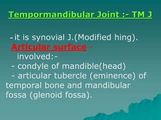
- 1. - it is synovial J.(Modified hing). -Articular surface -involved: - condyle of mandible(head) - articular tubercle (eminence) of temporal bone and mandibular fossa (glenoid fossa). Tempormandibular Joint :- TM J
- 4. - Fibrons capsule:- is loose , attaches to margin of articular area on temporal bone and around neck of mandible. - Articular disc:- divide the joint into two separate compartment, it is fibrous plate. - upper compart: permit gliding movement of protrusion and retrusion - lower compart: permit depression and elevation (gliding + rotatory)
- 6. Ligaments of Joint: - Lateral ligament (temporamandibular) - strengthen TMJ laterally - prevent post dislocation of joint - extend from root of zygomectic arch, attached to posterolate aspect of neck of mandible.
- 7. - stylomandibular ligament: - thicken part of deep cervical fascia - separted parotid from submandib. Gland - attached to lat.surface of styl. process and to angle of mandib. - sphenomandibular ligament: Run from spine of sphenoid to lingua of mandible.
- 11. Muscles Producing movement of mandible at TMJ are: * Depression (open mouth): - gravity - suprahyaid and infrahyaid muscles(Digastric,Mybhyaid,genaid) - lat. Pterygoid m. * Elevation (closed mouth): - temporal - masseter - medial pterygaid of both side
- 12. * Protrusion (protraction of chin): - lateral pterygoid m. - medial pterygoid m. - masseter (superficial) * Retrusion (retraction): - temporal (post., middle, fibres) - masseter (middle, deep fibres) * lateral or side to side movement produce by medial, lateral pterygaid of each side act alternatively.
- 13. Muscles of chewing + Grinding: - temporalis of the same side. - pterygoids of the opposite side.
- 14. Relation of T.M. joint: - Laterally___ skin , fascia Parotid gland Temporal branch of fascial - Medially___ tympanic plate which separate the joint from I.C.A. Sphine of sphenoid and sphenomandibular ligament middle meningal a.
- 15. - Anterior___ lateral pterygaid m. Masseteric n. and vessels - Posterior___ parotid separate joint from external auditory meatus Superfial temporal vessels Auriculo temporal vessels
- 16. - Superior____ middle cranial fossa Middle meningel vessels - Inferior___ axillary artery and nerves -Blood supply of joint: - superficial temporal a. - Maxillary a. -Nerves supply of joint: - auriculotemporal n. - Masseteric n.
- 17. Applied Anatomy - Dislocation of mandible - Dearrangement of articular dis (clicking) - Arthritis of TMJ
- 18. Pterygo palatine fossa * small pyramidal space inferior to the apex of orbit. Boundary: Anteriory ___ post. aspect of maxilla Posteriorly___ pterygaid process of sphenoid bone Medially___ palatine bone (vertical plate) Roof ___ greater using of sphenoid Floor ___ pyramidal process of palatine bone.
- 20. Communication of fossa *it is slilike open laterally into infratemporal fossa through pterygo- maxillary fissure. * Medially with nasal cavity through spheno-palatine foramen * Anterosuperiorly with the orbit through inferior orbital fissure * Posterosuperiorly with middle cranial fossa through foramen rotundom and pterygaid canal
- 21. Contents: Terminal (3rd)part of maxillary artery Maxillary nerves Nerve of pterygaid canal Pterygopalatine ganglia Maxillary artery: * pass over Lat. Pteryg. m. and enter pterygopal. fossa * pass through pterygomaxillary fissure where it lies anterior to pterygopalatine ganglia * Artery give branches that accompany all nerves in the fossa with same names.
- 25. *Branches of pterygopalatin part of maxillary artery are: 1- posterior superior alveolar a. 2- Descending palatine a. divided into:- greater palatine a.and lesser palatine a 3- Artery of pterygoid canal 4- spheno palatine artery divided into : - posterior lateral nasal a. to lat.wall of nasal cavity and paranasal sinus - posterior nasal septal a. 5- infra orbital a. which give rise to anterior superior alveolar a.
- 26. Maxillary nerves: * leave cranial cavity through foramen rotundum * cross pterygopal. fossa * continues forward through infer. orbital fissure into orbit. * terminate as infraorbital nerve traverse the infraorbital canal to reach the face * it has branches arising in :- - pterygopalatine fossa - in floor of orbit - on the face
- 27. * in pterygopalatine fossa:- 1- two branches suspend the pterygopataline ganglia called pterygopalatine nerve 2- posterior superior alvealor n. for upper molar teeth * in the floor of orbit: 1- middle superior alveolar n. 2- anterior superior alveolar n. In lateral , anterior wall of maxilla 3- zygomatic nerve which divided into zygomatico temporal, zygomaticofacial
- 28. •infra orbital nerve: emerge into face through infra orbital foramen supplies skin of cheek , lower eyelid , upper lib , lateral surface of nose.
- 33. Pterygo palatinr ganglion: * it is relay situation betw.superior salivary nucleus in pons and lacrimal gland and mucous and serous gland of palate , nose, and paranasal sinus. * it is ganglion of hay fever (running nose and eyes) * There is autonomic root called nerve of pterygoid canal(vidian nerve). This nerve is formed in foramen lacerum by union of greater(superf.) petrosal n. which containing parasympathetic, secretomotor fibres, with deep petrosal n. containing vasoconstrictor fibre. Both nerve pass forward in pterygoid canal and join the five branches of pterygopalatine ganglia:-
- 34. 1- nasopalatine(long sphenopalatine)n. Spheno palatin foramen ___ supply nasal septum 2- posterior superior lateral nasal n.(short sphenopalatine n.) Sphenopalatin foramen ___ posterosuperior quadrant of lateral wall of nose.
- 35. 3-anterior palatine n. (greater palatine n.) Greater palatine canal___ foramen mucous ml. of hard palate Medial wall of maxillary sinus 4- middle, posterior palatine n.(lesser palatine n.) Lesser palatine foramen ___ mucous ml. of soft palate supratonsillar racess 5- pharyngeal branch___ palatovaginal canal___ mucous ml. of nasopharynx
- 36. Digastric and styloid muscles Digastric muscles: * consist of anterior belly & posterior belly United by intermediate tendon Posterior belly: attached to medial surface of mastaid process inclined forward, downward, continuous with intermediate tendon close to hyaid bone.Tendon pierced the stylohyaid and is anchored by facial sling to hyaid bone.innervaled by facial nerve. Anterior belly: continuous forward from intermediate tendon to attached to inferior border of mandible near midline.innervated by mandibular division of trigeminal n. * digastric muscles elevate hyaid bone during swallowing and assist mylohyaid and lateral pteryg. in depressing mandible when opening the mouth
- 37. Muscles of styloid process: 3 muscles___ stylohyaid___ to hyaid bone Stylopharyngius___ pharynx Styloglossus___ tongue * 2 ligament attached to styloid process:- Stylohyaid ligament. Stylomandibular ligament.