
Recomendados
Mais conteúdo relacionado
Mais procurados
Mais procurados (20)
Semelhante a Eye Diseases
Semelhante a Eye Diseases (20)
Último
Último (20)
Eye Diseases
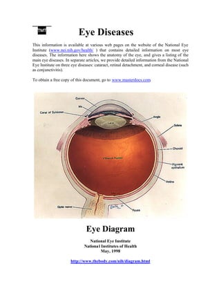
- 1. Eye Diseases This information is available at various web pages on the website of the National Eye Institute (www.nei.nih.gov/health/ ) that contains detailed information on most eye diseases. The information here shows the anatomy of the eye, and gives a listing of the main eye diseases. In separate articles, we provide detailed information from the National Eye Institute on three eye diseases: cataract, retinal detachment, and corneal disease (such as conjunctivitis). To obtain a free copy of this document, go to www.masterdocs.com. Eye Diagram National Eye Institute Nationa l Institutes of Health May, 1998 http://www.thebody.com/nih/diagram.html
- 2. List of Eye Diseases Detailed information on each disease is available at http://www.nei.nih.gov/health/ • Age-Related Macular Degeneration o Are You at Risk for Age-Related Macular Degeneration? o Facts About Age-Related Macular Degeneration o Results--Age-Related Eye Disease Study • Amblyopia • Anophthalmia and Microphthalmia • Behçet' s Disease of the Eye • Bietti's Crystalline Dystrophy • Blepharitis • Blepharospasm • Cataract o Are You at Risk for Cataract? o Facts About Cataract • Corneal Disease • Diabetic Eye Disease o Are You at Risk for Diabetic Eye Disease? o Facts About Diabetic Retinopathy o Diabetic Eye Disease: How Much Do You Know? Take this quiz and find out o Diabetic Eye Disease: How Much Do You Know? (PDF* file) o Diabetes: Think of all the beautiful things you wouldnít see if you lost your sight • Floaters • Glaucoma o Are You at Risk for Glaucoma? o Facts About Glaucoma o Glaucoma: How Much Do You Know? Take this quiz and find out o Glaucoma: How Much Do You Know? (PDF* file) o Library of Congress Mid-Day Lecture: Dr. Eve Higginbotham Discusses Glaucoma (cybercast) • Histoplasmosis o Low Visio n o Low Vision: Help Is Available o Do You Have Low Vision? Take this quiz and find out o What You Should Know About Low Vision • Macular Hole • Macular Pucker • Retinal Detachment • Retinopathy of Prematurity (ROP) • Usher Syndrome • Vitreous Detachment
- 3. What is age-related macular degeneration? Age-related macular degeneration (AMD) is a disease that blurs the sharp, central vision you need for "straight-ahead" activities such as reading, sewing, and driving. AMD affects the macula, the part of the eye that allows you to see fine detail. (See diagram below.) AMD causes no pain. In some cases, AMD advances so slowly that people notice little change in their vision. In others, the disease progresses faster and may lead to a loss of vision in both eyes. AMD is a leading cause of vision loss in Americans 60 years of age and older. What is amblyopia? The brain and the eye work together to produce vision. Light enters the eye and is changed into nerve signals that travel along the optic nerve to the brain. Amblyopia is the medical term used when the vision in one of the eyes is reduced because the eye and the brain are not working together properly. The eye itself looks normal, but it is not being used normally because the brain is favoring the other eye. This condition is also sometimes called lazy eye. What are anophthalmia and microphthalmia? Anophthalmia and microphthalmia are often used interchangeably. Microphthalmia is a disorder in which one or both eyes are abnormally small, while anophthalmia is the absence of one or both eyes. These rare disorders develop during pregnancy and can be associated with other birth defects. What is Behçet's disease? Behçet's disease is an autoimmune disease that results from damage to blood vessels throughout the body, particularly veins. In an autoimmune disease, the immune system attacks and harms the body's own tissues. What is Bietti's Crystalline Dystrophy? Bietti's crystalline dystrophy (BCD) is an inherited eye disease named for Dr. G. B. Bietti, an Italian ophthalmologist, who described three patients with similar symptoms in 1937. The symptoms of BCD include: crystals in the cornea (the clear covering of the eye); yellow, shiny deposits on the retina; and progressive atrophy of the retina, choriocapillaries and choroid (the back layers of the eye). This tends to lead to progressive night blindness and visual field constriction. BCD is a rare disease and appears to be more common in people with Asian ancestry. People with BCD have crystals in some of their white blood cells (lymphocytes) that can be seen by using an electron microscope. Researchers have been unable to determine
- 4. exactly what substance makes up these crystalline deposits. Their presence does not appear to harm the patient in any other way except to affect vision. What is blepharitis? Blepharitis is a common condition that causes inflammation of the eyelids. The condition can be difficult to manage because it tends to recur. What is Blepharospasm? Blepharospasm is an abnormal, involuntary blinking or spasm of the eyelids. What is a cataract? A cataract is a clouding of the lens in the eye that affects vision. Most cataracts are related to aging. Cataracts are very common in older people. By age 80, more than half of all Americans either have a cataract or have had cataract surgery. A cataract can occur in either or both eyes. It cannot spread from one eye to the other. What is the cornea? The cornea is the eye's outermost layer. It is the clear, dome-shaped surface that covers the front of the eye. What is diabetic retinopathy? Diabetic retinopathy is a complication of diabetes and a leading cause of blindness. It occurs when diabetes damages the tiny blood vessels inside the retina, the light-sensitive tissue at the back of the eye. A healthy retina is necessary for good vision. If you have diabetic retinopathy, at first you may notice no changes to your vision. But over time, diabetic retinopathy can get worse and cause vision loss. Diabetic retinopathy usually affects both eyes. Facts About Floaters Floaters are little "cobwebs" or specks that float about in your field of vision. They are small, dark, shadowy shapes that can look like spots, thread- like strands, or squiggly lines. They move as your eyes move and seem to dart away when you try to look at them directly. They do not follow your eye movements precisely, and usually drift when your eyes stop moving. In most cases, floaters are part of the natural aging process and simply an annoyance. They can be distracting at first, but eventually tend to "settle" at the bottom of the eye, becoming less bothersome. They usually settle below the line of sight and do not go away
- 5. completely. Most people have floaters and learn to ignore them; they are usually not noticed until they become numerous or more prominent. Floaters can become apparent when looking at something bright, such as white paper or a blue sky. What is glaucoma? Glaucoma is a group of diseases that can damage the eye's optic nerve and result in vision loss and blindness. However, with early treatment, you can often protect your eyes against serious vision loss. What is histoplasmosis? Histoplasmosis is a disease caused when airborne spores of the fungus Histoplasma capsulatum are inhaled into the lungs, the primary infection site. This microscopic fungus, which is found throughout the world in river valleys and soil where bird or bat droppings accumulate, is released into the air when soil is disturbed by plowing fields, sweeping chicken coops, or digging holes. Histoplasmosis is often so mild that it produces no apparent symptoms. Any symptoms that might occur are often similar to those from a common cold. In fact, if you had histoplasmosis symptoms, you might dismiss them as those from a cold or flu, since the body's immune system normally overcomes the infection in a few days without treatment. However, histoplasmosis, even mild cases, can later cause a serious eye disease called ocular histoplasmosis syndrome (OHS), a leading cause of vision loss in Americans ages 20 to 40. What is low vision? Low vision means that even with regular glasses, contact lenses, medicine, or surgery, people find everyday tasks difficult to do. Reading the mail, shopping, cooking, seeing the TV, and writing can seem challenging. Millions of Americans lose some of their vision every year. Irreversible vision loss is most common among people over age 65. What is a macular hole? A macular hole is a small break in the macula, located in the center of the eye's light- sensitive tissue called the retina. The macula provides the sharp, central vision we need for reading, driving, and seeing fine detail. A macular hole can cause blurred and distorted central vision. Macular holes are related to aging and usually occur in people over age 60.
- 6. What is a macular pucker? A macular pucker is scar tissue that has formed on the eye's macula, located in the center of the eye's light-sensitive tissue called the retina. The macula provides the sharp, central vision we need for reading, driving, and seeing fine detail. A macular pucker can cause blurred and distorted central vision. What is retinal detachment? The retina is the light-sensitive layer of tissue that lines the inside of the eye and sends visual messages through the optic nerve to the brain. When the retina detaches, it is lifted or pulled from its normal position. If not promptly treated, retinal detachment can cause permanent vision loss. In some cases there may be small areas of the retina that are torn. These areas, called retinal tears or retinal breaks, can lead to retinal detachment. What Is Usher Syndrome? Usher syndrome is an inherited condition that causes 1) a serious hearing loss that is usually present at birth or shortly thereafter and 2) progressive vision loss caused by retinitis pigmentosa (RP). RP is a group of inherited diseases that cause night-blindness and peripheral (side) vision loss through the progressive degeneration of the retina, the light-sensitive tissue at the back of the eye that is crucial for vision. Researchers have described three types of Usher syndrome-type I, type II and type III. Individuals with Usher syndrome type I are nearly or completely deaf and experience problems with balance from a young age. They usually begin to exhibit signs of RP in early adolescence. Individuals with Usher syndrome type II experience moderate to severe hearing impairment, have normal balance, and experience symptoms of RP later in adolescence. Individuals with Usher syndrome type III are born with normal hearing but develop RP and then progressive hearing loss. Vitreous Detachment Most of the eye's interior is filled with vitreous, a gel- like substance that helps the eye maintain a round shape. There are millions of fine fibers intertwined within the vitreous that are attached to the surface of the retina, the eye's light-sensitive tissue. As we age, the vitreous slowly shrinks, and these fine fibers pull on the retinal surface. Usually the fibers break, allowing the vitreous to separate and shrink from the retina. This is a vitreous detachment. In most cases, a vitreous detachment is not sight-threatening and requires no treatment.
- 7. As the vitreous shrinks, it becomes somewhat stringy, and the strands can cast tiny shadows on the retina that you may notice as floaters, which appear as little "cobwebs" or specks that seem to float about in your field of vision. If you try to look at these shadows they appear to quickly dart out of the way. One symptom of a vitreous detachment is a small but sudden increase in the number of new floaters. This increase in floaters may be accompanied by flashes of light (lightning streaks) in your peripheral, or side, vision. In most cases, either you will not notice a vitreous detachment, or you will find it merely annoying because of the increase in floaters. A vitreous detachment is a common condition that usually affects people over age 50, and is very common after age 80. People who are nearsighted are also at increased risk. Those who have a vitreous detachment in one eye are likely to have one in the other, although it may not happen until years later. Although a vitreous detachment does not threaten sight, once in a while some of the vitreous fibers pull so hard on the retina that they create a macular hole or lead to a retinal detachment. Both of these conditions are sight-threatening and should be treated immediately. If left untreated, a macular hole or detached retina can lead to permanent vision loss in the affected eye. Those who experience a sudden increase in floaters or an increase in flashes of light in peripheral vision should have an eye care professional examine their eyes as soon as possible. The only way to diagnose the cause of the problem is by a comprehensive dilated eye examination. If the vitreous detachment has led to a macular hole or detached retina, early treatment can help prevent loss of vision. This document is provided as a service to the public by TMT (Taylor MicroTechnology, Inc.). TMT does not provide medical advice to you. TMT does inform you of publicly available medical information. However, please realize that the possible diagnoses provided may not include the cause of your own pain, and that a reliable diagnosis can only be obtained by contacting your own health care provider. For details of the Content Disclaimer and Legal Disclaimers regarding materials provided by TMT, see www.masterdocs.com/disclaimer.htm.