SlideShare uma empresa Scribd logo
1 de 20
Baixar para ler offline
REVIEW ARTICLE


                               The Pathology of Endometriosis
      A Survey of the Many Faces of a Common Disease Emphasizing
      Diagnostic Pitfalls and Unusual and Newly Appreciated Aspects
                                                     Philip B. Clement, MD


                                                                      O     ur knowledge of the pathology and pathogenesis of
Abstract: Although the histologic diagnosis of endometriosis is             endometriosis largely stems from the work of Dr
usually straightforward, many diagnostic problems can arise as        John Albertson Sampson (1873 to 1946), the ‘‘Father of
a result of alterations or absence of its glandular or stromal        Endometriosis.’’ In a contribution to the History Series in
components. The diagnostic difficulty in such cases can be              the International Journal of Gynecological Pathology,1 I
compounded by tissue that is limited to a small biopsy specimen.      recounted in some detail Sampson’s remarkable career
The appearance of the glandular component can be altered by           and seminal contributions. That article was preceded by 2
hormonal and metaplastic changes, as well as cytologic atypia         others2,3 in which I reviewed the extensive post-Sampson
and hyperplasia. Although the last 2 findings are often referred       literature pertaining to the macroscopic and histologic
to collectively as ‘‘atypical endometriosis,’’ they should be         features of endometriosis in pelvic and extrapelvic sites.
separately recognized as their premalignant potential likely          One of those reviews appeared 17 years ago in a source
differs. In some cases, the endometriotic glands are sparse or         that is not always readily available2 and the other was
even absent (stromal endometriosis). The stromal component            written 7 years ago.3 Acccordingly, the current review is
can be obscured or effaced by infiltrates of foamy and pigmented        an update to those studies, but with a focus on diagnostic
histiocytes, fibrosis, elastosis, smooth muscle metaplasia, myx-       problems and unusual morphologic features of pelvic
oid change, and decidual change. Occasional findings in                endometriosis, some of which can lead to under-diagnosis
endometriosis that may raise concern for a neoplasm include           or misdiagnosis, even as a malignant tumor (Table 1).
necrotic pseudoxanthomatous nodules, polypoid growth (poly-           Endometriosis-associated neoplasms, which have been
poid endometriosis), bulky disease, and venous, lymphatic, or         the subject of 2 recent large series4,5 and 2 older
perineural invasion. Inflammatory and reactive changes within,         comprehensive reviews,6,7 will be touched upon only
adjacent to, or at a distance from foci of endometriosis can          briefly, mainly with regard to precancerous changes and
complicate the histologic findings and include infection within        recent observations regarding origin of endometrioid
endometriotic cysts, pseudoxanthomatous salpingitis, florid            carcinomas from vaginal and intestinal endometriosis.
mesothelial hyperplasia, peritoneal inclusion cysts, and Liese-              The pathologic diagnosis of endometriosis is not
gang rings. The histologic diagnosis of endometriosis can also be     only of obvious importance to the patient, but its
challenging when it involves an unusual or unexpected site. Five      recognition can also be of great help to the pathologist
such site-specific problematic areas considered are endometriosis      in accounting for synchronous findings that might
on or near the ovarian surface, superficial cervical endome-           otherwise be problematic. For example, the intimate
triosis, vaginal endometriosis, tubal endometriosis, and intest-      association of endometriosis with an ovarian or extra-
inal endometriosis, including the important distinction of an         ovarian neoplasm can suggest that the tumor is likely
endometrioid carcinoma arising from colonic endometriosis             primary rather than metastatic from a known or an occult
from a primary colonic adenocarcinoma. Finally, endometriotic         primary tumor elsewhere. The presence of endometriosis
foci can occasionally be intimately admixed with another              can also explain other potentially confusing or worrisome
process, such as peritoneal leiomyomatosis or gliomatosis,            findings such as florid mesothelial hyperplasia, pseudox-
resulting in a potentially confusing histologic appearance.           anthomatous salpingitis, and necrotic pseudoxanthoma-
                                                                      tous nodules (NPNs), lesions that will be discussed later
Key Words: endometriosis, pathologic diagnosis, precancerous
                                                                      in more detail.
changes, endometriosis-associated neoplasms
(Adv Anat Pathol 2007;14:241–260)
                                                                                   TYPICAL ENDOMETRIOSIS
                                                                           A definitive diagnosis of endometriosis continues to
                                                                      be based on histologic examination. One study8 found
                                                                      that only 50% of laparoscopic biopsy specimens from
From the Department of Pathology, Vancouver General Hospital,
   Vancouver, BC, Canada.                                             areas suspicious for endometriosis were proven micro-
Reprints: Philip B. Clement, MD, Department of Pathology, Vancouver
                                                                      scopically to be endometriosis. The histologic diagnosis of
   General Hospital, 910 W. 10th Avenue, Vancouver, BC, Canada
                                                                      endometriosis is usually straightforward and is based on
   V5Z 4E3 (e-mail: Phil.clement@vch.ca).
                                                                      the typical presence of both endometriotic glands and
Copyright r 2007 by Lippincott Williams & Wilkins


                                                                                                                            241
Adv Anat Pathol       Volume 14, Number 4, July 2007
Clement                                                                    Adv Anat Pathol       Volume 14, Number 4, July 2007


                                                                         The endometriotic stroma typically resembles, at
TABLE 1. Findings in Endometriosis That can be Associated
                                                                  least focally, eutopic inactive or proliferative endometrial
With Diagnostic Problems
                                                                  stroma, including the presence of a network of small
Alterations of the Stromal Component
                                                                  arterioles (Figs. 1A, B). The presence of these vessels,
   Foamy and pigmented histiocytes
                                                                  which may be engorged with erythrocytes, may provide a
   Fibrosis
                                                                  helpful initial clue to the endometriotic nature of the
   Elastosis
                                                                  lesion. In some cases, vessels are not conspicuous but
   Smooth muscle metaplasia
   Myxoid change                                                  focal stromal hemorrhage can be similarly diagnostically
   Decidual change, including signet-ring–like cells
                                                                  helpful. Metzger et al10 found recent hemorrhage within
Alterations of the Glandular Component
                                                                  53% of implants with stroma but in only 15% of those
   Postmenopausal and treatment-related changes
                                                                  with little or no stroma. Although hemorrhage in some
   Pregnancy-related changes
   Metaplastic changes                                            specimens might be related to the trauma of the biopsy,
   Cytologic atypia
                                                                  two-thirds of the biopsy specimens with hemorrhage in
   Hyperplasia
                                                                  the cited study10 also contained pigmented histiocytes, the
   Absence of glands (stromal endometriosis)
                                                                  characteristic inflammatory cell of endometriosis, as
Miscellaneous Tumorlike Findings
                                                                  discussed below.
   Necrotic pseudoxanthomatous nodules
   Polypoid endometriosis                                                Like normal and neoplastic endometrial stromal
   Vacular invasion
                                                                  cells, endometriotic stromal cells are typically immuno-
   Perineural invasion
                                                                  reactive for CD10 (Fig. 1C).12–14 CD10-positivity can
Unusual Inflammatory and Reactive Changes
                                                                  help confirm the endometriotic nature of stromal cells in
   Infected endometriotic cysts
   Pseudoxanthomatous salpingitis                                 problematic situations such as when dealing with limited
   Florid mesothelial hyperplasia
                                                                  material (as in a tiny laparoscopic biopsy specimen), when
   MPICs
                                                                  the stromal cells on routinely stained sections are of
   Liesegang rings
                                                                  uncertain nature, or when the glandular component is
Site-Related Diagnostic Problems
                                                                  absent, as discussed below. The role of CD10 staining in
   Endometriosis on or close to the ovarian surface
   Superficial endometriosis of the uterine cervix                 the diagnosis of superficial ovarian and cervical endome-
   Vaginal endometriosis
                                                                  triosis is discussed under the heading of ‘‘Selected Site-
   Tubal endometriosis
                                                                  related Issues.’’
   Intestinal endometriosis
Rare Associated Lesions
   Peritoneal leiomyomatosis
                                                                        DIAGNOSTIC ISSUES RELATED TO THE
   Peritoneal gliomatosis
                                                                             STROMAL COMPONENT
                                                                         Alterations in the typical microscopic appearance of
                                                                  endometriosis that can cause diagnostic problems for the
stroma, although as will be noted, the diagnosis can be           pathologist occur in both its glandular and stromal
made when only one of these components is present. The            components, but are more common in the latter and are
glands almost always have an overtly endometrioid                 accordingly considered first. Although these changes can
appearance (Figs. 1A, B) that may range from inactive             obscure the typical stromal component, their presence
to proliferative (or occasionally, secretory) to hyperplas-       should increase one’s diagnostic suspicion of the endome-
tic. The glandular cells may be ciliated, exemplifying the        triotic nature of the lesion. In such cases, endometriotic
close relationship of tubal-type and endometrioid epithe-         stroma, if present at all, may be very subtle and confined
lium in the female genital tract in both non-neoplastic and       to a barely perceptible and often discontinuous zone that
neoplastic states. The appearance of the glands may               is periglandular or that subtends the epithelial lining of an
reflect that of the eutopic endometrium, but often only to         endometriotic cyst. In the latter instance, if the epithelial
a limited degree.9–11 The lipofuscin and hemosiderin              cells are denuded, the stromal cells may directly abut the
pigment frequently present within histiocytes in endome-          cyst lumen. If the nature of the stromal cells is in doubt,
triosis (as discussed below) can occasionally also be found       immunostaining for CD10, as noted earlier, can be
within the endometriotic epithelial cells.                        helpful.


FIGURE 1. A, Typical endometriosis showing part of an endometriotic gland and a thin cuff of periglandular endometriotic
stroma containing dilated blood vessels. B, Endometriotic gland with only focal periglandular endometriotic stroma (extreme left
of field). The remaining stroma is edematous and has a nonspecific appearance. C, Typical endometriosis. The periglandular
endometriotic stroma shows immunoreactivity for CD10. D, Ovarian endometriosis. The endometriotic stroma is replaced by
foamy histiocytes (pseudoxanthoma cells), which also occupy gland lumens. Residual ovarian stroma is present at the top right.
E, Endometriotic cyst. The endometriotic stroma has been replaced by pseudoxanthoma cells. F, Endometriotic cyst with
numerous pigmented histiocytes within the endometriotic stroma. G, Presumptive endometriosis. An ovarian cyst is lined
by pigmented histiocytes and fibrous tissue without any diagnostic endometriotic epithelium or endometriotic stroma.
H, Pseudoxanthoma cells infiltrating omental fat in a patient with pelvic endometriosis.

242                                                                                              2007 Lippincott Williams  Wilkins
                                                                                           r
Adv Anat Pathol       Volume 14, Number 4, July 2007   The Pathology of Endometriosis


                                                                                243
    2007 Lippincott Williams  Wilkins
r
Clement                                                                         Adv Anat Pathol       Volume 14, Number 4, July 2007


Foamy and Pigmented Histiocytes                                       Fibrosis and Elastosis
       Hemorrhage and menstrual changes in endometrio-                       Endometriotic lesions often elicit a fibrotic reaction
sis often elicit an infiltrate of histiocytes that can obscure         that can lead to adhesions around the focus and focal to
the characteristic features of the stroma (Figs. 1D to G).            extensive replacement of the endometriotic stroma,
The histiocytes typically have abundant cytoplasm that                especially within the walls of endometriotic cysts
varies in appearance depending, at least in part, on the              (Figs. 2A, B). The fibroblasts within the fibrotic areas
age of the lesion. In early lesions, which tend to be                 are characteristically small and stellate (Fig. 2A). Long-
nonpigmented at laparoscopy or laparotomy,15 the                      standing cysts may have walls composed predominantly
histocytes are nonpigmented and have granular, eosino-                or entirely of hyalinized fibrous tissue (Fig. 2B); rarely
philic or foamy cytoplasm, the latter due to abundant                 foci of dystrophic calcification and/or heterotopic bone
lipid or glycolipid (‘‘pseudoxanthoma cells’’), some of               are present in such cases.
which likely represents ceroid precursors (Figs. 1D,                         A less common stromal response in endometriosis is
E).16–18 In contrast, the characteristic pigmented histio-            elastosis, which can sometimes be striking, resulting in
cytes of endometriosis are common in well-developed                   focal or extensive replacement of the typical endometrio-
                                                                      tic stroma (Figs. 2C, D).20 In some cases, circumscribed
lesions (Figs. 1F, G). Much of the pigment is ceroid or
lipofuscin that appears as fine, grayish-brown cytoplas-               foci of elastosis vaguely resemble a corpus albicans.
mic granules on routinely stained sections and that stains            Stromal elastosis can be a helpful diagnostic clue to the
with the periodic acid-Schiff (PAS) method after diastase              diagnosis of endometriosis, especially when accompanied
predigestion. When collections of ceroid-rich histiocytes             by characteristic endometriotic glands. Although we
associated with endometriosis (or other lesions) are well             initially believed that stromal elastosis in endometriosis
circumscribed, the term ‘‘ceroid granuloma’’ has some-                was a new observation, while our report on this finding
times been applied.19 Hemosiderin is also typically                   was in press, we found that Robert Meyer had made
present but is often less conspicuous than the lipofuscin             the same observation over 70 years earlier. In an article
                                                                      published in 1928, Jacobsen21 refers to one of Meyer’s
pigment and in contrast to the latter appears as coarse,
irregular, golden-brown cytoplasmic granules that stain               studies (unfortunately the exact citation is not given) by
with the Prussian blue method. Occasionally, the histio-              stating ‘‘Meyer ylays much stress on his own observa-
cytic infiltrate is massive (‘‘xanthomatous endometrio-                tion that elastic tissue is not found in the uterine mucosa,
sis’’) and can extensively or completely replace the                  but is present in ectopic endometrium-like tissue.’’
endometriotic stroma and fill the gland lumens (Figs.
                                                                      Smooth Muscle Metaplasia
1D, E). The presence of endometriotic glands allows for a
diagnosis of endometriosis when the endometriotic                           The stromal component of endometriosis can be
stroma has been effaced, but when both the endometriotic               focally or extensively replaced by smooth muscle, a
glands and stroma are obliterated by the histiocytes (often           finding consistent with the myofibroblastic poten-
accompanied by fibrosis, as noted below), only a                       tial of endometrial and endometriotic stromal cells
                                                                      (Fig. 2E).22,23 Smooth muscle metaplasia is most common
diagnosis of presumptive endometriosis is possible (Fig.
1G). Rarely, the lipid within endometriotic lesions can be            within ovarian endometriosis, being documented in 18%
                                                                      of cases in one study.22 In over half the cases in that
converted to cholesterol crystals that may evoke a
foreign-body giant-cell reaction. When this finding is                 study, the smooth muscle was within the wall of an
striking, the term ‘‘cholesteatomatous endometriosis’’ is             endometriotic cyst. When smooth muscle is prominent,
appropriate.                                                          the process has been referred to as ‘‘endomyometriosis.’’
       In some cases of endometriosis, pseudoxanthoma                 Striking examples of the latter have taken the form of
                                                                      uteruslike masses in the ovary,24,25 broad ligament,26 and
cells are found at a distance from the endometriotic foci,
                                                                      a pelvic lymph node.27 Some uteruslike masses, however,
such as within the mucosa of the fallopian tube (see
Pseudoxanthomatous Salpingitis), the pelvic peritoneum,               especially if associated with congenital malformations of
pelvic lymph nodes, and omentum (Fig. 1H).18                          the genitourinary tract, may represent a congenital


FIGURE 2. A, Endometriotic cyst with fibrosis. Beneath the endometriotic epithelium (top right) is a thin zone of endometriotic
stroma with foci of hemorrhage. The cyst wall is composed of loose fibrous tissue composed of small fibroblasts separated by
collagen. B, Endometriotic cyst (lumen at top of figure) with its wall composed of hyalinized fibrous tissue. Pseudoxanthoma cells
focally line the cyst, which has lost its epithelial lining. C, Endometriosis with prominent stromal elastosis. The brightly eosinophilic
elastotic tissue surrounds an endometriotic gland. No residual endometriotic stroma is present. D, Endometriosis with prominent
stromal elastosis, elastic tissue stain. E, Endometriosis with prominent smooth muscle metaplasia. At the extreme left is part of an
endometriotic gland with periglandular endometriotic stroma that merges with mature smooth muscle. F, Endometriosis with
prominent myxoid change. A typical endometriotic gland with periglandular stroma (left) lies adjacent to a large pool of acellular
mucin (right). This peritoneal lesion, which was found in a patient undergoing radial hysterectomy for adenocarcinoma of the
cervix, was misinterpreted on frozen section examination as metastatic mucinous adenocarcinoma. G, Endometriosis in
pregnancy with prominent decidual change. An atrophic endometriotic gland is lined by flattened epithelium, a finding that can
be mistaken for a vascular space. H, Endometriosis in pregnancy. Basophilic myxoid stroma separates the decidual cells. Note the
cytoplasmic vacuoles within the decidual cells.

244                                                                                                   2007 Lippincott Williams  Wilkins
                                                                                                 r
Adv Anat Pathol       Volume 14, Number 4, July 2007   The Pathology of Endometriosis


                                                                                245
    2007 Lippincott Williams  Wilkins
r
Clement                                                                        Adv Anat Pathol       Volume 14, Number 4, July 2007


mullerian-duct anomaly rather than a variant of endo-                secondary to progestin treatment (Fig. 3A). In pregnancy,
metriosis.25,28                                                      microscopic examination typically reveals a decidual
      Smooth muscle metaplasia in endometriosis should               reaction similar to that seen in the eutopic endometrium;
be distinguished from the much more common finding of                 the endometriotic glands are usually atrophic, as noted
endometriosis involving indigenous smooth muscle, such               below (Fig. 2G). Necrosis of the decidual cells, stromal
as the pelvic ligaments and the walls of the bowel and               myxoid change (as noted above) (Fig. 2H) or edema, and
bladder. In such cases, the endometriosis often stimulates           infiltration by lymphocytes may also be seen. Addition-
the indigenous smooth muscle to proliferate, analogous to            ally, the decidual cells in endometriosis can, like their
myometrial smooth muscle proliferation around foci of                eutopic counterparts, exhibit cytoplasmic vacuoles with
adenomyosis. The reactive smooth muscle proliferation in             displacement of the nucleus, resulting in a signet-ring–like
                                                                     appearance (Fig. 2H).29,33 Cells of the latter type can raise
these situations often accounts for the bulk of the lesion,
and if endometriosis is not identified in the initial slides,         a differential diagnosis with a metastatic signet-ring-cell
the findings may be suspicious enough for the diagnosis to            adenocarcinoma, but the merging of such cells with
prompt additional sampling.                                          typical decidual cells, the presence of acid rather than
                                                                     neutral mucin in the vacuoles, and the cytokeratin-
Myxoid Change                                                        negativity of the cells facilitate the correct diagnosis.
       In occasional cases, endometriotic stroma under-
goes striking myxoid change in which endometriotic                         DIAGNOSTIC ISSUES RELATED TO THE
stromal cells are separated by scanty to abundant                              GLANDULAR COMPONENT
acellular mucin; in the latter situation, pools of acellular
                                                                     Postmenopausal and Treatment-related
mucin may be seen (Figs. 2F, H).29–34 The mucin in these
                                                                     Changes
cases is stromal rather than epithelial mucin, and thus
stains positively for toluidine blue or Alcian blue (pH 2.5)               Diagnosing endometriosis in postmenopausal wo-
but not with PAS. Five of the 6 reported cases of myxoid             men can be more difficult than in those of reproductive
change in endometriosis have involved the skin or                    age because the endometriotic tissue is often atrophic.
superficial soft tissues and 4 of the 6 cases occurred                Inactive or atrophic glandular changes in endometriosis
during pregnancy or the puerperium, suggesting that the              can also occur in premenopausal patients treated with
location of the lesion and the hormonal milieu may be                oral contraceptives, danazol, antiprogesterone steroids
predisposing factors. In 2 cases, the histologic appearance          (gestrinone), and progestins (Fig. 3A).35,36 In atrophic
suggested the possibility of metastatic adenocarcinoma               endometriosis, the endometriotic glands may be small or
and/or pseudomyxoma peritonei,30,31 and in another case,             cystically dilated and are lined by flattened epithelial cells,
a diagnosis of a myxoid sarcoma of soft tissue was                   with an appearance that may be similar to simple or cystic
initially considered.33 In the 3 patients who were pregnant          atrophy of the eutopic endometrium (Fig. 3B). The
at the time of the removal of the lesion, the histologic             atrophic glands may be surrounded by stroma that is
interpretation of the specimen was further complicated by            often more fibrotic (Fig. 3B) than in younger patients,
the presence of a striking decidual transformation of the            although a thin cuff of typical endometriotic stroma will
endometriotic stroma (see below) (Fig. 2H). The correct              usually be visible around some of the glands. Immunos-
diagnosis is facilitated by awareness of these findings and           taining for CD10 can be helpful in establishing the
the presence of at least occasional endometriotic glands,            presence of endometriotic stroma in such cases.
although these may be atrophic if the patient is pregnant,                 In some postmenopausal women, endometriosis
as noted below.                                                      remains clinically active. In such cases, Toki et al37 found
                                                                     that the endometriotic lesions may have a more active
Decidual Change                                                      histologic appearance than the eutopic endometrium, and
      The endometriotic stroma usually exhibits striking             also more progesterone receptors and higher Ki-67
progestational changes during pregnancy (Figs. 2G, H);               activity in the endometriotic stromal cells as compared
similar but usually less striking changes can be seen                with the eutopic endometrial stromal cells.


FIGURE 3. A, Endometriosis in a patient treated with progestational agents. The endometriotic stromal cells are partly
decidualized and surround an atrophic gland. B, Endometriosis in a postmenopausal patient showing cystic atrophy of the glands
and fibrosis of the stroma. C, Endometriosis showing Arias-Stella reaction within the endometriotic glands in a pregnant patient.
D, Endometriosis with prominent mucinous metaplasia of a large irregular endometriotic gland. E, Endometriotic cyst with
atypical clear cell metaplasia of the lining epithelial cells. Most of the cyst was lined by this type of epithelium, which merged with
a clear cell carcinoma (not shown). F, Endometriotic cyst with prominent cytologic atypia of the lining epithelial cells. Note
prominent neutrophilic infiltrate. G, A patient, who had been on unopposed estrogen replacement therapy for 10 years following
a hysterectomy and salpingo-oophorectomy for endometriosis, presented with a pelvic mass. The mass was composed of typical
endometriosis with foci of atypical complex endometrial hyperplasia (left) that merged with grade 1 endometrioid
adenocarcinoma (right). Also see (H). H, Higher-power views of the atypical hyperplasia (left) and the grade 1 endometrioid
adenocarcinoma (right) depicted in (G).

246                                                                                                  2007 Lippincott Williams  Wilkins
                                                                                                r
Adv Anat Pathol       Volume 14, Number 4, July 2007   The Pathology of Endometriosis


                                                                                247
    2007 Lippincott Williams  Wilkins
r
Clement                                                                     Adv Anat Pathol       Volume 14, Number 4, July 2007


Pregnancy-related Changes                                          histologic types of ovarian epithelial tumors, and in a
                                                                   high proportion of ovarian clear cell carcinomas (40% to
      In pregnancy, endometriotic glands usually have an
                                                                   70% of cases) and endometrioid carcinomas (30% to
inactive appearance. They vary from small to large and
                                                                   40% of cases).43–46 A more specific association has been
cystic and are usually lined by cuboidal or flattened
                                                                   found between ‘‘atypical endometriosis’’ and synchro-
epithelial cells (Fig. 2G). We have seen cases of
                                                                   nous ovarian cancers. Of the ovarian cancers studied by
endometriosis in pregnant women in which the diagnosis
                                                                   Ogawa et al,45 29% had associated endometriosis, and it
was missed on microscopic examination because the
                                                                   was atypical in 78% of them. Similarly, Fukunaga et al44
flattened epithelial cells were mistaken for endothelial or
                                                                   found atypical endometriosis in approximately 60% of
mesothelial cells, resulting in a diagnosis of ectopic
                                                                   cases of endometriosis-associated carcinoma, whereas it
decidua rather than endometriosis. Occasionally, endo-
                                                                   was present in only 1.7% of cases of ovarian endome-
metriotic glands in women with an intrauterine or ectopic
                                                                   triosis not associated with an ovarian carcinoma.
pregnancy exhibit overt secretory changes that may
include the Arias-Stella reaction38,39 (Fig. 3C) and/or                   The term ‘‘atypical endometriosis’’ has been used in
optically clear nuclei40 similar to these pregnancy-related        the literature to refer to 2 different histologic find-
                                                                   ings.44,45,47–52 One of them, referred to here as ‘‘cytologic
findings in eutopic endometrial glands.
                                                                   atypia,’’ denotes the presence of cytologic atypia within
Metaplastic Changes                                                the lining of endometriotic cysts, whereas the other,
                                                                   referred to here as ‘‘hyperplasia,’’ refers to the same
       Metaplastic changes similar to those occurring in
                                                                   spectrum of hyperplasia (simple or complex, with or
eutopic endometrial glands are common in endometriotic
                                                                   without cytologic atypia) encountered in the endome-
epithelium.41,42 They were found in 68% of cases of
                                                                   trium.53 Most studies have used ‘‘atypical endometriosis’’
ovarian endometriosis in one study41 but in only 12% of
                                                                   to refer collectively to both cytologic atypia and
cases in another study,42 and include ciliated, eosinophi-
                                                                   hyperplasia, but an attempt should be made to distinguish
lic, hobnail, squamous, and mucinous metaplasia (Fig.
                                                                   between them because they almost certainly differ in their
3D); the last type may be composed of endocervical-type
                                                                   clinical significance,53 and each is considered here
cells or, less often, goblet cells. Additionally, we have seen
                                                                   separately.
several cases of clear cell and atypical clear cell metaplasia
in endometriotic cysts (Fig. 3E); in one such case, atypical
                                                                   Cytologic Atypia
clear cells lining an endometriotic cyst merged with a clear
cell carcinoma. In one of the studies of metaplasia in                   Cytologic atypia, especially mild to moderate
ovarian endometriosis cited above,41 17% of the endome-            atypia, within endometriotic cysts is a common find-
                                                                   ing.51,53–56 Its reported frequency obviously depends on
triotic cysts harbored a neoplasm, all of which had
associated epithelial metaplasia within the cyst, whereas          one’s diagnostic threshold for this diagnosis. Czerno-
                                                                   bilsky and Morris55 found mild atypia in 22% of their
only 63% of the cysts without a neoplasm showed
metaplasia. Furthermore, all 4 cases of endocervical-like          cases of ovarian endometriosis, whereas severe atypia was
mucinous borderline tumors (EMBTs) encountered in the              present in only 3.6% of cases. The corresponding
same study were contiguous with foci of hyperplastic               frequency from another study in which the atypia was
                                                                   not graded was 12%.57
mucinous epithelium. The distinction between papillary
mucinous metaplasia and an early EMBT in an endome-                      Atypia, when present, is almost always found in an
triotic cyst can be difficult and subjective, and is a not           endometriotic cyst, and is usually a focal or multifocal
infrequent issue in our referral material. When there is           finding in which the epithelial cells lining the cyst are
definite endometriotic stroma beneath the mucinous                  enlarged and polygonal with scanty to more commonly
proliferation, we consider it metaplastic endometriosis            abundant, dense eosinophilic cytoplasm (Fig. 3F). There
unless there are well-developed papillae.                          is usually no associated endometriotic glandular compo-
                                                                   nent. The nuclei of the atypical cells exhibit marked
Atypical Endometriosis                                             variation in size and shape and are variably hyperchro-
    A number of studies have found associated en-                  matic, often with smudged chromatin; nucleoi may be
dometriosis in a sizable minority (20% to 30%) of all              prominent (Fig. 3F). Hobnail-type cells are sometimes


FIGURE 4. A, Micronodular stromal endometriosis. Two nests of endometriotic stromal cells are present on the peritoneal surface.
B, Micronodular stromal endometriosis. Two nodules of endometriotic stroma are present within submesothelial connective
tissue. Note the pigmented histiocytes. C, Micronodular stromal endometriosis. A nodule of endometriotic stromal cells is present
on the surface of the omentum. Note the dilated arterioles within the nodule. D, Stromal endometriosis of cervix. Note prominent
hemorrhage. An endocervical gland is present on the extreme right. E, Necrotic pseudoxanthomatous nodule on the ovarian
surface. F, NPN. The center of the nodule (lower part of field), which consists of necrotic and calcified debris, is surrounded by
foamy and pigmented histiocytes and fibrous tissue. G, Polypoid endometriosis of colon. A polypoid mass projects from the
mucosal surface of the colon, potentially mimicking a colonic adenocarcinoma. H, Polypoid endometriosis of colon. Mucosal
polyps, which consist mostly of endometriotic glands and stroma, are covered by colonic epithelium. Islands of endometriosis are
also present within the muscularis propria.

248                                                                                               2007 Lippincott Williams  Wilkins
                                                                                            r
Adv Anat Pathol       Volume 14, Number 4, July 2007   The Pathology of Endometriosis


                                                                                249
    2007 Lippincott Williams  Wilkins
r
Clement                                                                 Adv Anat Pathol       Volume 14, Number 4, July 2007


present. The atypical cells are usually disposed in a single    of ovarian clear cell carcinomas, and 56% of endome-
layer but may be focally denuded or stratified, sometimes        triotic cysts. A number of studies have found loss of
as small papillae. If there is coexistent mucinous              heterozygosity (LOH) within ovarian endometriosis (in-
metaplasia, the appearance may overlap with papillary           cluding atypical endometriosis) at candidate ovarian
mucinous metaplasia as noted above. An associated               tumor suppressor gene loci and LOH events or other
stromal and/or epithelial neutrophilic infiltrate is com-        genetic alterations common to the endometriosis and
mon (Fig. 3F). Ballouk et al54 studied 6 endometriotic          synchronous ovarian carcinomas.65–70 Ali-Fehmi et al47
cysts with severe atypia and found that 3 of them were          found a significant difference in the frequency of LOH
aneuploid whereas endometriotic cysts with absent or            between endometriosis (4.3%) and ovarian carcinomas
only mild atypia were diploid.                                  (23.5%) at D10S608, suggesting that LOH at this locus
      In most cases, cytologic atypia in endometriotic          may be an important molecular event in the progression
cysts is likely a reactive or degenerative change, and when     of endometriosis to carcinoma. Additionally, Prefumo et
                                                                al71 found that endometriosis-associated ovarian carcino-
it is an isolated finding, the follow-up is typically
uneventful. Seidman53 obtained follow-up (mean 8.9 y)           mas had a higher expression of p53 and c-erb-2 than did
in 20 such cases, and it was uneventful in all of them          similar tumors without associated endometriosis, and that
except for persistent endometriosis in 1 patient. Never-        there was a significant degree of concordance for both
theless, this type of atypia can occasionally merge with an     oncogenes in the tumors and the adjacent endometriotic
EMBT (pure58 or mixed-cell type59) or an endometrioid           epithelium. Similarly, Sainz de la Cuesta et al48 found that
or clear cell carcinoma,51 suggesting that it is occasionally   p53 was present in all examples of atypical endometriosis
a premalignant change. We have recently seen a case of an       and 82% of endometriosis-associated ovarian carcino-
endometrioid adenocarcinoma arising in an endometrio-           mas, suggesting that the presence of p53 may be helpful in
tic cyst in which the endometriotic epithelium near the         identifying cases of endometriosis with a malignant
                                                                potential. Korner et al72 have recently found a higher
carcinoma exhibited cellular stratification and severe                         ¨
nuclear atypia with an appearance suggesting adenocar-          frequency of chromosomal aberrations in ovarian en-
cinoma in situ (AIS).                                           dometrioid carcinomas than in ovarian endometriosis,
                                                                and a higher frequency of the same aberrations in ovarian
Hyperplasia                                                     endometriosis compared with extraovarian endometriosis
      A variety of hyperplastic changes, with or without        and normal endometrium. These authors suggest that the
cytologic atypia, similar to these findings in the endome-       hormonal milieu of the ovary may induce genetic changes
trium have been described in endometriosis,43,51,53 some-       in endometriosis in the same site.
times but not always related to an endogenous or
                                                                Endometriosis Without Glands
exogenous estrogenic stimulus60,61 (Figs. 3G, H) or
                                                                (Stromal Endometriosis)
tamoxifen treatment.62,63 These changes are less common
that the pure cytologic atypia described in the preceding             In occasional cases of endometriosis, endometriotic
section, being encountered in only 2% of endometriotic          glands are rare or absent, a phenomenon that has been
cysts in one study55 (which is in accord with our               referred to as stromal endometriosis (Figs. 4A–D).20,73
experience) and 9.4% of cysts in another.42                     When reading the older literature, however, it is
      The infrequency of hyperplasia in endometriosis           important to be aware that the same term was used in
complicates the assessment of its premalignant potential,       the past to refer to low-grade endometrial stromal
but it is likely similar to that of endometrial hyperplasia     sarcoma. In some or perhaps most cases of stromal
in view of its occasional association with a synchronous        endometriosis, the apparent absence of endometriotic
or metachronous neoplasm in the same site.42,44,53 In one       glands is likely because of limited sampling of the lesion.
study in which 4% of endometriotic cysts harbored an            Indeed, it is quite common in small laparoscopic biopsy
endometrioid carcinoma, there was a significant associa-         specimens to find small superficial nodules or surface
tion between the presence of a carcinoma and synchro-           plaques that consist only of endometriotic stroma (Figs.
nous complex hyperplasia within the endometriosis.42            4A–C).73 Such foci can be misinterpreted as lymphoid
Accordingly, as in the endometrium, hyperplasia within          aggregates or a nonspecific finding, particularly if they
endometriosis should prompt a reexamination of the              are not examined at high-power magnification. An index
specimen and additional sampling to exclude a synchro-          of suspicion and also the presence of the characteristic
nous neoplasm.                                                  arterioles, foci of hemorrhage, pigmented histiocytes,
                                                                and CD10 immunoreactivity of the stromal cells facilitate
Molecular Evidence Supporting Precancerous                      the diagnosis. In some cases, deeper sections will dis-
Potential                                                       close occasional endometriotic glands, although cases of
       That endometriosis, particularly atypical endome-        bona fide pure stromal endometriosis almost certainly
triosis, may undergo malignant transformation (an               occur.
observation that dates back to Sampson’s work64) is also              Stromal endometriosis can also be encountered in
supported by recent molecular studies. Sato et al65 found       the ovary and uterine cervix, usually in patients without
PTEN mutations similar to those in endometrial carci-           typical pelvic endometriosis. In the ovary, it is usually
nomas in 42% of ovarian endometrioid carcinomas, 27%            an incidental microscopic finding within the stroma

250                                                                                           2007 Lippincott Williams  Wilkins
                                                                                        r
Adv Anat Pathol       Volume 14, Number 4, July 2007                                        The Pathology of Endometriosis


(‘‘benign stromatosis’’), and likely represents a metaplas-   history of previous cautery treatment, the typical presence
tic response of the ovarian stromal cells.74,75 Stromal       of black (carbon) or hematoidin pigment, and the absence
endometriosis of the cervix73 can occasionally be recog-      of foamy histiocytes help distinguish these granulomas
nized clinically as a small red mucosal discoloration.        from NPNs.
Microscopic examination reveals within the superficial
                                                              Polypoid Endometriosis
cervical stroma well-circumscribed rounded to plaquelike
foci composed of endometrial stromal cells, small                   This term is used to refer to rare cases of
blood vessels, and often large numbers of extravasated        endometriosis that form polypoid masses that can mimic
erythrocytes (Fig. 4D). The differential diagnosis is          a neoplasm on clinical, intraoperative, and gross patho-
                                                              logic examinations.7,78–81 In the only series of such
with cervical involvement by low-grade endometrial
                                                              cases,81 the patients were in the reproductive and
stromal sarcoma. Militating against that diagnosis are
the lesion’s small size, superficial location, extravasated    postmenopausal age groups (mean 52.5 y). Of the 24
erythrocytes, and absence of permeative growth and            patients, 7 were on unopposed estrogen and 4 were on
vascular invasion.                                            combined estrogen-progestin therapy; 1 patient had a
                                                              synchronous ovarian thecoma. Several cases of polypoid
                                                              endometriosis have also been reported in patients
     MISCELLANEOUS TUMORLIKE FINDINGS
                                                              receiving tamoxifen therapy78,82 and one case followed
      A variety of findings in endometriosis can suggest
                                                              withdrawal of GnRH-agonist treatment for severe
the presence of a neoplastic process. Some of these have
                                                              endometriosis.83
already been discussed, including myxoid change, signet-
                                                                    The most common clinical presentations are related
ring–like decidualized stromal cells, cytologic atypia and
                                                              to a pelvic mass, a polypoid vaginal mass, or large bowel
hyperplasia of the epithelial component, and stromal
                                                              obstruction. Sites of involvement in order of frequency in
endometriosis. Other tumorlike findings are considered in
                                                              the series of Parker et al81 were colonic mucosa, ovary
this section.
                                                              (involving the ovarian surface or lining an endometriotic
Necrotic Pseudoxanthomatous Nodules                           cyst), uterine serosa, cervical or vaginal mucosa, ureter,
      These lesions (NPNs) are an uncommon manifesta-         fallopian tube, omentum, bladder, paraurethral and
tion of endometriosis and tend to occur in the post-          paravaginal soft tissue, and retroperitoneum. In 30% of
menopausal and late reproductive age groups.18 They           cases, multiple sites were involved. Polypoid, pink, gray
take the form of multiple nodules that are usually            or tan, masses ranged up to 14 cm in maximal dimension
attached to the peritoneum or occasionally are free-          (Fig. 4G). On microscopic examination, the polypoid
floating within the peritoneal cavity. In most cases,          masses were composed of an admixture of endometriotic
typical endometriosis is present in the ovaries and in        glands and stroma (Fig. 4H). A variety of glandular
one case NPNs lined an endometriotic cyst.18 The              architectural patterns were observed, sometimes in
intraoperative appearance in some cases has suggested a       combination, most commonly cystic and non-cystic
disseminated malignant tumor. On histologic examina-          simple hyperplasia, but also simple or complex hyperpla-
tion, the nodules have a central necrotic zone that is        sia with atypia, disordered proliferative, and cystic
surrounded by pseudoxanthoma cells, often in a pali-          atrophy. Various types of epithelial metaplasia (tubal,
saded arrangement, hyalinized fibrous tissue, or both          mucinous, squamous, papillary syncytial metaplasia) were
(Figs. 4E, F). The presence of necrosis, particularly on      common. Hemorrhage, fibrosis, prominent thick-walled
frozen section examination in a patient with intraopera-      blood vessels, hemosiderin-laden histiocytes, and decidual
tive findings suspicious for cancer, can increase concern      change were also present in some cases. Most of the cases
for the latter. Endometriotic glands and stroma are           were associated with usual (nonpolypoid) endometriosis
usually absent or sparse within the nodules, but may be       (Fig. 4H). In 1 case, polypoid endometriosis merged with
seen elsewhere in the same specimen. Most NPNs likely         a mucinous borderline tumor of endocervical-type. Of the
represent ‘‘burned-out’’ or end-stage endometriotic foci.     17 patients with follow-up data, 15 were alive without
In 1 case, however, multiple peritoneal and omental           evidence of residual disease, 1 was alive with residual
NPNs that were not present at the time of salpingo-           endometriosis, and 1 died of other causes.
oophorectomy performed for a ruptured ovarian endo-                 Extrauterine mullerian adenosarcoma is the most
                                                              common differential diagnostic consideration.84,85 In
metriotic cyst, were found at reexploration 7 weeks later,
and were thus considered a unusual reaction to the            contrast to this neoplasm, however, the reported cases
contents of the endometriotic cyst.76                         of polypoid endometriosis, with 2 exceptions, lacked
      The importance of NPNs rests on their being             periglandular stromal hypercellularity, stromal atypia,
diagnostic or at least strongly suggestive of endometriosis   and intraglandular stromal papillae. Focal intraglandular
when its more typical features are absent, and their          stromal papillae were noted in 2 cases with focal mild
potential to be confused with an infectious process or        periglandular stromal hypercellularity in one of them, but
necrotic tumor that might prompt unnecessary investiga-       in contrast to adenosarcoma, stromal atypia was absent
tions. NPNs should also be distinguished from necrotiz-       in each case. Diagnosis may be particularly difficult if the
ing peritoneal granulomas that may be seen after              polyps of polypoid endometriosis undergo torsion with
diathermy ablation treatment for endometriosis.77 The         hemorrhagic infarction. In these cases, endometriotic

                                                                                                                     251
    2007 Lippincott Williams  Wilkins
r
Clement                                                                    Adv Anat Pathol       Volume 14, Number 4, July 2007


                                                                  Pseudoxanthomatous Salpingitis
stromal cells may exhibit reactive atypia and mitotic
activity, complicating the differential with adenosarcoma.               As noted earlier, pseudoxanthoma cells can be a
In rare cases in which a definite diagnosis is not possible,       striking finding in the tubal mucosa in patients with pelvic
the patient should receive clinical follow-up.                    endometriosis, a finding that has been referred to as
                                                                  pseudoxanthomatous salpingitis (or pseudoxanthoma-
                                                                  tous salpingiosis) (Figs. 5C, D).18,97,98 The process may
Rare Tumorlike Findings                                           result in a chocolate-brown discoloration of the tubal
       Rarely, endometriosis may be associated with               mucosa visible on macroscopic examination (Fig. 5C). In
worrisome findings that can include unusually extensive            our experience, pseudoxanthomatous salpingitis has al-
and/or bulky disease, vascular invasion, or a poor                ways been associated with pelvic endometriosis, and in
response to treatment.86–93 The designation ‘‘aggressive          such cases likely represents an inflammatory reaction to
endometriosis’’ has sometimes been applied to such cases,         endometriosis-derived blood or blood products that reach
but we discourage its use as a pathologic diagnosis               the tubal lumen. One otherwise similar case, however,
because the subsequent behavior of endometriosis cannot           reported as ‘‘pigmentosis tubae,’’ was found in a patient
be reliably predicted by its morphologic features. Further,       without endometriosis but who had been previously
such a diagnosis could prompt unwarranted physician or            treated with pelvic radiation.99
patient concern.
                                                                  Mesothelial Hyperplasia
       Vascular involvement by endometriosis (Fig. 5A) is
analogous to the more common phenomenon of endo-                         Endometriosis is frequently accompanied by me-
metrial glands and stroma (or stroma only) within                 sothelial hyperplasia of the pelvic or even extrapelvic
myometrial vessels in cases of adenomyosis.94 Scolyer et          peritoneum, which in some cases may be striking. The
al91 reported a case of an ovarian endometriotic cyst that        hyperplasia may occur adjacent to, or at a distance from,
                                                                  the endometriosis. Kerner et al100 documented mesothe-
was associated with florid involvement of large vascular
spaces (thick-walled lymphatics and veins) within both            lial hyperplasia (‘‘mesothelial inclusions’’) in 10% of
mesovaria. The endometriotic tissue completely occluded           women with ovarian endometriosis, but in none of the
some vessels or in others formed large polypoid tongues           patients from a control group without endometriosis.
that partly filled their lumina. A similar case was reported       Mesothelial hyperplasia in women with endometriosis
by Ooi and Valentine.90 In another report, Wuster and             most commonly involves the surface of the ovaries, the
Leu92 documented a case of abdominal wall endome-                 fallopian tubes, the pelvic peritoneum, and the omentum.
triosis in which several veins contained endometriotic            The hyperplastic mesothelial cells typically form small
tissue. Zardawi93 reported 2 cases of colonic endome-             tubules, papillae, nests, cords, or even single cells, often
triosis that were each associated with intravascular              entrapped within a reactive fibrous tissue, which in florid
endometriosis within the colonic wall. In one of them,            cases may create a pseudoinfiltrative pattern (Fig. 5E).
there was endometriosis within a pericolic lymph node             Retraction artifact around the congeries of mesothelial
and in the other there was a focus of hepatic endome-             cells is common and may incorrectly suggest lymphatic
triosis, findings supporting the hypothesis that some cases        invasion (Fig. 5E).
of endometriosis in unusual sites are a result of vascular               In our experience, the most striking cases of endo-
or lymphatic embolic spread.                                      metriosis-associated mesothelial hyperplasia are seen, not
       As has been reported in other non-neoplastic lesions       surprisingly, on the outer aspects of endometriotic cysts.
(sclerosing adenosis of the breast, vasitis nodosa), perineural   These mesothelial cells can become incorporated in the
invasion has been rarely encountered in otherwise typical,        wall of the cyst (Fig. 5E), often as small tubules in linear
benign endometriotic lesions95 (Fig. 5B) and seems to be          arrangements that parallel the overlying serosa, a finding
of no clinical significance.                                       identical to the mural mesothelial hyperplasia within the
                                                                  walls of multilocular peritoneal inclusion cysts (MPICs)
                                                                  (see below).101 Indeed, since the report of this finding in
                                                                  MPICs, we have seen more cases of mural mesothelial
  INFLAMMATORY AND REACTIVE CHANGES
                                                                  proliferation in the walls of endometriotic cysts than in
Infected Endometriotic Cysts                                      MPICs. In some such cases, the mesothelial cells may
       Schmidt et al96 found pathologic evidence of               even approach the cyst lining, the appearance sometimes
infection in 11 of 510 endometriotic cysts (2%). The              raising concern for an invasive adenocarcinoma arising
patients had a mean age of 34.7 years and typically               from the epithelial lining of the cyst. Awareness of the
presented with fever and lower abdominal pain; about              pitfalls associated with mesothelial hyperplasia in this
half had a history of pelvic inflammatory disease.                 setting is crucial in avoiding a serious diagnostic error. If
Histologic examination revealed endometriotic cysts that          necessary, immunohistochemical staining can be used to
contained a fibrinopurulent exudate, microabscesses, and           confirm that the cells are mesothelial and not epithelial.
inflammatory cells other than neutrophils, such as plasma
                                                                  Multilocular Peritoneal Inclusion Cysts
cells. All of the patients who had one or both fallopian
tubes removed at the same operation had histologic                      A substantial proportion of women with MPICs
evidence of acute and/or chronic salpingitis.                     (also referred to as ‘‘cystic mesotheliomas’’) have or have

252                                                                                              2007 Lippincott Williams  Wilkins
                                                                                          r
Adv Anat Pathol       Volume 14, Number 4, July 2007                                               The Pathology of Endometriosis


FIGURE 5. A, Endometriosis within the lumen of a thin-walled vein or lymphatic. B, Endometriosis showing perineural invasion.
Figure courtesy of Dr L Roth. C, Pseudoxanthomatous salpingitis. The fallopian tube has been opened to show a thickened
mucosa that varies from yellow to brown. D, Pseudoxanthomatous salpingitis. The tubal plicae are filled with foamy and
pigmented histiocytes. E, Florid mesothelia hyperplasia within the wall of an endometriotic cyst. The mesothelial cells form small
irregular aggregates within reactive fibrous tissue resulting in an infiltrative pattern. Note the retraction artifact around the
aggregates of mesothelial cells, a finding that can be misinterpreted as lymphatic invasion and thus increase suspicion for a
malignant tumor. F, Numerous Liesegang rings and necrotic debris occupy the lumen of an endometriotic cyst.

had endometriosis. In these cases, the cysts likely                study of MPICs had coexistent endometriosis or a history
represent a complication of endometriosis-related adhe-            of the disease. Additionally, several reports have docu-
sions. Ross et al102 found that 36% of women in their              mented an intimate admixture of the 2 lesions. Groisman

                                                                                                                             253
    2007 Lippincott Williams  Wilkins
r
Clement   Adv Anat Pathol       Volume 14, Number 4, July 2007


254                             2007 Lippincott Williams  Wilkins
                        r
Adv Anat Pathol       Volume 14, Number 4, July 2007                                               The Pathology of Endometriosis


and Kerner103 reported an MPIC that contained foci of              glands may be numerous and cystic, increasing their
typical endometriosis and NPNs within the cyst locules.            potential resemblance to cortical epithelial inclusion cysts
Additionally, Zotalis et al104 described a 35 cm omental           (Fig. 6A), and their often subtle cuffs of endometriotic
mass that consisted of an admixture of endometriosis,              stroma may not be distinguishable from ovarian stroma
peritoneal inclusion cysts, and nodules of smooth muscle           unless examined at high-power magnification.
(peritoneal leiomyomatosis, as discussed below).                         Helpful diagnostic features are the tendency for
                                                                   endometriotic stromal cells to be less spindled than
Liesegang Rings                                                    ovarian stromal cells and for the latter not to condense
      Liesegang rings are a rare microscopic finding and            around glands as in endometriosis. Ovarian stromal cells
are typically encountered within or adjacent to necrotic,          often have fascicular or storiform arrangements that are
inflamed, or fibrotic tissue. They take the form of                  not a feature of endometriotic stroma. Other findings, as
eosinophilic, PAS-positive, acellular, variably-sized, ring-       noted earlier, that should suggest the diagnosis of
like structures (Fig. 5F). Six of the 8 reported cases of          endometriosis are the presence of numerous arterioles
Liesegang rings in the female genital tract were associated        (some of which may be distended with erythrocytes),
with endometriosis, usually within the walls and lumens of         stromal hemorrhage, and foamy or pigmented histiocytes.
endometriotic cysts (5 of the cases) or less commonly              The latter may form aggregates only loosely attached to,
within endometriotic implants (1 case).105,106 These struc-        or even seemingly detached from, the ovarian surface.
tures have been histologically confused with, and should be        Immunoreactivity of the stromal cells for CD10 can also
distinguished from, parasites and foreign material.                be helpful distinguishing endometriotic stromal cells from
                                                                   ovarian stromal cells (Fig. 6D), which are CD10-
                                                                   negative,12–14 although immunostaining for this marker
           SELECTED SITE-RELATED ISSUES
                                                                   should rarely be needed.
Endometriosis on or Close to the
Ovarian Surface                                                    Superficial Endometriosis of the Uterine Cervix
       The ovary is the most common site for endome-                     Cervical endometriosis may be superficial (mucosal)
                                                                   or deep.107,108 Superficial cervical endometriosis is fre-
triosis, where its presence is usually obvious owing to
appreciable foci within the ovarian parenchyma that                quently localized to areas of prior biopsy or cautery,
contain obvious endometriotic glands or cysts and                  suggesting implantation of menstrual endometrium or
                                                                   trauma-induced metaplasia.108 Deep cervical endometrio-
endometriotic stroma. In contrast, endometriosis con-
fined to the ovarian surface or superficial cortex may be            sis is usually an extension of cul-de-sac involvement
under-diagnosed not only because of the often micro-               associated with typical pelvic endometriosis.
scopic size of the foci but also because endometriotic                   Superficial endometriosis is usually an incidental
stroma and glands are easily misinterpreted as ovarian             microscopic finding, but occasionally it can cause a
stroma and epithelial inclusion glands (Figs. 6A–C).               thickened, granular, or hemorrhagic mucosa, or be
Surface ovarian endometriosis is often embedded within             responsible for an abnormal cervicovaginal smear. The
adhesions or may form thin plaques or tiny tear-                   lesion is almost always found beneath the surface
drop–shaped surface polypoid projections (Fig. 6B). Both           epithelium and/or admixed with the normal endocervical
endometriotic glands and stroma are usually present but            glands (Figs. 6E, F). The endometriotic glands are
one component may predominate. When endometriotic                  typically well spaced and round to oval but occasionally
foci occupy the most superficial cortical stroma, the               may show irregularity in size and shape and crowding.


FIGURE 6. A, Endometriosis on the ovarian surface. The cystically dilated endometriotic glands are separated by endometriotic
stroma, the appearance of which differs from that of the underlying ovarian stroma (bottom). B, Endometriosis on the ovarian
surface. Polypoid projections are composed predominantly of endometriotic stroma that is congested and hemorrhagic, differing
in appearance from the underlying ovarian stroma at the bottom of the field. Two cystic endometriotic glands are also present
and can be potentially confused with ovarian inclusion glands, one of which is partly represented in the extreme bottom of the
field. C, Endometriosis in superficial ovarian cortex. A single endometriotic gland is surrounded by endometriotic stroma that
merges with ovarian stroma at the extreme left and extreme right of the figure. In a small focus such as this, the endometriotic
gland and endometriotic stroma can be misinterpreted as an epithelial inclusion gland and ovarian stroma, respectively.
D, Endometriosis in the superficial ovarian cortex showing immunoreactivity of the endometriotic stroma with CD10; the ovarian
stroma is CD10 negative. E, Superficial endometriosis of uterine cervix. Endometriotic glands are subjacent to the normal cervical
squamous epithelium and are separated by scanty endometriotic stroma that contains congested blood vessels. The
endometriotic stroma merges with the endocervical stroma at the lower right of the field. F, Superficial cervical endometriosis.
The endometriotic glands show cellular stratification and mitotic activity, a finding that can be misinterpreted as AIS if the
endometriotic nature of the focus is not recognized. The glands are separated by endometriotic stromal cells, the features of
which are somewhat obscured by edema and inflammatory cells. G, Intraluminal endometriosis within fallopian tube.
Endometriotic glands and stroma occlude the tubal lumen. The spaces between the endometriotic tissue and the myosalpinx are
dilated lymphatics. H, Endometriosis intimately admixed with peritoneal leiomyomatosis, two nodules of which are seen on the
right side of the field.

                                                                                                                             255
    2007 Lippincott Williams  Wilkins
r
Clement                                                                Adv Anat Pathol       Volume 14, Number 4, July 2007


They most commonly resemble the glands of a prolif-           with endometriosis in other sites. Deep endometriosis is
erative or weakly proliferative endometrium, including        more common, and is typically located in the posterior
the presence of occasional mitotic figures (Fig. 6F).          fornix. The latter may represent extension of cul-de-sac
Occasionally they have a secretory appearance. An             disease, and may be associated with involvement of the
                                                              rectovaginal septum.114
endometriotic stromal component is obvious in most
                                                                    Staats et al,115 in a recent study of 18 cases of
cases, usually as periglandular collections of cells with
small uniform, dark, naked, oval to round nuclei. In some     vaginal endometrioid adenocarcinoma, found a strong
cases, however, the stromal cells are difficult to appreciate   association with vaginal endometriosis, the latter being
because they are sparse and/or obscured by edema,             identified in 14 cases. Ten tumors were located at the
hemorrhage, inflammation, or combinations thereof              vaginal apex. The tumors contained an exclusive or
(Figs. 6E, F). The presence of the characteristic small       predominant component of endometrioid carcinoma;
arterioles and extravasated erythrocytes can aid the          unusual findings in some of the tumors included
recognition of the stromal component in such cases.           squamous metaplasia, mucinous metaplasia, and promi-
                                                              nent small nonvillous papillae.116 As noted at the very
Rarely, superficial cervical endometriosis is composed
only of endometriotic stroma (stromal endometriosis)          beginning of this review, the presence of endometriosis
(Fig. 4D), as considered earlier.73                           adjacent to a mullerian-type neoplasm can be very helpful
       The diagnosis of superficial cervical endometriosis     in indicating that the tumor is likely primary in that site.
and its distinction from the otherwise similar finding of      This observation is particularly important in establishing
tuboendometrioid metaplasia of the endocervical               a vaginal origin for endometrioid carcinomas because of
glands108 requires the recognition of endometriotic           their rarity compared to other sites in the female genital
stroma and its distinction from endocervical stroma. This     tract and because the vagina is a common site of
recognition can be facilitated by reticulin and trichrome     recurrence of endometrial adenocarcinoma.
staining, endometriotic stroma having dense reticulin but
                                                              Tubal Endometriosis
sparse collagen with the opposite findings in normal
endocervical stroma.109 McCluggage et al13 found that               An unadorned diagnosis of ‘‘tubal endometriosis’’ is
normal endocervical stromal cells are immunoreactive for      ambiguous as it has been applied to 3 different lesions of
CD10, especially those around endocervical glands,            the fallopian tube. The most common is endometriosis
limiting the usefulness of this marker in the diagnosis of    involving the tubal serosa or subserosa; it is usually
cervical endometriosis. In the study of Barroeta et al,110    associated with endometriosis elsewhere in the pelvis.
                                                              Sheldon et al,117 in a study of 23 such cases, found that
however, endocervical stromal cells were predominantly
CD34+/CD À (‘‘CD34 dominant phenotype’’), whereas             the foci were confined to the serosa in 16 cases, with the
endometrial stromal cells were predominantly CD34 À /         remainder showing some downgrowth into the subserosal
CD10+ (‘‘CD10 dominant phenotype’’). Additionally,            tissues; surprisingly, the myosalpinx was not involved in
Orlandi et al111 found that cellular retinol-binding          any of the cases. Ovarian endometriosis was present in
protein-1 is a more specific marker for endometrial            half the cases.
stromal cells than CD10 and could be helpful in this                The mucosa of the interstitial portion of one or both
context because it does not stain endocervical stromal        tubes can be of endometrial type in as many as 25% of
cells.                                                        women in the general population; the corresponding
       When superficial cervical endometriosis is over-        frequency of endometrial-type lining in the isthmic and/or
                                                              ampullary portions of the tube is 10%.118,119 The
looked on microscopic examination, reactive atypia and/
or mitotic activity in the endometriotic glands and their     endometrial-type lining in these cases can be diffuse or
occasional immunoreactivity for p16 and a high MIB1           interspersed with tubal-type mucosa. These findings are
index112 can result in a misdiagnosis of endocervical         considered to represent a normal morphologic variation,
glandular dysplasia or even AIS.107 An absence of both        but it is not clear if the ectopic endometrial tissue in these
the marked nuclear atypia and the numerous apoptotic          cases is developmental, metaplastic, or a result of direct or
bodies of AIS and recognition of the endometriotic            embolic spread of eutopic endometrium. The ectopic
nature of at least some of the stromal cells facilitate the   endometrial tissue may give rise to a variety of lesions,
                                                              including endometrial-type polyps,118 tubal adenomyosis
diagnosis. Negative or only focal p16-immunoreactivity
                                                              (analogous to salpingitis isthmica nodosa),119 and in-
also favors endometriosis in this context, although more
extensive staining does not exclude the diagnosis.112         traluminal endometriosis with occlusion of one or both
                                                              tubal lumens (Fig. 6G).120,121 Intraluminal endometriosis,
Vaginal Endometriosis                                         which may or may not be associated with endometriosis
       The vagina is not a common location for endome-        elsewhere, accounts for about 15% of tubal-related
triosis; Stern et al5 found that only 2% of over 1000 cases   infertility and may be associated with tubal pregnancy.120
of endometriosis were vaginal. As in the cervix, vaginal            The third type of tubal endometriosis, ‘‘postsalpin-
endometriosis can be superficial or deep.113,114 The           gectomy endometriosis,’’ occurs in the tip of the proximal
former most commonly involves the vaginal vault and           tubal stump, typically 1 to 4 years after tubal liga-
                                                              tion.122,123 It otherwise resembles tubal adenomyosis
shares with its more common cervical counterpart an
apparent relation to trauma and a lack of association         (see above), and may be admixed with salpingitis isthmica

256                                                                                          2007 Lippincott Williams  Wilkins
                                                                                      r
Adv Anat Pathol       Volume 14, Number 4, July 2007                                              The Pathology of Endometriosis


                                                               matosis have been reported (Fig. 6H)104,132,133; in 1 case,
nodosa. Endometrial glands and stroma extend from the
                                                               admixed peritoneal inclusion cysts were also present.104
endosalpinx into the myosalpinx and sometimes to the
serosa. Hysterosalpingography or India ink injection in        Two of the patients had taken oral contraceptives.
some cases can demonstrate tuboperitoneal fistulae,             Although the combination of endometriosis and perito-
which can lead to postligation pregnancies. Postsalpin-        neal leiomyomatosis might be coincidental, the intimate
gectomy endometriosis has been found in 20% to 50% of          relationship of the two lesions suggests a multidirectional
tubes examined after ligation. It is more common with          metaplasia, consistent with the mullerian potential of the
                                                               peritoneum (‘‘secondary mullerian system’’).134
long postligation intervals, the electrocautery method of
ligation, and with short proximal stumps.                             Three cases of peritoneal gliomatosis in which the
                                                               glial implants contained foci of endometriosis have been
Intestinal Endometriosis                                       described.135–137 In each case, the peritoneal lesions were
       The typical pathologic features of intestinal endo-     found a decade or more after removal of an ovarian
metriosis have been reviewed elsewhere,2,3,124 and only        teratoma that was immature in at least two of the cases.
more recent observations will be highlighted here. The         The foci of endometriosis were confined to the glial
lesions can vary macroscopically from small, clinically        implants in each case. Intriguingly, 2 recent studies138,139
insignificant serosal or mural lesions to large tumorlike       have suggested that the glial implants of peritoneal
mural masses that can cause kinking of the bowel               gliomatosis are genetically unrelated to the associated
and luminal obstruction. Endometriosis, including its          teratoma and are likely of metaplastic origin. The
polypoid variant, can also involve the colonic mucosa,         presence of endometriotic tissue within the glial tissue in
potentially simulating a primary colonic adenocarcinoma        these cases provides some support for this hypothesis.
on gross examination. The histologic diagnosis of
intestinal endometriosis is usually straightforward, but                         ACKNOWLEDGMENT
occasionally it may induce a variety of histologic changes           The author thanks Dr Robert H. Young who offered
that can mimic primary bowel disease, such as colitis,         constructive criticism of the manuscript that was most
mucosal prolapse, solitary rectal ulcer, and ischemic and      helpful.
adenomatous lesions.124–126 Thus caution is warranted in
rendering a diagnosis of primary bowel disease on biopsy                                 REFERENCES
material from patients with known intestinal endome-            1. Clement PB. History of gynecological pathology. IX. Dr. John
triosis and/or when the mucosal findings are in close               Albertson Sampson. Int J Gynecol Pathol. 2001;20:86–101.
                                                                2. Clement PB. Pathology of endometriosis. Pathol Annu. 1990;25:
proximity to foci of endometriosis in the same specimen.
                                                                   245–295.
       Several recent studies have focused on the variety of    3. Clement PB. Diseases of the peritoneum. In: Kurman RJ, ed.
neoplasms that can arise from intestinal endometriosis,            Blaustein’s Pathology of the Female Genital Tract. 5th ed. New
which are similar to those that arise from endometriosis           York: Springer Verlag; 2002:729–789.
elsewhere.127–131 About 80% of such tumors have arisen          4. Mondesitt SV, Tortolero-Luna G, Robinson JB, et al. Ovarian and
                                                                   extraovarian endometriosis-associated cancer. Obstet Gynecol.
in the rectosigmoid. Approximately 60% have been
                                                                   2002;100:788–795.
endometrioid carcinomas, 10% endometrioid stromal               5. Stern RC, Dash R, Bentley RC, et al. Malignancy in endometriosis:
sarcomas, 10% mullerian adenosarcomas, 5% malignant                frequency and comparison of ovarian and extraovarian types. Int J
mullerian mixed tumors (carcinosarcomas), and the                  Gynecol Pathol. 2001;20:133–139.
                                                                6. Brooks JJ, Wheeler JE. Malignancy arising in extragonadal
remainder comprised by other carcinomas (clear cell
                                                                   endometriosis. A case report and summary of the world literature.
adenocarcinoma, squamous cell carcinoma), a ‘‘spindle              Cancer. 1977;40:3065–3073.
cell’’ sarcoma, and a mixed germ cell tumor.                    7. Mostoufizadeh M, Scully RE. Malignant tumors arising in
       The studies cited above have noted the potential of         endometriosis. Clin Obstet Gynecol. 1980;23:951–963.
                                                                8. Walter AJ, Hentz JG, Magtibay PM, et al. Endometriosis:
endometrioid carcinomas arising in colonic endometriosis
                                                                   correlation between histologic and visual findings at laparoscopy.
to clinically and pathologically mimic a primary colonic
                                                                   Am J Obstet Gynecol. 2001;184:1407–1413.
adenocarcinoma. The distinction between the 2 tumors is         9. Bergqvist A, Ljungberg O, Myhre E. Human endometrium and
important because of the much better prognosis of                  endometriotic tissue obtained simultaneously: a comparative
endometrioid carcinomas compared with colonic carci-               histological study. Int J Gynecol Pathol. 1984;3:135–145.
                                                               10. Metzger DA, Olive DL, Haney AD. Limited hormonal respon-
nomas and because of potentially different treatment
                                                                   siveness of ectopic endometrium: histologic correlation with
requirements. Contiguity with or close proximity to                intrauterine endometrium. Hum Pathol. 1988;19:1417–1424.
endometriosis, a gross appearance atypical for colonic         11. Roddick JW, Conkey G, Jacobs EJ. The hormonal response of
carcinoma, no mucosal involvement, low-grade nuclear               endometrium in endometriotic implants and its relationship to
                                                                   symptomatology. Am J Obstet Gynecol. 1960;79:1173–1177.
features, squamous metaplasia, no dirty necrosis, and a
                                                               12. Groisman GM, Meir A. CD10 is helpful in detecting occult or
CK7+/CK20 À /CDX2 À immunoprofile are features
                                                                   inconspicuous endometrial stromal cells in cases of presumptive
that support or indicate a diagnosis of endometrioid               endometriosis. Arch Pathol Lab Med. 2003;127:1003–1006.
carcinoma rather than colonic carcinoma.                       13. McCluggage WG, Oliva E, Herrington CS, et al. CD10 and
                                                                   calretinin staining of endocervical glandular lesions, endocervical
                                                                   stroma and endometrioid adenocarcinomas of the uterine corpus:
              RARE ASSOCIATED LESIONS                              CD10 positivity is characteristic of, but not specific for, meso-
     Three case of endometriosis intimately admixed                nephric lesions and is not specific for endometrial stroma.
with the smooth muscle nodules of peritoneal leiomyo-              Histopathology. 2003;43:144–150.


                                                                                                                                257
    2007 Lippincott Williams  Wilkins
r
Clement                                                                            Adv Anat Pathol         Volume 14, Number 4, July 2007


14. Sumathi VP, McCluggage WG. CD10 is useful in demonstrating            41. Fukunaga M, Ushigome S. Epithelial metaplastic changes in
    endometrial stroma at ectopic sites and in confirming a diagnosis of       ovarian endometriosis. Mod Pathol. 1998;11:784–788.
    endometriosis. J Clin Pathol. 2002;55:391–392.                        42. Prefumo F, Todeschini F, Fulcheri E, et al. Epithelial abnormal-
15. Jansen RPS, Russell P. Nonpigmented endometriosis: clinical,              ities in cystic ovarian endometriosis. Gynecol Oncol. 2002;84:
    laparoscopic, and pathologic definition. Am J Obstet Gynecol.              280–284.
    1986;155:1154–1159.                                                   43. Sainz de la Cuesta R, Eichhorn JH, Rice LW, et al. Histologic
16. Hamperl H. Uber fluorescierende Kornchensellen (‘‘Fluorocyten’’).          transformation of benign endometriosis to early epithelial ovarian
    Virchows Arch (A). 1950;318:33–47.                                        cancer. Gynecol Oncol. 1996;60:238–244.
17. Novak ER. Pathology of endometriosis. Clin Obstet Gynecol.            44. Fukunaga M, Nomura K, Ishikawa E, et al. Ovarian atypical
    1960;3:413–428.                                                           endometriosis: its close association with malignant epithelial
18. Clement PB, Young RH, Scully RE. Necrotic pseudoxanthoma-                 tumors. Histopathology. 1997;30:249–255.
    tous nodules of the ovary and peritoneum in endometriosis. Am         45. Ogawa S, Kaku T, Amada S, et al. Ovarian endometriosis
    J Surg Pathol. 1988;12:390–397.                                           associated with ovarian carcinoma: a clinicopathological and
19. Ooi K, Riley C, Billson V, et al. Ceroid granulomas in the female         immunohistochemical study. Gynecol Oncol. 2000;77:298–304.
    genital system. J Clin Pathol. 1995;48:1057–1059.                     46. Toki T, Fujii S, Silverberg SG. A clinicopathologic study of the
20. Clement PB, Young RH. Two previously unemphasized features of             association of endometriosis and carcinoma of the ovary using a
    endometriosis: micronodular stromal endometriosis and endome-             scoring system. Int J Gynecol Cancer. 1996;6:68–75.
    triosis with stromal elastosis. Int J Surg Pathol. 2000;8:223–227.    47. Ali-Fehmi R, Khalifeh I, Bandyopadhyay S, et al. Patterns of loss
21. Jacobsen VC. Ectopic endometriosis. Arch Pathol. 1928;5:                  of heterozygosity at 10q23.3 and microsatellite instability in
    1054–1075.                                                                endometriosis, atypical endometriosis, and ovarian carcinoma
22. Fukunaga M. Smooth muscle metaplasia in ovarian endometriosis.            arising in association with endometriosis. Int J Gynecol Pathol.
    Histopathology. 2000;36:348–352.                                          2006;25:223–229.
23. Scully RE. Smooth-muscle differentiation in genital tract disorders.   48. Sainz de la Cuesta R, Izquierdo M, Canamero M, et al. Increased
    Arch Pathol Lab Med. 1981;105:505–507.                                    prevalence of p53 overexpression from typical endometriosis to
24. Pai SA, Desai SB, Borges AM. Uteruslike masses of the ovary               atypical endometriosis and ovarian cancer associated with en-
    associated with breast cnacer and raised serum CA 125. Am J Surg          dometriosis. Eur J Obstet Gynecol Reprod Biol. 2004;15:87–93.
    Pathol. 1998;22:333–337.                                              49. Chalas E, Chumas J, Barbieri R, et al. Nucleolar organizing
25. Shutter J. Uterus-like ovarian mass presenting near menarche. Int         regions in endometriosis, atypical endometriosis, and clear cell and
    J Gynecol Pathol. 2005;24:382–384.                                        endometrioid carcinomas. Gynecol Oncol. 1991;40:260–263.
26. Ahmed AA, Swan RW, Owen A, et al. Uterus-like mass arising in         50. Moll UM, Chumas JC, Chalas E, et al. Ovarian carcinoma arising
    the broad ligament: a metaplasia or mullerian duct anomaly? Int           in atypical endometriosis. Obstet Gynecol. 1990;75:537–539.
    J Gynecol Pathol. 1997;16:279–281.                                    51. LaGrenade A, Silverberg SG. Ovarian tumors associated with
27. Rohlfing MB, Kao KJ, Woddard BH. Endomyometriosis: possible                atypical endometriosis. Hum Pathol. 1988;19:1080–1084.
    association with leiomyomatosis disseminata and endometriosis         52. Oral E, Ilvan S, Tustas E, et al. Prevalence of endometriosis in
    [letter]. Arch Pathol Lab Med. 1981;105:556–557.                          malignant epithelial ovary tumours. Eur J Obstet Gynecol Reprod
28. Rosai J. Uteruslike mass replacing ovary. Letter. Arch Pathol Lab         Biol. 2003;109:97–101.
    Med. 1982;106:364.                                                    53. Seidman JD. Prognostic importance of hyperplasia and atypia in
       ´
29. Begin LR. Florid soft-tissue decidual reaction. A potential mimic         endometriosis. Int J Gynecol Pathol. 1996;15:1–9.
    of neoplasia. Am J Surg Pathol. 1997;21:348–353.                      54. Ballouk F, Ross JS, Wolf BC. Ovarian endometriotic cysts. An
30. Clement PB, Granai CO, Young RH, et al. Endometriosis with                analysis of cytologic atypia and DNA ploidy patterns. Am J Clin
    myxoid change: a case simulating pseudomyxoma peritonei. Am               Pathol. 1994;102:415–419.
    J Surg Pathol. 1994;18:849–853.                                       55. Czernobilsky B, Morris WJ. A histologic study of ovarian
31. Hameed A, Jafri N, Copeland LJ, et al. Endometriosis with myxoid          endometriosis with emphasis on hyperplastic and atypical changes.
    change simulating mucinous adenocarcinoma and pseudomyxoma                Obstet Gynecol. 1979;53:318–323.
    peritonei. Gynecol Oncol. 1996;62:317–319.                            56. Schuger L, Simon A, Okon E. Cytomegaly in benign ovarian cysts.
32. McCluggage WG, Kirk SJ. Pregnancy associated endometriosis                Arch Pathol Lab Med. 1986;110:928–929.
    with pronounced stromal myxoid change. J Clin Pathol. 2000;53:        57. Nishida M, Watanabe K, Sato N, et al. Malignant transformation
    241–242.                                                                  of ovarian endometriosis. Gynecol Obstet Invest. 2000;50(suppl 1):
33. Nogales FF, Martin F, Linares J, et al. Myxoid change in                  18–25.
    decidualized scar endometriosis mimicking malignancy. J Cutan         58. Rutgers JL, Scully RE. Ovarian mullerian mucinous papillary
    Pathol. 1993;20:87–91.                                                    cystadenomas of borderline malignancy. A clinicopathologic
34. Ying AJ, Copeland LJ, Hameed A. Myxoid change in nondeci-                 analysis. Cancer. 1988;61:340–348.
    dualized cutaneous endometriosis resembling malignancy. Gynecol       59. Rutgers JL, Scully RE. Ovarian mixed-epithelial papillary cysta-
    Oncol. 1998;68:301–303.                                                   denomas of borderline malignancy of mullerian type. A clinico-
35. Cornillie RJ, Brosens IA, Vasquez G, et al. Histologic and                pathologic analysis. Cancer. 1988;61:546–554.
    ultrastructural changes in human endometriotic implants treated       60. Reimnitz C, Brand E, Nieberg RK, et al. Malignancy arising in
    with the antiprogestrone steroid ethylnorgestrinone (Gestrinone)          endometriosis associated with unopposed estrogen replacement.
    during 2 months. Int J Gynecol Pathol. 1986;5:95–109.                     Obstet Gynecol. 1988;71:444–447.
36. Marchini M, Fedele L, Bianchi S, et al. Endometrial patterns          61. Zanetta GM, Webb MJ, Li H, et al. Hyperestrogenism: a relevant
    during therapy with danazol or gestrinone for endometriosis:              risk factor for the development of cancer from endometriosis.
    structural and ultrastructural study. Hum Pathol. 1992;23:51–56.          Gynecol Oncol. 2000;79:18–22.
37. Toki T, Horiuchi A, Li S, et al. Proliferative activity of            62. McCluggage WG, Bryson C, Lamki H, et al. Benign, borderline,
    postmenopausal endometriosis: a histopathologic and immunocy-             and malignant endometrioid neoplasia arising in endometriosis in
    tochemical study. Int J Gynecol Pathol. 1996;14:45–53.                    association with tamoxifen therapy. Int J Gynecol Pathol. 2000;19:
38. Birch HW, Collins CG. Atypical changes of genital epithelium              276–279.
    associated with ectopic pregnancy. Am J Obstet Gynecol. 1961;81:      63. Okugawa K, Hirakawa T, Ogawa S, et al. Ovarian endometrioid
    1198–1208.                                                                adenocarcinoma arising from an endometriotic cyst in a post-
39. Møller NE. The Arias-Stella phenomenon in endometriosis. Acta             menopausal woman under tamoxifen therapy for breast cancer: a
    Obstet Gynecol Scand. 1959;38:271–274.                                    case report. Gynecol Oncol. 2002;87:231–234.
40. Sobel HJ, Marquet MS, Schwarz R, et al. Optically clear               64. Sampson JA. Endometrial carcinoma of the ovary, arising in
    endometrial nuclei. Ultrastruct Pathol. 1984;6:229–231.                   endometrial tissue in that organ. Arch Surg. 1925;10:1–72.


258                                                                                                        2007 Lippincott Williams  Wilkins
                                                                                                      r
The Pathology Of Endometriosis

Mais conteúdo relacionado

Mais procurados (20)

DIAGNOSIS OF ENDOMETRIOSIS BY DR SHASHWAT JANI
DIAGNOSIS OF ENDOMETRIOSIS BY DR SHASHWAT JANIDIAGNOSIS OF ENDOMETRIOSIS BY DR SHASHWAT JANI
DIAGNOSIS OF ENDOMETRIOSIS BY DR SHASHWAT JANI
 
Endometriosis
EndometriosisEndometriosis
Endometriosis
 
Endometriosis
EndometriosisEndometriosis
Endometriosis
 
gyanaecology.endometriosis and adenomyosis.(dr.salama)
gyanaecology.endometriosis and adenomyosis.(dr.salama)gyanaecology.endometriosis and adenomyosis.(dr.salama)
gyanaecology.endometriosis and adenomyosis.(dr.salama)
 
ENDOMETRIOSIS
ENDOMETRIOSISENDOMETRIOSIS
ENDOMETRIOSIS
 
Endometriosis
EndometriosisEndometriosis
Endometriosis
 
Adenomyosis
AdenomyosisAdenomyosis
Adenomyosis
 
Endometriosis
EndometriosisEndometriosis
Endometriosis
 
BENIGN LESIONS OF UTERUS
BENIGN LESIONS OF UTERUSBENIGN LESIONS OF UTERUS
BENIGN LESIONS OF UTERUS
 
Endometriosis
EndometriosisEndometriosis
Endometriosis
 
adenomyosis
adenomyosisadenomyosis
adenomyosis
 
Endometriosis (ayesha sherzada)
Endometriosis (ayesha sherzada)Endometriosis (ayesha sherzada)
Endometriosis (ayesha sherzada)
 
Endometriosis
EndometriosisEndometriosis
Endometriosis
 
Gynecology 5th year, 3rd lecture (Dr. Sallama Kamil)
Gynecology 5th year, 3rd lecture (Dr. Sallama Kamil)Gynecology 5th year, 3rd lecture (Dr. Sallama Kamil)
Gynecology 5th year, 3rd lecture (Dr. Sallama Kamil)
 
Endometriosis
EndometriosisEndometriosis
Endometriosis
 
Adenomyosis
AdenomyosisAdenomyosis
Adenomyosis
 
ENDOMETRIOSIS
ENDOMETRIOSIS ENDOMETRIOSIS
ENDOMETRIOSIS
 
Endometriosis
EndometriosisEndometriosis
Endometriosis
 
Approach to endometrial biopsy
Approach to endometrial biopsyApproach to endometrial biopsy
Approach to endometrial biopsy
 
Benign ovarian tumours
Benign ovarian tumoursBenign ovarian tumours
Benign ovarian tumours
 

Destaque (15)

Endometriosis
EndometriosisEndometriosis
Endometriosis
 
Endometrial histopathology-Basics
Endometrial histopathology-BasicsEndometrial histopathology-Basics
Endometrial histopathology-Basics
 
Endometriosis
EndometriosisEndometriosis
Endometriosis
 
Endometriosis
EndometriosisEndometriosis
Endometriosis
 
Endometriosis
EndometriosisEndometriosis
Endometriosis
 
Endometriosis & adenomyosis
Endometriosis & adenomyosisEndometriosis & adenomyosis
Endometriosis & adenomyosis
 
Salivary gland pathology
Salivary gland pathologySalivary gland pathology
Salivary gland pathology
 
Endometriosis Ultrasonografia
Endometriosis UltrasonografiaEndometriosis Ultrasonografia
Endometriosis Ultrasonografia
 
Endometriosis and INFERTILITY an update Dr. Sharda Jain / Dr. Jyoti Agarwal, ...
Endometriosis and INFERTILITY an update Dr. Sharda Jain / Dr. Jyoti Agarwal, ...Endometriosis and INFERTILITY an update Dr. Sharda Jain / Dr. Jyoti Agarwal, ...
Endometriosis and INFERTILITY an update Dr. Sharda Jain / Dr. Jyoti Agarwal, ...
 
ENDOMETRIOSIS PRESENTACION 2014
ENDOMETRIOSIS PRESENTACION 2014ENDOMETRIOSIS PRESENTACION 2014
ENDOMETRIOSIS PRESENTACION 2014
 
ENDOMETRIOSIS
ENDOMETRIOSISENDOMETRIOSIS
ENDOMETRIOSIS
 
Polycystic Ovary Syndrome (PCOS)
Polycystic Ovary Syndrome (PCOS)Polycystic Ovary Syndrome (PCOS)
Polycystic Ovary Syndrome (PCOS)
 
Endometriosis
EndometriosisEndometriosis
Endometriosis
 
Polycystic ovary syndrome
Polycystic ovary syndromePolycystic ovary syndrome
Polycystic ovary syndrome
 
Endometriosis
EndometriosisEndometriosis
Endometriosis
 

Semelhante a The Pathology Of Endometriosis

Basal cell ameloblastoma a rare case report and review of literature
Basal cell ameloblastoma a rare case report and review of literatureBasal cell ameloblastoma a rare case report and review of literature
Basal cell ameloblastoma a rare case report and review of literatureQuách Bảo Toàn
 
Peritoneal leiomyomatosis – A rare case report
Peritoneal leiomyomatosis – A rare case reportPeritoneal leiomyomatosis – A rare case report
Peritoneal leiomyomatosis – A rare case reportApollo Hospitals
 
Pathogenesis of deep endometriosis (Re-TIAR)
Pathogenesis of deep endometriosis (Re-TIAR)Pathogenesis of deep endometriosis (Re-TIAR)
Pathogenesis of deep endometriosis (Re-TIAR)Mediana Sutopo L
 
ppt endometriosis final year class.pptx
ppt endometriosis final year class.pptxppt endometriosis final year class.pptx
ppt endometriosis final year class.pptxTejasAgarwal28
 
Odontogenic and Nonodontogenic Tumors of the Jaws
Odontogenic and Nonodontogenic Tumors of the JawsOdontogenic and Nonodontogenic Tumors of the Jaws
Odontogenic and Nonodontogenic Tumors of the JawsAndres Cardona
 
Dysmenorrhea - endometriosis
Dysmenorrhea - endometriosis Dysmenorrhea - endometriosis
Dysmenorrhea - endometriosis veerendrakumar cm
 
JOURNAL SOFT TISSUE updaates 2020...................................
JOURNAL SOFT TISSUE updaates 2020...................................JOURNAL SOFT TISSUE updaates 2020...................................
JOURNAL SOFT TISSUE updaates 2020...................................ameeemerald
 
Cystic Dysplasia of the Rete Testis
Cystic Dysplasia of the Rete TestisCystic Dysplasia of the Rete Testis
Cystic Dysplasia of the Rete Testismeducationdotnet
 
Primary Endometrial Stromal Sarcoma arising from Cervix
Primary Endometrial Stromal Sarcoma arising from CervixPrimary Endometrial Stromal Sarcoma arising from Cervix
Primary Endometrial Stromal Sarcoma arising from Cervixiosrjce
 
Fine needle aspiration biopsy of ameloblastic carcinoma of the mandible a cas...
Fine needle aspiration biopsy of ameloblastic carcinoma of the mandible a cas...Fine needle aspiration biopsy of ameloblastic carcinoma of the mandible a cas...
Fine needle aspiration biopsy of ameloblastic carcinoma of the mandible a cas...Quách Bảo Toàn
 
CLINICOPATHOLOGICAL FEATURES OF PERIPHERAL OSSIFYING FIBROMA IN A SERIES OF 4...
CLINICOPATHOLOGICAL FEATURES OF PERIPHERAL OSSIFYING FIBROMA IN A SERIES OF 4...CLINICOPATHOLOGICAL FEATURES OF PERIPHERAL OSSIFYING FIBROMA IN A SERIES OF 4...
CLINICOPATHOLOGICAL FEATURES OF PERIPHERAL OSSIFYING FIBROMA IN A SERIES OF 4...ishita1994
 
A Rare Case Report of Angiomyolipoma Kidney Associated with Tuberous Sclerosis
A Rare Case Report of Angiomyolipoma Kidney Associated with Tuberous SclerosisA Rare Case Report of Angiomyolipoma Kidney Associated with Tuberous Sclerosis
A Rare Case Report of Angiomyolipoma Kidney Associated with Tuberous Sclerosisiosrphr_editor
 
Gaint cell lesions of bone/oral surgery courses by indian dental academy
Gaint cell lesions of bone/oral surgery courses by indian dental academyGaint cell lesions of bone/oral surgery courses by indian dental academy
Gaint cell lesions of bone/oral surgery courses by indian dental academyIndian dental academy
 
Crimson Publishers-Cancerous Caricature, Fester, Transformation: Necrotizing ...
Crimson Publishers-Cancerous Caricature, Fester, Transformation: Necrotizing ...Crimson Publishers-Cancerous Caricature, Fester, Transformation: Necrotizing ...
Crimson Publishers-Cancerous Caricature, Fester, Transformation: Necrotizing ...CrimsonpublishersITERM
 

Semelhante a The Pathology Of Endometriosis (20)

Basal cell ameloblastoma a rare case report and review of literature
Basal cell ameloblastoma a rare case report and review of literatureBasal cell ameloblastoma a rare case report and review of literature
Basal cell ameloblastoma a rare case report and review of literature
 
Peritoneal leiomyomatosis – A rare case report
Peritoneal leiomyomatosis – A rare case reportPeritoneal leiomyomatosis – A rare case report
Peritoneal leiomyomatosis – A rare case report
 
Pathogenesis of deep endometriosis (Re-TIAR)
Pathogenesis of deep endometriosis (Re-TIAR)Pathogenesis of deep endometriosis (Re-TIAR)
Pathogenesis of deep endometriosis (Re-TIAR)
 
Mesothelioma Cell Types
Mesothelioma Cell TypesMesothelioma Cell Types
Mesothelioma Cell Types
 
Mesothelioma Help - Mesothelioma Cell Types
Mesothelioma Help - Mesothelioma Cell TypesMesothelioma Help - Mesothelioma Cell Types
Mesothelioma Help - Mesothelioma Cell Types
 
ppt endometriosis final year class.pptx
ppt endometriosis final year class.pptxppt endometriosis final year class.pptx
ppt endometriosis final year class.pptx
 
Odontogenic and Nonodontogenic Tumors of the Jaws
Odontogenic and Nonodontogenic Tumors of the JawsOdontogenic and Nonodontogenic Tumors of the Jaws
Odontogenic and Nonodontogenic Tumors of the Jaws
 
Fibrous Capsule Essay
Fibrous Capsule EssayFibrous Capsule Essay
Fibrous Capsule Essay
 
Dysmenorrhea - endometriosis
Dysmenorrhea - endometriosis Dysmenorrhea - endometriosis
Dysmenorrhea - endometriosis
 
JOURNAL SOFT TISSUE updaates 2020...................................
JOURNAL SOFT TISSUE updaates 2020...................................JOURNAL SOFT TISSUE updaates 2020...................................
JOURNAL SOFT TISSUE updaates 2020...................................
 
Cystic Dysplasia of the Rete Testis
Cystic Dysplasia of the Rete TestisCystic Dysplasia of the Rete Testis
Cystic Dysplasia of the Rete Testis
 
Primary Endometrial Stromal Sarcoma arising from Cervix
Primary Endometrial Stromal Sarcoma arising from CervixPrimary Endometrial Stromal Sarcoma arising from Cervix
Primary Endometrial Stromal Sarcoma arising from Cervix
 
Fine needle aspiration biopsy of ameloblastic carcinoma of the mandible a cas...
Fine needle aspiration biopsy of ameloblastic carcinoma of the mandible a cas...Fine needle aspiration biopsy of ameloblastic carcinoma of the mandible a cas...
Fine needle aspiration biopsy of ameloblastic carcinoma of the mandible a cas...
 
CLINICOPATHOLOGICAL FEATURES OF PERIPHERAL OSSIFYING FIBROMA IN A SERIES OF 4...
CLINICOPATHOLOGICAL FEATURES OF PERIPHERAL OSSIFYING FIBROMA IN A SERIES OF 4...CLINICOPATHOLOGICAL FEATURES OF PERIPHERAL OSSIFYING FIBROMA IN A SERIES OF 4...
CLINICOPATHOLOGICAL FEATURES OF PERIPHERAL OSSIFYING FIBROMA IN A SERIES OF 4...
 
A Rare Case Report of Angiomyolipoma Kidney Associated with Tuberous Sclerosis
A Rare Case Report of Angiomyolipoma Kidney Associated with Tuberous SclerosisA Rare Case Report of Angiomyolipoma Kidney Associated with Tuberous Sclerosis
A Rare Case Report of Angiomyolipoma Kidney Associated with Tuberous Sclerosis
 
Cholesteatoma
CholesteatomaCholesteatoma
Cholesteatoma
 
Jaw bone lesions
Jaw bone lesionsJaw bone lesions
Jaw bone lesions
 
Gaint cell lesions of bone
Gaint cell lesions of boneGaint cell lesions of bone
Gaint cell lesions of bone
 
Gaint cell lesions of bone/oral surgery courses by indian dental academy
Gaint cell lesions of bone/oral surgery courses by indian dental academyGaint cell lesions of bone/oral surgery courses by indian dental academy
Gaint cell lesions of bone/oral surgery courses by indian dental academy
 
Crimson Publishers-Cancerous Caricature, Fester, Transformation: Necrotizing ...
Crimson Publishers-Cancerous Caricature, Fester, Transformation: Necrotizing ...Crimson Publishers-Cancerous Caricature, Fester, Transformation: Necrotizing ...
Crimson Publishers-Cancerous Caricature, Fester, Transformation: Necrotizing ...
 

Mais de Celso Silva

Resposta celso caso hospital aliança pps red 09 2012
Resposta celso caso hospital aliança pps red 09 2012Resposta celso caso hospital aliança pps red 09 2012
Resposta celso caso hospital aliança pps red 09 2012Celso Silva
 
Pathology of the_placenta_-_lecture
Pathology of the_placenta_-_lecturePathology of the_placenta_-_lecture
Pathology of the_placenta_-_lectureCelso Silva
 
Caso extra de uropatologia
Caso extra de uropatologiaCaso extra de uropatologia
Caso extra de uropatologiaCelso Silva
 
Immortal Women Essays In Medical Eponyms Ii
Immortal Women Essays In Medical Eponyms IiImmortal Women Essays In Medical Eponyms Ii
Immortal Women Essays In Medical Eponyms IiCelso Silva
 
Dcis Breast Invasive
Dcis Breast InvasiveDcis Breast Invasive
Dcis Breast InvasiveCelso Silva
 
Caso GinecolóGico
Caso GinecolóGicoCaso GinecolóGico
Caso GinecolóGicoCelso Silva
 
Medical Foreign Bodies
Medical  Foreign  BodiesMedical  Foreign  Bodies
Medical Foreign BodiesCelso Silva
 
Mediastinal Pathology Compartmental Approach
Mediastinal  Pathology  Compartmental  ApproachMediastinal  Pathology  Compartmental  Approach
Mediastinal Pathology Compartmental ApproachCelso Silva
 
Unknow Primary Ca
Unknow Primary CaUnknow Primary Ca
Unknow Primary CaCelso Silva
 
Melanoma Lipoblastic Cell Type
Melanoma Lipoblastic Cell TypeMelanoma Lipoblastic Cell Type
Melanoma Lipoblastic Cell TypeCelso Silva
 

Mais de Celso Silva (13)

Resposta celso caso hospital aliança pps red 09 2012
Resposta celso caso hospital aliança pps red 09 2012Resposta celso caso hospital aliança pps red 09 2012
Resposta celso caso hospital aliança pps red 09 2012
 
Lobisomen
LobisomenLobisomen
Lobisomen
 
Pathology of the_placenta_-_lecture
Pathology of the_placenta_-_lecturePathology of the_placenta_-_lecture
Pathology of the_placenta_-_lecture
 
Caso extra de uropatologia
Caso extra de uropatologiaCaso extra de uropatologia
Caso extra de uropatologia
 
Immortal Women Essays In Medical Eponyms Ii
Immortal Women Essays In Medical Eponyms IiImmortal Women Essays In Medical Eponyms Ii
Immortal Women Essays In Medical Eponyms Ii
 
Dcis Breast Invasive
Dcis Breast InvasiveDcis Breast Invasive
Dcis Breast Invasive
 
Dorothy Reed
Dorothy ReedDorothy Reed
Dorothy Reed
 
Caso GinecolóGico
Caso GinecolóGicoCaso GinecolóGico
Caso GinecolóGico
 
Medical Foreign Bodies
Medical  Foreign  BodiesMedical  Foreign  Bodies
Medical Foreign Bodies
 
Mediastinal Pathology Compartmental Approach
Mediastinal  Pathology  Compartmental  ApproachMediastinal  Pathology  Compartmental  Approach
Mediastinal Pathology Compartmental Approach
 
Snc Who 2007
Snc Who 2007Snc Who 2007
Snc Who 2007
 
Unknow Primary Ca
Unknow Primary CaUnknow Primary Ca
Unknow Primary Ca
 
Melanoma Lipoblastic Cell Type
Melanoma Lipoblastic Cell TypeMelanoma Lipoblastic Cell Type
Melanoma Lipoblastic Cell Type
 

Último

97111 47426 Call Girls In Delhi MUNIRKAA
97111 47426 Call Girls In Delhi MUNIRKAA97111 47426 Call Girls In Delhi MUNIRKAA
97111 47426 Call Girls In Delhi MUNIRKAAjennyeacort
 
Nutrition of OCD for my Nutritional Neuroscience Class
Nutrition of OCD for my Nutritional Neuroscience ClassNutrition of OCD for my Nutritional Neuroscience Class
Nutrition of OCD for my Nutritional Neuroscience Classmanuelazg2001
 
April 2024 ONCOLOGY CARTOON by DR KANHU CHARAN PATRO
April 2024 ONCOLOGY CARTOON by  DR KANHU CHARAN PATROApril 2024 ONCOLOGY CARTOON by  DR KANHU CHARAN PATRO
April 2024 ONCOLOGY CARTOON by DR KANHU CHARAN PATROKanhu Charan
 
Case Report Peripartum Cardiomyopathy.pptx
Case Report Peripartum Cardiomyopathy.pptxCase Report Peripartum Cardiomyopathy.pptx
Case Report Peripartum Cardiomyopathy.pptxNiranjan Chavan
 
Presentation on General Anesthetics pdf.
Presentation on General Anesthetics pdf.Presentation on General Anesthetics pdf.
Presentation on General Anesthetics pdf.Prerana Jadhav
 
Giftedness: Understanding Everyday Neurobiology for Self-Knowledge
Giftedness: Understanding Everyday Neurobiology for Self-KnowledgeGiftedness: Understanding Everyday Neurobiology for Self-Knowledge
Giftedness: Understanding Everyday Neurobiology for Self-Knowledgeassessoriafabianodea
 
Presentation for Bella Mahl 2024-03-28-24-MW-Overview-Bella.pptx
Presentation for Bella Mahl 2024-03-28-24-MW-Overview-Bella.pptxPresentation for Bella Mahl 2024-03-28-24-MW-Overview-Bella.pptx
Presentation for Bella Mahl 2024-03-28-24-MW-Overview-Bella.pptxpdamico1
 
PERFECT BUT PAINFUL TKR -ROLE OF SYNOVECTOMY.pptx
PERFECT BUT PAINFUL TKR -ROLE OF SYNOVECTOMY.pptxPERFECT BUT PAINFUL TKR -ROLE OF SYNOVECTOMY.pptx
PERFECT BUT PAINFUL TKR -ROLE OF SYNOVECTOMY.pptxdrashraf369
 
Radiation Dosimetry Parameters and Isodose Curves.pptx
Radiation Dosimetry Parameters and Isodose Curves.pptxRadiation Dosimetry Parameters and Isodose Curves.pptx
Radiation Dosimetry Parameters and Isodose Curves.pptxDr. Dheeraj Kumar
 
Measurement of Radiation and Dosimetric Procedure.pptx
Measurement of Radiation and Dosimetric Procedure.pptxMeasurement of Radiation and Dosimetric Procedure.pptx
Measurement of Radiation and Dosimetric Procedure.pptxDr. Dheeraj Kumar
 
SGK HÓA SINH NĂNG LƯỢNG SINH HỌC 2006.pdf
SGK HÓA SINH NĂNG LƯỢNG SINH HỌC 2006.pdfSGK HÓA SINH NĂNG LƯỢNG SINH HỌC 2006.pdf
SGK HÓA SINH NĂNG LƯỢNG SINH HỌC 2006.pdfHongBiThi1
 
low cost antibiotic cement nail for infected non union.pptx
low cost antibiotic cement nail for infected non union.pptxlow cost antibiotic cement nail for infected non union.pptx
low cost antibiotic cement nail for infected non union.pptxdrashraf369
 
VarSeq 2.6.0: Advancing Pharmacogenomics and Genomic Analysis
VarSeq 2.6.0: Advancing Pharmacogenomics and Genomic AnalysisVarSeq 2.6.0: Advancing Pharmacogenomics and Genomic Analysis
VarSeq 2.6.0: Advancing Pharmacogenomics and Genomic AnalysisGolden Helix
 
Study on the Impact of FOCUS-PDCA Management Model on the Disinfection Qualit...
Study on the Impact of FOCUS-PDCA Management Model on the Disinfection Qualit...Study on the Impact of FOCUS-PDCA Management Model on the Disinfection Qualit...
Study on the Impact of FOCUS-PDCA Management Model on the Disinfection Qualit...MehranMouzam
 
COVID-19 (NOVEL CORONA VIRUS DISEASE PANDEMIC ).pptx
COVID-19  (NOVEL CORONA  VIRUS DISEASE PANDEMIC ).pptxCOVID-19  (NOVEL CORONA  VIRUS DISEASE PANDEMIC ).pptx
COVID-19 (NOVEL CORONA VIRUS DISEASE PANDEMIC ).pptxBibekananda shah
 
Big Data Analysis Suggests COVID Vaccination Increases Excess Mortality Of ...
Big Data Analysis Suggests COVID  Vaccination Increases Excess Mortality Of  ...Big Data Analysis Suggests COVID  Vaccination Increases Excess Mortality Of  ...
Big Data Analysis Suggests COVID Vaccination Increases Excess Mortality Of ...sdateam0
 
Hematology and Immunology - Leukocytes Functions
Hematology and Immunology - Leukocytes FunctionsHematology and Immunology - Leukocytes Functions
Hematology and Immunology - Leukocytes FunctionsMedicoseAcademics
 
Presentation on Parasympathetic Nervous System
Presentation on Parasympathetic Nervous SystemPresentation on Parasympathetic Nervous System
Presentation on Parasympathetic Nervous SystemPrerana Jadhav
 
Tans femoral Amputee : Prosthetics Knee Joints.pptx
Tans femoral Amputee : Prosthetics Knee Joints.pptxTans femoral Amputee : Prosthetics Knee Joints.pptx
Tans femoral Amputee : Prosthetics Knee Joints.pptxKezaiah S
 
ANTI-DIABETICS DRUGS - PTEROCARPUS AND GYMNEMA
ANTI-DIABETICS DRUGS - PTEROCARPUS AND GYMNEMAANTI-DIABETICS DRUGS - PTEROCARPUS AND GYMNEMA
ANTI-DIABETICS DRUGS - PTEROCARPUS AND GYMNEMADivya Kanojiya
 

Último (20)

97111 47426 Call Girls In Delhi MUNIRKAA
97111 47426 Call Girls In Delhi MUNIRKAA97111 47426 Call Girls In Delhi MUNIRKAA
97111 47426 Call Girls In Delhi MUNIRKAA
 
Nutrition of OCD for my Nutritional Neuroscience Class
Nutrition of OCD for my Nutritional Neuroscience ClassNutrition of OCD for my Nutritional Neuroscience Class
Nutrition of OCD for my Nutritional Neuroscience Class
 
April 2024 ONCOLOGY CARTOON by DR KANHU CHARAN PATRO
April 2024 ONCOLOGY CARTOON by  DR KANHU CHARAN PATROApril 2024 ONCOLOGY CARTOON by  DR KANHU CHARAN PATRO
April 2024 ONCOLOGY CARTOON by DR KANHU CHARAN PATRO
 
Case Report Peripartum Cardiomyopathy.pptx
Case Report Peripartum Cardiomyopathy.pptxCase Report Peripartum Cardiomyopathy.pptx
Case Report Peripartum Cardiomyopathy.pptx
 
Presentation on General Anesthetics pdf.
Presentation on General Anesthetics pdf.Presentation on General Anesthetics pdf.
Presentation on General Anesthetics pdf.
 
Giftedness: Understanding Everyday Neurobiology for Self-Knowledge
Giftedness: Understanding Everyday Neurobiology for Self-KnowledgeGiftedness: Understanding Everyday Neurobiology for Self-Knowledge
Giftedness: Understanding Everyday Neurobiology for Self-Knowledge
 
Presentation for Bella Mahl 2024-03-28-24-MW-Overview-Bella.pptx
Presentation for Bella Mahl 2024-03-28-24-MW-Overview-Bella.pptxPresentation for Bella Mahl 2024-03-28-24-MW-Overview-Bella.pptx
Presentation for Bella Mahl 2024-03-28-24-MW-Overview-Bella.pptx
 
PERFECT BUT PAINFUL TKR -ROLE OF SYNOVECTOMY.pptx
PERFECT BUT PAINFUL TKR -ROLE OF SYNOVECTOMY.pptxPERFECT BUT PAINFUL TKR -ROLE OF SYNOVECTOMY.pptx
PERFECT BUT PAINFUL TKR -ROLE OF SYNOVECTOMY.pptx
 
Radiation Dosimetry Parameters and Isodose Curves.pptx
Radiation Dosimetry Parameters and Isodose Curves.pptxRadiation Dosimetry Parameters and Isodose Curves.pptx
Radiation Dosimetry Parameters and Isodose Curves.pptx
 
Measurement of Radiation and Dosimetric Procedure.pptx
Measurement of Radiation and Dosimetric Procedure.pptxMeasurement of Radiation and Dosimetric Procedure.pptx
Measurement of Radiation and Dosimetric Procedure.pptx
 
SGK HÓA SINH NĂNG LƯỢNG SINH HỌC 2006.pdf
SGK HÓA SINH NĂNG LƯỢNG SINH HỌC 2006.pdfSGK HÓA SINH NĂNG LƯỢNG SINH HỌC 2006.pdf
SGK HÓA SINH NĂNG LƯỢNG SINH HỌC 2006.pdf
 
low cost antibiotic cement nail for infected non union.pptx
low cost antibiotic cement nail for infected non union.pptxlow cost antibiotic cement nail for infected non union.pptx
low cost antibiotic cement nail for infected non union.pptx
 
VarSeq 2.6.0: Advancing Pharmacogenomics and Genomic Analysis
VarSeq 2.6.0: Advancing Pharmacogenomics and Genomic AnalysisVarSeq 2.6.0: Advancing Pharmacogenomics and Genomic Analysis
VarSeq 2.6.0: Advancing Pharmacogenomics and Genomic Analysis
 
Study on the Impact of FOCUS-PDCA Management Model on the Disinfection Qualit...
Study on the Impact of FOCUS-PDCA Management Model on the Disinfection Qualit...Study on the Impact of FOCUS-PDCA Management Model on the Disinfection Qualit...
Study on the Impact of FOCUS-PDCA Management Model on the Disinfection Qualit...
 
COVID-19 (NOVEL CORONA VIRUS DISEASE PANDEMIC ).pptx
COVID-19  (NOVEL CORONA  VIRUS DISEASE PANDEMIC ).pptxCOVID-19  (NOVEL CORONA  VIRUS DISEASE PANDEMIC ).pptx
COVID-19 (NOVEL CORONA VIRUS DISEASE PANDEMIC ).pptx
 
Big Data Analysis Suggests COVID Vaccination Increases Excess Mortality Of ...
Big Data Analysis Suggests COVID  Vaccination Increases Excess Mortality Of  ...Big Data Analysis Suggests COVID  Vaccination Increases Excess Mortality Of  ...
Big Data Analysis Suggests COVID Vaccination Increases Excess Mortality Of ...
 
Hematology and Immunology - Leukocytes Functions
Hematology and Immunology - Leukocytes FunctionsHematology and Immunology - Leukocytes Functions
Hematology and Immunology - Leukocytes Functions
 
Presentation on Parasympathetic Nervous System
Presentation on Parasympathetic Nervous SystemPresentation on Parasympathetic Nervous System
Presentation on Parasympathetic Nervous System
 
Tans femoral Amputee : Prosthetics Knee Joints.pptx
Tans femoral Amputee : Prosthetics Knee Joints.pptxTans femoral Amputee : Prosthetics Knee Joints.pptx
Tans femoral Amputee : Prosthetics Knee Joints.pptx
 
ANTI-DIABETICS DRUGS - PTEROCARPUS AND GYMNEMA
ANTI-DIABETICS DRUGS - PTEROCARPUS AND GYMNEMAANTI-DIABETICS DRUGS - PTEROCARPUS AND GYMNEMA
ANTI-DIABETICS DRUGS - PTEROCARPUS AND GYMNEMA
 

The Pathology Of Endometriosis

  • 1. REVIEW ARTICLE The Pathology of Endometriosis A Survey of the Many Faces of a Common Disease Emphasizing Diagnostic Pitfalls and Unusual and Newly Appreciated Aspects Philip B. Clement, MD O ur knowledge of the pathology and pathogenesis of Abstract: Although the histologic diagnosis of endometriosis is endometriosis largely stems from the work of Dr usually straightforward, many diagnostic problems can arise as John Albertson Sampson (1873 to 1946), the ‘‘Father of a result of alterations or absence of its glandular or stromal Endometriosis.’’ In a contribution to the History Series in components. The diagnostic difficulty in such cases can be the International Journal of Gynecological Pathology,1 I compounded by tissue that is limited to a small biopsy specimen. recounted in some detail Sampson’s remarkable career The appearance of the glandular component can be altered by and seminal contributions. That article was preceded by 2 hormonal and metaplastic changes, as well as cytologic atypia others2,3 in which I reviewed the extensive post-Sampson and hyperplasia. Although the last 2 findings are often referred literature pertaining to the macroscopic and histologic to collectively as ‘‘atypical endometriosis,’’ they should be features of endometriosis in pelvic and extrapelvic sites. separately recognized as their premalignant potential likely One of those reviews appeared 17 years ago in a source differs. In some cases, the endometriotic glands are sparse or that is not always readily available2 and the other was even absent (stromal endometriosis). The stromal component written 7 years ago.3 Acccordingly, the current review is can be obscured or effaced by infiltrates of foamy and pigmented an update to those studies, but with a focus on diagnostic histiocytes, fibrosis, elastosis, smooth muscle metaplasia, myx- problems and unusual morphologic features of pelvic oid change, and decidual change. Occasional findings in endometriosis, some of which can lead to under-diagnosis endometriosis that may raise concern for a neoplasm include or misdiagnosis, even as a malignant tumor (Table 1). necrotic pseudoxanthomatous nodules, polypoid growth (poly- Endometriosis-associated neoplasms, which have been poid endometriosis), bulky disease, and venous, lymphatic, or the subject of 2 recent large series4,5 and 2 older perineural invasion. Inflammatory and reactive changes within, comprehensive reviews,6,7 will be touched upon only adjacent to, or at a distance from foci of endometriosis can briefly, mainly with regard to precancerous changes and complicate the histologic findings and include infection within recent observations regarding origin of endometrioid endometriotic cysts, pseudoxanthomatous salpingitis, florid carcinomas from vaginal and intestinal endometriosis. mesothelial hyperplasia, peritoneal inclusion cysts, and Liese- The pathologic diagnosis of endometriosis is not gang rings. The histologic diagnosis of endometriosis can also be only of obvious importance to the patient, but its challenging when it involves an unusual or unexpected site. Five recognition can also be of great help to the pathologist such site-specific problematic areas considered are endometriosis in accounting for synchronous findings that might on or near the ovarian surface, superficial cervical endome- otherwise be problematic. For example, the intimate triosis, vaginal endometriosis, tubal endometriosis, and intest- association of endometriosis with an ovarian or extra- inal endometriosis, including the important distinction of an ovarian neoplasm can suggest that the tumor is likely endometrioid carcinoma arising from colonic endometriosis primary rather than metastatic from a known or an occult from a primary colonic adenocarcinoma. Finally, endometriotic primary tumor elsewhere. The presence of endometriosis foci can occasionally be intimately admixed with another can also explain other potentially confusing or worrisome process, such as peritoneal leiomyomatosis or gliomatosis, findings such as florid mesothelial hyperplasia, pseudox- resulting in a potentially confusing histologic appearance. anthomatous salpingitis, and necrotic pseudoxanthoma- tous nodules (NPNs), lesions that will be discussed later Key Words: endometriosis, pathologic diagnosis, precancerous in more detail. changes, endometriosis-associated neoplasms (Adv Anat Pathol 2007;14:241–260) TYPICAL ENDOMETRIOSIS A definitive diagnosis of endometriosis continues to be based on histologic examination. One study8 found that only 50% of laparoscopic biopsy specimens from From the Department of Pathology, Vancouver General Hospital, Vancouver, BC, Canada. areas suspicious for endometriosis were proven micro- Reprints: Philip B. Clement, MD, Department of Pathology, Vancouver scopically to be endometriosis. The histologic diagnosis of General Hospital, 910 W. 10th Avenue, Vancouver, BC, Canada endometriosis is usually straightforward and is based on V5Z 4E3 (e-mail: Phil.clement@vch.ca). the typical presence of both endometriotic glands and Copyright r 2007 by Lippincott Williams & Wilkins 241 Adv Anat Pathol Volume 14, Number 4, July 2007
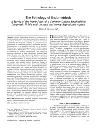
  • 2. Clement Adv Anat Pathol Volume 14, Number 4, July 2007 The endometriotic stroma typically resembles, at TABLE 1. Findings in Endometriosis That can be Associated least focally, eutopic inactive or proliferative endometrial With Diagnostic Problems stroma, including the presence of a network of small Alterations of the Stromal Component arterioles (Figs. 1A, B). The presence of these vessels, Foamy and pigmented histiocytes which may be engorged with erythrocytes, may provide a Fibrosis helpful initial clue to the endometriotic nature of the Elastosis lesion. In some cases, vessels are not conspicuous but Smooth muscle metaplasia Myxoid change focal stromal hemorrhage can be similarly diagnostically Decidual change, including signet-ring–like cells helpful. Metzger et al10 found recent hemorrhage within Alterations of the Glandular Component 53% of implants with stroma but in only 15% of those Postmenopausal and treatment-related changes with little or no stroma. Although hemorrhage in some Pregnancy-related changes Metaplastic changes specimens might be related to the trauma of the biopsy, Cytologic atypia two-thirds of the biopsy specimens with hemorrhage in Hyperplasia the cited study10 also contained pigmented histiocytes, the Absence of glands (stromal endometriosis) characteristic inflammatory cell of endometriosis, as Miscellaneous Tumorlike Findings discussed below. Necrotic pseudoxanthomatous nodules Polypoid endometriosis Like normal and neoplastic endometrial stromal Vacular invasion cells, endometriotic stromal cells are typically immuno- Perineural invasion reactive for CD10 (Fig. 1C).12–14 CD10-positivity can Unusual Inflammatory and Reactive Changes help confirm the endometriotic nature of stromal cells in Infected endometriotic cysts Pseudoxanthomatous salpingitis problematic situations such as when dealing with limited Florid mesothelial hyperplasia material (as in a tiny laparoscopic biopsy specimen), when MPICs the stromal cells on routinely stained sections are of Liesegang rings uncertain nature, or when the glandular component is Site-Related Diagnostic Problems absent, as discussed below. The role of CD10 staining in Endometriosis on or close to the ovarian surface Superficial endometriosis of the uterine cervix the diagnosis of superficial ovarian and cervical endome- Vaginal endometriosis triosis is discussed under the heading of ‘‘Selected Site- Tubal endometriosis related Issues.’’ Intestinal endometriosis Rare Associated Lesions Peritoneal leiomyomatosis DIAGNOSTIC ISSUES RELATED TO THE Peritoneal gliomatosis STROMAL COMPONENT Alterations in the typical microscopic appearance of endometriosis that can cause diagnostic problems for the stroma, although as will be noted, the diagnosis can be pathologist occur in both its glandular and stromal made when only one of these components is present. The components, but are more common in the latter and are glands almost always have an overtly endometrioid accordingly considered first. Although these changes can appearance (Figs. 1A, B) that may range from inactive obscure the typical stromal component, their presence to proliferative (or occasionally, secretory) to hyperplas- should increase one’s diagnostic suspicion of the endome- tic. The glandular cells may be ciliated, exemplifying the triotic nature of the lesion. In such cases, endometriotic close relationship of tubal-type and endometrioid epithe- stroma, if present at all, may be very subtle and confined lium in the female genital tract in both non-neoplastic and to a barely perceptible and often discontinuous zone that neoplastic states. The appearance of the glands may is periglandular or that subtends the epithelial lining of an reflect that of the eutopic endometrium, but often only to endometriotic cyst. In the latter instance, if the epithelial a limited degree.9–11 The lipofuscin and hemosiderin cells are denuded, the stromal cells may directly abut the pigment frequently present within histiocytes in endome- cyst lumen. If the nature of the stromal cells is in doubt, triosis (as discussed below) can occasionally also be found immunostaining for CD10, as noted earlier, can be within the endometriotic epithelial cells. helpful. FIGURE 1. A, Typical endometriosis showing part of an endometriotic gland and a thin cuff of periglandular endometriotic stroma containing dilated blood vessels. B, Endometriotic gland with only focal periglandular endometriotic stroma (extreme left of field). The remaining stroma is edematous and has a nonspecific appearance. C, Typical endometriosis. The periglandular endometriotic stroma shows immunoreactivity for CD10. D, Ovarian endometriosis. The endometriotic stroma is replaced by foamy histiocytes (pseudoxanthoma cells), which also occupy gland lumens. Residual ovarian stroma is present at the top right. E, Endometriotic cyst. The endometriotic stroma has been replaced by pseudoxanthoma cells. F, Endometriotic cyst with numerous pigmented histiocytes within the endometriotic stroma. G, Presumptive endometriosis. An ovarian cyst is lined by pigmented histiocytes and fibrous tissue without any diagnostic endometriotic epithelium or endometriotic stroma. H, Pseudoxanthoma cells infiltrating omental fat in a patient with pelvic endometriosis. 242 2007 Lippincott Williams Wilkins r
  • 3. Adv Anat Pathol Volume 14, Number 4, July 2007 The Pathology of Endometriosis 243 2007 Lippincott Williams Wilkins r
  • 4. Clement Adv Anat Pathol Volume 14, Number 4, July 2007 Foamy and Pigmented Histiocytes Fibrosis and Elastosis Hemorrhage and menstrual changes in endometrio- Endometriotic lesions often elicit a fibrotic reaction sis often elicit an infiltrate of histiocytes that can obscure that can lead to adhesions around the focus and focal to the characteristic features of the stroma (Figs. 1D to G). extensive replacement of the endometriotic stroma, The histiocytes typically have abundant cytoplasm that especially within the walls of endometriotic cysts varies in appearance depending, at least in part, on the (Figs. 2A, B). The fibroblasts within the fibrotic areas age of the lesion. In early lesions, which tend to be are characteristically small and stellate (Fig. 2A). Long- nonpigmented at laparoscopy or laparotomy,15 the standing cysts may have walls composed predominantly histocytes are nonpigmented and have granular, eosino- or entirely of hyalinized fibrous tissue (Fig. 2B); rarely philic or foamy cytoplasm, the latter due to abundant foci of dystrophic calcification and/or heterotopic bone lipid or glycolipid (‘‘pseudoxanthoma cells’’), some of are present in such cases. which likely represents ceroid precursors (Figs. 1D, A less common stromal response in endometriosis is E).16–18 In contrast, the characteristic pigmented histio- elastosis, which can sometimes be striking, resulting in cytes of endometriosis are common in well-developed focal or extensive replacement of the typical endometrio- tic stroma (Figs. 2C, D).20 In some cases, circumscribed lesions (Figs. 1F, G). Much of the pigment is ceroid or lipofuscin that appears as fine, grayish-brown cytoplas- foci of elastosis vaguely resemble a corpus albicans. mic granules on routinely stained sections and that stains Stromal elastosis can be a helpful diagnostic clue to the with the periodic acid-Schiff (PAS) method after diastase diagnosis of endometriosis, especially when accompanied predigestion. When collections of ceroid-rich histiocytes by characteristic endometriotic glands. Although we associated with endometriosis (or other lesions) are well initially believed that stromal elastosis in endometriosis circumscribed, the term ‘‘ceroid granuloma’’ has some- was a new observation, while our report on this finding times been applied.19 Hemosiderin is also typically was in press, we found that Robert Meyer had made present but is often less conspicuous than the lipofuscin the same observation over 70 years earlier. In an article published in 1928, Jacobsen21 refers to one of Meyer’s pigment and in contrast to the latter appears as coarse, irregular, golden-brown cytoplasmic granules that stain studies (unfortunately the exact citation is not given) by with the Prussian blue method. Occasionally, the histio- stating ‘‘Meyer ylays much stress on his own observa- cytic infiltrate is massive (‘‘xanthomatous endometrio- tion that elastic tissue is not found in the uterine mucosa, sis’’) and can extensively or completely replace the but is present in ectopic endometrium-like tissue.’’ endometriotic stroma and fill the gland lumens (Figs. Smooth Muscle Metaplasia 1D, E). The presence of endometriotic glands allows for a diagnosis of endometriosis when the endometriotic The stromal component of endometriosis can be stroma has been effaced, but when both the endometriotic focally or extensively replaced by smooth muscle, a glands and stroma are obliterated by the histiocytes (often finding consistent with the myofibroblastic poten- accompanied by fibrosis, as noted below), only a tial of endometrial and endometriotic stromal cells (Fig. 2E).22,23 Smooth muscle metaplasia is most common diagnosis of presumptive endometriosis is possible (Fig. 1G). Rarely, the lipid within endometriotic lesions can be within ovarian endometriosis, being documented in 18% of cases in one study.22 In over half the cases in that converted to cholesterol crystals that may evoke a foreign-body giant-cell reaction. When this finding is study, the smooth muscle was within the wall of an striking, the term ‘‘cholesteatomatous endometriosis’’ is endometriotic cyst. When smooth muscle is prominent, appropriate. the process has been referred to as ‘‘endomyometriosis.’’ In some cases of endometriosis, pseudoxanthoma Striking examples of the latter have taken the form of uteruslike masses in the ovary,24,25 broad ligament,26 and cells are found at a distance from the endometriotic foci, a pelvic lymph node.27 Some uteruslike masses, however, such as within the mucosa of the fallopian tube (see Pseudoxanthomatous Salpingitis), the pelvic peritoneum, especially if associated with congenital malformations of pelvic lymph nodes, and omentum (Fig. 1H).18 the genitourinary tract, may represent a congenital FIGURE 2. A, Endometriotic cyst with fibrosis. Beneath the endometriotic epithelium (top right) is a thin zone of endometriotic stroma with foci of hemorrhage. The cyst wall is composed of loose fibrous tissue composed of small fibroblasts separated by collagen. B, Endometriotic cyst (lumen at top of figure) with its wall composed of hyalinized fibrous tissue. Pseudoxanthoma cells focally line the cyst, which has lost its epithelial lining. C, Endometriosis with prominent stromal elastosis. The brightly eosinophilic elastotic tissue surrounds an endometriotic gland. No residual endometriotic stroma is present. D, Endometriosis with prominent stromal elastosis, elastic tissue stain. E, Endometriosis with prominent smooth muscle metaplasia. At the extreme left is part of an endometriotic gland with periglandular endometriotic stroma that merges with mature smooth muscle. F, Endometriosis with prominent myxoid change. A typical endometriotic gland with periglandular stroma (left) lies adjacent to a large pool of acellular mucin (right). This peritoneal lesion, which was found in a patient undergoing radial hysterectomy for adenocarcinoma of the cervix, was misinterpreted on frozen section examination as metastatic mucinous adenocarcinoma. G, Endometriosis in pregnancy with prominent decidual change. An atrophic endometriotic gland is lined by flattened epithelium, a finding that can be mistaken for a vascular space. H, Endometriosis in pregnancy. Basophilic myxoid stroma separates the decidual cells. Note the cytoplasmic vacuoles within the decidual cells. 244 2007 Lippincott Williams Wilkins r
  • 5. Adv Anat Pathol Volume 14, Number 4, July 2007 The Pathology of Endometriosis 245 2007 Lippincott Williams Wilkins r
  • 6. Clement Adv Anat Pathol Volume 14, Number 4, July 2007 mullerian-duct anomaly rather than a variant of endo- secondary to progestin treatment (Fig. 3A). In pregnancy, metriosis.25,28 microscopic examination typically reveals a decidual Smooth muscle metaplasia in endometriosis should reaction similar to that seen in the eutopic endometrium; be distinguished from the much more common finding of the endometriotic glands are usually atrophic, as noted endometriosis involving indigenous smooth muscle, such below (Fig. 2G). Necrosis of the decidual cells, stromal as the pelvic ligaments and the walls of the bowel and myxoid change (as noted above) (Fig. 2H) or edema, and bladder. In such cases, the endometriosis often stimulates infiltration by lymphocytes may also be seen. Addition- the indigenous smooth muscle to proliferate, analogous to ally, the decidual cells in endometriosis can, like their myometrial smooth muscle proliferation around foci of eutopic counterparts, exhibit cytoplasmic vacuoles with adenomyosis. The reactive smooth muscle proliferation in displacement of the nucleus, resulting in a signet-ring–like appearance (Fig. 2H).29,33 Cells of the latter type can raise these situations often accounts for the bulk of the lesion, and if endometriosis is not identified in the initial slides, a differential diagnosis with a metastatic signet-ring-cell the findings may be suspicious enough for the diagnosis to adenocarcinoma, but the merging of such cells with prompt additional sampling. typical decidual cells, the presence of acid rather than neutral mucin in the vacuoles, and the cytokeratin- Myxoid Change negativity of the cells facilitate the correct diagnosis. In occasional cases, endometriotic stroma under- goes striking myxoid change in which endometriotic DIAGNOSTIC ISSUES RELATED TO THE stromal cells are separated by scanty to abundant GLANDULAR COMPONENT acellular mucin; in the latter situation, pools of acellular Postmenopausal and Treatment-related mucin may be seen (Figs. 2F, H).29–34 The mucin in these Changes cases is stromal rather than epithelial mucin, and thus stains positively for toluidine blue or Alcian blue (pH 2.5) Diagnosing endometriosis in postmenopausal wo- but not with PAS. Five of the 6 reported cases of myxoid men can be more difficult than in those of reproductive change in endometriosis have involved the skin or age because the endometriotic tissue is often atrophic. superficial soft tissues and 4 of the 6 cases occurred Inactive or atrophic glandular changes in endometriosis during pregnancy or the puerperium, suggesting that the can also occur in premenopausal patients treated with location of the lesion and the hormonal milieu may be oral contraceptives, danazol, antiprogesterone steroids predisposing factors. In 2 cases, the histologic appearance (gestrinone), and progestins (Fig. 3A).35,36 In atrophic suggested the possibility of metastatic adenocarcinoma endometriosis, the endometriotic glands may be small or and/or pseudomyxoma peritonei,30,31 and in another case, cystically dilated and are lined by flattened epithelial cells, a diagnosis of a myxoid sarcoma of soft tissue was with an appearance that may be similar to simple or cystic initially considered.33 In the 3 patients who were pregnant atrophy of the eutopic endometrium (Fig. 3B). The at the time of the removal of the lesion, the histologic atrophic glands may be surrounded by stroma that is interpretation of the specimen was further complicated by often more fibrotic (Fig. 3B) than in younger patients, the presence of a striking decidual transformation of the although a thin cuff of typical endometriotic stroma will endometriotic stroma (see below) (Fig. 2H). The correct usually be visible around some of the glands. Immunos- diagnosis is facilitated by awareness of these findings and taining for CD10 can be helpful in establishing the the presence of at least occasional endometriotic glands, presence of endometriotic stroma in such cases. although these may be atrophic if the patient is pregnant, In some postmenopausal women, endometriosis as noted below. remains clinically active. In such cases, Toki et al37 found that the endometriotic lesions may have a more active Decidual Change histologic appearance than the eutopic endometrium, and The endometriotic stroma usually exhibits striking also more progesterone receptors and higher Ki-67 progestational changes during pregnancy (Figs. 2G, H); activity in the endometriotic stromal cells as compared similar but usually less striking changes can be seen with the eutopic endometrial stromal cells. FIGURE 3. A, Endometriosis in a patient treated with progestational agents. The endometriotic stromal cells are partly decidualized and surround an atrophic gland. B, Endometriosis in a postmenopausal patient showing cystic atrophy of the glands and fibrosis of the stroma. C, Endometriosis showing Arias-Stella reaction within the endometriotic glands in a pregnant patient. D, Endometriosis with prominent mucinous metaplasia of a large irregular endometriotic gland. E, Endometriotic cyst with atypical clear cell metaplasia of the lining epithelial cells. Most of the cyst was lined by this type of epithelium, which merged with a clear cell carcinoma (not shown). F, Endometriotic cyst with prominent cytologic atypia of the lining epithelial cells. Note prominent neutrophilic infiltrate. G, A patient, who had been on unopposed estrogen replacement therapy for 10 years following a hysterectomy and salpingo-oophorectomy for endometriosis, presented with a pelvic mass. The mass was composed of typical endometriosis with foci of atypical complex endometrial hyperplasia (left) that merged with grade 1 endometrioid adenocarcinoma (right). Also see (H). H, Higher-power views of the atypical hyperplasia (left) and the grade 1 endometrioid adenocarcinoma (right) depicted in (G). 246 2007 Lippincott Williams Wilkins r
  • 7. Adv Anat Pathol Volume 14, Number 4, July 2007 The Pathology of Endometriosis 247 2007 Lippincott Williams Wilkins r
  • 8. Clement Adv Anat Pathol Volume 14, Number 4, July 2007 Pregnancy-related Changes histologic types of ovarian epithelial tumors, and in a high proportion of ovarian clear cell carcinomas (40% to In pregnancy, endometriotic glands usually have an 70% of cases) and endometrioid carcinomas (30% to inactive appearance. They vary from small to large and 40% of cases).43–46 A more specific association has been cystic and are usually lined by cuboidal or flattened found between ‘‘atypical endometriosis’’ and synchro- epithelial cells (Fig. 2G). We have seen cases of nous ovarian cancers. Of the ovarian cancers studied by endometriosis in pregnant women in which the diagnosis Ogawa et al,45 29% had associated endometriosis, and it was missed on microscopic examination because the was atypical in 78% of them. Similarly, Fukunaga et al44 flattened epithelial cells were mistaken for endothelial or found atypical endometriosis in approximately 60% of mesothelial cells, resulting in a diagnosis of ectopic cases of endometriosis-associated carcinoma, whereas it decidua rather than endometriosis. Occasionally, endo- was present in only 1.7% of cases of ovarian endome- metriotic glands in women with an intrauterine or ectopic triosis not associated with an ovarian carcinoma. pregnancy exhibit overt secretory changes that may include the Arias-Stella reaction38,39 (Fig. 3C) and/or The term ‘‘atypical endometriosis’’ has been used in optically clear nuclei40 similar to these pregnancy-related the literature to refer to 2 different histologic find- ings.44,45,47–52 One of them, referred to here as ‘‘cytologic findings in eutopic endometrial glands. atypia,’’ denotes the presence of cytologic atypia within Metaplastic Changes the lining of endometriotic cysts, whereas the other, referred to here as ‘‘hyperplasia,’’ refers to the same Metaplastic changes similar to those occurring in spectrum of hyperplasia (simple or complex, with or eutopic endometrial glands are common in endometriotic without cytologic atypia) encountered in the endome- epithelium.41,42 They were found in 68% of cases of trium.53 Most studies have used ‘‘atypical endometriosis’’ ovarian endometriosis in one study41 but in only 12% of to refer collectively to both cytologic atypia and cases in another study,42 and include ciliated, eosinophi- hyperplasia, but an attempt should be made to distinguish lic, hobnail, squamous, and mucinous metaplasia (Fig. between them because they almost certainly differ in their 3D); the last type may be composed of endocervical-type clinical significance,53 and each is considered here cells or, less often, goblet cells. Additionally, we have seen separately. several cases of clear cell and atypical clear cell metaplasia in endometriotic cysts (Fig. 3E); in one such case, atypical Cytologic Atypia clear cells lining an endometriotic cyst merged with a clear cell carcinoma. In one of the studies of metaplasia in Cytologic atypia, especially mild to moderate ovarian endometriosis cited above,41 17% of the endome- atypia, within endometriotic cysts is a common find- ing.51,53–56 Its reported frequency obviously depends on triotic cysts harbored a neoplasm, all of which had associated epithelial metaplasia within the cyst, whereas one’s diagnostic threshold for this diagnosis. Czerno- bilsky and Morris55 found mild atypia in 22% of their only 63% of the cysts without a neoplasm showed metaplasia. Furthermore, all 4 cases of endocervical-like cases of ovarian endometriosis, whereas severe atypia was mucinous borderline tumors (EMBTs) encountered in the present in only 3.6% of cases. The corresponding same study were contiguous with foci of hyperplastic frequency from another study in which the atypia was not graded was 12%.57 mucinous epithelium. The distinction between papillary mucinous metaplasia and an early EMBT in an endome- Atypia, when present, is almost always found in an triotic cyst can be difficult and subjective, and is a not endometriotic cyst, and is usually a focal or multifocal infrequent issue in our referral material. When there is finding in which the epithelial cells lining the cyst are definite endometriotic stroma beneath the mucinous enlarged and polygonal with scanty to more commonly proliferation, we consider it metaplastic endometriosis abundant, dense eosinophilic cytoplasm (Fig. 3F). There unless there are well-developed papillae. is usually no associated endometriotic glandular compo- nent. The nuclei of the atypical cells exhibit marked Atypical Endometriosis variation in size and shape and are variably hyperchro- A number of studies have found associated en- matic, often with smudged chromatin; nucleoi may be dometriosis in a sizable minority (20% to 30%) of all prominent (Fig. 3F). Hobnail-type cells are sometimes FIGURE 4. A, Micronodular stromal endometriosis. Two nests of endometriotic stromal cells are present on the peritoneal surface. B, Micronodular stromal endometriosis. Two nodules of endometriotic stroma are present within submesothelial connective tissue. Note the pigmented histiocytes. C, Micronodular stromal endometriosis. A nodule of endometriotic stromal cells is present on the surface of the omentum. Note the dilated arterioles within the nodule. D, Stromal endometriosis of cervix. Note prominent hemorrhage. An endocervical gland is present on the extreme right. E, Necrotic pseudoxanthomatous nodule on the ovarian surface. F, NPN. The center of the nodule (lower part of field), which consists of necrotic and calcified debris, is surrounded by foamy and pigmented histiocytes and fibrous tissue. G, Polypoid endometriosis of colon. A polypoid mass projects from the mucosal surface of the colon, potentially mimicking a colonic adenocarcinoma. H, Polypoid endometriosis of colon. Mucosal polyps, which consist mostly of endometriotic glands and stroma, are covered by colonic epithelium. Islands of endometriosis are also present within the muscularis propria. 248 2007 Lippincott Williams Wilkins r
  • 9. Adv Anat Pathol Volume 14, Number 4, July 2007 The Pathology of Endometriosis 249 2007 Lippincott Williams Wilkins r
  • 10. Clement Adv Anat Pathol Volume 14, Number 4, July 2007 present. The atypical cells are usually disposed in a single of ovarian clear cell carcinomas, and 56% of endome- layer but may be focally denuded or stratified, sometimes triotic cysts. A number of studies have found loss of as small papillae. If there is coexistent mucinous heterozygosity (LOH) within ovarian endometriosis (in- metaplasia, the appearance may overlap with papillary cluding atypical endometriosis) at candidate ovarian mucinous metaplasia as noted above. An associated tumor suppressor gene loci and LOH events or other stromal and/or epithelial neutrophilic infiltrate is com- genetic alterations common to the endometriosis and mon (Fig. 3F). Ballouk et al54 studied 6 endometriotic synchronous ovarian carcinomas.65–70 Ali-Fehmi et al47 cysts with severe atypia and found that 3 of them were found a significant difference in the frequency of LOH aneuploid whereas endometriotic cysts with absent or between endometriosis (4.3%) and ovarian carcinomas only mild atypia were diploid. (23.5%) at D10S608, suggesting that LOH at this locus In most cases, cytologic atypia in endometriotic may be an important molecular event in the progression cysts is likely a reactive or degenerative change, and when of endometriosis to carcinoma. Additionally, Prefumo et al71 found that endometriosis-associated ovarian carcino- it is an isolated finding, the follow-up is typically uneventful. Seidman53 obtained follow-up (mean 8.9 y) mas had a higher expression of p53 and c-erb-2 than did in 20 such cases, and it was uneventful in all of them similar tumors without associated endometriosis, and that except for persistent endometriosis in 1 patient. Never- there was a significant degree of concordance for both theless, this type of atypia can occasionally merge with an oncogenes in the tumors and the adjacent endometriotic EMBT (pure58 or mixed-cell type59) or an endometrioid epithelium. Similarly, Sainz de la Cuesta et al48 found that or clear cell carcinoma,51 suggesting that it is occasionally p53 was present in all examples of atypical endometriosis a premalignant change. We have recently seen a case of an and 82% of endometriosis-associated ovarian carcino- endometrioid adenocarcinoma arising in an endometrio- mas, suggesting that the presence of p53 may be helpful in tic cyst in which the endometriotic epithelium near the identifying cases of endometriosis with a malignant potential. Korner et al72 have recently found a higher carcinoma exhibited cellular stratification and severe ¨ nuclear atypia with an appearance suggesting adenocar- frequency of chromosomal aberrations in ovarian en- cinoma in situ (AIS). dometrioid carcinomas than in ovarian endometriosis, and a higher frequency of the same aberrations in ovarian Hyperplasia endometriosis compared with extraovarian endometriosis A variety of hyperplastic changes, with or without and normal endometrium. These authors suggest that the cytologic atypia, similar to these findings in the endome- hormonal milieu of the ovary may induce genetic changes trium have been described in endometriosis,43,51,53 some- in endometriosis in the same site. times but not always related to an endogenous or Endometriosis Without Glands exogenous estrogenic stimulus60,61 (Figs. 3G, H) or (Stromal Endometriosis) tamoxifen treatment.62,63 These changes are less common that the pure cytologic atypia described in the preceding In occasional cases of endometriosis, endometriotic section, being encountered in only 2% of endometriotic glands are rare or absent, a phenomenon that has been cysts in one study55 (which is in accord with our referred to as stromal endometriosis (Figs. 4A–D).20,73 experience) and 9.4% of cysts in another.42 When reading the older literature, however, it is The infrequency of hyperplasia in endometriosis important to be aware that the same term was used in complicates the assessment of its premalignant potential, the past to refer to low-grade endometrial stromal but it is likely similar to that of endometrial hyperplasia sarcoma. In some or perhaps most cases of stromal in view of its occasional association with a synchronous endometriosis, the apparent absence of endometriotic or metachronous neoplasm in the same site.42,44,53 In one glands is likely because of limited sampling of the lesion. study in which 4% of endometriotic cysts harbored an Indeed, it is quite common in small laparoscopic biopsy endometrioid carcinoma, there was a significant associa- specimens to find small superficial nodules or surface tion between the presence of a carcinoma and synchro- plaques that consist only of endometriotic stroma (Figs. nous complex hyperplasia within the endometriosis.42 4A–C).73 Such foci can be misinterpreted as lymphoid Accordingly, as in the endometrium, hyperplasia within aggregates or a nonspecific finding, particularly if they endometriosis should prompt a reexamination of the are not examined at high-power magnification. An index specimen and additional sampling to exclude a synchro- of suspicion and also the presence of the characteristic nous neoplasm. arterioles, foci of hemorrhage, pigmented histiocytes, and CD10 immunoreactivity of the stromal cells facilitate Molecular Evidence Supporting Precancerous the diagnosis. In some cases, deeper sections will dis- Potential close occasional endometriotic glands, although cases of That endometriosis, particularly atypical endome- bona fide pure stromal endometriosis almost certainly triosis, may undergo malignant transformation (an occur. observation that dates back to Sampson’s work64) is also Stromal endometriosis can also be encountered in supported by recent molecular studies. Sato et al65 found the ovary and uterine cervix, usually in patients without PTEN mutations similar to those in endometrial carci- typical pelvic endometriosis. In the ovary, it is usually nomas in 42% of ovarian endometrioid carcinomas, 27% an incidental microscopic finding within the stroma 250 2007 Lippincott Williams Wilkins r
  • 11. Adv Anat Pathol Volume 14, Number 4, July 2007 The Pathology of Endometriosis (‘‘benign stromatosis’’), and likely represents a metaplas- history of previous cautery treatment, the typical presence tic response of the ovarian stromal cells.74,75 Stromal of black (carbon) or hematoidin pigment, and the absence endometriosis of the cervix73 can occasionally be recog- of foamy histiocytes help distinguish these granulomas nized clinically as a small red mucosal discoloration. from NPNs. Microscopic examination reveals within the superficial Polypoid Endometriosis cervical stroma well-circumscribed rounded to plaquelike foci composed of endometrial stromal cells, small This term is used to refer to rare cases of blood vessels, and often large numbers of extravasated endometriosis that form polypoid masses that can mimic erythrocytes (Fig. 4D). The differential diagnosis is a neoplasm on clinical, intraoperative, and gross patho- logic examinations.7,78–81 In the only series of such with cervical involvement by low-grade endometrial cases,81 the patients were in the reproductive and stromal sarcoma. Militating against that diagnosis are the lesion’s small size, superficial location, extravasated postmenopausal age groups (mean 52.5 y). Of the 24 erythrocytes, and absence of permeative growth and patients, 7 were on unopposed estrogen and 4 were on vascular invasion. combined estrogen-progestin therapy; 1 patient had a synchronous ovarian thecoma. Several cases of polypoid endometriosis have also been reported in patients MISCELLANEOUS TUMORLIKE FINDINGS receiving tamoxifen therapy78,82 and one case followed A variety of findings in endometriosis can suggest withdrawal of GnRH-agonist treatment for severe the presence of a neoplastic process. Some of these have endometriosis.83 already been discussed, including myxoid change, signet- The most common clinical presentations are related ring–like decidualized stromal cells, cytologic atypia and to a pelvic mass, a polypoid vaginal mass, or large bowel hyperplasia of the epithelial component, and stromal obstruction. Sites of involvement in order of frequency in endometriosis. Other tumorlike findings are considered in the series of Parker et al81 were colonic mucosa, ovary this section. (involving the ovarian surface or lining an endometriotic Necrotic Pseudoxanthomatous Nodules cyst), uterine serosa, cervical or vaginal mucosa, ureter, These lesions (NPNs) are an uncommon manifesta- fallopian tube, omentum, bladder, paraurethral and tion of endometriosis and tend to occur in the post- paravaginal soft tissue, and retroperitoneum. In 30% of menopausal and late reproductive age groups.18 They cases, multiple sites were involved. Polypoid, pink, gray take the form of multiple nodules that are usually or tan, masses ranged up to 14 cm in maximal dimension attached to the peritoneum or occasionally are free- (Fig. 4G). On microscopic examination, the polypoid floating within the peritoneal cavity. In most cases, masses were composed of an admixture of endometriotic typical endometriosis is present in the ovaries and in glands and stroma (Fig. 4H). A variety of glandular one case NPNs lined an endometriotic cyst.18 The architectural patterns were observed, sometimes in intraoperative appearance in some cases has suggested a combination, most commonly cystic and non-cystic disseminated malignant tumor. On histologic examina- simple hyperplasia, but also simple or complex hyperpla- tion, the nodules have a central necrotic zone that is sia with atypia, disordered proliferative, and cystic surrounded by pseudoxanthoma cells, often in a pali- atrophy. Various types of epithelial metaplasia (tubal, saded arrangement, hyalinized fibrous tissue, or both mucinous, squamous, papillary syncytial metaplasia) were (Figs. 4E, F). The presence of necrosis, particularly on common. Hemorrhage, fibrosis, prominent thick-walled frozen section examination in a patient with intraopera- blood vessels, hemosiderin-laden histiocytes, and decidual tive findings suspicious for cancer, can increase concern change were also present in some cases. Most of the cases for the latter. Endometriotic glands and stroma are were associated with usual (nonpolypoid) endometriosis usually absent or sparse within the nodules, but may be (Fig. 4H). In 1 case, polypoid endometriosis merged with seen elsewhere in the same specimen. Most NPNs likely a mucinous borderline tumor of endocervical-type. Of the represent ‘‘burned-out’’ or end-stage endometriotic foci. 17 patients with follow-up data, 15 were alive without In 1 case, however, multiple peritoneal and omental evidence of residual disease, 1 was alive with residual NPNs that were not present at the time of salpingo- endometriosis, and 1 died of other causes. oophorectomy performed for a ruptured ovarian endo- Extrauterine mullerian adenosarcoma is the most common differential diagnostic consideration.84,85 In metriotic cyst, were found at reexploration 7 weeks later, and were thus considered a unusual reaction to the contrast to this neoplasm, however, the reported cases contents of the endometriotic cyst.76 of polypoid endometriosis, with 2 exceptions, lacked The importance of NPNs rests on their being periglandular stromal hypercellularity, stromal atypia, diagnostic or at least strongly suggestive of endometriosis and intraglandular stromal papillae. Focal intraglandular when its more typical features are absent, and their stromal papillae were noted in 2 cases with focal mild potential to be confused with an infectious process or periglandular stromal hypercellularity in one of them, but necrotic tumor that might prompt unnecessary investiga- in contrast to adenosarcoma, stromal atypia was absent tions. NPNs should also be distinguished from necrotiz- in each case. Diagnosis may be particularly difficult if the ing peritoneal granulomas that may be seen after polyps of polypoid endometriosis undergo torsion with diathermy ablation treatment for endometriosis.77 The hemorrhagic infarction. In these cases, endometriotic 251 2007 Lippincott Williams Wilkins r
  • 12. Clement Adv Anat Pathol Volume 14, Number 4, July 2007 Pseudoxanthomatous Salpingitis stromal cells may exhibit reactive atypia and mitotic activity, complicating the differential with adenosarcoma. As noted earlier, pseudoxanthoma cells can be a In rare cases in which a definite diagnosis is not possible, striking finding in the tubal mucosa in patients with pelvic the patient should receive clinical follow-up. endometriosis, a finding that has been referred to as pseudoxanthomatous salpingitis (or pseudoxanthoma- tous salpingiosis) (Figs. 5C, D).18,97,98 The process may Rare Tumorlike Findings result in a chocolate-brown discoloration of the tubal Rarely, endometriosis may be associated with mucosa visible on macroscopic examination (Fig. 5C). In worrisome findings that can include unusually extensive our experience, pseudoxanthomatous salpingitis has al- and/or bulky disease, vascular invasion, or a poor ways been associated with pelvic endometriosis, and in response to treatment.86–93 The designation ‘‘aggressive such cases likely represents an inflammatory reaction to endometriosis’’ has sometimes been applied to such cases, endometriosis-derived blood or blood products that reach but we discourage its use as a pathologic diagnosis the tubal lumen. One otherwise similar case, however, because the subsequent behavior of endometriosis cannot reported as ‘‘pigmentosis tubae,’’ was found in a patient be reliably predicted by its morphologic features. Further, without endometriosis but who had been previously such a diagnosis could prompt unwarranted physician or treated with pelvic radiation.99 patient concern. Mesothelial Hyperplasia Vascular involvement by endometriosis (Fig. 5A) is analogous to the more common phenomenon of endo- Endometriosis is frequently accompanied by me- metrial glands and stroma (or stroma only) within sothelial hyperplasia of the pelvic or even extrapelvic myometrial vessels in cases of adenomyosis.94 Scolyer et peritoneum, which in some cases may be striking. The al91 reported a case of an ovarian endometriotic cyst that hyperplasia may occur adjacent to, or at a distance from, the endometriosis. Kerner et al100 documented mesothe- was associated with florid involvement of large vascular spaces (thick-walled lymphatics and veins) within both lial hyperplasia (‘‘mesothelial inclusions’’) in 10% of mesovaria. The endometriotic tissue completely occluded women with ovarian endometriosis, but in none of the some vessels or in others formed large polypoid tongues patients from a control group without endometriosis. that partly filled their lumina. A similar case was reported Mesothelial hyperplasia in women with endometriosis by Ooi and Valentine.90 In another report, Wuster and most commonly involves the surface of the ovaries, the Leu92 documented a case of abdominal wall endome- fallopian tubes, the pelvic peritoneum, and the omentum. triosis in which several veins contained endometriotic The hyperplastic mesothelial cells typically form small tissue. Zardawi93 reported 2 cases of colonic endome- tubules, papillae, nests, cords, or even single cells, often triosis that were each associated with intravascular entrapped within a reactive fibrous tissue, which in florid endometriosis within the colonic wall. In one of them, cases may create a pseudoinfiltrative pattern (Fig. 5E). there was endometriosis within a pericolic lymph node Retraction artifact around the congeries of mesothelial and in the other there was a focus of hepatic endome- cells is common and may incorrectly suggest lymphatic triosis, findings supporting the hypothesis that some cases invasion (Fig. 5E). of endometriosis in unusual sites are a result of vascular In our experience, the most striking cases of endo- or lymphatic embolic spread. metriosis-associated mesothelial hyperplasia are seen, not As has been reported in other non-neoplastic lesions surprisingly, on the outer aspects of endometriotic cysts. (sclerosing adenosis of the breast, vasitis nodosa), perineural These mesothelial cells can become incorporated in the invasion has been rarely encountered in otherwise typical, wall of the cyst (Fig. 5E), often as small tubules in linear benign endometriotic lesions95 (Fig. 5B) and seems to be arrangements that parallel the overlying serosa, a finding of no clinical significance. identical to the mural mesothelial hyperplasia within the walls of multilocular peritoneal inclusion cysts (MPICs) (see below).101 Indeed, since the report of this finding in MPICs, we have seen more cases of mural mesothelial INFLAMMATORY AND REACTIVE CHANGES proliferation in the walls of endometriotic cysts than in Infected Endometriotic Cysts MPICs. In some such cases, the mesothelial cells may Schmidt et al96 found pathologic evidence of even approach the cyst lining, the appearance sometimes infection in 11 of 510 endometriotic cysts (2%). The raising concern for an invasive adenocarcinoma arising patients had a mean age of 34.7 years and typically from the epithelial lining of the cyst. Awareness of the presented with fever and lower abdominal pain; about pitfalls associated with mesothelial hyperplasia in this half had a history of pelvic inflammatory disease. setting is crucial in avoiding a serious diagnostic error. If Histologic examination revealed endometriotic cysts that necessary, immunohistochemical staining can be used to contained a fibrinopurulent exudate, microabscesses, and confirm that the cells are mesothelial and not epithelial. inflammatory cells other than neutrophils, such as plasma Multilocular Peritoneal Inclusion Cysts cells. All of the patients who had one or both fallopian tubes removed at the same operation had histologic A substantial proportion of women with MPICs evidence of acute and/or chronic salpingitis. (also referred to as ‘‘cystic mesotheliomas’’) have or have 252 2007 Lippincott Williams Wilkins r
  • 13. Adv Anat Pathol Volume 14, Number 4, July 2007 The Pathology of Endometriosis FIGURE 5. A, Endometriosis within the lumen of a thin-walled vein or lymphatic. B, Endometriosis showing perineural invasion. Figure courtesy of Dr L Roth. C, Pseudoxanthomatous salpingitis. The fallopian tube has been opened to show a thickened mucosa that varies from yellow to brown. D, Pseudoxanthomatous salpingitis. The tubal plicae are filled with foamy and pigmented histiocytes. E, Florid mesothelia hyperplasia within the wall of an endometriotic cyst. The mesothelial cells form small irregular aggregates within reactive fibrous tissue resulting in an infiltrative pattern. Note the retraction artifact around the aggregates of mesothelial cells, a finding that can be misinterpreted as lymphatic invasion and thus increase suspicion for a malignant tumor. F, Numerous Liesegang rings and necrotic debris occupy the lumen of an endometriotic cyst. had endometriosis. In these cases, the cysts likely study of MPICs had coexistent endometriosis or a history represent a complication of endometriosis-related adhe- of the disease. Additionally, several reports have docu- sions. Ross et al102 found that 36% of women in their mented an intimate admixture of the 2 lesions. Groisman 253 2007 Lippincott Williams Wilkins r
  • 14. Clement Adv Anat Pathol Volume 14, Number 4, July 2007 254 2007 Lippincott Williams Wilkins r
  • 15. Adv Anat Pathol Volume 14, Number 4, July 2007 The Pathology of Endometriosis and Kerner103 reported an MPIC that contained foci of glands may be numerous and cystic, increasing their typical endometriosis and NPNs within the cyst locules. potential resemblance to cortical epithelial inclusion cysts Additionally, Zotalis et al104 described a 35 cm omental (Fig. 6A), and their often subtle cuffs of endometriotic mass that consisted of an admixture of endometriosis, stroma may not be distinguishable from ovarian stroma peritoneal inclusion cysts, and nodules of smooth muscle unless examined at high-power magnification. (peritoneal leiomyomatosis, as discussed below). Helpful diagnostic features are the tendency for endometriotic stromal cells to be less spindled than Liesegang Rings ovarian stromal cells and for the latter not to condense Liesegang rings are a rare microscopic finding and around glands as in endometriosis. Ovarian stromal cells are typically encountered within or adjacent to necrotic, often have fascicular or storiform arrangements that are inflamed, or fibrotic tissue. They take the form of not a feature of endometriotic stroma. Other findings, as eosinophilic, PAS-positive, acellular, variably-sized, ring- noted earlier, that should suggest the diagnosis of like structures (Fig. 5F). Six of the 8 reported cases of endometriosis are the presence of numerous arterioles Liesegang rings in the female genital tract were associated (some of which may be distended with erythrocytes), with endometriosis, usually within the walls and lumens of stromal hemorrhage, and foamy or pigmented histiocytes. endometriotic cysts (5 of the cases) or less commonly The latter may form aggregates only loosely attached to, within endometriotic implants (1 case).105,106 These struc- or even seemingly detached from, the ovarian surface. tures have been histologically confused with, and should be Immunoreactivity of the stromal cells for CD10 can also distinguished from, parasites and foreign material. be helpful distinguishing endometriotic stromal cells from ovarian stromal cells (Fig. 6D), which are CD10- negative,12–14 although immunostaining for this marker SELECTED SITE-RELATED ISSUES should rarely be needed. Endometriosis on or Close to the Ovarian Surface Superficial Endometriosis of the Uterine Cervix The ovary is the most common site for endome- Cervical endometriosis may be superficial (mucosal) or deep.107,108 Superficial cervical endometriosis is fre- triosis, where its presence is usually obvious owing to appreciable foci within the ovarian parenchyma that quently localized to areas of prior biopsy or cautery, contain obvious endometriotic glands or cysts and suggesting implantation of menstrual endometrium or trauma-induced metaplasia.108 Deep cervical endometrio- endometriotic stroma. In contrast, endometriosis con- fined to the ovarian surface or superficial cortex may be sis is usually an extension of cul-de-sac involvement under-diagnosed not only because of the often micro- associated with typical pelvic endometriosis. scopic size of the foci but also because endometriotic Superficial endometriosis is usually an incidental stroma and glands are easily misinterpreted as ovarian microscopic finding, but occasionally it can cause a stroma and epithelial inclusion glands (Figs. 6A–C). thickened, granular, or hemorrhagic mucosa, or be Surface ovarian endometriosis is often embedded within responsible for an abnormal cervicovaginal smear. The adhesions or may form thin plaques or tiny tear- lesion is almost always found beneath the surface drop–shaped surface polypoid projections (Fig. 6B). Both epithelium and/or admixed with the normal endocervical endometriotic glands and stroma are usually present but glands (Figs. 6E, F). The endometriotic glands are one component may predominate. When endometriotic typically well spaced and round to oval but occasionally foci occupy the most superficial cortical stroma, the may show irregularity in size and shape and crowding. FIGURE 6. A, Endometriosis on the ovarian surface. The cystically dilated endometriotic glands are separated by endometriotic stroma, the appearance of which differs from that of the underlying ovarian stroma (bottom). B, Endometriosis on the ovarian surface. Polypoid projections are composed predominantly of endometriotic stroma that is congested and hemorrhagic, differing in appearance from the underlying ovarian stroma at the bottom of the field. Two cystic endometriotic glands are also present and can be potentially confused with ovarian inclusion glands, one of which is partly represented in the extreme bottom of the field. C, Endometriosis in superficial ovarian cortex. A single endometriotic gland is surrounded by endometriotic stroma that merges with ovarian stroma at the extreme left and extreme right of the figure. In a small focus such as this, the endometriotic gland and endometriotic stroma can be misinterpreted as an epithelial inclusion gland and ovarian stroma, respectively. D, Endometriosis in the superficial ovarian cortex showing immunoreactivity of the endometriotic stroma with CD10; the ovarian stroma is CD10 negative. E, Superficial endometriosis of uterine cervix. Endometriotic glands are subjacent to the normal cervical squamous epithelium and are separated by scanty endometriotic stroma that contains congested blood vessels. The endometriotic stroma merges with the endocervical stroma at the lower right of the field. F, Superficial cervical endometriosis. The endometriotic glands show cellular stratification and mitotic activity, a finding that can be misinterpreted as AIS if the endometriotic nature of the focus is not recognized. The glands are separated by endometriotic stromal cells, the features of which are somewhat obscured by edema and inflammatory cells. G, Intraluminal endometriosis within fallopian tube. Endometriotic glands and stroma occlude the tubal lumen. The spaces between the endometriotic tissue and the myosalpinx are dilated lymphatics. H, Endometriosis intimately admixed with peritoneal leiomyomatosis, two nodules of which are seen on the right side of the field. 255 2007 Lippincott Williams Wilkins r
  • 16. Clement Adv Anat Pathol Volume 14, Number 4, July 2007 They most commonly resemble the glands of a prolif- with endometriosis in other sites. Deep endometriosis is erative or weakly proliferative endometrium, including more common, and is typically located in the posterior the presence of occasional mitotic figures (Fig. 6F). fornix. The latter may represent extension of cul-de-sac Occasionally they have a secretory appearance. An disease, and may be associated with involvement of the rectovaginal septum.114 endometriotic stromal component is obvious in most Staats et al,115 in a recent study of 18 cases of cases, usually as periglandular collections of cells with small uniform, dark, naked, oval to round nuclei. In some vaginal endometrioid adenocarcinoma, found a strong cases, however, the stromal cells are difficult to appreciate association with vaginal endometriosis, the latter being because they are sparse and/or obscured by edema, identified in 14 cases. Ten tumors were located at the hemorrhage, inflammation, or combinations thereof vaginal apex. The tumors contained an exclusive or (Figs. 6E, F). The presence of the characteristic small predominant component of endometrioid carcinoma; arterioles and extravasated erythrocytes can aid the unusual findings in some of the tumors included recognition of the stromal component in such cases. squamous metaplasia, mucinous metaplasia, and promi- nent small nonvillous papillae.116 As noted at the very Rarely, superficial cervical endometriosis is composed only of endometriotic stroma (stromal endometriosis) beginning of this review, the presence of endometriosis (Fig. 4D), as considered earlier.73 adjacent to a mullerian-type neoplasm can be very helpful The diagnosis of superficial cervical endometriosis in indicating that the tumor is likely primary in that site. and its distinction from the otherwise similar finding of This observation is particularly important in establishing tuboendometrioid metaplasia of the endocervical a vaginal origin for endometrioid carcinomas because of glands108 requires the recognition of endometriotic their rarity compared to other sites in the female genital stroma and its distinction from endocervical stroma. This tract and because the vagina is a common site of recognition can be facilitated by reticulin and trichrome recurrence of endometrial adenocarcinoma. staining, endometriotic stroma having dense reticulin but Tubal Endometriosis sparse collagen with the opposite findings in normal endocervical stroma.109 McCluggage et al13 found that An unadorned diagnosis of ‘‘tubal endometriosis’’ is normal endocervical stromal cells are immunoreactive for ambiguous as it has been applied to 3 different lesions of CD10, especially those around endocervical glands, the fallopian tube. The most common is endometriosis limiting the usefulness of this marker in the diagnosis of involving the tubal serosa or subserosa; it is usually cervical endometriosis. In the study of Barroeta et al,110 associated with endometriosis elsewhere in the pelvis. Sheldon et al,117 in a study of 23 such cases, found that however, endocervical stromal cells were predominantly CD34+/CD À (‘‘CD34 dominant phenotype’’), whereas the foci were confined to the serosa in 16 cases, with the endometrial stromal cells were predominantly CD34 À / remainder showing some downgrowth into the subserosal CD10+ (‘‘CD10 dominant phenotype’’). Additionally, tissues; surprisingly, the myosalpinx was not involved in Orlandi et al111 found that cellular retinol-binding any of the cases. Ovarian endometriosis was present in protein-1 is a more specific marker for endometrial half the cases. stromal cells than CD10 and could be helpful in this The mucosa of the interstitial portion of one or both context because it does not stain endocervical stromal tubes can be of endometrial type in as many as 25% of cells. women in the general population; the corresponding When superficial cervical endometriosis is over- frequency of endometrial-type lining in the isthmic and/or ampullary portions of the tube is 10%.118,119 The looked on microscopic examination, reactive atypia and/ or mitotic activity in the endometriotic glands and their endometrial-type lining in these cases can be diffuse or occasional immunoreactivity for p16 and a high MIB1 interspersed with tubal-type mucosa. These findings are index112 can result in a misdiagnosis of endocervical considered to represent a normal morphologic variation, glandular dysplasia or even AIS.107 An absence of both but it is not clear if the ectopic endometrial tissue in these the marked nuclear atypia and the numerous apoptotic cases is developmental, metaplastic, or a result of direct or bodies of AIS and recognition of the endometriotic embolic spread of eutopic endometrium. The ectopic nature of at least some of the stromal cells facilitate the endometrial tissue may give rise to a variety of lesions, including endometrial-type polyps,118 tubal adenomyosis diagnosis. Negative or only focal p16-immunoreactivity (analogous to salpingitis isthmica nodosa),119 and in- also favors endometriosis in this context, although more extensive staining does not exclude the diagnosis.112 traluminal endometriosis with occlusion of one or both tubal lumens (Fig. 6G).120,121 Intraluminal endometriosis, Vaginal Endometriosis which may or may not be associated with endometriosis The vagina is not a common location for endome- elsewhere, accounts for about 15% of tubal-related triosis; Stern et al5 found that only 2% of over 1000 cases infertility and may be associated with tubal pregnancy.120 of endometriosis were vaginal. As in the cervix, vaginal The third type of tubal endometriosis, ‘‘postsalpin- endometriosis can be superficial or deep.113,114 The gectomy endometriosis,’’ occurs in the tip of the proximal former most commonly involves the vaginal vault and tubal stump, typically 1 to 4 years after tubal liga- tion.122,123 It otherwise resembles tubal adenomyosis shares with its more common cervical counterpart an apparent relation to trauma and a lack of association (see above), and may be admixed with salpingitis isthmica 256 2007 Lippincott Williams Wilkins r
  • 17. Adv Anat Pathol Volume 14, Number 4, July 2007 The Pathology of Endometriosis matosis have been reported (Fig. 6H)104,132,133; in 1 case, nodosa. Endometrial glands and stroma extend from the admixed peritoneal inclusion cysts were also present.104 endosalpinx into the myosalpinx and sometimes to the serosa. Hysterosalpingography or India ink injection in Two of the patients had taken oral contraceptives. some cases can demonstrate tuboperitoneal fistulae, Although the combination of endometriosis and perito- which can lead to postligation pregnancies. Postsalpin- neal leiomyomatosis might be coincidental, the intimate gectomy endometriosis has been found in 20% to 50% of relationship of the two lesions suggests a multidirectional tubes examined after ligation. It is more common with metaplasia, consistent with the mullerian potential of the peritoneum (‘‘secondary mullerian system’’).134 long postligation intervals, the electrocautery method of ligation, and with short proximal stumps. Three cases of peritoneal gliomatosis in which the glial implants contained foci of endometriosis have been Intestinal Endometriosis described.135–137 In each case, the peritoneal lesions were The typical pathologic features of intestinal endo- found a decade or more after removal of an ovarian metriosis have been reviewed elsewhere,2,3,124 and only teratoma that was immature in at least two of the cases. more recent observations will be highlighted here. The The foci of endometriosis were confined to the glial lesions can vary macroscopically from small, clinically implants in each case. Intriguingly, 2 recent studies138,139 insignificant serosal or mural lesions to large tumorlike have suggested that the glial implants of peritoneal mural masses that can cause kinking of the bowel gliomatosis are genetically unrelated to the associated and luminal obstruction. Endometriosis, including its teratoma and are likely of metaplastic origin. The polypoid variant, can also involve the colonic mucosa, presence of endometriotic tissue within the glial tissue in potentially simulating a primary colonic adenocarcinoma these cases provides some support for this hypothesis. on gross examination. The histologic diagnosis of intestinal endometriosis is usually straightforward, but ACKNOWLEDGMENT occasionally it may induce a variety of histologic changes The author thanks Dr Robert H. Young who offered that can mimic primary bowel disease, such as colitis, constructive criticism of the manuscript that was most mucosal prolapse, solitary rectal ulcer, and ischemic and helpful. adenomatous lesions.124–126 Thus caution is warranted in rendering a diagnosis of primary bowel disease on biopsy REFERENCES material from patients with known intestinal endome- 1. Clement PB. History of gynecological pathology. IX. Dr. John triosis and/or when the mucosal findings are in close Albertson Sampson. Int J Gynecol Pathol. 2001;20:86–101. 2. Clement PB. Pathology of endometriosis. Pathol Annu. 1990;25: proximity to foci of endometriosis in the same specimen. 245–295. Several recent studies have focused on the variety of 3. Clement PB. Diseases of the peritoneum. In: Kurman RJ, ed. neoplasms that can arise from intestinal endometriosis, Blaustein’s Pathology of the Female Genital Tract. 5th ed. New which are similar to those that arise from endometriosis York: Springer Verlag; 2002:729–789. elsewhere.127–131 About 80% of such tumors have arisen 4. Mondesitt SV, Tortolero-Luna G, Robinson JB, et al. Ovarian and extraovarian endometriosis-associated cancer. Obstet Gynecol. in the rectosigmoid. Approximately 60% have been 2002;100:788–795. endometrioid carcinomas, 10% endometrioid stromal 5. Stern RC, Dash R, Bentley RC, et al. Malignancy in endometriosis: sarcomas, 10% mullerian adenosarcomas, 5% malignant frequency and comparison of ovarian and extraovarian types. Int J mullerian mixed tumors (carcinosarcomas), and the Gynecol Pathol. 2001;20:133–139. 6. Brooks JJ, Wheeler JE. Malignancy arising in extragonadal remainder comprised by other carcinomas (clear cell endometriosis. A case report and summary of the world literature. adenocarcinoma, squamous cell carcinoma), a ‘‘spindle Cancer. 1977;40:3065–3073. cell’’ sarcoma, and a mixed germ cell tumor. 7. Mostoufizadeh M, Scully RE. Malignant tumors arising in The studies cited above have noted the potential of endometriosis. Clin Obstet Gynecol. 1980;23:951–963. 8. Walter AJ, Hentz JG, Magtibay PM, et al. Endometriosis: endometrioid carcinomas arising in colonic endometriosis correlation between histologic and visual findings at laparoscopy. to clinically and pathologically mimic a primary colonic Am J Obstet Gynecol. 2001;184:1407–1413. adenocarcinoma. The distinction between the 2 tumors is 9. Bergqvist A, Ljungberg O, Myhre E. Human endometrium and important because of the much better prognosis of endometriotic tissue obtained simultaneously: a comparative endometrioid carcinomas compared with colonic carci- histological study. Int J Gynecol Pathol. 1984;3:135–145. 10. Metzger DA, Olive DL, Haney AD. Limited hormonal respon- nomas and because of potentially different treatment siveness of ectopic endometrium: histologic correlation with requirements. Contiguity with or close proximity to intrauterine endometrium. Hum Pathol. 1988;19:1417–1424. endometriosis, a gross appearance atypical for colonic 11. Roddick JW, Conkey G, Jacobs EJ. The hormonal response of carcinoma, no mucosal involvement, low-grade nuclear endometrium in endometriotic implants and its relationship to symptomatology. Am J Obstet Gynecol. 1960;79:1173–1177. features, squamous metaplasia, no dirty necrosis, and a 12. Groisman GM, Meir A. CD10 is helpful in detecting occult or CK7+/CK20 À /CDX2 À immunoprofile are features inconspicuous endometrial stromal cells in cases of presumptive that support or indicate a diagnosis of endometrioid endometriosis. Arch Pathol Lab Med. 2003;127:1003–1006. carcinoma rather than colonic carcinoma. 13. McCluggage WG, Oliva E, Herrington CS, et al. CD10 and calretinin staining of endocervical glandular lesions, endocervical stroma and endometrioid adenocarcinomas of the uterine corpus: RARE ASSOCIATED LESIONS CD10 positivity is characteristic of, but not specific for, meso- Three case of endometriosis intimately admixed nephric lesions and is not specific for endometrial stroma. with the smooth muscle nodules of peritoneal leiomyo- Histopathology. 2003;43:144–150. 257 2007 Lippincott Williams Wilkins r
  • 18. Clement Adv Anat Pathol Volume 14, Number 4, July 2007 14. Sumathi VP, McCluggage WG. CD10 is useful in demonstrating 41. Fukunaga M, Ushigome S. Epithelial metaplastic changes in endometrial stroma at ectopic sites and in confirming a diagnosis of ovarian endometriosis. Mod Pathol. 1998;11:784–788. endometriosis. J Clin Pathol. 2002;55:391–392. 42. Prefumo F, Todeschini F, Fulcheri E, et al. Epithelial abnormal- 15. Jansen RPS, Russell P. Nonpigmented endometriosis: clinical, ities in cystic ovarian endometriosis. Gynecol Oncol. 2002;84: laparoscopic, and pathologic definition. Am J Obstet Gynecol. 280–284. 1986;155:1154–1159. 43. Sainz de la Cuesta R, Eichhorn JH, Rice LW, et al. Histologic 16. Hamperl H. Uber fluorescierende Kornchensellen (‘‘Fluorocyten’’). transformation of benign endometriosis to early epithelial ovarian Virchows Arch (A). 1950;318:33–47. cancer. Gynecol Oncol. 1996;60:238–244. 17. Novak ER. Pathology of endometriosis. Clin Obstet Gynecol. 44. Fukunaga M, Nomura K, Ishikawa E, et al. Ovarian atypical 1960;3:413–428. endometriosis: its close association with malignant epithelial 18. Clement PB, Young RH, Scully RE. Necrotic pseudoxanthoma- tumors. Histopathology. 1997;30:249–255. tous nodules of the ovary and peritoneum in endometriosis. Am 45. Ogawa S, Kaku T, Amada S, et al. Ovarian endometriosis J Surg Pathol. 1988;12:390–397. associated with ovarian carcinoma: a clinicopathological and 19. Ooi K, Riley C, Billson V, et al. Ceroid granulomas in the female immunohistochemical study. Gynecol Oncol. 2000;77:298–304. genital system. J Clin Pathol. 1995;48:1057–1059. 46. Toki T, Fujii S, Silverberg SG. A clinicopathologic study of the 20. Clement PB, Young RH. Two previously unemphasized features of association of endometriosis and carcinoma of the ovary using a endometriosis: micronodular stromal endometriosis and endome- scoring system. Int J Gynecol Cancer. 1996;6:68–75. triosis with stromal elastosis. Int J Surg Pathol. 2000;8:223–227. 47. Ali-Fehmi R, Khalifeh I, Bandyopadhyay S, et al. Patterns of loss 21. Jacobsen VC. Ectopic endometriosis. Arch Pathol. 1928;5: of heterozygosity at 10q23.3 and microsatellite instability in 1054–1075. endometriosis, atypical endometriosis, and ovarian carcinoma 22. Fukunaga M. Smooth muscle metaplasia in ovarian endometriosis. arising in association with endometriosis. Int J Gynecol Pathol. Histopathology. 2000;36:348–352. 2006;25:223–229. 23. Scully RE. Smooth-muscle differentiation in genital tract disorders. 48. Sainz de la Cuesta R, Izquierdo M, Canamero M, et al. Increased Arch Pathol Lab Med. 1981;105:505–507. prevalence of p53 overexpression from typical endometriosis to 24. Pai SA, Desai SB, Borges AM. Uteruslike masses of the ovary atypical endometriosis and ovarian cancer associated with en- associated with breast cnacer and raised serum CA 125. Am J Surg dometriosis. Eur J Obstet Gynecol Reprod Biol. 2004;15:87–93. Pathol. 1998;22:333–337. 49. Chalas E, Chumas J, Barbieri R, et al. Nucleolar organizing 25. Shutter J. Uterus-like ovarian mass presenting near menarche. Int regions in endometriosis, atypical endometriosis, and clear cell and J Gynecol Pathol. 2005;24:382–384. endometrioid carcinomas. Gynecol Oncol. 1991;40:260–263. 26. Ahmed AA, Swan RW, Owen A, et al. Uterus-like mass arising in 50. Moll UM, Chumas JC, Chalas E, et al. Ovarian carcinoma arising the broad ligament: a metaplasia or mullerian duct anomaly? Int in atypical endometriosis. Obstet Gynecol. 1990;75:537–539. J Gynecol Pathol. 1997;16:279–281. 51. LaGrenade A, Silverberg SG. Ovarian tumors associated with 27. Rohlfing MB, Kao KJ, Woddard BH. Endomyometriosis: possible atypical endometriosis. Hum Pathol. 1988;19:1080–1084. association with leiomyomatosis disseminata and endometriosis 52. Oral E, Ilvan S, Tustas E, et al. Prevalence of endometriosis in [letter]. Arch Pathol Lab Med. 1981;105:556–557. malignant epithelial ovary tumours. Eur J Obstet Gynecol Reprod 28. Rosai J. Uteruslike mass replacing ovary. Letter. Arch Pathol Lab Biol. 2003;109:97–101. Med. 1982;106:364. 53. Seidman JD. Prognostic importance of hyperplasia and atypia in ´ 29. Begin LR. Florid soft-tissue decidual reaction. A potential mimic endometriosis. Int J Gynecol Pathol. 1996;15:1–9. of neoplasia. Am J Surg Pathol. 1997;21:348–353. 54. Ballouk F, Ross JS, Wolf BC. Ovarian endometriotic cysts. An 30. Clement PB, Granai CO, Young RH, et al. Endometriosis with analysis of cytologic atypia and DNA ploidy patterns. Am J Clin myxoid change: a case simulating pseudomyxoma peritonei. Am Pathol. 1994;102:415–419. J Surg Pathol. 1994;18:849–853. 55. Czernobilsky B, Morris WJ. A histologic study of ovarian 31. Hameed A, Jafri N, Copeland LJ, et al. Endometriosis with myxoid endometriosis with emphasis on hyperplastic and atypical changes. change simulating mucinous adenocarcinoma and pseudomyxoma Obstet Gynecol. 1979;53:318–323. peritonei. Gynecol Oncol. 1996;62:317–319. 56. Schuger L, Simon A, Okon E. Cytomegaly in benign ovarian cysts. 32. McCluggage WG, Kirk SJ. Pregnancy associated endometriosis Arch Pathol Lab Med. 1986;110:928–929. with pronounced stromal myxoid change. J Clin Pathol. 2000;53: 57. Nishida M, Watanabe K, Sato N, et al. Malignant transformation 241–242. of ovarian endometriosis. Gynecol Obstet Invest. 2000;50(suppl 1): 33. Nogales FF, Martin F, Linares J, et al. Myxoid change in 18–25. decidualized scar endometriosis mimicking malignancy. J Cutan 58. Rutgers JL, Scully RE. Ovarian mullerian mucinous papillary Pathol. 1993;20:87–91. cystadenomas of borderline malignancy. A clinicopathologic 34. Ying AJ, Copeland LJ, Hameed A. Myxoid change in nondeci- analysis. Cancer. 1988;61:340–348. dualized cutaneous endometriosis resembling malignancy. Gynecol 59. Rutgers JL, Scully RE. Ovarian mixed-epithelial papillary cysta- Oncol. 1998;68:301–303. denomas of borderline malignancy of mullerian type. A clinico- 35. Cornillie RJ, Brosens IA, Vasquez G, et al. Histologic and pathologic analysis. Cancer. 1988;61:546–554. ultrastructural changes in human endometriotic implants treated 60. Reimnitz C, Brand E, Nieberg RK, et al. Malignancy arising in with the antiprogestrone steroid ethylnorgestrinone (Gestrinone) endometriosis associated with unopposed estrogen replacement. during 2 months. Int J Gynecol Pathol. 1986;5:95–109. Obstet Gynecol. 1988;71:444–447. 36. Marchini M, Fedele L, Bianchi S, et al. Endometrial patterns 61. Zanetta GM, Webb MJ, Li H, et al. Hyperestrogenism: a relevant during therapy with danazol or gestrinone for endometriosis: risk factor for the development of cancer from endometriosis. structural and ultrastructural study. Hum Pathol. 1992;23:51–56. Gynecol Oncol. 2000;79:18–22. 37. Toki T, Horiuchi A, Li S, et al. Proliferative activity of 62. McCluggage WG, Bryson C, Lamki H, et al. Benign, borderline, postmenopausal endometriosis: a histopathologic and immunocy- and malignant endometrioid neoplasia arising in endometriosis in tochemical study. Int J Gynecol Pathol. 1996;14:45–53. association with tamoxifen therapy. Int J Gynecol Pathol. 2000;19: 38. Birch HW, Collins CG. Atypical changes of genital epithelium 276–279. associated with ectopic pregnancy. Am J Obstet Gynecol. 1961;81: 63. Okugawa K, Hirakawa T, Ogawa S, et al. Ovarian endometrioid 1198–1208. adenocarcinoma arising from an endometriotic cyst in a post- 39. Møller NE. The Arias-Stella phenomenon in endometriosis. Acta menopausal woman under tamoxifen therapy for breast cancer: a Obstet Gynecol Scand. 1959;38:271–274. case report. Gynecol Oncol. 2002;87:231–234. 40. Sobel HJ, Marquet MS, Schwarz R, et al. Optically clear 64. Sampson JA. Endometrial carcinoma of the ovary, arising in endometrial nuclei. Ultrastruct Pathol. 1984;6:229–231. endometrial tissue in that organ. Arch Surg. 1925;10:1–72. 258 2007 Lippincott Williams Wilkins r