
Recommended
More Related Content
What's hot
What's hot (20)
Viewers also liked
Viewers also liked (20)
Similar to Ideal implant positioning 1
Similar to Ideal implant positioning 1 (20)
Recently uploaded
Recently uploaded (20)
Ideal implant positioning 1
- 1. 43-61 Buser 11/23/04 4:04 PM Page 43 Optimizing Esthetics for Implant Restorations in the Anterior Maxilla: Anatomic and Surgical Considerations Daniel Buser, DMD, Prof Dr Med Dent1/William Martin, DMD, MS2/Urs C. Belser, DMD, Prof Dr Med Dent3 The placement of dental implants in the anterior maxilla is a challenge for clinicians because of patients’ exacting esthetic demands and difficult pre-existing anatomy. This article presents anatomic and surgical considerations for these demanding indications for implant therapy. First, potential causes of esthetic implant failures are reviewed, discussing anatomic factors such as horizontal or ver- tical bone deficiencies and iatrogenic factors such as improper implant selection or the malpositioning of dental implants for an esthetic implant restoration. Furthermore, aspects of preoperative analysis are described in various clinical situations, followed by recommendations for the surgical procedures in single-tooth gaps and in extended edentulous spaces with multiple missing teeth. An ideal implant position in all 3 dimensions is required. These mesiodistal, apicocoronal, and orofacial dimensions are well described, defining “comfort” and “danger” zones for proper implant position in the anterior max- illa. During surgery, the emphasis is on proper implant selection to avoid oversized implants, careful and low-trauma soft tissue handling, and implant placement in a proper position using either a peri- odontal probe or a prefabricated surgical guide. If missing, the facial bone wall is augmented using a proper surgical technique, such as guided bone regeneration with barrier membranes and appropriate bone grafts and/or bone substitutes. Finally, precise wound closure using a submerged or a semi-sub- merged healing modality is recommended. Following a healing period of between 6 and 12 weeks, a reopening procedure is recommended with a punch technique to initiate the restorative phase of ther- apy. INT J ORAL MAXILLOFAC IMPLANTS 2004;19(SUPPL):43–61 Key words: bone augmentation, endosseous dental implantation, esthetic failures, guided bone regeneration, implant esthetics, implant position, surgical procedures O ver the past 10 years, dental esthetics has been an important issue in implant dentistry. At major conferences it is common to see lectures addressing treatment outcomes can lead to disastrous clinical sit- uations that can only be corrected with removal of the implant and subsequent tissue augmentation pro- various techniques for obtaining esthetic implant cedures. With this in mind, it is important to estab- restorations. In the anterior maxilla, unsuccessful lish sound clinical concepts with clearly defined para- meters that lead to successful esthetics in the anterior maxilla, with long-term stability of the peri-implant tissues. This consensus article addresses these afore- 1Professor and Chairman, Department of Oral Surgery and Stom- mentioned concepts and parameters from an atology, School of Dental Medicine, University of Berne, Switzer- anatomic and surgical perspective. land. Initiation of therapy starts with an understanding 2Clinical Assistant Professor, University of Florida, Center for of the patient’s desires. In most cases, the patient’s Implant Dentistry, Departments of Oral and Maxillofacial Surgery and Prosthodontics, Gainesville, Florida. primary demand is an esthetic tooth replacement 3Professor and Chairman, Department of Fixed Prosthodontics offering a nice smile. For the dental clinician, the re- and Occlusion, School of Dental Medicine, University of Geneva, establishment of esthetics and function requires Switzerland. knowledge of all treatment modalities. Of the fixed Correspondence to: Dr Daniel Buser, School of Dental Medicine, options, conventional fixed partial dentures and University of Berne, Freiburgstrasse 7, CH-3010 Berne, Switzer- implant-supported restorations should be objectively land. Fax: +41-31-632-9884. E-mail: daniel.buser@zmk.unibe.ch evaluated for their potential to provide long-term The International Journal of Oral & Maxillofacial Implants 43 COPYRIGHT © 2004 BY QUINTESSENCE PUBLISHING CO, INC. PRINTING OF THIS DOCUMENT IS RESTRICTED TO PERSONAL USE ONLY. NO PART OF THIS ARTICLE MAY BE REPRODUCED OR TRANSMITTED IN ANY FORM WITHOUT WRITTEN PERMISSION FROM THE PUBLISHER.
- 2. 43-61 Buser 11/23/04 4:04 PM Page 44 BUSER ET AL Table 1 Clinical Conditions Presenting Tissue Deficiencies in the Anterior Maxilla Conditions Remarks Anatomic Narrow alveolar crest and/or Congenitally missing teeth facial undercut of alveolar process Pathologic Dental trauma Tooth avulsion with fracture of the facial bone plate Posttraumatic conditions Root ankylosis with infraocclusion, root resorption, root fractures Acute or chronic infections Periodontal disease, Fig 1 Various aspects of an esthetic implant restoration can be periapical lesions, influenced by the implant surgeon: a harmonious gingival line without abrupt changes in tissue height, intact papillae, and a endo/perio lesions convex contour of the facial aspect of the alveolar process. Disuse bone atrophy Long-standing tooth loss function and stability in a given situation. Today, resents simple, A advanced, and C complex treat- implant-supported restorations often represent the ment procedures. In the surgical classification, all best solution, because intact tooth structure and sup- esthetic indications have been placed in either the A porting tissues can be preserved. or C category, acknowledging the challenging clini- Esthetic parameters that have been defined for cal conditions often present in the anterior maxilla conventional dental restorations1,2 can also be used and the frequent need for bone augmentation pro- for implant patients during preoperative planning. cedures (Table 2). These parameters can help define potential risk fac- To successfully meet the challenges of esthetic tors for esthetic shortcomings. The main esthetic implant dentistry in daily practice, a team approach objectives of implant therapy from a surgical point of is advantageous and highly recommended. The view are the achievement of a harmonious gingival team includes an implant surgeon, a restorative margin without abrupt changes in tissue height, clinician, and a dental technician who preferably has maintaining intact papillae, and obtaining or preserv- advanced knowledge and clinical experience. In spe- ing a convex contour of the alveolar crest3–5 (Fig 1). cial situations, an orthodontist can supplement the Implant therapy in the anterior maxilla is chal- team. The successful implant surgeon working in lenging for the clinician because of the esthetic the esthetic zone should have a good biologic demands of patients and difficult pre-existing understanding of tissue response to implant place- anatomy. In this area of the mouth, the clinician is ment, a thorough surgical education enabling per- often confronted with tissue deficiencies caused by formance of precise and low-trauma surgical proce- various conditions. These conditions can be divided dures, and a large patient pool providing sufficient into 2 categories: anatomic and pathologic (Table 1). surgical experience with esthetic implant placement. Tissue deficiencies often require bone augmenta- tion procedures such as the guided bone regenera- tion (GBR) technique, which uses a simultaneous or POTENTIAL CAUSES OF ESTHETIC staged approach to regenerate adequate volumes of IMPLANT FAILURE bone to allow for implant placement.6 Soft tissue handling, precise implant placement in a restora- Anatomic Factors tive-driven 3-dimensional approach,7 and follow-up It is important for the clinician to understand that procedures represent a variety of challenges for the ridge anatomy includes the soft tissues and the sup- implant surgeon. porting bone in all dimensions, and that soft tissue To help categorize the difficulty level of a given contours around an implant are heavily influenced treatment, in 1999 the Swiss Society of Oral by the bone anatomy. In recent years, numerous Implantology proposed a system for classifying experimental studies have revealed that the concept implant patients from surgical and prosthetic points of biologic width, once described for natural teeth,8 of view. In the SAC classification system, the S rep- can also be applied to osseointegrated implants, 44 Volume 19, Supplement, 2004 COPYRIGHT © 2004 BY QUINTESSENCE PUBLISHING CO, INC. PRINTING OF THIS DOCUMENT IS RESTRICTED TO PERSONAL USE ONLY. NO PART OF THIS ARTICLE MAY BE REPRODUCED OR TRANSMITTED IN ANY FORM WITHOUT WRITTEN PERMISSION FROM THE PUBLISHER.
- 3. 43-61 Buser 11/23/04 4:04 PM Page 45 GROUP 2 Table 2 Surgical SAC Classification* of Implant Sites With or Without Bone Deficiencies Simple Advanced Complex Sites without • Edentulous mandible with • Edentulous mandible with 4 to 6 • Edentulous maxilla for a fixed bone defects 2 implants for a removable implants for a bar-supported full-arch prosthesis denture (ball attachment or bar) prosthesis or full-arch prosthesis • Distal-extension situation • Edentulous maxilla for removable maxilla/mandible denture • Extended edentulous gap in • Single-tooth gap in anterior maxilla posterior maxilla/ mandible • Extended edentulous gap in • Extended edentulous gap in anterior maxilla anterior mandible • Single-tooth gap in posterior area • Single-tooth gap in anterior mandible Sites with • None • Implants with simultaneous • All 2-stage bone augmentation bone defects membrane application procedures • Implants placed with osteotome • Sinus floor elevation with the technique window technique • Implants combined with "bone splitting" • Combined bone and soft tissue of the alveolar crest augmentation procedures *Classification of the Swiss Society of Oral Implantology (1999). because the soft tissues also demonstrate relatively alveolar crest to the contact point reduces the prob- constant dimensions around implants.9–13 These ani- ability of intact papillae (Fig 3). This observation mal studies have demonstrated a relatively constant has been confirmed with implant-supported restora- thickness of the peri-implant soft tissues of approxi- tions.18 It has also been shown that the height of mately 3 mm. The biologic width of the peri- peri-implant papillae in single-tooth gaps is inde- implant mucosa comprises the zone of supracrestal pendent of the proximal bone level next to the connective tissue, which measures approximately implant but is dependent on the interproximal bone 1 mm, and the epithelial structures, including the height of the adjacent teeth.15 Clinical situations junctional and sulcular epithelium, which measure with reduced vertical bone on adjacent teeth are about 2 mm in height.11,13,14 It should be noted that challenging, because there are currently no surgical the thickness of about 3 mm was measured around techniques available to predictably regain lost crest implants without adjacent teeth. In patients, the soft height. In an attempt to regain this lost tissue, tissues in interproximal areas are thicker because of orthodontic tooth extrusion techniques have been the papillae that form at the contact point to support proposed. 19,20 However, no clinical studies with the emerging restoration. In addition, clinical stud- long-term results have been presented to date. To ies have also demonstrated that there are some dif- detect patients at risk for short peri-implant papil- ferences in soft tissue thickness among different gin- lae, a detailed preoperative analysis of crest height gival biotypes. 15 A thin biotype, with a highly of the adjacent teeth is necessary. It is important to scalloped gingival architecture, has a reduced soft openly discuss treatment limitations with the patient tissue thickness when compared with a thick biotype prior to therapy to avoid unrealistic expectations. featuring blunted contours of the papillae.15,16 Having a facial bone wall of sufficient height and Keeping these relatively constant dimensions of thickness is important for long-term stability of har- peri-implant soft tissues in mind, the underlying monious gingival margins around implants and adja- bone structure plays a key role in the establishment cent teeth.4,21 In daily practice, implant patients fre- of esthetic soft tissues in the anterior maxilla. Two quently present with a bone wall that is missing or anatomic structures are important: the bone height of insufficient height and/or thickness because of of the alveolar crest in the interproximal areas and the various causes of tooth loss (Table 1). Attempts the height and thickness of the facial bone wall (Figs to place implants in sites with facial bone defects in 2a and 2b). The interproximal crest height plays a the absence of bone reconstruction will frequently role in the presence or absence of peri-implant result in soft tissue recession, potentially exposing papillae. A clinical study around teeth 17 demon- implant collars and leading to loss of the harmo- strated that a distance of 6 mm or more from the nious gingival margin. The International Journal of Oral & Maxillofacial Implants 45 COPYRIGHT © 2004 BY QUINTESSENCE PUBLISHING CO, INC. PRINTING OF THIS DOCUMENT IS RESTRICTED TO PERSONAL USE ONLY. NO PART OF THIS ARTICLE MAY BE REPRODUCED OR TRANSMITTED IN ANY FORM WITHOUT WRITTEN PERMISSION FROM THE PUBLISHER.
- 4. 43-61 Buser 11/23/04 4:04 PM Page 46 BUSER ET AL Fig 2 Esthetic peri-implant soft tissues significantly depend on 2 supporting bone structures: (a) the height of the alveolar crest at adjacent teeth, and (b) the height and thickness of the facial bone wall. a b view, the GBR technique is a well-documented pro- cedure that can be used with either a simultaneous or a staged approach.6,25 Clinical studies and experi- ence demonstrate that horizontal bone augmenta- tion can be predictably obtained with the GBR technique,30 whereas with vertical bone augmenta- tion, a clearly more difficult procedure, it is more difficult to obtain successful results.35,36 Iatrogenic Factors Esthetic failures can also be caused by inappropriate implant positioning and/or improper implant selec- tion. Placement of implants in a correct 3-dimen- sional position is a key to an esthetic treatment out- come regardless of the implant system used. This position is dependent on the planned restoration that the implant will support. The relationship of the position between the implant and the proposed restoration should be based on the position of the Fig 3 The presence or absence of a peri-implant papilla mainly implant shoulder, because this will influence the final depends on the distance (H) between the alveolar crest and the hard and soft tissue response. The implant shoulder contact point. In single-tooth gaps, the bone height at adjacent teeth determines the status of the papilla. position can be viewed in 3 dimensions: orofacial, mesiodistal, and apicocoronal. In the orofacial direc- tion, an implant shoulder placed too far facially will Various surgical techniques have been proposed result in a potential risk for soft tissue recession, in the past 15 years to correct such bone defects at because the thickness of the facial bone wall is clearly the facial aspect of potential implant sites, including reduced by the malpositioned implant (Fig 4). In onlay grafting, 22–24 GBR using barrier mem- addition, potential prosthetic complications could branes,25–29 a combination of block bone grafts and result in restoration–implant axis problems, making barrier membranes,30,31 and most recently distrac- the implant difficult to restore. Implants positioned tion osteogenesis.32–34 From a scientific point of too far palatally can result in emergence problems, as 46 Volume 19, Supplement, 2004 COPYRIGHT © 2004 BY QUINTESSENCE PUBLISHING CO, INC. PRINTING OF THIS DOCUMENT IS RESTRICTED TO PERSONAL USE ONLY. NO PART OF THIS ARTICLE MAY BE REPRODUCED OR TRANSMITTED IN ANY FORM WITHOUT WRITTEN PERMISSION FROM THE PUBLISHER.
- 5. 43-61 Buser 11/23/04 4:04 PM Page 47 GROUP 2 Fig 4a (Left) Esthetic failure of an implant crown. The implant was placed immediately into an extraction socket. Following implant restoration, significant soft tissue recession developed within a few months, exposing the implant surface. Fig 4b (Right) The occlusal view clearly demonstrates that the implant shoulder is located too far facially in the danger zone. This malposition was aggravated by the selection of a wide-platform implant. Fig 4c (Left) The periapical radiograph shows an osteolytic lesion at the mesial aspect of the implant. The diameter of the implant shoulder is clearly too large. Fig 5 (Right) Following implant restora- tion, some peri-implant bone resorption is routinely seen on periapical radiographs. This bone “saucer” has a vertical compo- nent of about 1.5 to 2.0 mm and a horizon- tal component of at least 1.0 mm. seen with ridge-lap restorations. These restorations This saucerization can also play a role with regard can be unesthetic and extremely difficult to maintain, to the apicocoronal position of the implant shoulder. and should therefore be avoided.3,4,37,38 If the implant is placed too far apically using exten- Improper mesiodistal positioning of implants can sive countersinking procedures, the vertical dimen- have a substantial effect on the generation of inter- sion of the bone saucerization will lead to unneces- proximal papillary support as well as on the osseous sary bone loss. This vertical dimension amounts to crest on the adjacent natural tooth. Placement of the approximately 2 mm in interproximal areas when implant too close to the adjacent tooth can cause measured from the implant shoulder (Figs 3 and 5). resorption of the interproximal alveolar crest to the This radiographic observation routinely seen in level of that on the implant.39,40 With this loss of the patients 39 was confirmed by experimental stud- interproximal crest height comes a reduction in the ies.14,42–44 These studies demonstrated that the posi- papillary height. Restorative problems exist as well. tion of the implant/abutment interface, often called Poor embrasure form and emergence profile will the microgap, has an important influence on the result in a restoration with a long contact zone and hard and soft tissue reactions around osseointegrated compromised clinical outcomes. The loss of crest implants. The more apically the microgap was height on adjacent teeth is caused by the bone saucer- located, the more bone resorption was observed. ization routinely found around the implant shoulder The extent of vertical bone resorption measured of osseointegrated implants. This saucerization com- between 1.3 and 1.8 mm in these animal studies. prises 2 dimensions: horizontal and vertical. Radi- Clinically, if an implant is placed with an excessive ographs demonstrate that the horizontal dimension countersinking procedure, an unnecessary amount of of the proximal bone saucerization measures about bone loss will occur. Because this resorption will 1.0 to 1.5 mm from the implant surface.41 This mini- take place circumferentially (Fig 6), it will affect not mal distance needs to be respected on implant place- only the proximal bone structure but also the height ment to prevent vertical bone loss on adjacent teeth. of the facial bone wall and can lead to undesired soft The International Journal of Oral & Maxillofacial Implants 47 COPYRIGHT © 2004 BY QUINTESSENCE PUBLISHING CO, INC. PRINTING OF THIS DOCUMENT IS RESTRICTED TO PERSONAL USE ONLY. NO PART OF THIS ARTICLE MAY BE REPRODUCED OR TRANSMITTED IN ANY FORM WITHOUT WRITTEN PERMISSION FROM THE PUBLISHER.
- 6. 43-61 Buser 11/23/04 4:04 PM Page 48 BUSER ET AL Fig 6 (Above) This peri-implant bone “saucer” is a circumferential phenomenon and can lead to a partial resorption of the facial bone wall, with subsequent soft tis- sue recession. Fig 7a (Top center) Compromised esthetic result in a young female patient with a high lip line. Clinical status 4 months following implant restoration. Fig 7b (Right) The detailed view clearly shows the loss of a harmonious gingival line fol- lowing soft tissue recession at the implant crown. Fig 7c (Far right) The periapical radiograph demonstrates the cause of the esthetic fail- ure: The implant shoulder was positioned too far apically, which led to the resorption of the facial bone wall. Fig 8a (Top left) Schematic diagram of a situation with two adjacent implants. The “saucer” developed around both implants and led to a flattening between both implants, resulting in a distance (H) that clearly exceeds 5 mm. Fig 8b (Bottom left) Clinical status of 2 adjacent implants in central incisors. The interimplant papilla is approximately 2 mm shorter than adjacent papillae because of the bone resorption between the 2 implants. The short papilla is nicely com- pensated with prosthetic means, such as a long interproximal contact line. Fig 8c (Right) The periapical radiograph 6 years following implant placement shows the reduction in crest height between the 2 implants. tissue recession. Restoratively, long clinical crowns, implant diameters based solely on the mesiodistal pink porcelain, or visible metal margins will result, dimension of the tooth to be replaced should be compromising the esthetic treatment outcome (Figs avoided. With such wide-platform or wide-neck 7a to 7c). This phenomenon is also important in implants, the implant shoulder may be too close to sites with 2 adjacent implants because the interim- adjacent teeth and too far facially, leading to the plant bone will be resorbed, leading to a shortened above-mentioned complications. In the case of interimplant papilla41 (Figs 8a to 8c). adjacent implant placement, wide-platform Esthetic failures can also be caused by improper implants will reduce the amount of interimplant implant selection, mainly because of the use of bone and increase the risk of extensive interimplant oversized implants. The use of “tooth-analogous” bone loss. 48 Volume 19, Supplement, 2004 COPYRIGHT © 2004 BY QUINTESSENCE PUBLISHING CO, INC. PRINTING OF THIS DOCUMENT IS RESTRICTED TO PERSONAL USE ONLY. NO PART OF THIS ARTICLE MAY BE REPRODUCED OR TRANSMITTED IN ANY FORM WITHOUT WRITTEN PERMISSION FROM THE PUBLISHER.
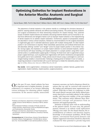
- 7. 43-61 Buser 11/23/04 4:04 PM Page 49 GROUP 2 Fig 9a Correct implant position in the Fig 9b Correct implant position in the oro- Fig 9c Correct implant position in the mesiodistal dimension. The implant shoul- facial dimension. The implant shoulder is apicocoronal dimension. The implant shoul- der should be positioned within the comfort positioned about 1 mm palatal to the point der is positioned about 1 mm apical to the zone, avoiding the danger zones, which are of emergence at adjacent teeth. The danger CEJ of the contralateral tooth in patients located close to adjacent root surfaces. The zone is clearly entered when the implant is without gingival recession. The danger zone danger zone is about 1.0 to 1.5 mm wide. placed too facially; this can cause resorp- is entered when the implant is placed too tion of the facial bone wall with subsequent far apically using excessive countersinking, recession. A second danger zone is located or too far coronally, which results in implant too far palatally, which can require an shoulder exposure at the mucosa. implant crown with a ridge-lap design. IDEAL IMPLANT PLACEMENT IN esthetic shortcomings. Implants positioned in the THE ANTERIOR MAXILLA comfort zones provide the basis for an esthetic restoration. Comfort and danger zones are defined As previously mentioned, esthetic implant place- in mesiodistal, orofacial, and apicocoronal dimen- ment is based on a restorative-driven philoso- sions. In the mesiodistal dimension, the danger phy.3–5,7,45 Correct 3-dimensional positioning of the zones are located next to adjacent teeth. At present, planned implant restoration is the driving force in it is not clear how wide these danger zones are. Pre- implant placement. This will allow for optimal sup- vious publications recommended that the implant port and stability of the peri-implant hard and soft shoulder and the adjacent root surface be at least tissues. In the anterior maxilla, the following 1 mm apart.21 With the tulip shape of the implant implant types are recommended for clinical use: shoulder on Straumann implants, this would place standard screw, wide body, narrow neck, TE 4.1/4.8, the implant body surface no closer than 1.5 mm to and TE 3.3/4.8 (Institut Straumann, Waldenburg, the adjacent root surface (Fig 9a). With this in Switzerland). These implants differ in restorative mind, the minimal gap size for implant selection in shoulder and implant thread dimensions. To utilize the anterior maxilla, based upon the implant shoul- these implants successfully in the anterior maxilla, der, can be defined (Table 3). Wide-neck implants, correct implant selection relative to the mesiodistal with their 6.5-mm shoulder diameter, are not rec- dimension of the tooth to be replaced is critical. In ommended for use in the anterior maxilla. Their this article, this dimension is referred to as gap size. implant shoulder margin is likely to be located too When planning for an ideal 3-dimensional close to adjacent teeth or too far facially, entering implant position, a distinction is made between so- into the respective danger zones. called “comfort” and “danger” zones in each dimen- With regard to the orofacial dimension, it has sion. Selection and placement of the dental implant been proposed that the position of the implant should be based on the planned restoration in these shoulder margin should be at the ideal point of zones. If the implant shoulder is positioned within emergence.3,4 The facial danger zone is located any- the danger zones, one of the above-mentioned com- where facially to the imaginary line highlighted plications could occur, potentially resulting in from the point of emergence of the adjacent teeth The International Journal of Oral & Maxillofacial Implants 49 COPYRIGHT © 2004 BY QUINTESSENCE PUBLISHING CO, INC. PRINTING OF THIS DOCUMENT IS RESTRICTED TO PERSONAL USE ONLY. NO PART OF THIS ARTICLE MAY BE REPRODUCED OR TRANSMITTED IN ANY FORM WITHOUT WRITTEN PERMISSION FROM THE PUBLISHER.
- 8. 43-61 Buser 11/23/04 4:04 PM Page 50 BUSER ET AL Table 3 Relationship Between the Mesiodistal depending on the tooth to be replaced, and must be Gap Size and the Diameter of the Implant taken into consideration. 46 In particular, lateral Shoulder (Straumann Dental Implant System) incisors are smaller and their CEJ is normally Shoulder Minimal Ideal located more coronally than the CEJs of central Implant diameter gap size gap size incisors or canines. Implant placement within the type (mm) (mm) (mm) apical danger zone (located anywhere 3 mm or Standard screw (S 4.1) 4.8 7.0 8.0 to 9.0 more apical to the proposed gingival margin) can Wide-body (S 4.8) 4.8 7.0 8.0 to 9.0 result in undesired facial bone resorption and subse- Narrow-neck (NN 3.3) 3.5 5.5 6.0 to 7.0 quent gingival recession. The coronal danger zone TE (TE 3.3/4.8) 4.8 7.0 8.0 to 9.0 is invaded with a supragingival shoulder position, TE (TE 4.1/4.8) 4.8 7.0 8.0 to 9.0 leading to a visible metal margin and poor emer- gence profile (Fig 9c). Respecting the comfort zones in 3 dimensions results in an implant shoulder Table 4 Risk Factors in Implant Patients located in an ideal position, allowing for an esthetic Risk factors Remarks implant restoration with stable, long-term peri- implant tissue support. Medical Severe bone disease causing impaired bone healing Immunologic disease Medication with steroids PREOPERATIVE ANALYSIS Uncontrolled diabetes mellitus Irradiated bone Risk Assessment Others In each patient, a detailed preoperative analysis Periodontal Active periodontal disease History of refractory periodontitis should be performed to assess the individual risk Genetic disposition profile and the level of difficulty of the planned ther- Smoking habits Light smoking (< 10 cigarettes per d) apy. Risk assessment in the anterior maxilla of poten- Heavy smoking (≥ 10 cigarettes per d) tial implant patients includes several aspects (Table Oral hygiene/ Home care measured by gingival 4). The goal of risk assessment is to identify patients compliance indices Personality, intellectual aspects whose implant therapy carries a high risk of a nega- Occlusion Bruxism tive outcome. Among the listed factors, patients with increased periodontal susceptibility and/or a history of a rapidly progressing or refractory periodontitis should be identified, because there is increasing evi- and/or planned restoration (Fig 9b). The palatal dence in the literature that these patients have an danger zone starts about 2 mm from this point of increased risk of biologic complications around emergence and leads to an increased risk of a ridge- osseointegrated implants.47,48 In the past 5 years, lap restoration. Placement of the implant orofacially genetic testing using a swab has been recommended in the comfort zone, which is located anywhere in to identify positive interleukin-1 (IL-1) genotype between these areas, will allow for a restoration patients, because these patients have an increased with the proper emergence profile to maintain the risk of developing periodontitis.49–51 It seems that harmonious scalloping of the gingival margins. the combination of an IL-1-positive genotype and The apicocoronal positioning of the implant smoking further increases this risk.52,53 Smoking is shoulder follows the philosophy “as shallow as pos- also an important risk factor for implant complica- sible, as deep as necessary,” as a compromise tions. Several clinical studies have demonstrated between esthetic and biologic principles. As agreed increased failure rates for smokers either during the upon at the last ITI consensus meeting, the position healing or the follow-up period.54–57 Recently pub- of the implant shoulder should be approximately lished studies have provided the first evidence that 2 mm apical to the midfacial gingival margin of the the combination of positive IL-1 genotype and planned restoration.21 This can be accomplished smoking also has a negative synergistic effect on through the use of surgical templates that highlight peri-implant tissues, because increased bone loss and the gingival margin of the planned restoration. In a higher frequency of biologic complications have patients without vertical tissue deficiencies, the use been noted.58–60 Thus, the identification of patients of periodontal probes leveled on the adjacent with a history of periodontitis combined with smok- cementoenamel junction (CEJ) in single-tooth gaps ing is important during preoperative analysis, has proven to be a valid alternative.21 It is important because these patients are clearly at risk for the to note that the CEJs of adjacent teeth can vary, development of biologic peri-implant complications. 50 Volume 19, Supplement, 2004 COPYRIGHT © 2004 BY QUINTESSENCE PUBLISHING CO, INC. PRINTING OF THIS DOCUMENT IS RESTRICTED TO PERSONAL USE ONLY. NO PART OF THIS ARTICLE MAY BE REPRODUCED OR TRANSMITTED IN ANY FORM WITHOUT WRITTEN PERMISSION FROM THE PUBLISHER.
- 9. 43-61 Buser 11/23/04 4:04 PM Page 51 GROUP 2 Anatomic Site Analysis: General Remarks Table 5 Anatomic Site Analysis in the An optimal esthetic implant restoration depends on Anterior Maxilla 4 anatomic and surgical parameters: (1) submucosal Factor Areas for analysis positioning of the implant shoulder, (2) adequate 3- dimensional implant positioning, (3) long-term sta- Location of the smile line • High lip line bility of esthetic and peri-implant soft tissue con- • Medium lip line • Low lip line tours, and (4) symmetry of clinical crown volumes Gingival morphotype • Thin with highly scalloped between the implant site and contralateral teeth.3,4 gingiva With this in mind, implant placement in an optimal • Thick with shallow scalloped position begins with a restorative plan and an gingiva anatomic assessment of the single- or multiple- Interocclusal relationship • Horizontal overlap • Vertical overlap tooth gap (Table 5). Dimensions of edentulous • Mesiodistal gap size Assessment begins extraorally and includes the gap • Multiple missing tooth patient’s smile. A keen eye is needed to determine if dimensions the smile is natural. Patients with unacceptable tooth Anatomy of alveolar crest • Horizontal bone deficiency health, shade, or position may not give a full smile • Vertical bone deficiency Status of adjacent dentition • Crown integrity when asked. Previous photographs of the patient and • Endodontic status family interviews may help to determine the natural • Periodontal status position of the patient’s lip during a smile. As Radiographic status • Vertical bone height expected, patients with a high lip line will show more • Anatomic structures (eg, tissue and will require maximal efforts to maintain nasopalatal canal) • Position and axis of adjacent peri-implant tissue support throughout the planning, roots provisional, surgical, and restorative phases. • Radiolucencies in alveolar The dental midline, tooth size, and shade should process be recorded. The intraoral exam should document • Foreign bodies in alveolar excessive or irregular gingival tissue, crowding, and process asymmetric teeth (eg, peg laterals), in addition to including a thorough periodontal and radiographic charting. It is paramount that orthodontic and peri- definitive implant-supported restoration is vital for odontal esthetic problems be addressed either prior the diagnosis of hard and soft tissue deficiencies to or during implant rehabilitation. Tissue shaping— prior to implant placement. Retention of the restora- whether excessive or deficient—should be managed tion, whether with cement or screws, will play a role with a restorative plan by experienced clinicians. in positioning of the shoulder of the implant to allow Characteristics of the soft tissue biotype15,16 will for sufficient peri-implant tissue support and proper play a prominent role in planning for final shoulder crown emergence. The use of diagnostic waxups and position of the implant. A thin biotype with highly templates for determination of anatomic comfort scalloped tissue will require the implant body and and danger zones in the planning process will pro- shoulder to be placed more palatal to mask any tita- vide the team members with information that can nium show-through. When implants are placed help maximize esthetic outcomes. With this vision of toward the palate, a slightly deeper placement the definitive restoration in hand, a comprehensive (within the apicocoronal comfort zone) is required anatomic site analysis is possible. to allow for a proper emergence profile of the restoration. Adjacent implant placement challenges Anatomic Site Analysis in Single-Tooth Gaps the treatment team’s ability to place dental implants As mentioned earlier, the single-tooth gap in the in a position that allows for subgingival shoulder anterior maxilla is assessed in 3 dimensions based on location and an ideal emergence profile while maxi- a planned restoration and the surrounding teeth. mizing the osseous crest height. In general, a patient Single-tooth sites offer less of a challenge because with the combination of a high lip line and a thin of the ability to use the adjacent teeth as landmarks biotype is extremely difficult to treat and should be in planning. With this in mind, several key analyses considered an anatomic risk. Patients who fit into must still take place prior to commencing with these treatment categories should be made aware of implant placement. A diagnostic waxup highlighting the challenges involved in obtaining an esthetic tissue deficiencies and final tooth positioning can result before treatment begins. assist in this planning process. Once the extraoral examination has been com- One of the first things to be assessed is orofacial pleted, a vision of the emergence and position of the ridge anatomy, including whether there is sufficient The International Journal of Oral & Maxillofacial Implants 51 COPYRIGHT © 2004 BY QUINTESSENCE PUBLISHING CO, INC. PRINTING OF THIS DOCUMENT IS RESTRICTED TO PERSONAL USE ONLY. NO PART OF THIS ARTICLE MAY BE REPRODUCED OR TRANSMITTED IN ANY FORM WITHOUT WRITTEN PERMISSION FROM THE PUBLISHER.
- 10. 43-61 Buser 11/23/04 4:04 PM Page 52 BUSER ET AL crest width and the presence or absence of facial Determination of the location of the nasopalatine bone atrophy. Deficient alveolar crest width and/or foramen and the distance to the adjacent teeth and facial bone atrophy require a bone augmentation the floor of the nose are necessary for proper procedure so that the implant can be positioned in a implant selection. If sectional imaging is not neces- correct orofacial position. Depending on the extent sary, the periapical radiograph will generally pro- and morphology of the bone defect, a simultaneous vide sufficient information, with greater accuracy or staged approach is necessary. Clinical sounding than a panoramic radiograph.61 Proper anatomic and sophisticated radiographic techniques such as site analysis in conjunction with restorative-driven conventional tomograms, dental computerized planning will optimize predictable esthetic results in tomograms (CTs) or volume CTs can assist in diag- the maxillary single-tooth gap. nosing deficiencies in this dimension. Mesiodistally, the space should be equal to that Anatomic Site Analysis in of the adjacent tooth (centrals) or the contralateral Extended Edentulous Spaces tooth (laterals and canines). Excesses or deficiencies Patients with extended edentulous spaces present in these dimensions must be addressed through the additional anatomic challenges, making it even use of orthodontics, enameloplasty, or restorative more difficult to produce an esthetic result with any materials prior to implant placement. For patients certainty. Varying clinical situations such as missing with diastemas, it is necessary to decide whether to centrals, central and lateral, lateral and canine, or eliminate or maintain the space prior to implant even several anterior teeth are possible, leading to placement, as this will affect the mesiodistal shoul- an array of treatment obstacles. With the loss of an der position. Guidelines for implant selection based adjacent tooth or teeth, planning for implant place- on gap size can be found in Table 3. ment will require a diagnostic waxup based on The most critical assessment remains the apico- sound esthetic principles, tooth morphology, and coronal dimension. Deficient tissue in this dimen- occlusal schemes. Understanding the fundamental sion can result from several factors: periodontal dis- objectives in the anterior esthetic zone—such as ease of the adjacent tooth/teeth, atrophy, trauma, tooth axis, interdental closure, gingival contours, infection, or a congenital abnormality. A tissue balance of gingival levels, interdental contacts, deficit in this dimension must be addressed and tooth dimensions, and tooth form—will help pro- managed carefully throughout the course of treat- duce a waxup that will dictate to the surgeon the ment. Because of the complexity of vertical goals necessary for replacement of the missing teeth hard/soft tissue grafting, patients with this condi- and tissue.46 tion are placed in a high anatomic risk group. The replacement of 2 missing central incisors Patients with excess tissue height require attention with dental implants can often lead to an acceptable as well. A bone-scalloping procedure will be esthetic result because of the symmetric gingival required to allow placement of the implant shoulder margin positions and the ability to form an interim- in a subgingival position. The most efficient way to plant papilla with the redundant nasopalatine tissue examine this position is through the use of a tem- commonly found in that region. Placement of the plate highlighting the proposed gingival margin implants in a strict apicocoronal position honoring position of the implant restoration. the maxim “as shallow as possible, as deep as neces- Interocclusal space must be addressed for reasons sary” will help maintain the interimplant crest in addition to the obvious ones. Placing the long height and provide support for the peri-implant tis- axis of the implant through the incisal edge of ante- sues (Figs 10a to 10c). rior teeth is beneficial for patients with excessive Patients with a thin gingival morphotype will be vertical overlap. A diagnostic waxup will highlight challenging because the implants will need to be the potential difficulties in restoring the proposed placed closer to the palate and deeper to provide for implant and managing the patient’s occlusion. Prior proper emergence, thus increasing the potential for to placement of the dental implant, a radiographic loss of interimplant tissue and resulting in a “black survey should be performed. A radiographic tem- triangle” and/or broad contact points. Patients who plate outlining the proposed implant position in the are missing a central and a lateral incisor or a lateral orofacial and mesiodistal dimensions with a metallic incisor and a canine are clinically more challenging rod will help determine if the implant will interfere because the edentulous space is smaller and the with adjacent tooth structure or vital anatomy.45 interimplant soft tissue tends to be less voluminous Magnification and distortion of the imaging tech- (Figs 11a to 11c). nique can be taken into account by inserting the Replacement of several missing teeth with known dimensions of the rod into the template. implants allows for the use of fixed partial dentures 52 Volume 19, Supplement, 2004 COPYRIGHT © 2004 BY QUINTESSENCE PUBLISHING CO, INC. PRINTING OF THIS DOCUMENT IS RESTRICTED TO PERSONAL USE ONLY. NO PART OF THIS ARTICLE MAY BE REPRODUCED OR TRANSMITTED IN ANY FORM WITHOUT WRITTEN PERMISSION FROM THE PUBLISHER.
- 11. 43-61 Buser 11/23/04 4:04 PM Page 53 GROUP 2 Fig 10a (Above) Two implants were placed in central incisor positions following the princi- ple “as shallow as possible, as deep as necessary” to maintain as much of the interimplant papilla as possible. Fig 10b (Center) Final treatment outcome shows a pleasing result, since the interimplant papilla was well maintained. Fig 10c (Right) Periapical radiograph, taken 3 years after implant placement, shows min- imal bone resorption around both implants. Fig 11a (Left) The esthetic result with 2 adjacent implants in the central and lateral posi- tions is compromised because of a short interimplant papilla. Fig 11b (Center) The esthetic result was still acceptable since the patient had a low lip line. Fig 11c (Right) The radiograph at the 5-year examination shows stable bone crest levels with a flat bone crest between both implants. and the opportunity to use ovate pontics to help Following the template and planning procedures support the tissues and form pseudo-papillae. Ques- previously mentioned should allow the clinician to tions arise when bone augmentation procedures maximize the potential for an acceptable esthetic have been performed previously and pontics are result in a difficult clinical situation. Future implant used to restore the sites. Will the bone remain, or is designs with anatomically contoured implant shoul- there a need to place an implant in these sites to ders may benefit treatments of this type by improv- maintain the bone? Replacement of several missing ing interproximal tissue support.5,62,63 teeth—eg, lateral-central-central-lateral—with implants requires maximizing placement in all 3 dimensions, avoiding embrasures, supragingival SURGICAL PROCEDURES shoulders, and irregular gingival margins. Implant selection becomes critical, because the implant Implant Selection needs to provide for emergence as well as maintain Based on the anatomic site analysis, the appropriate peri-implant hard tissue support. implant type is selected to best fit a single-tooth gap. The International Journal of Oral & Maxillofacial Implants 53 COPYRIGHT © 2004 BY QUINTESSENCE PUBLISHING CO, INC. PRINTING OF THIS DOCUMENT IS RESTRICTED TO PERSONAL USE ONLY. NO PART OF THIS ARTICLE MAY BE REPRODUCED OR TRANSMITTED IN ANY FORM WITHOUT WRITTEN PERMISSION FROM THE PUBLISHER.
- 12. 43-61 Buser 11/23/04 4:04 PM Page 54 BUSER ET AL Fig 12a Correct implant positioning using Fig 12b The spiral drill is guided by the Fig 12c Status following implant place- a surgical template. The template imitates surgical template for proper alignment. ment. The implant shoulder is located the future soft tissue margin at the implant about 2 mm apical to the future soft tissue crown. margin. In central incisors and canines, implants with a regu- generates a template that will place the implant in a lar-neck configuration (shoulder diameter of position that will support the planned restoration 4.8 mm) are most often used. The minimal mesiodis- (Figs 12a to 12c) and make restoring it easier. tal gap size for such a standard-neck implant is 7 It is clear that templates can be helpful in making mm, whereas 8 to 9 mm are ideal to allow a sufficient anterior esthetics more predictable and reliable. distance to adjacent roots (Table 3). The narrow- However, they are only as good as the team that uses neck implant with a shoulder diameter of 3.5 mm is them. Communication between the restorative clini- most often used in lateral incisor areas with a mini- cian making the template and the surgeon using it is mal gap size of 5.5 mm. The TE implants, mainly imperative, so that they can agree on a design that will developed for placement in extraction socket defects, make the placement process efficient and accurate. are offered with 2 different neck diameters: regular and wide. In the anterior maxilla, the 2 TE implant Surgical Procedures in Single-Tooth Gaps types with the regular-neck configuration (diameter Under local anesthesia, the mucosa is opened with a of 4.8 mm) are used for standard prosthetic proce- crestal incision located approximately 2 to 3 mm dures. The wide-neck configuration (shoulder diam- toward the palatal aspect and extended through the eter of 6.5 mm) should only be used in exceptional sulcus of adjacent teeth to the facial aspect of the clinical situations because of its potential for reach- alveolar crest. This incision avoids the formation of ing too far facially and/or proximally. scar tissue in the midcrestal area and ensures suffi- cient vascularity of the facial flap in the area of the Surgical Templates future papillae. Facial line-angle relieving incisions The use of surgical templates in the anterior maxilla are most often necessary to allow sufficient access to can be valuable to properly place the implant shoul- the surgical site (Figs 13a and 13b). In patients who der in a position that will allow for an ideal emer- need a bone augmentation procedure, this flap gence profile and long-term peri-implant hard and design also allows for tension-free wound closure soft tissue support.3,4,7 Templates are mandatory for with the release of the periosteum and a coronal implant treatment of extended edentulous spaces. mobilization of the flap. As an alternative, a para- Many variations of surgical templates exist. Good papillary incision technique may be used. Implant templates should have the following features: they placement without flap elevation (often called “flap- should be easy to place and remove, they should be less implant placement”) is considered experimen- rigid and stable, they must allow for placement and tal, because no clinical studies with sufficient data removal of bite blocks when possible, and they must have been published yet. not interfere with tissue reflection and visualization After the incisions have been made, the facial and of the depth indicators or the cooling of the surgical palatal mucoperiosteal flaps are elevated with a fine drills. A key feature of a surgical template used in tissue elevator to guarantee low-trauma soft tissue the anterior maxilla is designation of the final apico- handling. This is followed by an intrasurgical site coronal, mesiodistal, and orofacial positioning of analysis to evaluate the facial aspect of the alveolar the implant shoulder. The best way to indicate these crest. For implant sites in the central incisor area, positions is to complete a diagnostic waxup high- location of the nasopalatal foramen must be deter- lighting the final gingival margin position, facial mined. A crest-flattening or bone-scalloping proce- surface, and embrasure form of the proposed dure is recommended, since this facilitates easier restoration. Working backward from this waxup and more precise preparation of the implant bed 54 Volume 19, Supplement, 2004 COPYRIGHT © 2004 BY QUINTESSENCE PUBLISHING CO, INC. PRINTING OF THIS DOCUMENT IS RESTRICTED TO PERSONAL USE ONLY. NO PART OF THIS ARTICLE MAY BE REPRODUCED OR TRANSMITTED IN ANY FORM WITHOUT WRITTEN PERMISSION FROM THE PUBLISHER.
- 13. 43-61 Buser 11/23/04 4:04 PM Page 55 GROUP 2 and the natural shape of the alveolar crest is imi- Today, the GBR procedure, ie, applying barrier tated. However, the surgeon should not remove any membranes in combination with bone grafts and/or bone in the proximal area of adjacent teeth, because bone substitutes, is routinely used (Figs 13g to 13j). this bone is important for the support and mainte- The goal of GBR is to establish a thick facial bone nance of the papillae. wall of at least 2 to 3 mm to achieve sufficient and The precise position of the implant site is long-lasting bone support for the facial soft tissues. marked with small round burs. Correct 3-dimen- Improvement of soft tissue esthetics can also be sional implant placement can be determined by achieved with soft tissue grafting at implant place- using either a periodontal probe and landmarks of ment.21 In patients with thin soft tissues and/or a adjacent teeth21 or a prefabricated surgical template concave contour of the facial mucosa, a connective with a built-in gingival margin for the future tissue graft can be used to improve the thickness implant crown.45 Both techniques provide sufficient and contour of the soft tissues. These grafts are har- guidance in single-tooth gaps. vested in the premolar area of the palate and can be Preparation of the implant bed is carried out sutured to the periosteum of the mucoperiosteal with standard spiral drills of increasing diameter flap to avoid displacement of the graft during (2.2 mm, 2.8 mm, and 3.5 mm). This technique wound closure. reduces the trauma to the bone tissue and gives the Prior to completion of the surgical procedure, surgeon a chance to change the position of the the mucoperiosteal flap is repositioned precisely, implant and/or the direction of the implant axis particularly in the area of the future papillae. The between drill steps. As previously outlined, the surgeon has to make sure that wound closure is pre- objective is to position the implant shoulder within cise and tension-free. To achieve this, an incision of the comfort zones in all 3 dimensions. To ensure the periosteum is often necessary to release the flap correct esthetic implant placement, the entrance of in a coronal direction (Fig 13k). For suturing, fine the bone cavity has to be prepared with the profile atraumatic suture material (5-0) is recommended. drill to allow deeper implant placement. In addi- Following surgery, a periapical radiograph is taken tion, implants with a short neck configuration are to examine the position and direction of the placed most often used to limit the amount of bone resorp- implant and its relationship to the roots of adjacent tion in the crestal area. teeth (Fig 13l). During bone preparation, different depth gauges help the surgeon to control the future implant posi- Surgical Procedures in tion in the mesiodistal, orofacial, and apicocoronal Extended Edentulous Spaces directions, as well as the implant axis (Figs 13c and In implant sites with multiple missing teeth, the 13d). Pretapping of the thread is rarely done in the surgical procedure is clearly more demanding and anterior maxilla. Most often, self-tapping implants requires optimal preoperative planning and an are used, since the bone structure in the anterior implant surgeon with sufficient experience. The use maxilla is rather spongy. Implant placement is per- of an appropriate surgical template is mandatory to formed either with an adapter attached to a special enable correct 3-dimensional implant positioning in contra-angle handpiece (at 15 rpm) or with the the mesiodistal, orofacial, and apicocoronal direc- hand ratchet. Following implant placement, pri- tions. In sites with adjacent implants, an additional mary stability of the implant is carefully checked. aspect needs to be considered: the interimplant dis- An appropriate healing cap is then selected. It is tance. In such sites, bone resorption of 1 to 2 mm at recommended that a healing cap be used that covers the proximal aspects of the implant leads to a flat- the implant shoulder, such as the 1.5-mm cover tening of the interimplant bone and consequently a screw (Figs 13e and 13f) or an esthetic healing cap short interimplant papilla. A distance of at least with a buccal bevel, which is available in 2 heights 3 mm has been recommended between 2 adjacent (2 mm and 3.5 mm). All these healing caps have the implants to minimize this bone resorption.41 This advantage that no bone can grow on top of the recommendation seems logical based on current implant shoulder during healing, and the caps sup- knowledge, but no clinical and radiographic studies port the soft tissues in the proximal area. The buc- are yet available to support it. cal bevel of the esthetic healing cap will also allow The surgical procedures with regard to incision for additional space for the interim restoration dur- technique, flap design, bone preparation, and implant ing the healing phase. placement in extended edentulous spaces follow In the case of a peri-implant bone defect, either the same guidelines as previously outlined. Such with an intact or a deficient facial bone wall, a local sites most often also have horizontal and/or vertical bone augmentation procedure is recommended. bone deficiencies. Therefore, bone augmentation The International Journal of Oral & Maxillofacial Implants 55 COPYRIGHT © 2004 BY QUINTESSENCE PUBLISHING CO, INC. PRINTING OF THIS DOCUMENT IS RESTRICTED TO PERSONAL USE ONLY. NO PART OF THIS ARTICLE MAY BE REPRODUCED OR TRANSMITTED IN ANY FORM WITHOUT WRITTEN PERMISSION FROM THE PUBLISHER.
- 14. 43-61 Buser 11/23/04 4:04 PM Page 56 BUSER ET AL Fig 13a Single-tooth gap in the central Fig 13b The surgical site is exposed with Fig 13c Following preparation with the incisor area. Status 8 weeks following tooth a full-thickness flap using 2 distal-line-angle first round burs and drills, the depth gauge extraction. A palatal incision will be used relieving incisions. is inserted to examine the future implant about 3 mm from the middle of the crest. position and axis. Note the palatal position of the pin in relation to the extraction socket. Fig 13d The second depth gauge, with a Fig 13e Status following implant place- Fig 13f The occlusal view confirms the built-in 5-mm ring, is used to check the ment and insertion of 1.5-mm large healing correct orofacial position of the implant proximity of the future implant shoulder to cap to cover the implant shoulder. Note the shoulder being slightly palatal to the point adjacent root surfaces. correct apicocoronal position of the implant of emergence of the contralateral central shoulder, about 1 mm apical to the CEJ of incisor (line). Note the minor bone defect at the adjacent contralateral tooth (line). the facial aspect, which requires bone graft- ing. Fig 13g The facial bone defect is filled Fig 13h A second layer of bone substitute Fig 13i The augmentation material is cov- with autogenous bone chips harvested in is used to overaugment the surgical site. A ered with a collagen-based barrier mem- the vicinity, such as the anterior nasal bone filler with a low substitution rate (Bio- brane using the principles of GBR. Two spine. Oss) is preferred. membrane strips are used (“double layer technique”) to improve membrane stability. 56 Volume 19, Supplement, 2004 COPYRIGHT © 2004 BY QUINTESSENCE PUBLISHING CO, INC. PRINTING OF THIS DOCUMENT IS RESTRICTED TO PERSONAL USE ONLY. NO PART OF THIS ARTICLE MAY BE REPRODUCED OR TRANSMITTED IN ANY FORM WITHOUT WRITTEN PERMISSION FROM THE PUBLISHER.
- 15. 43-61 Buser 11/23/04 4:04 PM Page 57 GROUP 2 Fig 13j (Left) The occlusal view clearly shows how the alveolar crest was locally overaug- mented. Fig 13k (Center) Following incision of the periosteum, the flap is mobilized coronally, and a tension-free primary wound closure is obtained. To close the wound, 5-0 and 6-0 nonre- sorbable suture material is used. Fig 13l (Right) Periapical postsurgical radiograph. Note the minor radiolucency in the middle of the implant. Fig 13m Soft tissue status at 8 weeks of Fig 13n The reopening was done with a Fig 13o Status a few weeks following healing. The site is ready for reopening to 12b blade, removing some keratinized placement of the provisional crown based gain access to the implant shoulder and ini- mucosa slightly palatal to the healing cap. A on a titanium coping. The shape of the pro- tiate the restorative phase. larger healing cap was inserted to com- visional restoration was used for soft tissue press the soft tissues slightly to the facial conditioning. aspect. Fig 13p (Above) Status 12 months follow- Fig 13r Final treatment outcome of this ing implant placement. The definitive cer- 27-year-old female patient with a high lip amometallic crown has been seated. The line. esthetic result is pleasing with a harmo- nious gingival margin and intact papillae. Fig 13q (Right) The periapical radiograph at 12 months with the definitive crown indi- cates minimal bone resorption. The International Journal of Oral & Maxillofacial Implants 57 COPYRIGHT © 2004 BY QUINTESSENCE PUBLISHING CO, INC. PRINTING OF THIS DOCUMENT IS RESTRICTED TO PERSONAL USE ONLY. NO PART OF THIS ARTICLE MAY BE REPRODUCED OR TRANSMITTED IN ANY FORM WITHOUT WRITTEN PERMISSION FROM THE PUBLISHER.
- 16. 43-61 Buser 11/23/04 4:04 PM Page 58 BUSER ET AL Fig 14a Intrasurgical status following Fig 14b Submerged implant healing for 3 Fig 14c Three months following implant placement of a standard screw implant in months was chosen for this patient in placement, a thin facial mucosa was appar- area 13 in a correct 3-dimensional position 1992. ent, requiring soft tissue graft at the re- and an intact facial bone wall in the crestal opening procedure. area. An apical fenestration defect was aug- mented with locally harvested autogenous bone grafts. Fig 14d At reopening, a free connective tissue graft was applied to improve the thickness of the facial soft tissues. Fig 14e Clinical status duing the phase of provisionsal restoration demonstrates the convex facial soft tissue margin at the right level. Fig 14f (Left) Clinical status at 12 years following implant placement (2004) demon- strates remarkable soft tissue stability at the mid-facial margin and nice convex contour of the facial mucosa. Fig 14g (Center) Esthetic result with the lip line. Fig 14h (Right) The periapical radiograph 12 years following implant placement confirms stable bone crest values around the standard screw implant. procedures are common in sites with multiple miss- restoration that will not place intermittent pressure ing teeth, using either a simultaneous or a staged on the healing cap and tissues is recommended. For approach. this reason, removable partial dentures should be adjusted to prevent these contacts, which can cause Interim Restoration difficulty in patients with limited interocclusal space Delivery of an appropriate interim restoration at or excessive vertical overlap. Interim restorations the time of implant placement in the anterior max- that are fixed to the adjacent teeth or that com- illa is paramount for patient satisfaction and peri- pletely eliminate the possibility for soft tissue con- implant tissue protection. Fabrication of an interim tacts are more beneficial for implant integration and 58 Volume 19, Supplement, 2004 COPYRIGHT © 2004 BY QUINTESSENCE PUBLISHING CO, INC. PRINTING OF THIS DOCUMENT IS RESTRICTED TO PERSONAL USE ONLY. NO PART OF THIS ARTICLE MAY BE REPRODUCED OR TRANSMITTED IN ANY FORM WITHOUT WRITTEN PERMISSION FROM THE PUBLISHER.