

Recomendados
Mais conteúdo relacionado
Mais procurados
Mais procurados (20)
Destaque
Destaque (16)
Semelhante a Zeiss Opmi Pico
Semelhante a Zeiss Opmi Pico (20)
Mais de Bojidar Kafelov
Último
Último (20)
Zeiss Opmi Pico
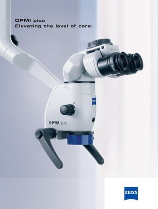
- 1. OPMI pico Elevating the level of care.
- 3. Microscope technology from Carl Zeiss makes the most minute details and finest structures clearly visible, enabling you to visualize brilliant, high- contrast, true-color images. Most importantly: better vision is the key to improving the quality of the diagnosis and the treatment. ® Designed for today’s practice. OPMI pico. OPMI pico is a compact, high-performance, easy- to-use microscope. In addition to its ergonomic design, it features many innovative functions that optimize the quality of diagnosis and treatment. Complete integration of cables, light source, light guide, video camera and control console is a practical design decision that allows OPMI pico to compliment the overall image of any practice while eliminating any interference with your work. The integrated video camera option for OPMI pico facilitates one-touch, on-demand documentation essential to the communication process with both patients and staff during treatment. Carl Zeiss is renowned for their superior optics. With apochromatic coatings, more light is trans- ferred to the viewer’s eye, ensuring a significant increase in image resolution, contrast and depth perception. The result is more brilliant, true-color images. The integrated product design is as unique as the superior optics, and is part of the countless func- tions that Carl Zeiss delivers with OPMI pico. Photo: Dr. Stephane Browet, Koekelberg, Belgium 3
- 4. For better ergonomics. OPMI pico is a true advancement in the prevention of neck strain and back problems. Experience it for yourself: only a microscope makes it possible to work in a comfortable and ergonomically correct position. The five-step magnification changer delivers brilliant images – from an overview of the working field down to the finest details. No matter your region of interest, you sit upright the entire time. Objective lenses with focal lengths of 200 mm, 250 mm and 300 mm are available to precisely match the microscope to your personal working distance. The ergonomically designed grips fit perfectly and securely in your hands, enabling you to easily place OPMI pico in the desired position. 4
- 5. 5
- 6. Create more trust. A picture is worth a thousand words. Explain your diagnosis, the treatment and the results to the patient to ensure a thorough understanding of the benefits of the procedure. Through your step-by- step explanation, you foster a higher level of trust between you and your patient. After all, seeing is believing. OPMI pico provides all the necessary options needed to carry out effective communication, including the digital MediLive® solutions that enable you to document your work using videos and photos. There are also MediLive systems to edit, archive and present your work, allowing for easy presentation to a group of colleagues or to a referrer. Photo: Practice of Dr. Wolfgang Bolz, Prof. Dr. Hannes Wachtel, Prof. Dr. Markus Hürzeler, Dr. Otto Zuhr, 6 Dr. Wolf Richter, Munich, Germany
- 7. 7
- 8. For more integration. OPMI pico is a compact microscope that optimally fits into your practice workflow. It is characterized Integrated light by its elegant form, which was honored with the source with swing-in red dot award for its design quality and very easy- backup lamp module. to-use functions. Simply plug in the cable and You have the choice of OPMI pico is ready for use. There are no exposed halogen or xenon illumina- cables or external components to interfere with tion. Xenon has a light your work. All functional elements such as the temperature characteristic video control console, video camera, cables, light of natural daylight. sources, and light guides have been completely integrated into the microscope. 8
- 9. The light guide and the power cable are integrated. No hanging cables to interfere with your work. Integrated video control console. Intuitive system adjustments allow you to fine tune the video image. Compact microscope body with integrated MediLive Primo video camera (optional). A freeze function captures still shots for patient and staff communication during treatment. A rotatable video image enables your patient and staff to always view the image upright. We would like to thank Dr. Mohr in Neu-Isenburg for kindly allowing us to take photographs in his practice. 9
- 10. For more operating comfort. The design focus during development of OPMI pico was extreme functionality. The microscope is precisely tailored to the needs of dentistry. There are numerous functions designed to support your comfort in every phase of treatment. Everything can be intuitively operated from the user’s posi- tion without ever having to divert your attention from the field of treatment. The unique optical system of OPMI pico offers magnifications finely tuned to each other, contin- uous brightness adjustment and a fine focusing feature that provides you with a new quality of vision and work. MORA interface – the pinnacle of ergonomics. The MORA interface enables you to easily and precisely move OPMI pico into the desired position. Even considering the unique, smooth operation of the MORA interface, you’ll find the most im- pressive feature to be the upright viewing position that remains constant regardless of the angle of the microscope. This allows you to remain in a comfortable position at all times without taking your eyes off the subject. MORA Interface. Enhanced mobility and comfort. You always remain upright and comfortable, regardless of how OPMI pico is positioned. 10
- 11. Continuous brightness regulation at your fingertips. Allows you to easily adjust the light intensity for any clinical situation. The 180° tiltable tube. Widefield eyepieces Allows you to work in (12.5x or 10x). maximum comfort in Large visual field with 3-D, a variety of positions stereoscopic image. Special and on difficult-to-reach diopter settings also make areas. Your back will them suitable for eyeglass thank you – particularly wearers. during longer procedures. 5-step magnification changer. Optimally matched magni- fications for brilliant images – from an overview of the working field to ultra-fine details. Fine focusing Adjust the focus to your personal needs. Focusing objective lens. Easy operation for both left and right-handed users. A variety of work- ing distances are available. 11
- 12. For even more possibilities. The large selection of accessories enable you to configure OPMI pico for any conceivable treat- ment situation. Everything is as easy and intuitive to operate as the microscope itself. Ergonomic design and Angled optics and tube dove- tail. For a comfortable position during work on difficult-to-reach areas. Digital visualization MediLive Primo 1 CCD video MediLive Trio Dent 3 CCD camera (integrated). video camera. 12
- 13. Optics and illumination Orange filter. Prevents premature Double iris diaphragm. Xenon or halogen illumination. Continuous brightness control. curing of composite materials. For optimum depth of field. Includes integrated backup lamp Adjustment at your fingertips. module. operating comfort Foot control panel. To comfort- VisionGuard® Drape. Sterilizable caps and handgrip ably operate the video freeze For sterile working environments. drapes. For asepsis. function. Adapter for a SLR (single Adapter for a digital still lens reflex) camera. compact camera. 13
- 14. OPMI pico by the OPMI pico microscope numbers. Magnification system - Manual apochromatic magnification changer - Five click stop positions: 0.4x; 0.6x; 1.0x; 1.6x; 2.5x Tubes - 45° inclined binocular tube, f = 170 mm - 0° - 180° tiltable binocular tube; f = 170 or f = 200 mm Eyepieces - 12.5x and 10x widefield eyepieces, also suitable for eyeglass wearers Magnification range example with f = 250 mm lens and 12.5x eyepiece: Magnification/Field-of-view diameter: 3.4x 5.1x 8.5x 13.6x 21.3x 65 mm 43 mm 26 mm 16 mm 10 mm Focusing - Manual focusing, focusing range 13 mm - 3 focusing lenses available: f = 200 mm, f = 250 mm, f = 300 mm, interchangeable Microscope mount, mobility Microscope body adapted to S100 suspension systems via - 120° coupler - MORA Interface (optional) - Mechanical Optical Rotating Assembly - Rotatable coupling for OPMI pico with +/-25° rotational range - Integrated, swing-in angular stop to increase depth of field - Patented, n. Dr. Assad F. Mora Handgrips: 4 versions Additional optics - Angled optics with tube dovetail (optional) - Double iris diaphragm to increase the depth of field (optional) 14
- 15. Suspension systems Accessories to attach external cameras S100 carrier system for user-friendly operation of - Splitter: the OPMI pico - Angled optics with left and right documenta- - Matched to a weight capacity of 2.5 to 7 kg tion ports, optional with tube dovetail S 100 floor stand - Beam Splitter 20 with documentation ports - Mobile solutions for the practice - Beam Splitter 50 with documentation ports - Dimensions base: 650 x 625 mm - MORA interface with documentation port - Weight: approx. 102 kg - Adapters: S100 wall mount - Video lens with C-mount interface for the - The space-saving alternative for small rooms attachment of external video cameras - Weight: Short arm: approx. 47 kg, - Lens to attach external digital cameras long arm: approx. 52 kg S100 ceiling mount Accessories - Creates free space around the treatment chair - Splash protection for the objective lens - Weight: approx. 54 kg - High-quality cover lens with brilliant optical quality, easy to change Illumination systems - Reflection-free vision through slanted - Integrated coaxial cold-light illumination arrangement of the lens - Control knob to adjust brightness directly - VisionGuard drapes for sterile work above the viewing tube - Easy-to-reach, swing-in orange filter for Electrical data composite materials - Rated voltage - Integrated 12V 100W halogen illumination 115 VAC (100…120 VAC ± 10%) with 2 halogen reflector lamps in the quick- 230 VAC (220…240 VAC ± 10%) change module - Power consumption Halogen illumination: - Integrated 12V 180W xenon illumination with 115 VAC max. 2.0 A; 230 VAC max. 1.0 A daylight characteristic including 2 xenon lamps - Power consumption Xenon illumination: in the quick-change module (optional) 115 VAC max. 5.0 A; 230 VAC max. 2.5 A - Rated frequency 50…60 Hz Camera systems (optional) - Fuses Halogen illumination: Integrated 1 CCD video camera 115 VAC T 6.3 A/H 250 V; - Functioning and ready for use at any time 230 VAC T 3.15 A/H 250 V - Freeze function for capturing still shots Xenon illumination: automatic circuit breaker - Image rotate - Electrical standard Protection class I, - Customizable user settings Degree of protection IPX0, Product classifica- - Video standard: PAL/NTSC tion I in acc. with 93/42/EEC Annex IX - Ports: Y/C (S-Video) and VBS composite External MediLive Trio Dent 3 CCD video camera Compliance - Customizable user-specific settings - DIN EN ISO 9001, DIN EN ISO 13485 - Video standard: PAL/NTSC DIN EN 60601-1, IEC 601-1 - Ports: Y/C (S-Video) and VBS composite UL 60601-1, CAN/CSA-C22.2 No. 601.1 RGB, Progressive Scan ® C US DV, DVI 15
- 16. DS-Nr.: 30-186/I-e Printed in Germany AW-TS-III/2007 Uoo Subject to change in design and scope of delivery and as a result of ongoing technical development. OPMI, MediLive, SpeedFokus and VisionGuard are registered trademarks of Carl Zeiss. Germany 73446 Oberkochen Carl Zeiss Surgical GmbH Fax: +49 (0) 7364 / 20-4823 A Carl Zeiss Meditec Company www.meditec.zeiss.com/loupes www.meditec.zeiss.com/contacts Email: surgical@meditec.zeiss.com