
Recomendados
Mais conteúdo relacionado
Mais procurados
Mais procurados (20)
Semelhante a Trauma Rounds, 1:3; Evaluating The Cervical Spine
Semelhante a Trauma Rounds, 1:3; Evaluating The Cervical Spine (20)
Mais de Arun Shanbhag
Mais de Arun Shanbhag (15)
Último
Último (20)
Trauma Rounds, 1:3; Evaluating The Cervical Spine
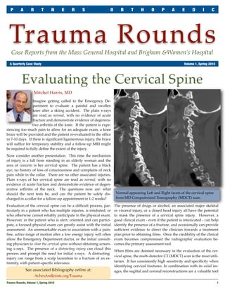
- 1. P A R T N E R S O R T H O P A E D I C Trauma Rounds Case Reports from the Mass General Hospital and Brigham &Women’s Hospital A Quarterly Case Study Volume 1, Spring 2010 Evaluating the Cervical Spine Mitchel Harris, MD Imagine getting called to the Emergency De- partment to evaluate a painful and swollen knee after a skiing accident. The plain x-rays are read as normal, with no evidence of acute fracture and demonstrate evidence of degenera- tive arthritis of the knee. If the patient is expe- riencing too much pain to allow for an adequate exam, a knee brace will be provided and the patient re-evaluated in the office in 7-10 days. If there is significant ligamentous injury, the brace will suffice for temporary stability and a follow-up MRI might be required to fully define the extent of the injury. Now consider another presentation. This time the mechanism of injury is a fall from standing in an elderly woman and the area of concern is her cervical spine. The patient has a black eye, no history of loss of consciousness and complains of neck pain while in the collar. There are no other associated injuries. Plain x-rays of her cervical spine are read as normal, with no evidence of acute fracture and demonstrate evidence of degen- erative arthritis of the neck. The questions now are: what should the next tests be, and can the patient be safely dis- Normal appearing Left and Right facets of the cervical spine charged in a collar for a follow-up appointment in 1-2 weeks? from MD Computerized Tomography (MDCT) scan. Evaluation of the cervical spine can be a difficult process, par- The presence of drugs or alcohol, an associated major skeletal ticularly in a patient who has multiple injuries, is intubated, or or visceral injury, or a closed head injury all have the potential who otherwise cannot reliably participate in the physical exam. to mask the presence of a cervical spine injury. However, a However, in the patient who is alert, oriented and can partici- good clinical exam - even if the patient is intoxicated - can help pate, a focused physical exam can greatly assist with the initial identify the presence of a fracture, and occasionally can provide assessment. An unremarkable exam in association with a pain- sufficient evidence to direct the clinician towards a treatment free, active range of motion after a low energy injury will often plan prior to obtaining films. Once the credibility of the clinical allow the Emergency Department doctor, or the initial consult- exam becomes compromised the radiographic evaluation be- ing physician to clear the cervical spine without obtaining screen- comes the primary assessment tool. ing x-rays. The presence of a distracting injury can cloud this When films are deemed necessary in the evaluation of the cer- process and prompt the need for initial x-rays. A distracting vical spine, the multi-detector CT (MDCT) scan is the most utili- injury can range from a scalp laceration to a fracture of an ex- tarian. It has consistently high sensitivity and specificity when tremity, with patient-specific relevance. identifying cervical fractures. In combination with its axial im- See associated Bibliography online at: ages, the sagittal and coronal reconstructions are a valuable tool AchesAndJoints.org/Trauma Trauma Rounds, Volume 1, Spring 2010 1
- 2. P A R T N E R S O R T H O P A E D I C T R A U M A R O U N D S to assess traumatic soft tissue injuries including posterior ligamentous injuries and facet subluxa- tion. So in the second example above, if films are deemed necessary to assess for a cervical spine injury, MDCT should be preferentially or- dered over the traditional c-spine trauma series consisting of an AP/ Lateral/ Open mouth dens view. Once the MDCT has been reviewed, the man- agement controversy begins. In the setting of a positive scan a spine consult should be obtained. However, if the MDCT is read as normal with no evidence of an acute fracture and demonstrates evidence of degenerative arthritis of the neck, can the collar be safely discontinued? At this juncture, the safest management option is to carefully perform (or repeat) a focused physical Left: Ligamentum falvum disruption observed in MRI - T2 image. exam. It should start with the evaluation of the Right: C-Spine Evaluation Algorithm; Read about the 2/3 Rule in Text midline structures, both bony and ligamentous. In the setting where the examination remains difficult to inter- If there are no palpable defects and no appreciable tenderness pret or the patient is unable to provide feedback (intubated, to palpation, the patient should be asked to laterally rotate his/ intoxicated, closed head injury, drugs, electrolyte imbalance, her head from side-to-side. If this can be performed without post-seizure, etc.) an MRI will be valuable to safely allow for pain the patient should be asked to lift his/her head off the bed removal of the collar. An MRI without evidence of an acute and bring chin to chest. If this too can be performed without injury and a negative MDCT should consistently provide the pain, then in the presence of a negative MDCT the collar can be greatest assurance that it is safe to remove the collar. safely discontinued. However, if there is pain with motion or tenderness with palpa- The key to navigating this often-difficult evaluation algorithm tion despite the presence of a negative CT, the collar should stay is to remember the 2/3 Rule. If two of the three key evaluation on until a follow-up visit with x-rays 10-14 days later. An up- studies (MDCT, MRI and credible physical exam) are negative, right lateral x-ray in the collar should be carefully reviewed at a clinically relevant injury will not be missed. this follow-up. If there is no sign of deformity, the focused exam should be repeated with the collar off. If uncertainty still While a spine surgery consult is not often necessary after your exists, active flexion-extension films would be helpful to further initial evaluation, it will be beneficial for you to have a comfort- assess for an occult ligamentous injury. Flexion-extension films able relationship with your local spine surgeon. These surgeons will be optimally useful if the patient is capable of actively often have defined protocols for this controversial area of man- moving his/her neck through a full range of motion. agement and can help guide you toward the best practices. Trauma Faculty David Ring, MD — 617-724-3953 Editor in Chief MGH Hand & Upper Extremity Service Mark Vrahas, MD — 617-726-2943 Mark Vrahas, MD dring@partners.org Partners Chief of Orthopaedic Trauma mvrahas@partners.org George Dyer, MD — 617-732-6607 Program Director Mitchel B Harris, MD — 617-732-5385 BWH Hand & Upper Extremity Service Suzanne Morrison, MPH Chief, BWH Orthopedic Trauma gdyer@partners.org (617) 525-8876 mbharris@partners.org Please send correspondence to: smmorrison@partners.org R Malcolm Smith, MD, FRCS — 617-726-2794 Mark Vrahas, MD / Trauma Rounds Chief, MGH Orthopaedic Trauma Yawkey Center for Outpatient Care, Suite 3C Editor, Publisher 55 Fruit Street, Boston, MA 02114 Arun Shanbhag, PhD, MBA rmsmith1@partners.org David Lhowe, MD — 617-724-2800 MGH Orthopaedic Trauma Linda Honeycutt: May 5, 1944 - April 10, 2010 dlhowe@partners.org Thank you for all the years you took such good care of our patients & staff. We miss your smile, wit and easy manner. 2 Trauma Rounds, Volume 1, Spring 2010