SlideShare uma empresa Scribd logo
1 de 75
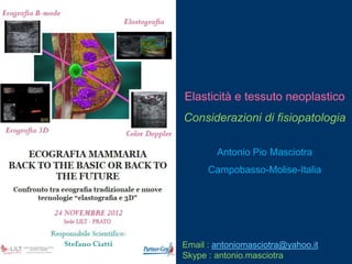
Elasticità e tessuto neoplastico
Considerazioni di fisiopatologia

        Antonio Pio Masciotra
      Campobasso-Molise-Italia


Email : antoniomasciotra@yahoo.it
Skype : antonio.masciotra
Mechanical (elastic) properties
    of neoplastic tissue
         Physiopathology

        Antonio Pio Masciotra
      Campobasso-Molise-Italy


Email : antoniomasciotra@yahoo.it
Skype : antonio.masciotra
Elastografia mammaria :
    quantitativa o qualitativa?

         Antonio Pio Masciotra
             Campobasso


Email : antoniomasciotra@yahoo.it
Skype : antonio.masciotra
Breast sonoelastography :
    quantitative or qualitative?

         Antonio Pio Masciotra
       Campobasso-Molise-Italy


Email : antoniomasciotra@yahoo.it
Skype : antonio.masciotra
PRINCIPAL MECHANICAL PROPERTIES

    Those characteristics of the materials which describe their behaviour under external loads are known as
                                           Mechanical Properties.
                           The most important and useful mechanical properties are:

Strength
It is the resistance offered by a material when subjected to external loading.
So, stronger the material the greater the load it can withstand.
Depending upon the type of load applied the strength can be tensile, compressive, shear or torsional.
The maximum stress that any material will withstand before destruction is called its ultimate strength.

Elasticity
Elasticity of a material is its power of coming back to its original position after deformation when the stress
or load is removed.
Elasticity is a tensile property of its material.
The greatest stress that a material can endure without taking up some permanent set is called elastic limit.

Stiffness (Rigidity)
The resistance of a material to deflection is called stiffness or rigidity.
Steel is stiffer or more rigid than aluminium.
Stiffness is measured by Young‟s modulus E.
The higher the value of the Young‟s modulus, the stiffer the material.

Hardness
It is the ability of a material to resist scratching, abrasion, indentation or penetration.
PRINCIPALI           PROPRIETA’ MECCANICHE

   Le caratteristiche dei materiali che descrivono il loro comportamento quando vengono sottoposti a carichi
                           esterni vengono definite PROPRIETA’ MECCANICHE.
                                          Le più importanti di esse sono:

FORZA
E‟ la resistenza offerta da un materiale quando viene sottoposto ad un carico esterno.
Pertanto, quanto più forte è un materiale tanto maggiore sarà il carico che esso può sorreggere.


ELASTICITA’
E‟ la capacità di un materiale a recuperare le sue posizione e forma iniziali dopo la rimozione di un carico od una
forza, la cui applicazione ne aveva indotto la deformazione.


STIFFNESS (RIGIDITA’)
E‟ la resistenza che un materiale oppone al suo „piegamento‟.
L‟acciaio è più rigido dell‟alluminio.
La stiffness viene misurata dal Modulo di Young E.
Quanto maggiore è il valore del modulo di Young tanto maggiore è la stiffness del materiale.


DUREZZA
E‟ la capacità di un materiale a resistere al graffio, all‟abrasione, alla scalfittura od alla penetrazione
ATOMIC FORCE MICROSCOPE
Stiffness distribution of cells and results of
                                           migration and invasion test


Citation: Xu W, Mezencev R, Kim B, Wang L, McDonald J, et al. (2012)
Cell Stiffness Is a Biomarker of the Metastatic Potential of Ovarian Cancer Cells.
PLoS ONE 7(10): e46609. doi:10.1371/journal.pone.0046609
The distribution of the actin network plays an important role in
determining the mechanical properties of single cells.
As cells transform from non-malignant to cancerous states, their
cytoskeletal structure changes from an organized to an irregular
network, and this change subsequently reduces the stiffness of single
cells.
Further progressive reduction of stiffness corresponds to an increase
in invasive and migratory capacity of malignant cells.
                                                            Less invasive


                            Normal cell toward cancer cell


                            Single cell stiffness reduction


                                                           More invasive
Mammary epithelial growth and morphogenesis is
regulated by matrix stiffness.
(A) 3D cultures of normal mammary epithelial cells
within collagen gels of different concentration.
Stiffening the ECM through an incremental increase in
collagen concentration (soft gels: 1 mg/ml Collagen I,
140 Pa; stiff gels 3.6 mg/ml Collagen I, 1200 Pa) results
in the progressive perturbation of morphogenesis, and
the increased growth and modulated survival of MECs.

Altered mammary acini morphology is illustrated by the
destabilization of cell–cell adherens junctions and
disruption of basal tissue polarity indicated by the
gradual loss of cell–cell localized β-catenin (green) and
disorganized β4 integrin (red) visualized through
immunofluorescence and confocal imaging.

Kass et al. Page 9
Int J Biochem Cell Biol. Author manuscript; available in
PMC 2009 March 19.
NIH-PA
Tumor cells‟ stiffness decreases


Extracellular matrix‟s stiffness increases
La rigidità delle cellule neoplastiche diminuisce


La rigidità della matrice extracellulare aumenta
Cellularità
 HES
                NV
            V


CD 31                Densità dei vasi


                         Fibrosis
Masson‟s
Trichrome
Cellularity
 HES
                NV
            V


CD 31                Microvascular density


                           Fibrosis
Masson‟s
Trichrome
Stiffness in funzione del volume

         5 mm                  7 mm              11 mm                 16 mm


a) Molto „molle‟ (9 kPa)   „Molle‟ (22 kPa)   „Duro‟ (50 kPa)   Molto „duro‟ (108 kPa)
Stiffness depending on volume

       5 mm                7 mm          11 mm                  16 mm


a) Very soft (9 kPa)   Soft (22 kPa)   Stiff (50 kPa)   Very stiff (108 kPa)
Cellularità


                                                  Densità dei vasi


                                                    Fibrosi


Molto „molle‟   „Molle‟   „Duro‟   Molto „duro‟
Cellularity


                                        Microvascular
                                           density


                                        Fibrosis


Very soft   Soft   Stiff   Very stiff
20


18


16


14                                           MVD score

12
                                             Cellularity score
10


8                                            Fibrosis score

6
                                             "Pathological
4                                            Stiffness Score

2


0
     Very soft   Soft   Stiff   Very stiff
Transizione da un ‘imaging’ ‘morfologico’ ad
        un’imaging fisiopatologico?
Going from a morphologic to a
 physiopathologic ‘imaging’?
Transizione da un ‘imaging’ ‘morfologico’
     ad un’imaging fisiopatologico?
Going from a morphologic to a
 physiopathologic ‘imaging’?
 Nell‟Antico Egitto il riscontro di una massa dura
  nel corpo veniva correlata ad uno stato di
  malattia.


 Nella Medicina Ippocratica la palpazione era
  parte essenziale dell‟esame fisico del paziente.


 Nel Terzo Millennio la «Palpazione Remota»
  sta diventando    realtà grazie all‟ Imaging
  Elastografico.
 In ancient Egypt, a link was established between
 a hard mass within the human body & pathology.


 In Hippocratic medicine, palpation was
  an essential part of a physical examination.


 In the 21st century, «remote palpation» by means
 of elastographic imaging is becoming a reality.
 Many R& D techniques have emerged since the 1990s, based on the
  Ultrasound and Magnetic Resonance imaging modalities.
        Sonoelasticity:                     KJ Parker et al, 1990
        Ultrasound Strain Elastography:     J Ophir et al, 1991
        MR Elastography:                    R Sinkus et al, 2000
        Shear Wave Elastography:            J Bercoff et al, 2004


 All techniques are based on the same principle:
     Generate a stress, and then use an imaging technique to map the
      tissue response to this stress in every point of the image.
                  but differ substantially in terms of their performance
   characteristics:
     Qualitative / quantitative nature, absolute / relative quantification.
     Accuracy / precision / reproducibility, …
     Spatial / temporal resolution, sensitivity / penetration, …


    28
 Initially introduced by Hitachi, and later on
       Siemens, in the early 2000s.

      More manufacturers have followed in the last
       year(s).


       The basic principle used is the one proposed
        by Ophir‟s group in the early 1990s:
      1. Tissue compression (Stress) is induced
         manually by the user.

      2. Multiple images are recorded using
         conventional imaging at standard frame rates.
      3. The relative deformation (Strain) is estimated
         using Tissue Doppler techniques.
      4. The derived strains are displayed as
29
         a qualitative elasticity image.
Strain Elastography Summary
 Stress Source             Manual Compression (user-dependent).
 Stress Frequency          Static (user-induced vibration < 2 Hz).
 Result Type               Qualitative image (E=Stress/Strain, but Stress is
                             unknown).
                            Relative quantification (Background-to-Lesion-Ratio).

   Straightforward implementation on
    current scanners (standard acquisition
    architecture, plus Tissue-Doppler-like processing)..


   Stress penetration / uniformity issues.
    User-applied compression is attenuated by
    soft objects & depth and cannot penetrate hard-shelled lesions.

   User-dependence.
     User-applied compression is attenuated by soft objects & depth, and
    cannot penetrate hard-shelled lesions.
     30
External                  Natural


 Mechanical force               Heart


  SuperSonic Imagine has developed a novel method called
                         SonicTouch,
 which is based on focused ultrasound, and can remotely generate
Shear Wave-fronts providing uniform coverage of a 2D area interest.
Esempio di viscosità
La sostanza in basso ha maggior viscosità
      della sostanza acquosa in alto
Viscosity demonstration
The bottom substance has higher viscosity
        than the clear liquid above
Strain vs. Shear Wave Elastography


Strain Elastography tends to
produce a
binary classification, where the
whole lesion is either hard or soft.
                                                Shear Wave Elastography provides
                                                richer & more complex information with
                                                many cases of hard borders plus soft
                                                centers.


       The differences between Strain and Shear Wave Elastography are not
  34   surprising, given the very different principles on which they are based.
Shear Wave Elastography
 Phantom with liquid center inside hard lesion

                                                     Highly-localized estimation
                                                     of tissue elasticity
                                                     • Especially, inside hard lesions


                                                 Shear Wave Elastography can “see” inside
                                                 the hard lesion, because the shear waves
                                                 can propagate through the hard shell.


Strain Elastography interprets the whole
lesion as hard, because the applied manual
     35
compression cannot penetrate the hard shell.
Tipo di tessuto/organo    Young‟s modulus    Densità
                                             E (kPa)         (kg/L)

Mammella   Tessuto adiposo normale           18-24

           Tessuto ghiandolare normale       28-66

           Tessuto fibroso                   96-244

           Carcinoma                         22-560


Prostata   Parte anteriore normale           55-63         1.0 10%
                                                            ~ Acqua
           Parte posteriore normale          62-71

           Iperplasia benigna                36-41

           Carcinoma                         96-241

Muscolo                                        6-7

Fegato     Parenchima sano                    0.4-6

Rene       Tessuto fibroso                   10-55
Breast multiple fibroadenomas – Directional PD


• Mother (58 years old)              • Daughter (29 years old)
Breast multiple fibroadenomas – SW Elastography


• Mother (58 years old)            • Daughter (29 years old)
Breast SWE – Normal
  •   Fat     53.5 kPa
  •   Gland   29.0 kPa
Breast SWE – Hyperechoic nodule in fat
           •   Fat      7.8 kPa
           •   Nodule   4.8 kPa
Breast SWE – unilateral gynecomastia 16 years

           •   Nodule       14.8 kPa
           •   Parenchima   21.3 kPa
RT induced effects on breast
             Bidimensional US


6 months after RT           13 years after RT
RT induced effects on breast
                SW Elastography

6 months after RT 135 kPa   13 years after RT 25 kPa
RT induced breast subacute effects
             3D US
RT induced breast subacute effects
            3D SWE
Breast complicated cyst
          Bidimensional US


First study           7 days after therapy
Breast complicated cyst
           Powerdoppler


First study           7 days after therapy
Breast complicated cyst
          SW Elastography


First study           7 days after therapy
Breast complicated cyst
                3D US


First study              7 days after therapy
Breast complicated cyst
              3D SWE


First study           7 days after therapy
Breast complicated cyst
         SWE different settings


Resolution mode         Penetration mode
Breast fibroadenomas
            Bidimensional US


Almost homogeneous       Inhomogeneous
Breast fibroadenomas
            SW Elastography

  Different kPa         Similar elasticity ratio
26kPa Vs 83 kPa               2.1 Vs 2.5
Breast papillary carcinoma


2008


2009


2010


         2009
        2008
       2011
        2010
2011
Breast carcinoma – Mammography


 Benign                Malignant
Breast carcinoma – US

Bidimensional – 0.89 cm       3D – 1.86 xm
Breast carcinoma – SWE

Bidimensional              3D
Breast carcinoma – SWE

• High transparence            • Low transparence
Breast carcinoma Vs Fibroadenoma
                      SWE
• High transparence           • High transparence
2 more nodules in the same breast
        Bidimensional US

Nodule n. 1             Nodule n. 2
2 more nodules in the same breast
SW Elastography (both benign at histology)

    Nodule n. 1              Nodule n. 2
Breast carcinoma – Axilla US

Bidimensional                3D
Breast carcinoma – Axilla SWE

Bidimensional               3D
Lymphnodes 2D US


B cell Lymphoma   Breast cancer metastasis
Lymphnodes US 3D


B cell Lymphoma      Breast cancer metastasis
Lymphnodes SWE


B cell Lymphoma    Breast cancer metastasis
Lymphnodes in different sites in the same patient
              Bidimensional US

B cell Lymphoma inguinal   B cell Lymphoma ext. iliac
Lymphnodes in different sites in the same patient
              SW Elastography

B cell Lymphoma inguinal   B cell Lymphoma ext. iliac
Lymphnodes SWE
            Different stiffness depending on histology
•   B cell Lymphoma - 21 kPa                   •   Breast cancer metastasis – 16 kPa


                          •    NET metastasis -209 kPa
Aims of elastography


Correct tissue elasticity quantification


Identification of „cut off‟ elasticity values
   for the right diagnostic workup of
        diffuse and focal diseases
Breast lipomas
       SW Elastography precision and repeatibility
Fat 19.9 kPa Lipoma 20.5 kPa    Fat 8.0 kPa Lipoma 7.8 kPa
       SW Ratio 1.03                     SW Ratio 1.03
         Ore 10:07:09                     Ore 10:07:34
Breast sonoelastography :
Question n. 1 :
quantitative or qualitative?

Answer n. 1 Quantitative!


Question n. 2 :
SW or Strain Elastography?

Answer n. 2 SW Elastography


            Antonio Pio Masciotra
          Campobasso-Molise-Italy

Email : antoniomasciotra@yahoo.it
Skype : antonio.masciotra
Email : antoniomasciotra@yahoo.it
Skype : antonio.masciotra

Mais conteúdo relacionado

Semelhante a Masciotra prato 2012 compresso

Pavia 2013 dr. masciotra sonoelastography of the testis
Pavia 2013 dr. masciotra sonoelastography of the testisPavia 2013 dr. masciotra sonoelastography of the testis
Pavia 2013 dr. masciotra sonoelastography of the testisantonio pio masciotra
 
Lecture 8_Soft connective tissue (1).pptx
Lecture 8_Soft connective tissue (1).pptxLecture 8_Soft connective tissue (1).pptx
Lecture 8_Soft connective tissue (1).pptxTohfatulJinan1
 
Ultrasound Elastography
Ultrasound Elastography Ultrasound Elastography
Ultrasound Elastography Sahil Chaudhry
 
Shoulder Tendinopathy.31.12.09
Shoulder Tendinopathy.31.12.09Shoulder Tendinopathy.31.12.09
Shoulder Tendinopathy.31.12.09Paulo Santos
 
Shoulder Tendinopathy.31.12.09
Shoulder Tendinopathy.31.12.09Shoulder Tendinopathy.31.12.09
Shoulder Tendinopathy.31.12.09Paulo Santos
 
Shoulder Tendinopathy.31.12.09
Shoulder Tendinopathy.31.12.09Shoulder Tendinopathy.31.12.09
Shoulder Tendinopathy.31.12.09Paulo Santos
 
Shoulder Tendinopathy.31.12.09
Shoulder Tendinopathy.31.12.09Shoulder Tendinopathy.31.12.09
Shoulder Tendinopathy.31.12.09Paulo Santos
 
Ultrasound Elastography: Principles, techniques & clinical applications
Ultrasound Elastography: Principles, techniques & clinical applications Ultrasound Elastography: Principles, techniques & clinical applications
Ultrasound Elastography: Principles, techniques & clinical applications Abhishek Kaushik
 
Biomechanics of dental implants/dental implant courses by Indian dental academy
Biomechanics of dental implants/dental implant courses by Indian dental academyBiomechanics of dental implants/dental implant courses by Indian dental academy
Biomechanics of dental implants/dental implant courses by Indian dental academyIndian dental academy
 
Elastography
ElastographyElastography
ElastographyThoi Pham
 
Physical properties of dental materials
Physical properties of dental materialsPhysical properties of dental materials
Physical properties of dental materialsIndian dental academy
 
2002 Dr. Keith Wesley on spinal immob
2002 Dr. Keith Wesley on spinal immob2002 Dr. Keith Wesley on spinal immob
2002 Dr. Keith Wesley on spinal immobRobert Cole
 

Semelhante a Masciotra prato 2012 compresso (20)

Pavia 2013 dr. masciotra sonoelastography of the testis
Pavia 2013 dr. masciotra sonoelastography of the testisPavia 2013 dr. masciotra sonoelastography of the testis
Pavia 2013 dr. masciotra sonoelastography of the testis
 
Lecture 8_Soft connective tissue (1).pptx
Lecture 8_Soft connective tissue (1).pptxLecture 8_Soft connective tissue (1).pptx
Lecture 8_Soft connective tissue (1).pptx
 
Pernas_Badrdin
Pernas_BadrdinPernas_Badrdin
Pernas_Badrdin
 
SONOELASTOGRAPHY
SONOELASTOGRAPHYSONOELASTOGRAPHY
SONOELASTOGRAPHY
 
Ultrasound Elastography
Ultrasound Elastography Ultrasound Elastography
Ultrasound Elastography
 
Mechanical properties
Mechanical propertiesMechanical properties
Mechanical properties
 
Shoulder Tendinopathy.31.12.09
Shoulder Tendinopathy.31.12.09Shoulder Tendinopathy.31.12.09
Shoulder Tendinopathy.31.12.09
 
Shoulder Tendinopathy.31.12.09
Shoulder Tendinopathy.31.12.09Shoulder Tendinopathy.31.12.09
Shoulder Tendinopathy.31.12.09
 
Shoulder Tendinopathy.31.12.09
Shoulder Tendinopathy.31.12.09Shoulder Tendinopathy.31.12.09
Shoulder Tendinopathy.31.12.09
 
Shoulder Tendinopathy.31.12.09
Shoulder Tendinopathy.31.12.09Shoulder Tendinopathy.31.12.09
Shoulder Tendinopathy.31.12.09
 
Tissue biomechanics
Tissue biomechanicsTissue biomechanics
Tissue biomechanics
 
Elastography dr vineet
Elastography  dr vineetElastography  dr vineet
Elastography dr vineet
 
Ultrasound Elastography: Principles, techniques & clinical applications
Ultrasound Elastography: Principles, techniques & clinical applications Ultrasound Elastography: Principles, techniques & clinical applications
Ultrasound Elastography: Principles, techniques & clinical applications
 
abm final presentation
abm final presentationabm final presentation
abm final presentation
 
Biomechanics of dental implants/dental implant courses by Indian dental academy
Biomechanics of dental implants/dental implant courses by Indian dental academyBiomechanics of dental implants/dental implant courses by Indian dental academy
Biomechanics of dental implants/dental implant courses by Indian dental academy
 
Short case...Spinal metastasis
Short case...Spinal metastasisShort case...Spinal metastasis
Short case...Spinal metastasis
 
Elastography
ElastographyElastography
Elastography
 
Elastography
ElastographyElastography
Elastography
 
Physical properties of dental materials
Physical properties of dental materialsPhysical properties of dental materials
Physical properties of dental materials
 
2002 Dr. Keith Wesley on spinal immob
2002 Dr. Keith Wesley on spinal immob2002 Dr. Keith Wesley on spinal immob
2002 Dr. Keith Wesley on spinal immob
 

Mais de antonio pio masciotra

Dr. masciotra a clinical case of renal angiomyolipoma
Dr. masciotra   a clinical case of renal angiomyolipomaDr. masciotra   a clinical case of renal angiomyolipoma
Dr. masciotra a clinical case of renal angiomyolipomaantonio pio masciotra
 
Dr. masciotra sonoelastography and us in the diagnosis of small thyroid pap...
Dr. masciotra   sonoelastography and us in the diagnosis of small thyroid pap...Dr. masciotra   sonoelastography and us in the diagnosis of small thyroid pap...
Dr. masciotra sonoelastography and us in the diagnosis of small thyroid pap...antonio pio masciotra
 
Dr. masciotra new emerging tools in us technology in a case of thyroid fol...
Dr. masciotra   new emerging tools in us technology in a case of  thyroid fol...Dr. masciotra   new emerging tools in us technology in a case of  thyroid fol...
Dr. masciotra new emerging tools in us technology in a case of thyroid fol...antonio pio masciotra
 
Dr. masciotra case of the day n.1 - breast cancer
Dr. masciotra  case of the day n.1 - breast cancerDr. masciotra  case of the day n.1 - breast cancer
Dr. masciotra case of the day n.1 - breast cancerantonio pio masciotra
 
Dr masciotra clinical case of intrahepatic cholelithiasis
Dr masciotra   clinical case of intrahepatic cholelithiasisDr masciotra   clinical case of intrahepatic cholelithiasis
Dr masciotra clinical case of intrahepatic cholelithiasisantonio pio masciotra
 
Dr. masciotra swe in a selection of primitive and secondary lymphnodes mali...
Dr. masciotra   swe in a selection of primitive and secondary lymphnodes mali...Dr. masciotra   swe in a selection of primitive and secondary lymphnodes mali...
Dr. masciotra swe in a selection of primitive and secondary lymphnodes mali...antonio pio masciotra
 
Dr. masciotra shear wave elastography and more in a clinical case of multip...
Dr. masciotra   shear wave elastography and more in a clinical case of multip...Dr. masciotra   shear wave elastography and more in a clinical case of multip...
Dr. masciotra shear wave elastography and more in a clinical case of multip...antonio pio masciotra
 
Dr. masciotra breast swe clinical cases
Dr. masciotra   breast swe clinical casesDr. masciotra   breast swe clinical cases
Dr. masciotra breast swe clinical casesantonio pio masciotra
 
Dr. masciotra speed of sound role in us studies
Dr. masciotra   speed of sound role in us studiesDr. masciotra   speed of sound role in us studies
Dr. masciotra speed of sound role in us studiesantonio pio masciotra
 
Swe on breast cancer in dense breast with considerations on tnm staging
Swe on breast cancer in dense breast with considerations on tnm stagingSwe on breast cancer in dense breast with considerations on tnm staging
Swe on breast cancer in dense breast with considerations on tnm stagingantonio pio masciotra
 
Shear waves elastography movies compresso
Shear waves elastography movies compressoShear waves elastography movies compresso
Shear waves elastography movies compressoantonio pio masciotra
 
Carotid and vertebral arteries cd, pd, ultrafast doppler, cimt and pulsed wav...
Carotid and vertebral arteries cd, pd, ultrafast doppler, cimt and pulsed wav...Carotid and vertebral arteries cd, pd, ultrafast doppler, cimt and pulsed wav...
Carotid and vertebral arteries cd, pd, ultrafast doppler, cimt and pulsed wav...antonio pio masciotra
 
D. antonio pio masciotra breast cancer seen on chest ct
D. antonio pio masciotra   breast cancer seen on chest ctD. antonio pio masciotra   breast cancer seen on chest ct
D. antonio pio masciotra breast cancer seen on chest ctantonio pio masciotra
 
Dr. masciotra liver shear wave elastography clinical cases
Dr. masciotra liver shear wave elastography clinical casesDr. masciotra liver shear wave elastography clinical cases
Dr. masciotra liver shear wave elastography clinical casesantonio pio masciotra
 
Sono elastography meeting pavia 2011 may 29-30 - presentation of dr. masciotra
Sono elastography meeting pavia 2011 may 29-30 - presentation of dr. masciotraSono elastography meeting pavia 2011 may 29-30 - presentation of dr. masciotra
Sono elastography meeting pavia 2011 may 29-30 - presentation of dr. masciotraantonio pio masciotra
 
Masciotra pavia 2012 clinical cases
Masciotra pavia 2012 clinical cases Masciotra pavia 2012 clinical cases
Masciotra pavia 2012 clinical cases antonio pio masciotra
 

Mais de antonio pio masciotra (17)

Dr. masciotra a clinical case of renal angiomyolipoma
Dr. masciotra   a clinical case of renal angiomyolipomaDr. masciotra   a clinical case of renal angiomyolipoma
Dr. masciotra a clinical case of renal angiomyolipoma
 
Dr. masciotra sonoelastography and us in the diagnosis of small thyroid pap...
Dr. masciotra   sonoelastography and us in the diagnosis of small thyroid pap...Dr. masciotra   sonoelastography and us in the diagnosis of small thyroid pap...
Dr. masciotra sonoelastography and us in the diagnosis of small thyroid pap...
 
Dr. masciotra new emerging tools in us technology in a case of thyroid fol...
Dr. masciotra   new emerging tools in us technology in a case of  thyroid fol...Dr. masciotra   new emerging tools in us technology in a case of  thyroid fol...
Dr. masciotra new emerging tools in us technology in a case of thyroid fol...
 
Dr. masciotra case of the day n.1 - breast cancer
Dr. masciotra  case of the day n.1 - breast cancerDr. masciotra  case of the day n.1 - breast cancer
Dr. masciotra case of the day n.1 - breast cancer
 
Dr masciotra clinical case of intrahepatic cholelithiasis
Dr masciotra   clinical case of intrahepatic cholelithiasisDr masciotra   clinical case of intrahepatic cholelithiasis
Dr masciotra clinical case of intrahepatic cholelithiasis
 
Dr. masciotra swe in a selection of primitive and secondary lymphnodes mali...
Dr. masciotra   swe in a selection of primitive and secondary lymphnodes mali...Dr. masciotra   swe in a selection of primitive and secondary lymphnodes mali...
Dr. masciotra swe in a selection of primitive and secondary lymphnodes mali...
 
Dr. masciotra shear wave elastography and more in a clinical case of multip...
Dr. masciotra   shear wave elastography and more in a clinical case of multip...Dr. masciotra   shear wave elastography and more in a clinical case of multip...
Dr. masciotra shear wave elastography and more in a clinical case of multip...
 
Dr. masciotra breast swe clinical cases
Dr. masciotra   breast swe clinical casesDr. masciotra   breast swe clinical cases
Dr. masciotra breast swe clinical cases
 
Dr. masciotra speed of sound role in us studies
Dr. masciotra   speed of sound role in us studiesDr. masciotra   speed of sound role in us studies
Dr. masciotra speed of sound role in us studies
 
Swe on breast cancer in dense breast with considerations on tnm staging
Swe on breast cancer in dense breast with considerations on tnm stagingSwe on breast cancer in dense breast with considerations on tnm staging
Swe on breast cancer in dense breast with considerations on tnm staging
 
Shear waves elastography movies compresso
Shear waves elastography movies compressoShear waves elastography movies compresso
Shear waves elastography movies compresso
 
Carotid and vertebral arteries cd, pd, ultrafast doppler, cimt and pulsed wav...
Carotid and vertebral arteries cd, pd, ultrafast doppler, cimt and pulsed wav...Carotid and vertebral arteries cd, pd, ultrafast doppler, cimt and pulsed wav...
Carotid and vertebral arteries cd, pd, ultrafast doppler, cimt and pulsed wav...
 
D. antonio pio masciotra breast cancer seen on chest ct
D. antonio pio masciotra   breast cancer seen on chest ctD. antonio pio masciotra   breast cancer seen on chest ct
D. antonio pio masciotra breast cancer seen on chest ct
 
Dr. masciotra liver shear wave elastography clinical cases
Dr. masciotra liver shear wave elastography clinical casesDr. masciotra liver shear wave elastography clinical cases
Dr. masciotra liver shear wave elastography clinical cases
 
Sono elastography meeting pavia 2011 may 29-30 - presentation of dr. masciotra
Sono elastography meeting pavia 2011 may 29-30 - presentation of dr. masciotraSono elastography meeting pavia 2011 may 29-30 - presentation of dr. masciotra
Sono elastography meeting pavia 2011 may 29-30 - presentation of dr. masciotra
 
Masciotra pavia 2012 clinical cases
Masciotra pavia 2012 clinical cases Masciotra pavia 2012 clinical cases
Masciotra pavia 2012 clinical cases
 
Breast elastography
Breast elastographyBreast elastography
Breast elastography
 

Masciotra prato 2012 compresso

  • 1. Elasticità e tessuto neoplastico Considerazioni di fisiopatologia Antonio Pio Masciotra Campobasso-Molise-Italia Email : antoniomasciotra@yahoo.it Skype : antonio.masciotra
  • 2. Mechanical (elastic) properties of neoplastic tissue Physiopathology Antonio Pio Masciotra Campobasso-Molise-Italy Email : antoniomasciotra@yahoo.it Skype : antonio.masciotra
  • 3. Elastografia mammaria : quantitativa o qualitativa? Antonio Pio Masciotra Campobasso Email : antoniomasciotra@yahoo.it Skype : antonio.masciotra
  • 4. Breast sonoelastography : quantitative or qualitative? Antonio Pio Masciotra Campobasso-Molise-Italy Email : antoniomasciotra@yahoo.it Skype : antonio.masciotra
  • 5. PRINCIPAL MECHANICAL PROPERTIES Those characteristics of the materials which describe their behaviour under external loads are known as Mechanical Properties. The most important and useful mechanical properties are: Strength It is the resistance offered by a material when subjected to external loading. So, stronger the material the greater the load it can withstand. Depending upon the type of load applied the strength can be tensile, compressive, shear or torsional. The maximum stress that any material will withstand before destruction is called its ultimate strength. Elasticity Elasticity of a material is its power of coming back to its original position after deformation when the stress or load is removed. Elasticity is a tensile property of its material. The greatest stress that a material can endure without taking up some permanent set is called elastic limit. Stiffness (Rigidity) The resistance of a material to deflection is called stiffness or rigidity. Steel is stiffer or more rigid than aluminium. Stiffness is measured by Young‟s modulus E. The higher the value of the Young‟s modulus, the stiffer the material. Hardness It is the ability of a material to resist scratching, abrasion, indentation or penetration.
  • 6. PRINCIPALI PROPRIETA’ MECCANICHE Le caratteristiche dei materiali che descrivono il loro comportamento quando vengono sottoposti a carichi esterni vengono definite PROPRIETA’ MECCANICHE. Le più importanti di esse sono: FORZA E‟ la resistenza offerta da un materiale quando viene sottoposto ad un carico esterno. Pertanto, quanto più forte è un materiale tanto maggiore sarà il carico che esso può sorreggere. ELASTICITA’ E‟ la capacità di un materiale a recuperare le sue posizione e forma iniziali dopo la rimozione di un carico od una forza, la cui applicazione ne aveva indotto la deformazione. STIFFNESS (RIGIDITA’) E‟ la resistenza che un materiale oppone al suo „piegamento‟. L‟acciaio è più rigido dell‟alluminio. La stiffness viene misurata dal Modulo di Young E. Quanto maggiore è il valore del modulo di Young tanto maggiore è la stiffness del materiale. DUREZZA E‟ la capacità di un materiale a resistere al graffio, all‟abrasione, alla scalfittura od alla penetrazione
  • 7.
  • 9. Stiffness distribution of cells and results of migration and invasion test Citation: Xu W, Mezencev R, Kim B, Wang L, McDonald J, et al. (2012) Cell Stiffness Is a Biomarker of the Metastatic Potential of Ovarian Cancer Cells. PLoS ONE 7(10): e46609. doi:10.1371/journal.pone.0046609
  • 10. The distribution of the actin network plays an important role in determining the mechanical properties of single cells. As cells transform from non-malignant to cancerous states, their cytoskeletal structure changes from an organized to an irregular network, and this change subsequently reduces the stiffness of single cells. Further progressive reduction of stiffness corresponds to an increase in invasive and migratory capacity of malignant cells. Less invasive Normal cell toward cancer cell Single cell stiffness reduction More invasive
  • 11. Mammary epithelial growth and morphogenesis is regulated by matrix stiffness. (A) 3D cultures of normal mammary epithelial cells within collagen gels of different concentration. Stiffening the ECM through an incremental increase in collagen concentration (soft gels: 1 mg/ml Collagen I, 140 Pa; stiff gels 3.6 mg/ml Collagen I, 1200 Pa) results in the progressive perturbation of morphogenesis, and the increased growth and modulated survival of MECs. Altered mammary acini morphology is illustrated by the destabilization of cell–cell adherens junctions and disruption of basal tissue polarity indicated by the gradual loss of cell–cell localized β-catenin (green) and disorganized β4 integrin (red) visualized through immunofluorescence and confocal imaging. Kass et al. Page 9 Int J Biochem Cell Biol. Author manuscript; available in PMC 2009 March 19. NIH-PA
  • 12.
  • 13. Tumor cells‟ stiffness decreases Extracellular matrix‟s stiffness increases
  • 14. La rigidità delle cellule neoplastiche diminuisce La rigidità della matrice extracellulare aumenta
  • 15. Cellularità HES NV V CD 31 Densità dei vasi Fibrosis Masson‟s Trichrome
  • 16. Cellularity HES NV V CD 31 Microvascular density Fibrosis Masson‟s Trichrome
  • 17. Stiffness in funzione del volume 5 mm 7 mm 11 mm 16 mm a) Molto „molle‟ (9 kPa) „Molle‟ (22 kPa) „Duro‟ (50 kPa) Molto „duro‟ (108 kPa)
  • 18. Stiffness depending on volume 5 mm 7 mm 11 mm 16 mm a) Very soft (9 kPa) Soft (22 kPa) Stiff (50 kPa) Very stiff (108 kPa)
  • 19. Cellularità Densità dei vasi Fibrosi Molto „molle‟ „Molle‟ „Duro‟ Molto „duro‟
  • 20. Cellularity Microvascular density Fibrosis Very soft Soft Stiff Very stiff
  • 21. 20 18 16 14 MVD score 12 Cellularity score 10 8 Fibrosis score 6 "Pathological 4 Stiffness Score 2 0 Very soft Soft Stiff Very stiff
  • 22. Transizione da un ‘imaging’ ‘morfologico’ ad un’imaging fisiopatologico?
  • 23. Going from a morphologic to a physiopathologic ‘imaging’?
  • 24. Transizione da un ‘imaging’ ‘morfologico’ ad un’imaging fisiopatologico?
  • 25. Going from a morphologic to a physiopathologic ‘imaging’?
  • 26.  Nell‟Antico Egitto il riscontro di una massa dura nel corpo veniva correlata ad uno stato di malattia.  Nella Medicina Ippocratica la palpazione era parte essenziale dell‟esame fisico del paziente.  Nel Terzo Millennio la «Palpazione Remota» sta diventando realtà grazie all‟ Imaging Elastografico.
  • 27.  In ancient Egypt, a link was established between a hard mass within the human body & pathology.  In Hippocratic medicine, palpation was an essential part of a physical examination.  In the 21st century, «remote palpation» by means of elastographic imaging is becoming a reality.
  • 28.  Many R& D techniques have emerged since the 1990s, based on the Ultrasound and Magnetic Resonance imaging modalities.  Sonoelasticity: KJ Parker et al, 1990  Ultrasound Strain Elastography: J Ophir et al, 1991  MR Elastography: R Sinkus et al, 2000  Shear Wave Elastography: J Bercoff et al, 2004  All techniques are based on the same principle:  Generate a stress, and then use an imaging technique to map the tissue response to this stress in every point of the image. but differ substantially in terms of their performance characteristics:  Qualitative / quantitative nature, absolute / relative quantification.  Accuracy / precision / reproducibility, …  Spatial / temporal resolution, sensitivity / penetration, … 28
  • 29.  Initially introduced by Hitachi, and later on Siemens, in the early 2000s.  More manufacturers have followed in the last year(s).  The basic principle used is the one proposed by Ophir‟s group in the early 1990s: 1. Tissue compression (Stress) is induced manually by the user. 2. Multiple images are recorded using conventional imaging at standard frame rates. 3. The relative deformation (Strain) is estimated using Tissue Doppler techniques. 4. The derived strains are displayed as 29 a qualitative elasticity image.
  • 30. Strain Elastography Summary  Stress Source  Manual Compression (user-dependent).  Stress Frequency  Static (user-induced vibration < 2 Hz).  Result Type  Qualitative image (E=Stress/Strain, but Stress is unknown). Relative quantification (Background-to-Lesion-Ratio).  Straightforward implementation on current scanners (standard acquisition architecture, plus Tissue-Doppler-like processing)..  Stress penetration / uniformity issues. User-applied compression is attenuated by soft objects & depth and cannot penetrate hard-shelled lesions.  User-dependence. User-applied compression is attenuated by soft objects & depth, and cannot penetrate hard-shelled lesions. 30
  • 31. External Natural Mechanical force Heart  SuperSonic Imagine has developed a novel method called SonicTouch, which is based on focused ultrasound, and can remotely generate Shear Wave-fronts providing uniform coverage of a 2D area interest.
  • 32. Esempio di viscosità La sostanza in basso ha maggior viscosità della sostanza acquosa in alto
  • 33. Viscosity demonstration The bottom substance has higher viscosity than the clear liquid above
  • 34. Strain vs. Shear Wave Elastography Strain Elastography tends to produce a binary classification, where the whole lesion is either hard or soft. Shear Wave Elastography provides richer & more complex information with many cases of hard borders plus soft centers. The differences between Strain and Shear Wave Elastography are not 34 surprising, given the very different principles on which they are based.
  • 35. Shear Wave Elastography Phantom with liquid center inside hard lesion Highly-localized estimation of tissue elasticity • Especially, inside hard lesions Shear Wave Elastography can “see” inside the hard lesion, because the shear waves can propagate through the hard shell. Strain Elastography interprets the whole lesion as hard, because the applied manual 35 compression cannot penetrate the hard shell.
  • 36. Tipo di tessuto/organo Young‟s modulus Densità E (kPa) (kg/L) Mammella Tessuto adiposo normale 18-24 Tessuto ghiandolare normale 28-66 Tessuto fibroso 96-244 Carcinoma 22-560 Prostata Parte anteriore normale 55-63 1.0 10% ~ Acqua Parte posteriore normale 62-71 Iperplasia benigna 36-41 Carcinoma 96-241 Muscolo 6-7 Fegato Parenchima sano 0.4-6 Rene Tessuto fibroso 10-55
  • 37. Breast multiple fibroadenomas – Directional PD • Mother (58 years old) • Daughter (29 years old)
  • 38. Breast multiple fibroadenomas – SW Elastography • Mother (58 years old) • Daughter (29 years old)
  • 39. Breast SWE – Normal • Fat 53.5 kPa • Gland 29.0 kPa
  • 40. Breast SWE – Hyperechoic nodule in fat • Fat 7.8 kPa • Nodule 4.8 kPa
  • 41. Breast SWE – unilateral gynecomastia 16 years • Nodule 14.8 kPa • Parenchima 21.3 kPa
  • 42. RT induced effects on breast Bidimensional US 6 months after RT 13 years after RT
  • 43. RT induced effects on breast SW Elastography 6 months after RT 135 kPa 13 years after RT 25 kPa
  • 44. RT induced breast subacute effects 3D US
  • 45. RT induced breast subacute effects 3D SWE
  • 46.
  • 47. Breast complicated cyst Bidimensional US First study 7 days after therapy
  • 48. Breast complicated cyst Powerdoppler First study 7 days after therapy
  • 49. Breast complicated cyst SW Elastography First study 7 days after therapy
  • 50. Breast complicated cyst 3D US First study 7 days after therapy
  • 51. Breast complicated cyst 3D SWE First study 7 days after therapy
  • 52. Breast complicated cyst SWE different settings Resolution mode Penetration mode
  • 53. Breast fibroadenomas Bidimensional US Almost homogeneous Inhomogeneous
  • 54. Breast fibroadenomas SW Elastography Different kPa Similar elasticity ratio 26kPa Vs 83 kPa 2.1 Vs 2.5
  • 55. Breast papillary carcinoma 2008 2009 2010 2009 2008 2011 2010 2011
  • 56. Breast carcinoma – Mammography Benign Malignant
  • 57. Breast carcinoma – US Bidimensional – 0.89 cm 3D – 1.86 xm
  • 58. Breast carcinoma – SWE Bidimensional 3D
  • 59. Breast carcinoma – SWE • High transparence • Low transparence
  • 60. Breast carcinoma Vs Fibroadenoma SWE • High transparence • High transparence
  • 61. 2 more nodules in the same breast Bidimensional US Nodule n. 1 Nodule n. 2
  • 62. 2 more nodules in the same breast SW Elastography (both benign at histology) Nodule n. 1 Nodule n. 2
  • 63. Breast carcinoma – Axilla US Bidimensional 3D
  • 64. Breast carcinoma – Axilla SWE Bidimensional 3D
  • 65. Lymphnodes 2D US B cell Lymphoma Breast cancer metastasis
  • 66. Lymphnodes US 3D B cell Lymphoma Breast cancer metastasis
  • 67. Lymphnodes SWE B cell Lymphoma Breast cancer metastasis
  • 68. Lymphnodes in different sites in the same patient Bidimensional US B cell Lymphoma inguinal B cell Lymphoma ext. iliac
  • 69. Lymphnodes in different sites in the same patient SW Elastography B cell Lymphoma inguinal B cell Lymphoma ext. iliac
  • 70. Lymphnodes SWE Different stiffness depending on histology • B cell Lymphoma - 21 kPa • Breast cancer metastasis – 16 kPa • NET metastasis -209 kPa
  • 71.
  • 72. Aims of elastography Correct tissue elasticity quantification Identification of „cut off‟ elasticity values for the right diagnostic workup of diffuse and focal diseases
  • 73. Breast lipomas SW Elastography precision and repeatibility Fat 19.9 kPa Lipoma 20.5 kPa Fat 8.0 kPa Lipoma 7.8 kPa SW Ratio 1.03 SW Ratio 1.03 Ore 10:07:09 Ore 10:07:34
  • 74. Breast sonoelastography : Question n. 1 : quantitative or qualitative? Answer n. 1 Quantitative! Question n. 2 : SW or Strain Elastography? Answer n. 2 SW Elastography Antonio Pio Masciotra Campobasso-Molise-Italy Email : antoniomasciotra@yahoo.it Skype : antonio.masciotra