SlideShare uma empresa Scribd logo
1 de 60
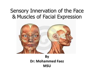
Sensory Innervationof the Face &Muscles of Facial Expression By Dr: Mohammed Faez MSU
Scalp The scalp is the part of the head that extends from the superciliary arches anteriorly to the external occipital protuberance and superior nuchal lines posteriorly.  Laterally it continues inferiorly to the zygomatic arch.
Scalp
Scalp The scalp is a multilayered structure with layers that can be defined by the word itself: S-skin C-connective tissue (dense) A-aponeurotic layer (galeaaponeurotica) L-loose connective tissue P-pericranium
Scalp The first three layers are tightly held together, forming a single unit. This unit is sometimes referred to as the scalp proper and is the tissue torn away during serious 'scalping' injuries.
Scalp
Scalp Skin is thick, hair bearing and contains numerous sebaceous glands. Connective tissue is fibrofatty, the fibrous septa uniting the skin to the underlying aponeurosis of the occipitofrontalis muscle. Numerous arteries and veins are found in this layer. The arteries are branches of the external and internal carotid arteries, and a free anastomosis takes place between them. Aponeurosis (epicranial) is a thin, tendinous sheet that unites the occipital and frontal bellies of the occipitofrontalis muscle. The lateral margins of the aponeurosis are attached to the temporal fascia.
Scalp Loose areolar tissue occupies the subaponeurotic space and loosely connects the epicranialaponeurosis to the periosteum of the skull (the pericranium). The areolar tissue contains a few small arteries, but it also contains some important emissary veins. The emissary veins are valveless and connect the superficial veins of the scalp with the diploic veins of the skull bones and with the intracranial venous sinuses.  Called dangerous layer of scalp-emissary veins open here and carry any infections inside the brain (venous sinus). Bleeding lead to black eye. Pericranium, which is the periosteum covering the outer surface of the skull bones.
Muscle of the Scalp Occipitofrontalis Muscle  It has a frontal belly anteriorly, an occipital belly posteriorly, and an aponeurotic tendon (epicranialaponeurosis) connecting the two.
Occipitofrontalis Muscle
Sensory innervations of the scalp It is from two major sources, cranial nerves or cervical nerves, depending on whether it is anterior or posterior to the ears and the vertex of the head. Anterior to the ears and the vertex Posterior to the ears and the vertex
Sensory innervations of the scalp Anterior to the ears and the vertex Supratrochlear nerve Supraorbital nerve Zygomaticotemporal nerve Auriculotemporal nerve By branches of all four divisions of the trigeminal nerve Posterior to the ears and the vertex Great auricular nerve Lesser occipital nerve Greater occipital nerve Third occipital nerve By branches of all four divisions of the spinal cutaneous nerves (C2 and C3)
Sensory innervations of the scalp
Arterial Supply of the Scalp Arteries supplying the scalp are branches of either the external carotid artery or the ophthalmic artery which is a branch of the internal carotid artery.
Arterial Supply of the Scalp External carotid arteries Occipital arteries Posterior auricular arteries Superficial temporal arteries Internal carotid arteries Supratrochlear arteries Supraorbital arteries
Arterial Supply of the Scalp
Venous Drainage of the Scalp Veins draining the scalp follow a pattern similar to the arteries. Of the deep parts of the scalp  Via emissary veins that communicates with the dural sinuses.
Lymphatic Drainage of the Scalp Lymphatic drainage of the scalp generally follows the pattern of arterial distribution. The lymphatics in the occipital region initially drain to occipital nodes which drain into upper deep cervical nodes.
Lymphatic Drainage of the Scalp Lymphatics from the upper part of the scalp drain in two directions: Posterior to the vertex of the head they drain to mastoid nodes. Anterior to the vertex of the head they drain to pre-auricular and parotid nodes.
Lymphatic Drainage of the Scalp
Face Boundaries Extends superiorly to the hair line, inferiorly to the chin and base of mandible, and on each side to auricle Forehead is common to both scalp and face.
Face Very vascular Due to rich vascularity face blush and blanch. Facial skin is rich in sebaceous gland and sweat gland. Wounds of face bleed profusely but heal rapidly. Sebaceous gland keep the skin oily but also cause acne in adult.
Face Called muscle of facial expression and lie in superficial fascia. Embryologically they develop from mesoderm of 2ndbranchial arch, therefore supplied by facial  nerve. No deep fascia is present in the face.
Bones of the Face The facial skeleton consists of 14 stationary bones and  the mandible.  These 14 bones form the basic shape of the face, and are responsible for providing attachments for muscles that make the jaw move and control facial expressions.
Bones of the Face
Muscles of the Face (Muscles of Facial Expression) The muscles of the face develop from the 2nd pharyngeal arch and are innervated by branches of the facial nerve [VII].  They are in the superficial fascia, with origins from either bone or fascia, and insertions into the skin. these muscles control expressions of the face. They act as sphincters and dilators of the orifices of the face (i.e. the orbits, nose, and mouth).
Muscles of the Face Orbital group Nasal group Oral group Other muscle groups
Muscles of the Face
Orbital group Two muscles are Orbicularisoculi Corrugatorsupercilii
Orbicularisoculi 3 parts- Orbital part(outer) Originate from medial part of medial palpebral ligament and form concentric rings, return to point of origin     Action –closes the lids tightly Palpebralpart(Inner) Originate from lateral part of medial palpebral ligament Insert into lateral palpebralraphe      Action-closes the lids gently Lacrimalpart(Small) Originate from lacrimal fascia& lacrimal bone Insert into upper &lower tarsi      Action-dilate lacrimal sac
Orbicularisoculi
Orbicularisoculi Palpebral part Orbital part
Orbicularisoculi
Corrugatorsupercillii Origin : superciliary arch Insertion: skin of the eyebrow Action: produces vertical wrinkles of the forehead in frowning as an expression of annoyance
Nasal group Three muscles are associated with the nasal group: Nasalis Procerus Depressor septinasi
Nasal group
Compressor naris Origin: Frontal process of the maxilla Insertion: Aponeurosis which crosses the bridge of the nose Action: Compresses the mobile nasal cartilages
Dilator naris Origin : Maxilla bone Insertion: Ala of the nose Action: Widens the nasal aperture (by pulling the alar laterally) in deep inspiration; is also a sign of anger
Procerus Origin: nasal bone and lateral nasal cartilage Insertion: skin between the eyebrows Action:  pulls down the medial end of the eyebrow wrinkles the skin of the nose transversely in 		  frowning
Oral group The muscles in the oral group move the lips and cheek: Orbicularisoris Buccinator Lower group of oral muscles depressor angulioris depressor labiiinferioris Mentalis Upper group of oral muscles  risorius zygomaticus major and zygomaticus minor  levatorlabiisuperioris levatorlabiisuperiorisalaequenasi levatorangulioris
Orbicularisoris Origin: from maxilla above incisor teeth  Insertion: into skin of lip. Action: closes the mouth
Buccinator Upper fibers Origin- from maxilla opposite molar teeth Insertion-upper lip Lower fibers Origin-from mandible opposite molar teeth Insertion-lower lip Middle fibers Origin –from pterigomandibularraphe Insertion-decussate before passing to lips Action- it aids in mastication by prevent accumulation of food in vestibule of mouth. It is used every time air expanding the cheeks is forcefully expelled
Other muscle groups They include: Platysma Auricular (anterior, superior, and posterior auricular muscles)  Occipitofrontalis
Platysma Origin– upper part of pectoral and deltoid fascia Insertion– base of mandible, skin of lower face and lip Action– releases pressure of skin on the subjacent veins, depress mandible, pulls angle of mouth downwards.
Muscles of the Face
Sensory Nerves of the Face The skin of the face is supplied by the trigeminal nerve (V), except for the small area over the angle of the mandible and the parotid gland which is supplied by the great auricular nerve (C2 and 3). The trigeminal nerve (V) divides into three major divisions-the ophthalmic (V1), maxillary (V2), and mandibular (V3) nerves
Sensory Nerves of the Face
Testing the Integrity of the Trigeminal Nerve (temperature & pain)
Motor Nerves of the Face Motor supply: Facial nerve ,[object Object],Temporal Zygomatic Buccal Marginal mandibular Cervical
Applied  Trigeminal neuralgia Maxillary and mandibular nerve are involved Excruciating pain in the region of distribution of these nerve In infranuclear lesions of facial nerve (eg, bell’s palsy)- whole face is paralyzed c/f Affected side is motionless Loss of wrinkles Eye cannot be closed In smiling the mouth is drawn to normal side During mastication food accumulates in vestibule of mouth
bell’s palsy
Facial nerve lesion ( Bell’s palsy )
Read about Arterial supply of the face Venous drainage of the face Lymphatic drainage of the face.
Scalp + face

Mais conteúdo relacionado

Mais procurados (20)

Mandibular nerve
Mandibular nerveMandibular nerve
Mandibular nerve
 
Anterior triangle of the neck
Anterior triangle of the neckAnterior triangle of the neck
Anterior triangle of the neck
 
cervical fascia
cervical fasciacervical fascia
cervical fascia
 
Muscles of facial expression
Muscles of facial expressionMuscles of facial expression
Muscles of facial expression
 
venous supply of head & neck
venous supply of head & neckvenous supply of head & neck
venous supply of head & neck
 
Oral cavity palate - pharynx
Oral cavity   palate - pharynxOral cavity   palate - pharynx
Oral cavity palate - pharynx
 
Submandibular salivary gland dr chithra
Submandibular salivary gland dr chithraSubmandibular salivary gland dr chithra
Submandibular salivary gland dr chithra
 
Maxilla anatomy, development & surgical anatomy
Maxilla  anatomy, development & surgical anatomyMaxilla  anatomy, development & surgical anatomy
Maxilla anatomy, development & surgical anatomy
 
Mandible
MandibleMandible
Mandible
 
Paranasal sinuses
Paranasal sinusesParanasal sinuses
Paranasal sinuses
 
Facial nerve
Facial nerveFacial nerve
Facial nerve
 
FACIAL ARTERY.pptx
FACIAL ARTERY.pptxFACIAL ARTERY.pptx
FACIAL ARTERY.pptx
 
Blood supply of face
Blood supply of faceBlood supply of face
Blood supply of face
 
Trigeminal Nerve Anatomy
Trigeminal Nerve AnatomyTrigeminal Nerve Anatomy
Trigeminal Nerve Anatomy
 
Anatomy of infratemporal region
Anatomy of infratemporal regionAnatomy of infratemporal region
Anatomy of infratemporal region
 
Parotid gland
Parotid gland Parotid gland
Parotid gland
 
Scalp
Scalp Scalp
Scalp
 
DEEP CERVICAL FASCIA.pptx
DEEP CERVICAL FASCIA.pptxDEEP CERVICAL FASCIA.pptx
DEEP CERVICAL FASCIA.pptx
 
Face
FaceFace
Face
 
Alveolar bone
Alveolar boneAlveolar bone
Alveolar bone
 

Destaque

Forensic medicine post mortem artefact
Forensic medicine   post mortem artefactForensic medicine   post mortem artefact
Forensic medicine post mortem artefactMBBS IMS MSU
 
Hema practical 05 hema staining
Hema practical 05 hema stainingHema practical 05 hema staining
Hema practical 05 hema stainingMBBS IMS MSU
 
anatomy - Scalp + face
anatomy - Scalp + faceanatomy - Scalp + face
anatomy - Scalp + faceMBBS IMS MSU
 

Destaque (6)

mbbs ims msu
mbbs ims msumbbs ims msu
mbbs ims msu
 
9781841101866ws
9781841101866ws9781841101866ws
9781841101866ws
 
Forensic medicine post mortem artefact
Forensic medicine   post mortem artefactForensic medicine   post mortem artefact
Forensic medicine post mortem artefact
 
Face Anatomy PowerPoint Templates
Face Anatomy PowerPoint TemplatesFace Anatomy PowerPoint Templates
Face Anatomy PowerPoint Templates
 
Hema practical 05 hema staining
Hema practical 05 hema stainingHema practical 05 hema staining
Hema practical 05 hema staining
 
anatomy - Scalp + face
anatomy - Scalp + faceanatomy - Scalp + face
anatomy - Scalp + face
 

Semelhante a Scalp + face

Scalp, temple and face
Scalp, temple and faceScalp, temple and face
Scalp, temple and faceAreebFatimaPT
 
Muscles of facial expression By dr. vijay Makasana
Muscles of facial expression By dr. vijay MakasanaMuscles of facial expression By dr. vijay Makasana
Muscles of facial expression By dr. vijay MakasanaHardik Rupareliya
 
Scalpface scalp to face muscles and nerve supply
Scalpface scalp to face muscles and nerve supply Scalpface scalp to face muscles and nerve supply
Scalpface scalp to face muscles and nerve supply mehermoinkhan
 
Anatomy(2) The skull and face The temporal region Infratemporal and pterygopa...
Anatomy(2) The skull and face The temporal region Infratemporal and pterygopa...Anatomy(2) The skull and face The temporal region Infratemporal and pterygopa...
Anatomy(2) The skull and face The temporal region Infratemporal and pterygopa...Hamzeh AlBattikhi
 
Muscles of head_&_neck[1]
Muscles of head_&_neck[1]Muscles of head_&_neck[1]
Muscles of head_&_neck[1]spmc bikaner
 
Nerves of the Head and Neck: Intro, CNS, PNS, Cranial Nerves, Trigeminal
Nerves of the Head and Neck: Intro, CNS, PNS, Cranial Nerves, TrigeminalNerves of the Head and Neck: Intro, CNS, PNS, Cranial Nerves, Trigeminal
Nerves of the Head and Neck: Intro, CNS, PNS, Cranial Nerves, TrigeminalHeatherSeghi
 
Osteology of head and face
Osteology of head and faceOsteology of head and face
Osteology of head and facesweetdil
 
Muscles of fasical expression
Muscles of fasical expressionMuscles of fasical expression
Muscles of fasical expressionDr. Nasir Mustafa
 
Head & neck-scalp anatomy for study purpose
Head & neck-scalp anatomy for study purposeHead & neck-scalp anatomy for study purpose
Head & neck-scalp anatomy for study purposeparkviewinternending
 
MUSCLES OF FACIAL EXPRESSION.pptx
MUSCLES OF FACIAL EXPRESSION.pptxMUSCLES OF FACIAL EXPRESSION.pptx
MUSCLES OF FACIAL EXPRESSION.pptxNikhilSuresh47
 

Semelhante a Scalp + face (20)

Scalp, temple and face
Scalp, temple and faceScalp, temple and face
Scalp, temple and face
 
Muscles of facial expression By dr. vijay Makasana
Muscles of facial expression By dr. vijay MakasanaMuscles of facial expression By dr. vijay Makasana
Muscles of facial expression By dr. vijay Makasana
 
Face and Scalp.pdf
Face and Scalp.pdfFace and Scalp.pdf
Face and Scalp.pdf
 
Scalpface scalp to face muscles and nerve supply
Scalpface scalp to face muscles and nerve supply Scalpface scalp to face muscles and nerve supply
Scalpface scalp to face muscles and nerve supply
 
Anatomy(2) The skull and face The temporal region Infratemporal and pterygopa...
Anatomy(2) The skull and face The temporal region Infratemporal and pterygopa...Anatomy(2) The skull and face The temporal region Infratemporal and pterygopa...
Anatomy(2) The skull and face The temporal region Infratemporal and pterygopa...
 
Face
FaceFace
Face
 
Muscles of facial expressions
Muscles of facial expressionsMuscles of facial expressions
Muscles of facial expressions
 
orofacial muscles.pptx
orofacial muscles.pptxorofacial muscles.pptx
orofacial muscles.pptx
 
Eye + ear
Eye + earEye + ear
Eye + ear
 
Face
FaceFace
Face
 
الاسراء 2015
الاسراء 2015الاسراء 2015
الاسراء 2015
 
Muscles of head_&_neck[1]
Muscles of head_&_neck[1]Muscles of head_&_neck[1]
Muscles of head_&_neck[1]
 
Nerves of the Head and Neck: Intro, CNS, PNS, Cranial Nerves, Trigeminal
Nerves of the Head and Neck: Intro, CNS, PNS, Cranial Nerves, TrigeminalNerves of the Head and Neck: Intro, CNS, PNS, Cranial Nerves, Trigeminal
Nerves of the Head and Neck: Intro, CNS, PNS, Cranial Nerves, Trigeminal
 
the scalp by dr. haydar muneer salih
the scalp by dr. haydar muneer salihthe scalp by dr. haydar muneer salih
the scalp by dr. haydar muneer salih
 
Osteology of head and face
Osteology of head and faceOsteology of head and face
Osteology of head and face
 
The face by dr. haydar muneer salih
The face by dr. haydar muneer salihThe face by dr. haydar muneer salih
The face by dr. haydar muneer salih
 
Muscles of fasical expression
Muscles of fasical expressionMuscles of fasical expression
Muscles of fasical expression
 
Head & neck-scalp anatomy for study purpose
Head & neck-scalp anatomy for study purposeHead & neck-scalp anatomy for study purpose
Head & neck-scalp anatomy for study purpose
 
The Scalp.pptx
The Scalp.pptxThe Scalp.pptx
The Scalp.pptx
 
MUSCLES OF FACIAL EXPRESSION.pptx
MUSCLES OF FACIAL EXPRESSION.pptxMUSCLES OF FACIAL EXPRESSION.pptx
MUSCLES OF FACIAL EXPRESSION.pptx
 

Mais de MBBS IMS MSU

Hema practical 03 coagulation
Hema practical 03 coagulationHema practical 03 coagulation
Hema practical 03 coagulationMBBS IMS MSU
 
Hema practical 02 hematology
Hema practical 02 hematologyHema practical 02 hematology
Hema practical 02 hematologyMBBS IMS MSU
 
Pharmacology anticoagulation
Pharmacology   anticoagulationPharmacology   anticoagulation
Pharmacology anticoagulationMBBS IMS MSU
 
Microbiology hiv-yf
Microbiology   hiv-yfMicrobiology   hiv-yf
Microbiology hiv-yfMBBS IMS MSU
 
Forensic medicine firearms and firearm injuries
Forensic medicine   firearms and firearm injuriesForensic medicine   firearms and firearm injuries
Forensic medicine firearms and firearm injuriesMBBS IMS MSU
 
Forensic medicine firearms and firearm injuries
Forensic medicine   firearms and firearm injuriesForensic medicine   firearms and firearm injuries
Forensic medicine firearms and firearm injuriesMBBS IMS MSU
 
Forensic medicine firearms and firearm injuries
Forensic medicine   firearms and firearm injuriesForensic medicine   firearms and firearm injuries
Forensic medicine firearms and firearm injuriesMBBS IMS MSU
 
Forensic medicine changes after death
Forensic medicine   changes after deathForensic medicine   changes after death
Forensic medicine changes after deathMBBS IMS MSU
 
Pharmacology cvs medicine
Pharmacology   cvs medicinePharmacology   cvs medicine
Pharmacology cvs medicineMBBS IMS MSU
 
Pharmacology antiarrhythmias
Pharmacology   antiarrhythmiasPharmacology   antiarrhythmias
Pharmacology antiarrhythmiasMBBS IMS MSU
 
Pharmacology angina
Pharmacology   anginaPharmacology   angina
Pharmacology anginaMBBS IMS MSU
 
Pathology hematology 3
Pathology   hematology 3Pathology   hematology 3
Pathology hematology 3MBBS IMS MSU
 
Forensic medicine the medico-legal autopsy
Forensic medicine   the medico-legal autopsyForensic medicine   the medico-legal autopsy
Forensic medicine the medico-legal autopsyMBBS IMS MSU
 
Pharmacology anemia and its treatment
Pharmacology   anemia and its treatmentPharmacology   anemia and its treatment
Pharmacology anemia and its treatmentMBBS IMS MSU
 
Pharmacology neuromuscular blockers & anemia
Pharmacology   neuromuscular blockers & anemiaPharmacology   neuromuscular blockers & anemia
Pharmacology neuromuscular blockers & anemiaMBBS IMS MSU
 
Pharmacology - Parkinsonism
Pharmacology - ParkinsonismPharmacology - Parkinsonism
Pharmacology - ParkinsonismMBBS IMS MSU
 
Forensic medicine medical negligence
Forensic medicine   medical negligenceForensic medicine   medical negligence
Forensic medicine medical negligenceMBBS IMS MSU
 
Forensic medicine medical negligence 2-bolam principle
Forensic medicine   medical negligence 2-bolam principleForensic medicine   medical negligence 2-bolam principle
Forensic medicine medical negligence 2-bolam principleMBBS IMS MSU
 
Forensic medicine medical negligence
Forensic medicine   medical negligenceForensic medicine   medical negligence
Forensic medicine medical negligenceMBBS IMS MSU
 
Pathology environmental & nutritional disease
Pathology   environmental & nutritional diseasePathology   environmental & nutritional disease
Pathology environmental & nutritional diseaseMBBS IMS MSU
 

Mais de MBBS IMS MSU (20)

Hema practical 03 coagulation
Hema practical 03 coagulationHema practical 03 coagulation
Hema practical 03 coagulation
 
Hema practical 02 hematology
Hema practical 02 hematologyHema practical 02 hematology
Hema practical 02 hematology
 
Pharmacology anticoagulation
Pharmacology   anticoagulationPharmacology   anticoagulation
Pharmacology anticoagulation
 
Microbiology hiv-yf
Microbiology   hiv-yfMicrobiology   hiv-yf
Microbiology hiv-yf
 
Forensic medicine firearms and firearm injuries
Forensic medicine   firearms and firearm injuriesForensic medicine   firearms and firearm injuries
Forensic medicine firearms and firearm injuries
 
Forensic medicine firearms and firearm injuries
Forensic medicine   firearms and firearm injuriesForensic medicine   firearms and firearm injuries
Forensic medicine firearms and firearm injuries
 
Forensic medicine firearms and firearm injuries
Forensic medicine   firearms and firearm injuriesForensic medicine   firearms and firearm injuries
Forensic medicine firearms and firearm injuries
 
Forensic medicine changes after death
Forensic medicine   changes after deathForensic medicine   changes after death
Forensic medicine changes after death
 
Pharmacology cvs medicine
Pharmacology   cvs medicinePharmacology   cvs medicine
Pharmacology cvs medicine
 
Pharmacology antiarrhythmias
Pharmacology   antiarrhythmiasPharmacology   antiarrhythmias
Pharmacology antiarrhythmias
 
Pharmacology angina
Pharmacology   anginaPharmacology   angina
Pharmacology angina
 
Pathology hematology 3
Pathology   hematology 3Pathology   hematology 3
Pathology hematology 3
 
Forensic medicine the medico-legal autopsy
Forensic medicine   the medico-legal autopsyForensic medicine   the medico-legal autopsy
Forensic medicine the medico-legal autopsy
 
Pharmacology anemia and its treatment
Pharmacology   anemia and its treatmentPharmacology   anemia and its treatment
Pharmacology anemia and its treatment
 
Pharmacology neuromuscular blockers & anemia
Pharmacology   neuromuscular blockers & anemiaPharmacology   neuromuscular blockers & anemia
Pharmacology neuromuscular blockers & anemia
 
Pharmacology - Parkinsonism
Pharmacology - ParkinsonismPharmacology - Parkinsonism
Pharmacology - Parkinsonism
 
Forensic medicine medical negligence
Forensic medicine   medical negligenceForensic medicine   medical negligence
Forensic medicine medical negligence
 
Forensic medicine medical negligence 2-bolam principle
Forensic medicine   medical negligence 2-bolam principleForensic medicine   medical negligence 2-bolam principle
Forensic medicine medical negligence 2-bolam principle
 
Forensic medicine medical negligence
Forensic medicine   medical negligenceForensic medicine   medical negligence
Forensic medicine medical negligence
 
Pathology environmental & nutritional disease
Pathology   environmental & nutritional diseasePathology   environmental & nutritional disease
Pathology environmental & nutritional disease
 

Scalp + face

  • 1. Sensory Innervationof the Face &Muscles of Facial Expression By Dr: Mohammed Faez MSU
  • 2. Scalp The scalp is the part of the head that extends from the superciliary arches anteriorly to the external occipital protuberance and superior nuchal lines posteriorly. Laterally it continues inferiorly to the zygomatic arch.
  • 4. Scalp The scalp is a multilayered structure with layers that can be defined by the word itself: S-skin C-connective tissue (dense) A-aponeurotic layer (galeaaponeurotica) L-loose connective tissue P-pericranium
  • 5. Scalp The first three layers are tightly held together, forming a single unit. This unit is sometimes referred to as the scalp proper and is the tissue torn away during serious 'scalping' injuries.
  • 7. Scalp Skin is thick, hair bearing and contains numerous sebaceous glands. Connective tissue is fibrofatty, the fibrous septa uniting the skin to the underlying aponeurosis of the occipitofrontalis muscle. Numerous arteries and veins are found in this layer. The arteries are branches of the external and internal carotid arteries, and a free anastomosis takes place between them. Aponeurosis (epicranial) is a thin, tendinous sheet that unites the occipital and frontal bellies of the occipitofrontalis muscle. The lateral margins of the aponeurosis are attached to the temporal fascia.
  • 8. Scalp Loose areolar tissue occupies the subaponeurotic space and loosely connects the epicranialaponeurosis to the periosteum of the skull (the pericranium). The areolar tissue contains a few small arteries, but it also contains some important emissary veins. The emissary veins are valveless and connect the superficial veins of the scalp with the diploic veins of the skull bones and with the intracranial venous sinuses. Called dangerous layer of scalp-emissary veins open here and carry any infections inside the brain (venous sinus). Bleeding lead to black eye. Pericranium, which is the periosteum covering the outer surface of the skull bones.
  • 9.
  • 10. Muscle of the Scalp Occipitofrontalis Muscle It has a frontal belly anteriorly, an occipital belly posteriorly, and an aponeurotic tendon (epicranialaponeurosis) connecting the two.
  • 12. Sensory innervations of the scalp It is from two major sources, cranial nerves or cervical nerves, depending on whether it is anterior or posterior to the ears and the vertex of the head. Anterior to the ears and the vertex Posterior to the ears and the vertex
  • 13. Sensory innervations of the scalp Anterior to the ears and the vertex Supratrochlear nerve Supraorbital nerve Zygomaticotemporal nerve Auriculotemporal nerve By branches of all four divisions of the trigeminal nerve Posterior to the ears and the vertex Great auricular nerve Lesser occipital nerve Greater occipital nerve Third occipital nerve By branches of all four divisions of the spinal cutaneous nerves (C2 and C3)
  • 15. Arterial Supply of the Scalp Arteries supplying the scalp are branches of either the external carotid artery or the ophthalmic artery which is a branch of the internal carotid artery.
  • 16. Arterial Supply of the Scalp External carotid arteries Occipital arteries Posterior auricular arteries Superficial temporal arteries Internal carotid arteries Supratrochlear arteries Supraorbital arteries
  • 17. Arterial Supply of the Scalp
  • 18. Venous Drainage of the Scalp Veins draining the scalp follow a pattern similar to the arteries. Of the deep parts of the scalp Via emissary veins that communicates with the dural sinuses.
  • 19. Lymphatic Drainage of the Scalp Lymphatic drainage of the scalp generally follows the pattern of arterial distribution. The lymphatics in the occipital region initially drain to occipital nodes which drain into upper deep cervical nodes.
  • 20. Lymphatic Drainage of the Scalp Lymphatics from the upper part of the scalp drain in two directions: Posterior to the vertex of the head they drain to mastoid nodes. Anterior to the vertex of the head they drain to pre-auricular and parotid nodes.
  • 22. Face Boundaries Extends superiorly to the hair line, inferiorly to the chin and base of mandible, and on each side to auricle Forehead is common to both scalp and face.
  • 23. Face Very vascular Due to rich vascularity face blush and blanch. Facial skin is rich in sebaceous gland and sweat gland. Wounds of face bleed profusely but heal rapidly. Sebaceous gland keep the skin oily but also cause acne in adult.
  • 24. Face Called muscle of facial expression and lie in superficial fascia. Embryologically they develop from mesoderm of 2ndbranchial arch, therefore supplied by facial nerve. No deep fascia is present in the face.
  • 25. Bones of the Face The facial skeleton consists of 14 stationary bones and the mandible. These 14 bones form the basic shape of the face, and are responsible for providing attachments for muscles that make the jaw move and control facial expressions.
  • 26. Bones of the Face
  • 27. Muscles of the Face (Muscles of Facial Expression) The muscles of the face develop from the 2nd pharyngeal arch and are innervated by branches of the facial nerve [VII]. They are in the superficial fascia, with origins from either bone or fascia, and insertions into the skin. these muscles control expressions of the face. They act as sphincters and dilators of the orifices of the face (i.e. the orbits, nose, and mouth).
  • 28.
  • 29. Muscles of the Face Orbital group Nasal group Oral group Other muscle groups
  • 31. Orbital group Two muscles are Orbicularisoculi Corrugatorsupercilii
  • 32. Orbicularisoculi 3 parts- Orbital part(outer) Originate from medial part of medial palpebral ligament and form concentric rings, return to point of origin Action –closes the lids tightly Palpebralpart(Inner) Originate from lateral part of medial palpebral ligament Insert into lateral palpebralraphe Action-closes the lids gently Lacrimalpart(Small) Originate from lacrimal fascia& lacrimal bone Insert into upper &lower tarsi Action-dilate lacrimal sac
  • 36. Corrugatorsupercillii Origin : superciliary arch Insertion: skin of the eyebrow Action: produces vertical wrinkles of the forehead in frowning as an expression of annoyance
  • 37. Nasal group Three muscles are associated with the nasal group: Nasalis Procerus Depressor septinasi
  • 39. Compressor naris Origin: Frontal process of the maxilla Insertion: Aponeurosis which crosses the bridge of the nose Action: Compresses the mobile nasal cartilages
  • 40. Dilator naris Origin : Maxilla bone Insertion: Ala of the nose Action: Widens the nasal aperture (by pulling the alar laterally) in deep inspiration; is also a sign of anger
  • 41. Procerus Origin: nasal bone and lateral nasal cartilage Insertion: skin between the eyebrows Action: pulls down the medial end of the eyebrow wrinkles the skin of the nose transversely in frowning
  • 42. Oral group The muscles in the oral group move the lips and cheek: Orbicularisoris Buccinator Lower group of oral muscles depressor angulioris depressor labiiinferioris Mentalis Upper group of oral muscles risorius zygomaticus major and zygomaticus minor levatorlabiisuperioris levatorlabiisuperiorisalaequenasi levatorangulioris
  • 43. Orbicularisoris Origin: from maxilla above incisor teeth Insertion: into skin of lip. Action: closes the mouth
  • 44. Buccinator Upper fibers Origin- from maxilla opposite molar teeth Insertion-upper lip Lower fibers Origin-from mandible opposite molar teeth Insertion-lower lip Middle fibers Origin –from pterigomandibularraphe Insertion-decussate before passing to lips Action- it aids in mastication by prevent accumulation of food in vestibule of mouth. It is used every time air expanding the cheeks is forcefully expelled
  • 45. Other muscle groups They include: Platysma Auricular (anterior, superior, and posterior auricular muscles) Occipitofrontalis
  • 46. Platysma Origin– upper part of pectoral and deltoid fascia Insertion– base of mandible, skin of lower face and lip Action– releases pressure of skin on the subjacent veins, depress mandible, pulls angle of mouth downwards.
  • 48.
  • 49. Sensory Nerves of the Face The skin of the face is supplied by the trigeminal nerve (V), except for the small area over the angle of the mandible and the parotid gland which is supplied by the great auricular nerve (C2 and 3). The trigeminal nerve (V) divides into three major divisions-the ophthalmic (V1), maxillary (V2), and mandibular (V3) nerves
  • 50. Sensory Nerves of the Face
  • 51. Sensory Nerves of the Face
  • 52. Testing the Integrity of the Trigeminal Nerve (temperature & pain)
  • 53.
  • 54.
  • 55. Applied Trigeminal neuralgia Maxillary and mandibular nerve are involved Excruciating pain in the region of distribution of these nerve In infranuclear lesions of facial nerve (eg, bell’s palsy)- whole face is paralyzed c/f Affected side is motionless Loss of wrinkles Eye cannot be closed In smiling the mouth is drawn to normal side During mastication food accumulates in vestibule of mouth
  • 57.
  • 58. Facial nerve lesion ( Bell’s palsy )
  • 59. Read about Arterial supply of the face Venous drainage of the face Lymphatic drainage of the face.