Vascular system
•Transferir como PPT, PDF•
2 gostaram•1,310 visualizações

patho

Recomendados
Mais conteúdo relacionado
Mais procurados
Mais procurados (20)
Destaque
Destaque (20)
Semelhante a Vascular system
Semelhante a Vascular system (20)
Mais de Rawalpindi Medical College
Mais de Rawalpindi Medical College (20)
Vascular system
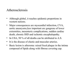
- 1. Atherosclerosis • Although global, it reaches epidemic proportions in western nations. • Major consequences are myocardial infarction, CVA, aortic aneurysms,less important are gangrene of lower extremities, mesenteric complications, sudden cardiac death, chronic IHD and ischemic encephalopathy. • In USA, 50 % of all deaths can be attributed to A.S. • It is the disease of elastic and muscular arteries. • Basic lesion is atheroma: raised focal plaque in the intima composed of lipids along with fibrous covering cap.
- 2. Atherosclerosis • At first the atheromas are sparse, which increase in no with progressive disease and may involve the entire circumference of the vessel encroaching on the lumen as well as tunica media of the vessel. • In the small arteries the plaques are occlusive causing ischemic injury. • In large vessels, the plaques are destructive causing weakening and aneurysms, may also rupture which favours thrombosis and embolism.
- 3. Hyperlipidemia • Patients with familial hyperlipidemia are most prone. • Higher the cholesterol, higher the risk especially LDL cholesterol, hypertriglyceridemia with VLDL. • Treatment with diet and cholesterol lowering drugs significantly decrease cardiovascular mortality. • HDL cholesterol reduce the risk. • High dietary intake of cholesterol raises the plasma levels of cholesterol. • Paradoxically in Finland, the Eskimos, in spite of high fat intake, incidence of IHD is low because of a specific FA in fish and fish oil, that decreases LDL, increases HDL, and.
- 4. • And modifies various mediators favoring platelet aggregates.
- 5. Hypertension • Mechanism of role in atherosclerosis in unknown. Higher the chronic hypertension, higher the risk of IHD and CVA.
- 6. Smoking • One of the established risk factor in the incidence and severity of atherosclerosis.>10-20 cigarettes/day for many years increases risk to >200%. Cessation of smoking reverts the risk. • Diabetes: diabetics have two fold increased risk of dying from MI, CVA and up to 100 times increased tendency of lower extremity gangrene. • Other rare risk factors include elevated homocysteine levels causing endothelial dysfunction, raised levels of plasminogen activator inhibitors and inflammation such as plasma fibrinogen and CRP.
- 7. Morphological Features • The key process is intimal thickening and lipid accumulation. Fatty streaks appear in young children which are the possible precursors of atheromas. • Atheromas are the raised focal plaques which consist of cholesterol and esters surrounded by fibrous cap. • Grossly appear white to yellow, measuring 0.3-1.5 cm, encroach on to lumen, may coalesce to form large lesions. • Superficial part is firm white, deep soft and yellow. • Distribution: Abdominal aorta, thoracic aorta, more often near ostia of major branches; coronary, popliteal, circle of Willis; upper limbs, renal and mesenteric vessels spared. Usually patchy and part of circumference is involved.
- 8. • 3 parts of plaque are smooth muscle cells, macrophages and other inflammatory cells, extracellular matrix e.g. collagen, elastic tissue and proteoglycans, intracellular and extracellular lipid deposits. • Fibrous cap consists of smooth muscle cells, dense connective tissue, cellular area consists of macrophages and muscle cells, T lymphocytes, deep necrotic core contains lipid material, fibrinous material and other plasma proteins, foamy macrophages which may be transformed macrophages and smooth muscle cells. • Around the plaque there is neovascularization. • Non atheromatous intimal thickening in coronary arteries may be due to hemodynamic stress in adults having no significance. • In late stages, atheromas may get converted to fibrous scar.
- 9. Complicated Atheromas • Calcification, vessel may be like a brittle pipe. • Focal rupture and ulceration causing thrombosis and cholesterol emboli. • Hemorrhage in to the atheromas especially in coronaries. • Superimposed thrombosis and organization. • May cause atrophy of underlying media, weakening and aneurysmal dilatation.
- 10. Fatty Streaks • Not raised, no flow disturbances, precursors of atherosclerosis. • Begin as small yellow spots, may coalesce to form 1 cm lesions, composed of foamy macrophages, lymphocytes and minimum amount of lipids. • Start at 1 year age, most of 10 years aged children have aortic lesions. • No relation with sex, geography, race and environment. • Distribution same as atherosclerosis. • Not all the streaks develop the ominous disease. • Types of atheromatous lesions: isolated foam cells, fatty streaks, small extracellular lipid collections, fibrofatty atheroma and complicated lesions.
- 11. Pathogenesis of Atherosclerosis • Its clinical implications are grave consequences has stimulated the scientists to explore and discover the cause. • Hypothesis 1: cellular proliferation in intima as a reaction to insudation of plasma proteins and lipids. Organization and repetitive growth of thrombi result in plaque formation. • Current theory: it is a chronic inflammatory response of the arterial wall initiated by some form of injury to the endothelium. • 1- chronic focal endothelial injury- dysfunction- increased permeability and leukocyte adhesion. • 2- lipoprotein insudation asp LDL, VLDL, their oxidation. • 3- adhesion of monocytes, transformation to foamy macrophages • 4- platelet adhesion • 5- activated platelets and macrophage release factors that cause migration of smooth muscle cells from media.
- 12. • 6- smooth muscle proliferation in intima, elaboration of extracellular matrix leading to accumulation of collagen and proteoglycans. • 7- accumulation of intracellular and extracellular lipids.
- 13. Endothelial Injury • Human lesion develop in intact endothelium, so endothelial dysfunction and activation are more important in causing permeability, leukocyte adhesion and alterations, in expression of a number of endothelial gene products which mediate adhesion of monocyte and lymphocytes. • Endothelial dysfunction may be due to endotoxins, hypoxia, products of cigarette smoking, homocysteine and viruses. • Currently it is postulated that hemodynamic disturbances (ostia and posterior aortic wall, which are adversely affected by shear stress) and adverse effects of hypercholesterolemia are the main factors in endothelial injury.
- 14. Role of Lipids • Increased lipids cause endothelial dysfunction through superoxide and other oxygen free radicals that deactivate NO. • Increased lipids result in lipoproteins accumulation in intima. • Lipid oxidation yields oxidized LDL ingested readily by macrophages to form foam cells which is chemotactic for monocytes and increased their adhesion. It inhibits macrophage mobility favoring retention. It stimulates release of growth factors and cytokines. It is cytotoxic to endothelial cells and smooth muscle cells. It is immunogenic with formation of antibodies to lipoproteins.
- 15. Role of Macrophages • Monocytes adhere to endothelium, proliferate and migrate to subendothelial tissues, change to macrophages, engulf lipoproteins esp. oxidized LDL, transform to foamy macrophages. • Macrophages have secretory and biological functions via IL-1, TNF which cause adhesion of leukocytes and monocytes, chemoattractant proteins, produce toxic oxygen species that cause smooth muscle proliferation, T lymphocytes are also present by unknown mechanism. • As long as hypercholesterolemia persists, monocyte adhesion, subendothelial migration of smooth muscle cells and accumulation of lipids within macrophages and smooth muscle cells continues eventually leading to form fatty streaks. May regress if hypercholesterolemia is ameliorated.
- 16. Role of Smooth Muscle Cell Proliferation • If hypercholesterolemia persists, smooth muscle proliferation and extracellular matrix deposition, in intima continues, which is one of the major factors converting streaks in to atheromas. • Growth factors implicated in vascular smooth muscle cell proliferation are: PDGF (Platelets, macrophages, endothelial cells, smooth muscle cells), FGF and TGF- alpha. • inhibitors are heparin like molecules, TGF-beta.
- 17. Progression of Lesions • Initial intimal plaque: central aggregates of foamy macrophages and foamy smooth muscle cells. • If progression occurs, cellular fatty atheroma is modified by further deposition of collagen and proteoglycans. Some plaques may be fibrous by increased connective tissue. • Many of the atheromas undergo disruption and thrombosis which may be associated with catastrophic consequences.
- 18. Clinical Features and Prevention • May be due to thrombosis, calcification, aneurysmal dilatations, distal ischemic events in heart, brain and lower extremities. • So measures to prevent the toll are both primary and secondary. • Primary are meant to delay the atheroma formation and regression. Smoking, hypertension, weight, exercise, alcohol, lowering of LDL and increasing HDL. • Secondary to prevent recurrences of serious events. Decreased lipids and anti platelet drugs.
- 19. Normal Aorta with no Fatty Streaks
- 20. Fatty Streaks
- 21. Fatty Streaks, Coronary Artery with Increased Fat
- 22. Coronary Artery Atherosclerosis with Thrombosis and Narrowing
- 25. Coronary Atherosclerosis with Thrombosis and Cholesterol Clefts
- 26. Atheroma, Showing Foamy Macrophages and Cholesterol Clefts.
- 29. Aortic Atherosclerosis, Mild, Moderate, Severe
- 30. Atherosclerosis with Hemorrhage and Cholesterol Clefts, Aorta
- 32. Hypertensive Vascular Disease • It affects both structure and function of small muscular arteries and arterioles. • It has devastating consequences and remains asymptomatic for long periods. • One of the major risk factor for heart disease, CVA, cardiac hypertrophy with failure, aortic dissections and renal failure esp. if systolic blood pressure is sustained above 140 mm and diastolic more than 90 mm. • Prevalence is more with increasing age, blacks are affected twice more often, more vulnerable to complications.
- 33. Causes • In 90% primary. • Secondary: Renal. Ac Glomerulonephritis. Ch Renal Disease. Renal Artery Stenosis and Vasculitis. Renin producing Tumours. Endocrine. Cushing Syndrome. Primary Aldosteronism. Exogenous Hormones. Sympathomimetic Drugs. MAO inhibitors. Phaeochromocytoma. Acromegaly.
- 34. Causes • Cardiovascular. • Coarctation of Aorta. • Increased Blood Volume. • Aortic Rigidity. • Neurologic. • Increased Intracranial Pressure. • Acute Stress, Operative Procedures.
- 35. • Accelerated Hypertension: >120 mm diastolic, if untreated, death occurs within 1-2 years. • Complications include: renal failure, retinal hemorrhage, exudates and papilloedema.
- 36. Regulation of Blood Pressure • Cardiac Out Put x Peripheral Resistance. • Cardiac Out Put: Blood volume (Na, Mineralocorticoids, Atrial Natriuretic Peptides, Heart rate, Contractility). • Peripheral Resistance: Humoral constrictors (Angio II, Catecholamines, Thromboxane, Leucotrienes, Endothelin). • Humoral Dilators: Prostaglandins, Kinins, NO/ EDRF. • Peripheral Resistance: Local Autoregulation by pH and Hypoxia; Neural Regulation is by alpha adrenergic drugs.
- 37. Pathogenesis of Hypertension • Changes that alter the relationship between blood volume and resistance. • Mechanism of development: (1) • Genetic factors: Twins, Family History (Mostly Polygenic and heterogeneous disorder). • Environmental: Chinese in China have low BP, Stress, Obesity, Physical Inactivity, increased salt intake. • Mechanism of development: (2) • Renal retention of excess Na, increased volume, increased cardiac output, vasoconstriction to prevent over perfusion of tissues. At increased BP, kidneys cab excrete more Na, called reseting of pressure natriuresis.
- 38. • Second hypothesis suggests increased resistance as primary event; factors may be functional vasoconstriction by increased sensitivity to vasoconstrictors due to genetic defect in transport of Na and Ca, Neurogenic release of vasoconstrictors. • Structural changes in vessel walls causing thickened walls and narrowed lumina.