1. CHAPTER 7. REPRODUCTIVE BIOLOGY
Christina L. Conrath, Virginia Institute of Marine Science, College of William and Mary, PO Box
1346, Gloucester Point, VA 23062 USA
7.1 INTRODUCTION: MODES OF REPRODUCTION
7.1.1 Oviparity
7.1.2 Aplacental viviparity
7.1.2.1 Aplacental yolk sac
7.1.2.2 Oophagy and adelphophagy
7.1.2.3 Placental analogues: histotrophe and trophonemata
7.1.3 Placental viviparity
7.2 BASIC ANATOMY
7.2.1 Male
7.2.2 Female
7.3 MATURITY
7.3.1 Assessing maturity
7.3.1.1 Male
7.3.1.2 Female
7.3.2 Determining the size or age at maturity
7.3.2.1 Male
7.3.2.2 Female
7.3.3 Age at first maturity vs. age at first reproduction
7.4 TIMING OF THE REPRODUCTIVE CYCLE
7.4.1 Mating
7.4.2 Male reproductive cycle
7.4.2.1 Gonad size indices
7.4.2.2 Histological examination of the reproductive tract
7.4.3 Female reproductive cycle
7.4.3.1 Ovulation cycle
7.4.3.2 Gestation cycle and time of birth
7.4.3.3 Reproductive interval
7.4.3.4 Reproductive cycle examples and embryonic diapause
7.5 FECUNDITY
7.6 SPERM STORAGE IN FEMALE ELASMOBRANCHS AND OVIDUCAL
GLAND STRUCTURE
7.7 ADDITIONAL RESOURCES
7.7.1 Literature
7.7.2 Web-based resources
7.7.3 Field data collection
7.8 REFERENCES
133
3. 7.1 INTRODUCTION: MODES OF REPRODUCTION
Several reproductive specializations are found within the elasmobranchs. All elasmobranchs
utilize internal fertilization and produce a relatively small number of large eggs. Elasmobranch fecun-
dity generally ranges from one to two offspring produced per year up to a possible maximum of 300 in
the whale shark (Compagno, 1990; Joung et al., 1996). Elasmobranch reproductive strategies include
oviparity, aplacental viviparity, and placental viviparity (Wourms, 1977). Oviparous species enclose
eggs in an egg case and deposit them into the environment, where embryos will develop external to the
body of the mother. Embryos remain in the egg case to develop for a period of time ranging from less
than two months to over one year (Compagno, 1990). Viviparous species retain eggs within the uteri
where the embryos will develop. The yolk sac of placental viviparous species interdigitates with the
uterine wall to form a placenta in which nutrients from the mother are transferred to the embryo
directly. In most species the egg envelope is retained and incorporated into the uteroplacental complex
(Hamlett et al., 1985). Gestation for viviparous species ranges from less than six months to greater
than two years (Compagno, 1990). Viviparous species may have either lecithotrophic or matrotrophic
development. Lecithotrophic development occurs when embryos derive their nutrition solely from yolk
reserves and occurs in many aplacental viviparous species. Matrotrophic development occurs when
embryos supplement the yolk reserves by obtaining maternally derived nutrients during gestation and
also occurs in many aplacental species and all placental viviparous species (Wourms and Lombardi,
1992). The advantage of matrotrophy may be the increase in juvenile size at birth and therefore
increased survivorship of young. Another important consideration in the evolution of elasmobranch
reproductive strategies is the presence or absence of uterine compartments. Uterine compartments
are formed in all species with placental development and some species with aplacental development
and are proposed to be an important step in the evolution of placental viviparity (Otake, 1990).
7.1.1 Oviparity
Oviparity occurs in all batoids of the family Rajidae and six families and over 100 species of
sharks in the orders Heterodontiformes, Orectolobiformes and Carcharhiniformes (Compagno, 1990;
Compagno, 2001). In oviparous species, eggs are enclosed within an egg case and deposited in the
environment. Two types of oviparity occur, extended oviparity and retained oviparity. Almost all
oviparous species have extended oviparity in which large egg cases are fertilized, enclosed in an egg
case, deposited and, after a period of up to 15 months, hatch out. In this reproductive mode almost all
of the embryonic development occurs within the egg case outside of the mother’s body. Retained
oviparity occurs much more rarely and refers to species in which cased eggs are retained in the
oviduct and development proceeds for a longer period before the eggs are released into the environ-
ment. One form of retained oviparity occurs in some scyliorhinid catsharks when multiple egg cases
are retained within the oviduct before being released (Compagno, 1988; Compagno, 1990).
135
4. The egg case generally has tendrils and sticky filaments that aide in attaching the egg to some
sort of structure or substrate where the eggs incubate. The egg case also hardens after being depos-
ited to protect the embryos from predation. All oviparous chondrichthyan eggs are laid in pairs
(Mellinger, 1983). Oviparous embryos tend to be relatively smaller than viviparous embryos as growth
of the embryo is constrained by the amount of yolk initially present in the yolk sac (Hamlett, 1997).
Compagno (1990) suggests egg-laying elasmobranchs may select appropriate substrates for egg
deposition as occurs in the bullhead shark in which the female picks up the egg after it is laid and
wedges it into rocks or marine vegetation. Development time within the egg case is likely dependent
on external temperatures. Differences in the length of the incubation period of eggs laid by female
thornback rays, Raja clavata, held at different temperatures have been noted in at least two aquarium
experiments (Clark, 1922; Ellis and Shackley, 1995).
7.1.2 Aplacental viviparity
Embryos from species with aplacental viviparous development are retained within the mother
for the duration of development, but no placental connection is formed between the mother and the
embryo. A wide range of developmental forms occur within this reproductive mode and Wourms
(1977) separated them into three groups: those dependent entirely on yolk reserves, those which feed
on other eggs or embryos, and those which possess placental analogues. Animals within the first group
are considered lecithotrophic as the embryo receives no extra nutrition from the mother, and animals in
the last two groups are considered matrotrophic as the embryo’s nutrition is supplemented with either
ovulated eggs or uterine milk (histotrophe).
7.1.2.1 Aplacental yolk sac
Embryos from species within this group are entirely dependent on yolk reserves to complete
development. This type of development is the most common reproductive strategy employed by
sharks. It occurs in the orders Hexanchiformes, Squaliformes, Pristiophoriformes, Squatiniformes,
Rhinobatiformes, Pristiformes, Torpediniformes and some species in the orders Orectolobiformes and
Carcharhiniformes (Compagno, 1990). This form of development offers protection from predators for
a longer period of time than oviparous development (Hamlett, 1997).
7.1.2.2 Oophagy and adelphophagy
Oophagy occurs when embryos within the uterus hatch out of the egg capsule after a few
months and then consume additional eggs that continue to be ovulated while the embryo develops.
Oophagy is thought to occur in all sharks in the order Lamniformes (Compagno, 1990; Gilmore, 1993),
specific examples described within the literature include the bigeye thresher shark, Alopias
superciliosus, the pelagic thresher shark, A. pelagicus, the shortfin mako shark, Isurus oxyrinchus
and the porbeagle shark, Lamna nasus (Moreno and Moron, 1992; Francis and Stevens, 1999; Liu et
136
5. al., 1999; Mollet et al., 2000). In the sand tiger shark, Carcharias taurus, the first embryo to develop
in each uterus consumes all the other embryos within that uterus (adelphophagy or intrauterine
cannabilism), as well as additional ovulated eggs (Gilmore et al., 1983). This type of development may
facilitate the development of very large embryos and may prepare the embryo for a predatory life style
(Wourms, 1977). Yano (1992) found that embryos of the false catshark, Pseudotriakis microdon, also
ingest yolk material from other ova but that they transfer ingested yolk to an external yolk sac rather
than forming the extended stomach of lamniform oophagous embryos. Female slender smooth-hounds,
Gollum attenuatus, form egg capsules which contain 30-80 ova, and only one ovum within each egg
capsule develops with all other ova ingested and packed to an external yolk sac (Yano, 1993). While
ova are ingested by the slender smooth-hound embryo during development, this form of reproduction
may or may not be considered oophagy as after the initial consumption of ova within the egg sac, the
embryo then develops without any additional ova or maternal investment.
7.1.2.3 Placental analogues: histotrophe and trophonemata
This type of development occurs in all rays of the order Myliobatiformes. Trophonemata are
long villous extensions of the uterine epithelium that secrete histotrophe or “uterine milk” which can be
ingested or absorbed by the embryo. The quantity and composition of the histotrophe varies widely
between species. Trophonemata envelope the embryo and may occasionally enter the embryo through
the spiracles. As yolk reserves are depleted, trophonemata increase in size and release uterine secre-
tions rich in proteins and lipids (histotrophe) (Wourms, 1981). White et al. (2001) found that trophone-
mata of the stingaree, Urolophus lobatus, increase in length and enter the gill, spiracles and mouths
of developing embryos in the uterus about six months after ovulation when yolk reserves from the
external yolk sac have been utilized. Trophonemata are also formed in the Atlantic stingray, Dasyatis
sabina, and increase in length in the late stages of gestation while the developing young are bathed in
histotrophe (Snelson et al., 1988). The transfer of nutrients has been found to be much more efficient
in species with trophonemata than in species with a yolk sac placenta (Wourms, 1981).
7.1.3 Placental viviparity
Placental viviparity occurs when during the course of embryonic development after an initial
period of reliance on yolk from a yolk sac, the yolk sac attaches to the uterine wall and forms a yolk
sac placenta and the associated yolk stalk forms the umbilical cord. In most species the egg envelope
is retained and incorporated into the uteroplacental complex (Hamlett et al., 1985). Thirty percent of
viviparous sharks form a yolk sac placenta (Hamlett, 1997). This type of reproductive development
only occurs in sharks of the order Carcharhinformes (Compagno, 1990), and can occur within the
same family or genus as aplacental viviparous species. The genus Mustelus includes several aplacen-
tal viviparous species such as the spotted estuary smooth-hound, M. lenticulatus, the gummy shark,
M. antarcticus, and the starspotted smooth-hound, M. manazo, and several placental viviparous
137
6. species such as the dusky smooth-hound, M. canis, and the spotless smooth-hound, M. griseus
(Francis and Mace, 1980; Teshima, 1981; Lenanton et al., 1990; Conrath and Musick, 2002). Wourms
and Lombardi (1992) estimate the yolk sac placenta has evolved independently 11-20 times within the
elasmobranchs. This has led to a large diversity in placental structure. After ovulation placental species
undergo a period of dependency on yolk reserves that may last for several weeks to months before the
placenta is formed. Teshima (1981) divides the placental species into two groups, those in which it
forms in mid-gestation and those in which it forms soon after ovulation.
7.2 BASIC ANATOMY
7.2.1 Male
The male reproductive system is composed of the testes, genital ducts (ductus efferens,
epididymis, ductus deferens and seminal vesicle), accessory glands and secondary sex organs (Figure
7.01). Male reproductive organs and tissues have been described and defined using various terminolo-
gies, and this account will follow the terminology of Hamlett (1999). The testes are paired structures
supported by a mesorchium and in some species enveloped by the epigonal organ. A pre-germinal fold
runs the length of the testis and is the origin of the spermatogenesis process. The testes are the
location of spermatogenesis and also play a role in creating and secreting steroid hormones. Pratt
(1988) identified three types of testes in elasmobranchs: radial, diametric, and compound, defined by
their pattern of seminiferous follicle origin and propagation. The epididymis is connected to the testis
via the ductus efferens, which are fine tubules which cross the mesorchium at the anterior edge of the
testis. Mature sperm are discharged from the testis through the ductus efferens (Wourms, 1977). The
efferent ducts join the epididymis, which expands to form a long tube with complex convolutions. The
epididymis is continuous with the next section of the genital duct, the ductus deferens also known as
the vas deferens or Wolffian duct. The ductus deferens is continuous with the seminal vesicle or
ampulla ductus deferens. The ductus deferens and seminal vesicle function as storage areas for
seminal products, and in some species sperm is packaged into either spermatozeugmata or spermato-
phores here (Wourms, 1977). The ureter becomes entwined with the terminal portion of the seminal
vesicle, and both end in the anterior wall of the urogenital sinus. The urogenital sinus vents into a
common cloaca by means of a single large papilla. Two accessory glands are present, Leydig glands
and the alkaline gland. Leydig glands are a series of branched tubular glands that secrete seminal
fluids into the epididymis and ductus deferens. The alkaline gland of batoids may be involved in sperm
protection (Hamlett, 1999).
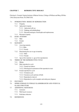
The secondary sex organs include the claspers and the associated siphon sacs. Claspers are
modified regions of the pelvic fin that act as copulatory organs to transfer sperm and seminal matrix
from the male to the female (Figure 7.02). All elasmobranchs have internal fertilization and possess
138
7. claspers, but clasper structure varies widely. All claspers have a dorsal longitudinal groove through
which semen passes to the female during mating. The clasper consists of two intermediate elements—
known as the joint and beta cartilages that extend down from the metapterygium of the pelvic fin, the
main stem cartilage to which two marginal cartilages are fused, and four terminal cartilages, the claw,
rhipidion, the distal basal and the spur. The two marginal cartilages help to form the clasper groove
with a terminal end opening, the hypopyle, and an anterodorsal opening, the apopyle (Compagno,
1988). A good diagram of clasper skeletal structure can be found in Compagno (2001). Most male
elasmobranchs possess siphon sacs which are subcutaneous muscular, epithelium-lined bladders
situated on each side of the midline between the skin and belly
musculature. Each sac ends blindly anteriorly and opens into the
clasper groove posteriorly through the apopyle (Gilbert and Heath,
1972). Gilbert and Heath (1972) examined the structure and function
of the siphon sacs in piked dogfish, Squalus acanthias, and dusky
testis
smooth-hounds, Mustelus canis, and determined that the siphon
sacs’ function is to hold seawater, which is used to wash sperm from
the clasper groove into the oviduct of the female.
ep
dd
Figure 7.02 Male and female little skates, Leucoraja erinacea,
female on the left, male on the right.
sv
7.2.2 Female
The female reproductive system is composed of either a
paired or single ovary and oviducts, which are differentiated into an
Figure 7.01 The male
reproductive tract of a ostium, the anterior oviduct, the oviducal gland, the isthmus, a dilated
spiny dogfish, Squalus
acanthias, ep = epididymis, terminal region/uterus, a cervix and the urogenital sinus (Hamlett and
dd = ductus deferens, and Koob, 1999) (Figure 7.03). The ovary and the oviducts are in close
sv = seminal vesicle.
139
8. association but are not continuous. The female reproductive tract begins as paired ovaries and ovi-
ducts, but in many adults the reproductive tract becomes asymmetrical as the animal develops. In
many viviparous sharks species only the right ovary develops fully, and in many ray species the right
ovary and oviduct are reduced to varying degrees. The ovaries are attached to the body wall by a
mesovarium (Wourms, 1977). Pratt (1988) described two types of ovaries: one found in lamniforms in
which the ovary was hollow and contained within the epigonal organ, the other found in other elasmo-
branch species in which the ovary was external and borne on the flat surface of the epigonal organ or
suspended directly from the mesovarium. The ovary functions in the generation of germ cells, the
acquisition and accumulation of yolk and the biosynthesis and secretion of hormones. The ovary
consists of oocytes, developing follicles and embedded loose connective tissue stroma. The epigonal
organ is present in most species and supports the ovary or ovaries. The ostium is the anterior funnel-
shaped opening of the oviduct which functions to collect the ovulated eggs. The oviducal gland is a
specialized portion of the oviduct where egg capsule and egg jelly formation occur and where fertiliza-
tion may take place although it may occur in the upper oviduct (Hamlett
et al., 1998). (The oviducal gland is described more completely in
section 7.6.) An isthmus may occur before the widening of the oviduct
into a posterior oviducal section or a uterus and may function to isolate
the contents of the uterus. The uterus in oviparous species is special-
ized for egg capsule formation and provides structural modifications for
movement of the capsule through the uterine lumen (Koob and
Hamlett, 1998). In viviparous species the uterus is highly developed and
modified for retention of eggs and the developing embryos. The cervix
occurs at the junction of the uterus and urogenital sinus and is a con-
striction in this area. The uteri independently join the urogenital sinus
(Hamlett and Koob, 1999).
7.3 MATURITY
7.3.1 Assessing maturity
The meaning of the term maturity in recent elasmobranch
Figure 7.03 The female
reproductive tract dis- literature ranges from defining the onset of maturation to the period of
sected out of a spiny time when a female elasmobranch undergoes parturition and produces
dogfish, Squalus
acanthias, og = oviducal a litter of pups. Since in many elasmobranch species the period be-
gland, ova = ovary, tween the beginning of the maturation process until pupping can last a
i = isthmus, e = uterine
embryo, ys = embryonic period of years, it is important to specifically define the term maturity in
yolk sac, ut = uterus, and an elasmobranch reproductive study. For the purposes of this manual a
c = cervix.
140
9. mature animal is defined as one that is immediately capable of mating and producing viable offspring
or one that has already done so. Therefore, in order to be considered mature, an animal must have
previously mated or possess fully developed gametes and all of the secondary structures necessary for
successful mating and fertilization. For female elasmobranchs that have not previously mated this
requires the presence of fully developed ova in the ovaries that are ready to be ovulated. For male
elasmobranchs that have not previously mated this requires not only the presence of mature sperm
within the reproductive tract but also the presence of fully developed claspers and siphon sacs. Matu-
rity in sharks is determined by either observation of the reproductive tract organs or secondary sex
structures or by noting the presence or absence of reproductive products within the reproductive tract.
7.3.1.1 Male
In order for male elasmobranchs to successfully mate they must have fully developed and
functional claspers, and they must have mature sperm ready to be transported by the claspers into the
female. Therefore male maturity can be assessed by determining if the claspers are calcified and if
sperm products are found within the seminal vesicles of the reproductive tract. Clark and von Schmidt
(1965) considered males mature when the clasper head (rhipidion) could be spread open, the clasper
proximal to the head was rigid due to calcification of the supporting cartilage, the base of the clasper
rotated easily, and when the siphon sacs were fully elongated. Clasper calcification can be a simple
and quick way to determine if male elasmobranchs are mature; however, maturity assessments based
on calcification alone may be inaccurate as claspers may have developed before spermatogenesis is
complete. Pratt (1979), in a reproductive study on blue sharks, Prionace glauca, stated that many
sharks with claspers that appeared mature lacked sperm aggregations and had small ductus deferentia
and were therefore still immature.
Histological evidence or direct observation will confirm the presence of sperm within the
reproductive tract. Sperm products can be located and viewed by cutting a cross section of the
reproductive tract, or smears of the reproductive tract can be taken, stained, and viewed under a
ut
microscope to determine if viable sperm or sperm products are present. Pratt (1979) found the most
accurate way to determine maturity in male blue sharks was to note the presence or absence of sperm
in the ampulla ductus deferens (seminal vesicle). He found a field test to look for the presence of
mature sperm aggregates could be done by cross-sectioning the thickest part of the kidney of the male
blue shark. When the cross-section was made four ducts were visible; the largest two were the
seminal vesicles, and the presence or absence of spermatophores in a white supportive tissue could be
observed with the aid of a magnifying glass. This technique was used to assess the male maturity of
smalleye hammerheads, Sphyrna tudes, and Pacific angelsharks, Squatina californica (Natanson
and Cailliet, 1986; Castro, 1989). Pratt and Tanaka (1994) stated that mature male elasmobranchs in a
141
10. resting stage may not possess sperm within the ampullae of the reproductive tract but that the size and
the shape of the ampullae should be a good indicator of maturity as mature males will have large
ampullae. Assessments based on the presence of sperm in the reproductive tract alone may also be
somewhat inaccurate as sperm may be present within the reproductive tract before the claspers are
fully functional. Clark and von Schmidt (1965) found that individuals of at least two species (the
blacktip shark, Carcharhinus limbatus, and the tiger shark, Galeocerdo cuvier) possessed mature
sperm that were produced and present in the seminal vesicles before the claspers and siphon sacs
were fully developed. The best approach to determining maturity in male elasmobranchs should
therefore combine an examination of clasper calcification and development with a simple field or
laboratory test to determine if sperm are present within the seminal vesicles of the reproductive tract
or to determine if the seminal vesicles are enlarged indicating a previous mating event.
7.3.1.2 Female
Female elasmobranchs are considered mature if there is evidence of a current or previous
pregnancy or evidence that they will be ready to reproduce within a short period of time. For females
that are not or have not been pregnant previously, maturity can be determined by assessing the condi-
tion of the ova in the ovary and the size of the oviduct. Mature females will have well developed yolky
eggs in the ovary, and the oviduct may start to expand and detach from the body wall. Females that
have previously been pregnant will have an expanded oviduct containing expanded oviducal glands and
well developed uteri. Female maturity can therefore be determined by assessing the condition of the
reproductive tract and noting the presence or absence of well developed ova in the ovary, eggs or
embryos within the reproductive tract, or expanded oviducts. Bass et al. (1973) defined female sharks
with distinct ova in the ovary and an expanded uteri to be mature. In doubtful cases the presence or
absence of an intact hymen was used to show if the female was still an adolescent or was in between
pregnancies. The hymen is a circular transverse fold that separates the vagina from the cloaca; in
virgin elasmobranchs the vagina is sealed by a membrane which is an extension of the hymen (Pratt,
1979). The condition of the reproductive tract has been used to determine maturity in female sandbar
sharks, Carcharhinus plumbeus, and piked dogfish, Squalus acanthias (Springer, 1960; Jones and
Geen, 1977).
In many studies an intermediate maturing stage is identified. During this stage the oviduct
begins to expand or the ova within the ovary begin to undergo vitellogenesis. Jones and Geen (1977) in
their reproductive study of piked dogfish, Squalus acanthias, defined a maturing phase and plotted the
proportion of animals in this stage versus length to determine the size at the onset of maturity.
Natanson and Cailliet (1986) also define three stages of maturity for the Pacific angelshark, Squatina
californica—immature, maturing and mature—based on the condition of ova in the ovary and the
142
11. condition of the oviduct. Also, in many studies mature females are classified according to what stage
of the reproductive cycle they are currently undergoing. Jones and Geen (1977) defined three stages
of mature females: those between pregnancies, those with candles within the uteri, and those with free
embryos within the uteri. Determining maturity in female elasmobranchs is largely dependent on
observation, and, therefore, assessing maturity will be most accurate when a large enough number of
immature, maturing and mature animals can be observed.
7.3.2 Determining the size or age at maturity
Size or age at maturity is usually determined by either analyzing the growth of reproductive
organs relative to size or age or by quantifying the proportion of mature animals at each length or age
group and determining the length at which 50% of a class is mature.
7.3.2.1 Male
Clasper length measurements have been used in many studies to estimate the size at maturity
because there is a known correlation between the development of secondary sex characters and the
reproductive organs, and maturity. Clasper length is most commonly measured from the posterior
margin of the anus to the tip of the clasper (clasper inner length) or from the base of the pelvic fin to
the tip of the clasper (clasper outer length) (Compagno, 1984). The length of the clasper as a propor-
tion of the precaudal, fork or total length is plotted against the corresponding length. This usually
results in a plot that shows a sharp increase in the slope for a range of lengths before leveling off. This
portion of the plot with the steeper slope corresponds to the range of lengths at which the shark is
becoming mature (Figure 7.04). While the most common reproductive measurement to plot against
length is the clasper length in male elasmobranchs, the size or weight of other reproductive structures
like the testis and siphon sac are often used and plotted in the same manner (Parsons, 1981; Teshima,
1981; Yano, 1993).
The other method commonly used to determine the size at maturity for male elasmobranchs is
to use a maturity ogive. Using this method, the reproductive condition of a large enough sample size of
males of different sizes is first determined as in section 7.3.1.1. Then the proportion of mature animals
found in each length group is determined. These data can then be fitted with a logistic regression and
the length at the point of the curve corresponding to 50% is often used as an indicator of the size at
which these animals mature. The logistic equation can take the following form: proportion mature at a
specific length or age = 1/(1+ea+(b*length or age)), where a and b are coefficients estimated by fitting the
data to the logistic curve. This equation can then be solved to determine at what length or age 50% of
the population is mature (Conrath and Musick, 2002) (Figure 7.05).
143
12. Figure 7.04 The relationship
between clasper length (as % total
length) and total length of M.
canis.
Proportion Mature
Total Length (cm)
a
Proportion Mature
Figure 7.05 a - Maturity
ogives for total length (TL)
of male and female M.
canis, b - maturity ogives for
Age (years)
b age of male and female
M. canis.
144
13. 7.3.2.2 Female
The size of oviducal gland or other structures of the female reproductive tract is often used to
assess the size at which animals become mature. The size or weight of the ovary, or the size of the
oviducal gland, uterus, or other reproductive structure is often plotted against the length of the animal
to determine if there is a size range at which the structure in question begins to develop very quickly
before growth tapers off again. Similar to the clasper length versus total length plot discussed above
the length range at which an elasmobranch population matures is determined by a change in the slope
of the plot (Wass, 1973; Parsons, 1981; Castro et al., 1988; Yano, 1993).
As with male elasmobranchs, the other method used to determine length at maturity in female
elasmobranchs is to create a maturity ogive. The same procedures and equations as explained above
in section 7.3.1.2. are used for females (Figure 7.05) (Conrath and Musick, 2002).
7.3.3 Age at maturity vs. age at first reproduction
It is important to distinguish the difference between the age or length at first maturity with the
age or length of first reproduction. This distinction becomes very important in demographic models.
The time a fish matures is generally understood as the size or age of first mating, and this needs to be
distinguished from the size or age at which the animal actually produces pups. If the species being
considered is oviparous or has a very short gestation time there may be no discrepancy or only a slight
one. However, if the animal has a gestation time of several months or longer, the delay must be
accounted for in the model, and the fecundity term should not be included at the size or age when the
animal is first mature but at the age or size when the animal actually undergoes parturition. Dusky
smooth-hound, Mustelus canis, females mature at four to five years of age, and therefore in a age-
based model a fecundity term would not be added until age five to six as gestation lasts nearly one
year.
7.4 TIMING OF THE REPRODUCTIVE CYCLE
Wourms (1977) defined three types of reproductive cycles exhibited by elasmobranchs:
reproduction continuously throughout the year, a prolonged annual cycle that is not well defined with
one or two peaks in activity, or a well defined annual or biennial cycle. Chen et al. (1996) found
encapsulated fertilized eggs in the uteri of female blacktip sawtail catsharks, Galeus sauteri, all year
round indicating this species reproduces throughout the year without a well-defined breeding season.
This may be characteristic of some deep sea elasmobranch species as this also occurs in the deep sea
black dogfish, Centroscyllium fabricii (Yano, 1995). The small-spotted catshark, Scyliorhinus
canicula, is proposed to have a very extended breeding season, but peak reproductive activity occurs
during the winter and spring months (Sumpter and Dodd, 1979). Dusky smooth-hounds, Mustelus
canis, have a very well-defined annual reproductive season with an 11 to 12 month gestation followed
145
14. by ovulation of the next year class of eggs within a period of days to weeks (Conrath and Musick,
2002).
7.4.1 Mating
Determining what time of year a species mates can be quite difficult and is often inferred by
assuming mating occurs sometime between parturition of one year class of embryos and ovulation of
the next year class of eggs to be fertilized. The timing of mating can also be inferred from viewing
female specimens with mating scars. Pratt (1979) found that the skin of mature female blue sharks
was twice as thick as that of male blue sharks to accommodate the biting that occurs during mating.
Pratt and Carrier (2001) found that biting by males during mating seems universal among elasmo-
branchs, and therefore during the mating season female elasmobranchs frequently bear mating marks
on their bodies with the most common being tooth cuts and abrasions on the pectoral fins. They further
found that in some elasmobranch species there is a sexual dimorphism of the teeth with males having
teeth designed to make courtship biting more effective. Tricas and LeFeuvre (1985) proposed that
biting in the whitetip reef shark, Triaenodon obesus, functions as a precopulatory releasing mecha-
nism for females and to maintain contact during copulation. The timing of the reproductive cycle
before and after mating is generally considered separately for male and female elasmobranchs.
7.4.2 Male reproductive cycle
The timing of the reproductive cycle of male elasmobranchs is generally determined by using
various gonad size indices, through histological examination of the testes, or by noting the presence and
amount of sperm products in the reproductive tract throughout the year. Since the contribution of the
male to the reproductive effort in elasmobranchs primarily ends with mating and inseminating the
female, the following two sections of the chapter are primarily concerned with the timing of mating.
The next two sections are not included in the previous mating section as most of the techniques listed
track the reproductive condition of the male throughout the year or reproductive cycle.
7.4.2.1 Gonad size indices
A gonadosomatic index (GSI)—or some other relationship between the size of the male
reproductive organs and the total size of the animal—is often used to determine when sperm and
sperm products are being produced. The GSI is the testis weight expressed as a percentage of the
total body weight, GSI = (testis weight/total body weight) * 100. By comparing the GSI from mature
males caught during various times of the year, a mating season can be estimated by assuming mating is
occurring when the GSI reaches its highest value. This will correlate to the time of year when sperm
production has reached its highest level. Peak GSI values may not always coincide exactly with mating
season, as sperm products must move down the reproductive tract before mating can occur and sperm
may be stored in the reproductive tract for a period of time. Simpfendorfer (1992) found that the peak
GSI for male Australian sharpnose sharks, Rhizoprionodon taylori, occurred approximately a month
146
15. before the mating season. While a GSI can give valuable information about the timing of sperm
production in male elasmobranchs, caution should be used when trying to use this data as an approxi-
mation of when mating season begins. GSI data is best used combined with other supporting data, like
examinations of sperm presence and quantity in the lower portions of the reproductive tract or other
evidence of mating activity like the presence of sperm products in the female or the presence of
courtship wounds on the female. Stevens and Wiley (1986) defined the mating season by determining
the monthly GSI of two carcharhinid shark species and also examined the quantity of sperm in the
seminal vesicles, mating scars of females captured during the appropriate time of year and the mean
maximum ova diameter of the females.
7.4.2.2 Histological examination of the reproductive tract
A more detailed way to track the formation of sperm in the testis through time is by making
histological sections of the testis. The functional unit of the testis is defined by Callard (1991) as, “the
germ cell clone plus associated Sertoli cells within a closed spherical unit bounded by a basement
membrane.” Parsons and Grier (1992) name this unit the spermatocyst and define the sequence of
development from the germinal zone to the degenerate zone which includes zones of spermatocysts in
various stages of development. Parsons and Grier (1992) define seven stages of development as do
Maruska et al. (1996) also. While these two papers differ slightly in the definition of stages, both track
the spermatogenesis process from loosely organized germ cell, to spermatogonia, spermocytes,
spermatids, and mature spermatozoa.
In order to use this technique a section is removed from the middle of the testis and preserved
in either 10% formalin or Bouin’s solution. The section is processed using standard histological tech-
niques and stained with hematoxylin and eosin. The section is rinsed in a series of water washes,
placed in a tissue cassette, and the Bouin’s fixed tissues are rinsed with a solution of 50% ethanol
(ETOH) saturated with lithium carbonate to remove soluble picrates, then rinsed in 70% ETOH. The
cassettes are then placed in a tissue processor to dehydrate them and infiltrate them with paraffin. A
rotary microtome is used to cut 5 µm thick sections of the tissue, which are then stained with hema-
toxylin and eosin and cover-slipped with a synthetic mounting media. The testis section is then viewed
under a compound microscope and the proportion of the testis occupied by each different stage is
measured along a straight-line distance across the cross section of the testis, starting from the germinal
zone. Or the number of spermotocysts occupied by each stage can be counted across the straight-line
distance and compared for various times of the year. The mean proportion of the testis occupied by
each stage or the mean number of spermatocysts in each stage throughout different months of the
year can then be compared to determine if there is a recognizable seasonal pattern in testis develop-
ment. Parsons and Grier (1992) suggest using caution with this technique as the peak testicular
development may not coincide with the mating season.
147
16. Mating season has been determined for the male piked dogfish, Squalus acanthias, (Jones
and Geen, 1977) and two smooth-hounds, Mustelus griseus and Mustelus manazo (Teshima, 1981)
by examining what percent of the ampullae contain each defined spermatogenic stage throughout the
year. The timing and duration of spermatogenesis was determined for the Port Jackson shark,
Heterodontus portusjacksoni, by examining the migration of the degenerative zone throughout the
year (Jones and Jones, 1982).
In the dusky smooth-hound, Mustelus canis, the mean proportion of the testis occupied by the
seven stages defined by Maruska et al. (1996) was measured and compared for different months of
the year to determine if there was a recognizable seasonal pattern in testis development. A cross
section of the testis is shown in Figure 7.06, and the stages of the sperm development, modeled after
Maruska et al. (1996), are shown in Figure 7.07. Stage one consists of spermatogonia and loosely
organized germ cells not yet bound by a basement membrane into a spermatocyst. During stage two a
layer of spermatogonia and associated Sertoli cells divide and surround a central lumen and are
bounded by a basement membrane forming the spermatocyst. In stage three the spermatogonia
undergo mitosis to become primary spermatocytes, which will then undergo the first meiotic division to
become secondary spermatocytes. In stage four the secondary spermatocytes have undergone the
second meiotic division to become spermatids. Stage five consists of immature sperm, which are
spermatids that have undergone spermiogenesis and possess a head and tail region, but individual
sperm have not organized into bundles yet. During stage six these spermatozoa organize into tightly
shaped packets arranged spirally along the outside of the spermatocysts. Unlike Maruska et al. (1996),
the seventh “degenerate” stage was classified by Conrath and Musick (2002) as the area of the testis
posterior to stage six, which consists of empty spermatocysts, free spermatagonia and free spermato-
zoa. During September through October the majority of the testes were primarily occupied by
spermatocysts in the spermatocyte stage (stage 3). During November the majority of the testes were
occupied primarily by spermatocysts in the spermatid stage (stage 4). By March and continuing
through May the majority of the testes
were occupied by spermatocysts in the
mature spermatozoa stage (stage 6). Thus
mating most likely occurs sometime
between the months of May and Septem-
ber for this species (Figure 7.08).
Figure 7.06 Cross section of a M. canis
testis, stained with hematoxylin and eosin.
148
17. Figure 7.07 Sperm stages of the testis: Stages 1 – 7, SG = spermatogonia, SC = spermatocytes,
ST = spermatids, IS = immature sperm, MS = mature spermatozoa, ES = empty spermatocyst,
SG = spermatogonia.
149
18. 45 1 1 7.4.3 Female
30
15 reproductive cycle
0
In female
M a y u
J n e u
J y
l A u g u t
s S e p e
t mb e r O c o
t b e r N o v e mb e r D e c e mb e r a
J n u a y
r F e b r
u a y
r M a c
r h A p r
l
i
2
45
elasmobranchs the
30
15
timing of reproductive
0 M a y u
J n e u
J y
l A u g u t
s S e p e
t mb e r O c o
t b e r N o v e mb e r D e c e mb e r a
J n u a y
r F e b r
u a y
r M a c
r h A p l
i
r
3 events is usually
45
30 determined by direct
15
0 M a y u
J n e u
J y
l A u g u t
s S e p e
t mb e r O c o
t b e r N o v e mb e r D e c e mb e r a
J n u a y
r F e b r
u a y
r M a c
r h A p l
i
r
observation of the
44 reproductive tract, by
45
30
15 tracking the size of
0
ovarian eggs throughout
M a y u
J n e u
J y
l A u g u t
s S e p e
t mb e r O c o
t b e r N o v e mb e r D e c e mb e r a
J n u a r
y F e b r
u a r
y M a r
c h A p l
i
r
5
45 the year (ovulation
30
15 cycle), and by tracking
0 M a y u
J n e u
J y
l A u g u t
s S e p e
t mb e r O c o
t b e r N o v e mb e r D e c e mb e r a
J n u a y
r F e b r
u a y
r M a c
r h A p l
i
r
6 the size of pups within
45
30 the uterus throughout the
15
0 M a y u
J n e u
J y
l A u g u t
s S e p e
t mb e r O c o
t b e r N o v e mb e r D e c e mb e r a
J n u a r
y F e b r
u a r
y M a c
r h A p l
i
r
year (gestation cycle). A
45 7 7 comparison of the timing
30
15 of the ovulation and
0
gestation cycles can help
st
y
er
ri l
ay
y
e
r
ry
ch
r
r
be
be
l
ar
n
be
gu
Ju
ob
Ap
ua
M
Ju
ar
nu
m
m
em
Au
ct
br
M
ve
ce
Ja
O
Fe
pt
determine the repro-
No
De
Se
Figure 7.08 The mean proportion of the testis occupied by each ductive resting interval.
stage for May through April (N=62, error bars are standard error).
7.4.3.1 Ovulation cycle
The ovulation cycle is determined by measuring the largest developing ova in the ovary and
comparing their size throughout the year. Usually anywhere from two to five of the largest ova in the
ovary are isolated and their diameter is measured using calipers. Then a mean maximum ova diameter
(MOD) is calculated and compared for various animals captured throughout the year. Capape et al.
(1990) studied two angel shark species and plotted the diameter of oocytes and uterine ova against
time to determine the timing of reproductive events. For dusky smooth-hounds the maximum ova
diameter was measured and the mean MOD was calculated for each month of sampling. Ova sizes
increased until May and then became much smaller by July, indicating ovulation occurs between May
and July (Conrath and Musick, 2002) (Figure 7.09a).
7.4.3.2 Gestation cycle and time of birth
For viviparous species the timing and length of gestation is usually determined by following
through time the size of eggs and embryos found within the uterus. The length and timing of gestation
150
19. has been determined by compar-
a. 18 N = 141
17 ing the length and weight of
16
15 uterine eggs and embryos
Maximum ova diameter (mm)
14
13
12
throughout the year for Atlantic
11
10 sharpnose sharks,
9
8 Rhizoprionodon terraenovae
7
6 and dusky smooth-hounds,
5
4
3
Mustelus canis (Parsons, 1981;
2
1 Conrath and Musick, 2002)
0
(Figure 7.09b). This can also be
t
y
r
ril
ay
y
e
r
ry
ch
r
us
r
be
be
be
l
ar
n
be
Ju
Ap
ua
M
Ju
ar
g
o
nu
em
m
em
Au
ct
br
M
ce
Ja
used to determine the size of
O
v
Fe
pt
No
De
Se
b. 40 N = 1598
embryos at birth and the timing of
35
birth. If each period of the year is
30
Total length (cm)
25
adequately sampled, the largest
20 size embryos will give a minimum
15 size estimate for size at birth, and
10 the time between the capture of
5 females with the largest uterine
0
embryos and of females with the
e l y st r r r r ry ry ch ril ay
n Ju gu be be be be ua ua ar
Ju o Ap M
Au em ct em em Ja n br M
smallest uterine eggs can give
pt O v c Fe
Se No De
some indication of when parturi-
Figure 7.09 a - Mean maximum ova diameter (MOD), June
through May, b - mean M. canis pup length for May through tion is occurring. This approach
April (error bars are standard deviation). will give a minimum estimate of
size at birth and may underestimate the size at birth and not accurately reflect the timing of mating if
all time periods are not sampled adequately. A more accurate way to determine the size at birth and
timing of birth is to compare the size of the largest embryos found within the uterus with the size of the
smallest free living animals captured throughout the year. This approach has been taken for determin-
ing the size at birth of blue sharks, Prionace glauca, and of spotted estuary smooth-hounds, Mustelus
lenticulatus (Pratt, 1979; Francis and Mace, 1980). Females containing the largest embryos should be
captured just prior to and possibly during the period of time when the smallest free living animals are
captured depending on the length of the parturition period. In studies of the common guitarfish,
Rhinobatos rhinobatos, and the finetooth shark, Carcharhinus isodon, the timing of parturition has
been estimated or verified by comparing the time between the capture of females with the largest
embryos and the capture of the smallest free living specimens (Abdel-Aziz et al., 1993; Castro, 1993).
151