SlideShare uma empresa Scribd logo
1 de 10
Baixar para ler offline
A microfluidic platform for complete mammalian cell culture (DOI: 10.1039/c002147d)                                                     05/05/10 10:19 a.m.


      Lab on a Chip
   Home      Publishing     Journals    Lab on a Chip      Advance Articles      DOI: 10.1039/c002147d
   Lab Chip, 2010   DOI: 10.1039/c002147d     Paper


   A microfluidic platform for complete mammalian cell culture
   Irena Barbulovic-Nad ab, Sam H. Au ab and Aaron R. Wheeler *abc
   a Institute for Biomaterials and Biomedical Engineering, University of Toronto, 164 College St, Toronto, ON
   M5S 3G9, Canada. E-mail: awheeler@chem.utoronto.ca; Fax: +1 416 946 3865; Tel: +1 416 946 3864
   b Donnelly Centre for Cellular and Biomolecular Research, 160 College St, Toronto, ON M5S 3E1, Canada
   cDepartment of Chemistry, University of Toronto, 80 St George St, Toronto, ON M5S 3H6, Canada


   Received 1st February 2010 , Accepted 31st March 2010

   First published on the web 15th April 2010


   We introduce the first lab-on-a-chip platform for complete mammalian cell culture. The new method is
   powered by digital microfluidics (DMF), a technique in which nanolitre-sized droplets are manipulated on an
   open surface of an array of electrodes. This is the first application of DMF to adherent cell culture and
   analysis, and more importantly, represents the first microfluidic platform capable of implementing all of the
   steps required for mammalian cell culture—cell seeding, growth, detachment, and re-seeding on a fresh
   surface. Three key innovations were required to implement complete cell culture on a microfluidic device:
   (1) a technique for growing cells on patterned islands (or adhesion pads ) positioned on an array of DMF
   actuation electrodes; (2) a method for rapidly and efficiently exchanging media and other reagents on cells
   grown on adhesion pads; and (3) a system capable of detachment and collection of cells from an (old) origin
   site and delivery to a (new) destination site for subculture. The new technique was applied to cells from
   several different lines which were seeded and repeatedly subcultured for weeks at a time in 150 nL droplets.
   Cells handled in this manner exhibited growth characteristics and morphology comparable to those cultured
   in standard tissue culture vessels. To illustrate an application for this system, a microfluidic method was
   developed to implement transient transfection—we propose that the combination of this technique with
   multigenerational culture allows for on-demand generation of transiently transfected cells. Broadly, we
   anticipate that the automated cell microculture technique presented here will be useful in myriad applications
   that would benefit from automated mammalian cell culture.


   Introduction
   In the past decade, applications involving mammalian cells have been extremely popular in the microfluidics
   and lab-on-a-chip communities.1–4 For example, microfluidic methods have been developed for cell
   sorting, 5–9 single cell analysis,10–14 and cell-based screening assays,15–20 capitalizing on the advantages of
   reduced reagent consumption and the favorable scaling of physical phenomena at micron length-dimensions.
   But despite this intense interest (and thousands of publications), there are no reports of a direct microfluidic
   analogue to the widely practiced technique of in vitro mammalian cell subculture (as observed by Kim et
   al. 21). In the microfluidic literature, the methods reported have consistently used mammalian cells that were
   grown and analyzed during a single subculture (i.e., passage), after which the devices and cells were
   discarded (or, the cells were analyzed outside of the device). While microfluidic systems enable continuous
   media exchange and hence improved culture conditions, they have no capacity for cell passaging—the
   experiment is terminated when cells reach confluency.
        The closest microfluidic analogue to conventional mammalian cell culture that we are aware of was
   reported by Lee and coworkers,22 who developed methods to periodically strip cells from a culture chamber
   in a microchannel by exposure to trypsin. This is a useful and interesting technique but is not equivalent to
   conventional in vitro culture—in the microfluidic technique, cells are grown, culled, and grown again on the
   same site, a method likely limited to only a few generations because of build-up of surface contaminants
   and/or susceptibility to infection. In contrast, conventional (macro-scale) cell culture relies on repeated re-
   plating of cells in fresh, sterile vessels. 23 We speculate that the conventional format of microfluidics is not an
   ideal match for complete cell culture, because all of the reagents and cells are positioned in an interconnected
   network of enclosed microchannels, making it difficult to establish fresh, sterile sites for seeding new

http://0-www.rsc.org.fama.us.es/delivery/_ArticleLinking/DisplayHTML…m?JournalCode=LC&Year=2010&ManuscriptID=c002147d&Iss=Advance_Article    Página 1 de 10
A microfluidic platform for complete mammalian cell culture (DOI: 10.1039/c002147d)                                                     05/05/10 10:19 a.m.


   generations of cells.
      Recently, digital microfluidics (DMF) has emerged as an alternative to the conventional format of
   enclosed microchannels. 24,25 As depicted in Fig. 1, digital microfluidic devices are formed from an array of
   electrodes which are used to manipulate discrete fluidic droplets. We recently reported the first application of
   DMF to cell-based analysis, generating dose–response toxicity data for cells suspended in 150 nL droplets.26
   Under these conditions, cell vitality was shown to be unaffected by digital microfluidic manipulation. Other
   DMF/cell studies have followed,27–29 but we are not aware of any reports of the application of DMF to
   adherent cells (i.e., cells grown on surfaces).


                                    Fig. 1 Photo of a digital microfluidic platform developed
                                    for complete cell culture. The device comprised seven
                                    reservoirs (left), an array of actuation electrodes (2.22 mm
                                    ! 2.22 mm) used to manipulate 1 µL droplets (center),
                                    and a series of specially designated sites for cell culture in
                                    150 nL droplets (right and inset).

      Here, we report the first application of digital microfluidics to adherent cell studies. The system is
   powered by several key innovations including a heterogeneous device format with dedicated cell culture sites,
   a passive reagent dispensing mechanism for seeding and feeding cells, and a new scheme for collection and
   delivery of cells in nanolitre droplets. This work represents the first true microfluidic analogue to macro-scale
   mammalian cell culture—cells are seeded, grown, and passaged onto fresh culture sites ad nauseam. We
   anticipate that the microfluidic technique presented here will be useful in myriad applications that would
   benefit from automated, miniaturized mammalian cell culture.

   Results and discussion
   Cell seeding and growth

   Adherent cells are typically cultured on substrates bearing a hydrophilic charged surface, which is required
   for cell attachment and spreading. For example, commercial cell culture flasks are formed from polystyrene
   which is treated with plasma oxidation to generate a charged surface. In some cases, this characteristic is
   enhanced by coating surfaces with extracellular matrix (ECM) proteins like fibronectin or collagen. Surfaces
   formed in this manner are more biomimetic than those formed by plasma oxidation and such techniques are
   required for certain cell types including some primary cells, 30 and for the maintenance of (feeder layer-free)
   undifferentiated embryonic stem cells. 31 In the current work, we used ECM protein coatings to make digital
   microfluidic devices amenable to adherent cell growth.
      Droplet actuation by digital microfluidics requires devices with hydrophobic surfaces. To make DMF
   devices compatible with both cell growth and droplet actuation, we developed a new strategy in which each
   device is globally coated with Teflon-AF (which features a contact angle for water of 115°), and is then
   locally modified with islands of ECM proteins, forming adhesion pads (Fig. 2a). The adhesion pads were
   formed by dispensing droplets containing fibronectin and allowing them to dry to the surface. For the
   purposes of imaging, devices were designed with transparent windows in the electrode array, such that cells
   growing on adhesion pads could be monitored by microscopy. This had the added advantage of isolating the
   cells from long-term exposure to electrical fields. Fig. 2b shows HeLa cells on an adhesion pad on a device
   immediately after seeding, and after 24 h of incubation in a 150 nL droplet of media. As shown, the cells
   shifted from round to spread morphology, as is observed for cells grown in multiwell plates (Fig. 2c). Similar
   effects were observed for many different cell lines, including CHO-K1, NIH-3T3 and INS-1 (not shown).
   During cell growth, evaporation was controlled by storing devices in humidified chambers in a tissue culture

http://0-www.rsc.org.fama.us.es/delivery/_ArticleLinking/DisplayHTML…m?JournalCode=LC&Year=2010&ManuscriptID=c002147d&Iss=Advance_Article    Página 2 de 10
A microfluidic platform for complete mammalian cell culture (DOI: 10.1039/c002147d)                                                     05/05/10 10:19 a.m.


   incubator.


                       Fig. 2 Digital microfluidic device with adhesion pad for culturing adherent
                       cells. (a) Top- and side-view schematics of a DMF device with an adhesion
                       pad. (b) Pictures of HeLa cells on an adhesion pad immediately after seeding
                       (left) and after 24 h (right). In the pictures, the top plate is not observed
                       because it is transparent. (c) Picture of control HeLa cells after 24 h incubation
                       in a well coated with fibronectin. The cells in droplets showed similar
                       morphology and spreading characteristics to the cells grown in wells. Scale
                       bars are 200 µm and 80 µm for the main panels and insets, respectively.


   Media exchange

   Cell seeding and growth is the first of many requirements for a complete cell culture platform; the next
   requirement is a method to remove old cell growth media and replace it with fresh media. In macro-scale
   tissue culture, this is accomplished by aspiration and pipetting, and care must be taken to dispense the new
   media quickly to avoid drying out the cells. Here, we introduce a new DMF-driven method for solution
   replacement for cell culture, which we have termed passive dispensing. In this method, solution removal
   and replacement are simultaneous processes; hence, cells are continuously hydrated, with no concern for cell
   drying or death between removal and replacement of media.
       Passive dispensing is depicted in Fig. 3a. A large source droplet of new media is driven across an adhesion
   pad bearing a smaller droplet of old media. As the large droplet crosses the adhesion pad, the old media are
   swept away, leaving a portion of the new media behind. We call this process passive dispensing because the
   act of splitting the small droplet (which remains on the pad) from the source droplet (which moves away from
   the pad) is driven by the difference in hydrophilicities of the surface rather than by actuation of electrodes. As
   depicted in Fig. 1S in the ESI , this method works both for delivery of an initial droplet to a dry contact pad
   as well as for exchanging fluid on a pad that already bears a droplet. In the work reported here, passive
   dispensing was used to generate 150 nL droplets on the adhesion pads, while active dispensing (i.e.,
   charging adjacent electrodes to dispense a droplet from a reservoir by necking and splitting 32) was used to
   generate the 1 µL source droplets actuated on the array of electrodes. We note that passive dispensing is
   similar to what was developed independently by Yang et al. 33 for non-biological applications.


http://0-www.rsc.org.fama.us.es/delivery/_ArticleLinking/DisplayHTML…m?JournalCode=LC&Year=2010&ManuscriptID=c002147d&Iss=Advance_Article    Página 3 de 10
A microfluidic platform for complete mammalian cell culture (DOI: 10.1039/c002147d)                                                     05/05/10 10:19 a.m.


    Fig. 3 Passive dispensing for long-term cell culture. (a) Schematics depicting passive media exchange. A new source
    droplet sweeps the old media away from the culture site while simultaneously delivering fresh media to the cells. (b)
    Passive dispensing efficiency evaluated by exchanging droplets of 10 nM fluorescein with consecutive droplets of PBS.
    The fluorescence is reduced to 3% of its initial value after exchange with a single droplet, and is completely extinguished
    after exchange with three source droplets. (c) Pictures of HeLa cells on a device from day 2 to day 5 of culture, with
    passive exchange of culture medium every 24 h. Scale bars are 200 µm.

       In developing this technique, we hypothesized that the solution transfer efficiency of passive dispensing
   (i.e., the degree of exchange of new media for old) would be high, because the Reynold's number (Re) in
   such systems is low (Re < 100 in the device reported here). To test this hypothesis, we evaluated the
   persistence of fluorescence in droplets containing fluorescein that were replaced with droplets containing
   phosphate buffered saline (PBS). Fig. 3b shows the results of these experiments, which verify that the
   solution transfer efficiency is high. After a single passive exchange, less than 3% of the initial fluorescence
   persists in the droplet on the adhesion pad. And after three passive exchanges, the dye is completely removed.
   Thus, in the work described here, three passive exchanges were used to deliver new media and other reagents
   to cells growing on adhesion pads.
       Passive dispensing was used to facilitate all aspects of cell culture and growth, including the initial seeding
   of cells from suspension, and delivery of fresh media to cells growing on adhesion pads. Several different
   types of cells were cultured in nanolitre droplets for multiple days with media exchange every 24 h (Fig. 3c)
   with proliferation rates comparable to those observed in macro-scale culture.
   Cell subculture

   In addition to cell seeding and media exchange, a complete cell culture system must be able to facilitate cell
   harvesting and passaging (i.e. subculture), which comprises separation of a portion of cells from a source
   population and seeding on a fresh surface. In conventional cell culture, this process is tedious and time-
   consuming, involving multiple aspiration, dispensing, and centrifugation steps. As described in the
   introduction, despite intense interest in the lab-on-a-chip community for applications involving cells, 1–4 no
   methods have been reported that are analogous to conventional mammalian cell passaging.
       Here, we introduce the first microfluidic platform for mammalian cell subculture; the four steps in this
   process are outlined in Fig. 4. Cells were first grown in 150 nL droplets on primary (1°) adhesion pads (Fig.
   4a) with regular media exchange via passive dispensing. In the second half of the exponential growth phase,
   cells were detached by passively exchanging the media with trypsin (Fig. 4b). After cells detached, a fresh
   droplet of cell culture media was translated over the 1° pad, blocking trypsin and collecting some of the cells.
   The droplet was translated to the secondary (2°) adhesion pad, where a second passive dispensing step
   delivered some of the cells in the droplet to the new site (Fig. 4c). In the devices described here, we observed
   that 2 to 3% of the 1° cells were delivered to the 2° adhesion pad per droplet; thus, on average four 1 µL
   droplets were used to collect and deliver 10% of the 1° cells to the 2° pad. Finally, the subculture of cells
   was grown on the 2° adhesion pad (Fig. 4d) before they were subcultured again. We applied this technique to
   several cell lines (CHO-K1, HeLa, and NIH-3T3) for up to 4 subcultures, which suggests that this process is
   broadly applicable to long-term cell subculture.


http://0-www.rsc.org.fama.us.es/delivery/_ArticleLinking/DisplayHTML…m?JournalCode=LC&Year=2010&ManuscriptID=c002147d&Iss=Advance_Article    Página 4 de 10
A microfluidic platform for complete mammalian cell culture (DOI: 10.1039/c002147d)                                                     05/05/10 10:19 a.m.


                                    Fig. 4 Schematic and pictures depicting subculture of
                                    CHO-K1 cells in droplets by digital microfluidics. (a)
                                    Monolayer of the first generation of cells on the primary
                                    (1°) adhesion pad. (b) Cells dissociating from the surface
                                    of the 1° pad after delivery of trypsin. (c) Trypsinized cells
                                    being harvested in a droplet of media containing serum,
                                    and then seeded on the secondary (2°) pad. (d) Monolayer
                                    of subcultured cells on the 2° pad after 72 h. Scale bars are
                                    200 µm.

       In addition to the advantage of being automated, the new microfluidic cell subculture technique is faster
   and more straightforward than conventional macro-scale methods. These improvements are a result of the
   efficiency of passive dispensing. For example, the standard cell culture technique of washing cells with PBS
   before trypsinization can be omitted because passive dispensing efficiently removes the old solution (media)
   in addition to delivering the new one (trypsin) (see Fig. 3b). Likewise, after exposure to trypsin, separate
   blocking and washing steps are not needed in the microfluidic technique because as the first droplet of media
   efficiently replaces the trypsin, most of the cells remain on the surface of the device. This eliminates the
   requirement for centrifugation.
       To evaluate the differences between the new microfluidic method and conventional techniques, two cell
   lines, CHO-K1 and HeLa, were subcultured on microfluidic devices and in 96 well plates, and their
   proliferation was monitored for 6–8 days. As shown in Fig. 5, CHO-K1 cell proliferation was nearly identical
   in the micro- and macro-scale formats but HeLa cell proliferation exhibited differences. In the microfluidic
   technique, HeLa cells experienced a longer lag phase prior to entering exponential growth, and, once begun,
   the doubling time in wells (1.28 ± 0.09 days) was shorter than the doubling time in droplets (1.81 ± 0.27
   days).


http://0-www.rsc.org.fama.us.es/delivery/_ArticleLinking/DisplayHTML…m?JournalCode=LC&Year=2010&ManuscriptID=c002147d&Iss=Advance_Article    Página 5 de 10
A microfluidic platform for complete mammalian cell culture (DOI: 10.1039/c002147d)                                                     05/05/10 10:19 a.m.


                                    Fig. 5 Growth curves for CHO-K1 and HeLa cells in
                                    macro-scale (wells, open circles) and micro-scale
                                    (droplets, closed circles) formats. (a) CHO-K1 cells seeded
                                    at 20 cells per mm2 were cultured for 3 days. Upon
                                    reaching a density of 200 cells per mm2 they were split
                                    and cultured in a new well/adhesion pad for another 3
                                    days. (b) HeLa cells seeded at 100 cells per mm2 were
                                    cultured for 4 days, and then split and cultured for another
                                    4 days. A one-day lag was observed in growth of cells in
                                    droplets whereas cells in wells almost immediately entered
                                    exponential growth phase. The exponential growth rates in
                                    the two systems were similar.

      While the data suggest that the proliferation rates in micro- and macro-scale formats are not identical, their
   similarities are striking, given the many differences in physical parameters. For example, the density of cells
   per unit area was kept constant in the micro- and macro-scale systems, which led to a volumetric density of
   cells at seeding in droplets ( 133 to 666 cells per µL) 20 times higher than that in wells ( 6 to 32 cells per
   µL). To compensate for this difference, media in microfluidic devices were exchanged more frequently than
   in wells (every 24 h vs. every 3–4 days), but the initial 20-fold difference in nutrients and cell growth factors
   (as well as differences in accumulated cell excretions) is a likely source of differing growth conditions. In the
   future, we may design microfluidic devices with volumetric densities closer to that of macro-scale systems;
   however, we note that this would sacrifice some of the advantages of miniaturization (reduced reagent use,
   etc.). Another potential source of variation between the macro- and micro-scale systems is the integrity of the
   ECM coating: fibronectin densities and adhesion characteristics may be slightly different on Teflon-AF
   (microfluidic device) relative to plasma-treated polystyrene (macro-scale). In on-going work, we are
   evaluating different kinds of local surface treatments for devices, including plasma oxidation and various
   types of ECM coatings. Regardless, both CHO-K1 and HeLa cells were observed to grow robustly,
   suggesting that the digital microfluidic environment is suitable for long-term culture and analysis.
   On-demand transfection

   As an example of an application of the new techniques reported here, we developed means to implement
   transient transfections in cells grown in the digital microfluidic system. Gene transfer and manipulation of

http://0-www.rsc.org.fama.us.es/delivery/_ArticleLinking/DisplayHTML…m?JournalCode=LC&Year=2010&ManuscriptID=c002147d&Iss=Advance_Article    Página 6 de 10
A microfluidic platform for complete mammalian cell culture (DOI: 10.1039/c002147d)                                                     05/05/10 10:19 a.m.


   eukaryotic genomes are used widely to develop understanding of gene functions and regulatory sequences,
   post-translational events, and for the development of gene therapy.34,35 Most transfections are performed
   transiently, such that DNA introduced into cells is not passed onto progeny; hence, each time a researcher
   wants to use such cells, a new batch must be generated. By integrating routine transfections with subculture
   on an automated digital microfluidic device, we propose that researchers will have access to transfected cells
    on-demand.
      To illustrate this concept, we performed transient transfection of CHO-K1 cells on microfluidic devices
   with an expression vector encoding green fluorescent protein (GFP). Prior to transfection, cells were grown
   and subcultured on the microfluidic platform. Fig. 6a (left) is a picture of CHO-K1 cells 24 h after
   transfection in a device (in a 150 nL droplet) while Fig. 6a (right) shows CHO-K1 cells similarly transfected
   in wells (100 µL). As shown in Fig. 6b, the transfection efficiency in the micro-system is comparable to that
   in wells. While cell transfection in microchannels has been established, 36,37 the work reported here is the first
   report of transfection by digital microfluidics, and more importantly, represents the first combination of
   automated multigenerational subculture techniques with transfection (i.e., transfection on-demand) in any
   microfluidic format.


                                    Fig. 6 On-demand transient transfection of CHO-K1 cells
                                    in droplets. (a) Pictures of CHO-K1 cells transfected to
                                    express GFP in a droplet (left) and in a well (right).
                                    Transfected cells appear green as a consequence of GFP
                                    emission. (b) Comparison of transfection efficiency in
                                    droplets and wells. Approximately 20% of cells were
                                    typically transfected in both systems (n = 3). Scale bars are
                                    200 µm.


   Conclusion
   We present the first microfluidic analogue to in vitro mammalian cell culture. Development of this system
   required several key innovations, including techniques for growing cells on adhesion pads on an array of
   electrodes, a passive system for exchanging media on cells grown on devices, and a method for detachment,
   collection, and delivery of cells to fresh and sterile sites for subculture. Cells grown using this method exhibit
   growth characteristics comparable to those found in conventional tissue culture, and are compatible with on-

http://0-www.rsc.org.fama.us.es/delivery/_ArticleLinking/DisplayHTML…m?JournalCode=LC&Year=2010&ManuscriptID=c002147d&Iss=Advance_Article    Página 7 de 10
A microfluidic platform for complete mammalian cell culture (DOI: 10.1039/c002147d)                                                     05/05/10 10:19 a.m.


   chip genetic transformation. In addition, the new methods developed for media exchange can be used to
   deliver reagents and staining solutions to cells cultured on DMF devices for cell-based assays. We anticipate
   that these new methods may be useful for a wide range of applications in cell biology, tissue engineering, -
   omics, screening and beyond.

   Experimental
   Reagents and materials

   Unless otherwise indicated, general-use reagents were from Sigma-Aldrich (Oakville, ON) and cells and cell
   culture reagents were from American Type Culture Collection (ATCC, Manassas, VA). Details relating to
   device fabrication and operation can be found in the ESI .
   Macro-scale cell culture

   For macro-scale work, cell lines CHO-K1, NIH-3T3, HeLa, and INS-1 were maintained in cell culture flasks
   in a humidified incubator (5% CO 2 , 37 °C). The growth media included DMEM (NIH-3T3 and HeLa), F-
   12/DMEM (50/50) (CHO-K1), and RPMI 1640 with 20 µM glucose (INS1). All media formulations were
   supplemented with 10% fetal bovine serum (Invitrogen Canada, Burlington, ON), and all but F-12/DMEM
   were supplemented with penicillin (100 IU mL "1) and streptomycin (100 µg mL "1). CHO-K1, NIH-3T3, and
   HeLa were subcultured every 3–4 days at 5 ! 105 cells per mm2 , and INS-1 was subcultured every 7 days at
   the same density. Prior to experiments, cells were harvested and resuspended in fresh media, and cell
   numbers and viability were quantified using a hemocytometer and trypan blue exclusion (Invitrogen Canada).
   All reagents, washing solutions and media contained 0.05% Pluronic F68 (w/v).

   Microfluidic cell culture

   For micro-scale work, CHO-K1, NIH-3T3, HeLa, and INS-1 cells were seeded on adhesion pads in 150 nL
   droplets of media containing 20–100 cells. For long-term experiments, the media were exchanged by passive
   dispensing (using three source droplets) every 24 h, and devices were stored in a humidified chamber in an
   incubator (5% CO 2 , 37 °C). For subculture experiments, cells were grown to a high density ( 200 to 300
   cells) on a primary (1°) adhesion pad. The old media were then replaced with a 150 nL droplet containing
   trypsin (0.25% vol/vol) and EDTA (1 mM) by passive dispensing (using three source droplets), and cells
   were allowed to dissociate for 3–5 min. To harvest cells, a droplet of fresh media (1 µL) was translated over
   the 1° adhesion pad, collecting dissociated cells from the surface. The droplet was then driven to a secondary
   (2°) pad, where some of the cells were seeded in a 150 nL droplet by passive dispensing. The procedure was
   repeated four times such that the droplet on the 2° pad contained 20–100 cells. In some cases, the process was
   repeated for up to four generations of cells (on four adhesion pads). Cell numbers and densities per mm2 were
   determined using ImageJ (US National Institutes of Health, http://rsb.info.nih.gov/ij/).
       For comparison, some cells were grown and subcultured in 96 well plates (Corning Inc., USA). In these
   experiments, 50 µL of fibronectin (33 µg mL "1 in DI water) was dispensed into each well and then incubated
   in the tissue culture incubator (1 h). After aspirating and drying, wells were seeded with 20–100 cells per
   mm2 in 100 µL of media, and were monitored as described above (densities per mm2 were measured in three
   locations per well and then averaged). For subculture experiments, cells were rinsed in PBS, incubated in
   trypsin/EDTA (3–5 min), rinsed again in cell media, and then split into new fibronectin-coated wells at
   10% of their original density (in 100 µL of media). All solution exchanges in macro-scale experiments were
   carried out by manual pipetting, and all cell culture experiments in micro- and macro-scale systems were
   repeated in at least four replicates.
   Cell transfection

   In transfection experiments, pCAG–GFP plasmid DNA38 (Addgene, Cambridge, MA) was extracted and
   purified from E. coli using GenElute HP Plasmid Miniprep Kits (Sigma-Aldrich, Oakville, ON). A stock
   solution for transfection was prepared by mixing 0.25 µg DNA and 1 µL Dreamfect cationic lipid (Oz
   Biosciences, Marseille, France) in 25 µL PBS. That mixture was allowed to complex for 20 minutes and was
   subsequently diluted 1 : 4 in CHO-K1 culture media for use in experiments (supplemented 0.05% w/v
   Pluronic F68). CHO-K1 cells were seeded in 150 nL droplets on DMF devices on a 1° adhesion pad,
   incubated until they reached high density, and then subcultured on a 2° adhesion pad, as described above.
   After incubating overnight, cell media were exchanged with 150 nL of transfection solution by passive
   dispensing, such that the cells were exposed to approximately 20 nL lipid reagent and 5 ng DNA. After
   incubating 3 h, the transfection solution was replaced with fresh media by passive dispensing, and the cells

http://0-www.rsc.org.fama.us.es/delivery/_ArticleLinking/DisplayHTML…m?JournalCode=LC&Year=2010&ManuscriptID=c002147d&Iss=Advance_Article    Página 8 de 10
A microfluidic platform for complete mammalian cell culture (DOI: 10.1039/c002147d)                                                     05/05/10 10:19 a.m.


   were incubated overnight. For comparison, cells were also seeded in 96 well plates and then transfected using
   the same reagents according to the manufacturer's protocol for the transfection of CHO-K1 cells.

   Acknowledgements
   We thank Evan Mills and Prof. Kevin Truong (Institute of Biomaterials and Biomedical Engineering,
   University of Toronto) for assistance with cell transfection and Connie Cepko for kindly providing plasmids.
   We also thank Dr Alessandro Datti (Mount Sinai Hospital, Toronto) for helpful discussions on automation of
   cell culture. We acknowledge the Natural Sciences and Engineering Council (NSERC), the Canada
   Foundation for Innovation (CFI), and the Canadian Institutes of Health Research (CIHR) for financial
   support. IBN and SHA thank NSERC for graduate fellowships, and ARW thanks the CRC for a Canada
   Research Chair.

   References

    1 H. Andersson and A. van den Berg, Sens. Actuators, B, 2003, 92, 315–325 [Links].
    2 I. Barbulovic-Nad and A. R. Wheeler, in Encyclopedia of Microfluidics and Nanofluidics, ed. D. Q. Li,
      Springer, New York, 1st edn, 2008, pp. 209–216.
    3 J. El-Ali, P. K. Sorger and K. F. Jensen, Nature, 2006, 442, 403–411 [Links].
    4 I. Meyvantsson and D. J. Beebe, Annu. Rev. Anal. Chem., 2008, 1, 423–449.
    5 A. Y. Fu, C. Spence, A. Scherer, F. H. Arnold and S. R. Quake, Nat. Biotechnol., 1999, 17, 1109–1111
      [Links].
    6 A. Wolff, I. R. Perch-Nielsen, U. D. Larsen, P. Friis, G. Goranovic, C. R. Poulsen, J. P. Kutter and P.
      Telleman, Lab Chip, 2003, 3, 22–27 [Links].
    7 J. Kruger, K. Singh, A. O'Neill, C. Jackson, A. Morrison and P. O'Brien, J. Micromech. Microeng., 2002,
      12, 486–494 [Links].
    8 M. M. Wang, E. Tu, D. E. Raymond, J. M. Yang, H. C. Zhang, N. Hagen, B. Dees, E. M. Mercer, A. H.
      Forster, I. Kariv, P. J. Marchand and W. F. Butler, Nat. Biotechnol., 2005, 23, 83–87 [Links].
    9 X. B. Wang, J. Yang, Y. Huang, J. Vykoukal, F. F. Becker and P. R. C. Gascoyne, Anal. Chem., 2000,
      72, 832–839 [Links].
   10 M. Khine, A. Lau, C. Ionescu-Zanetti, J. Seo and L. P. Lee, Lab Chip, 2005, 5, 38–43 [Links].
   11 S. Takayama, E. Ostuni, P. LeDuc, K. Naruse, D. E. Ingber and G. M. Whitesides, Nature, 2001, 411,
      1016 [Links].
   12 A. R. Wheeler, W. Throndset, R. J. Whelan, A. M. Leach, R. N. Zare, Y.-H. Liau, K. Farrell, I. Manger
      and A. Daridon, Anal. Chem., 2003, 75, 3581–3586 [Links].
   13 D. Di Carlo, L. Y. Wu and L. P. Lee, Lab Chip, 2006, 6, 1445–1449 [Links].
   14 P. C. H. Li, L. de Camprieu, J. Cai and M. Sangar, Lab Chip, 2004, 4, 174–180 [Links].
   15 T. Thorsen, S. J. Maerkl and S. R. Quake, Science, 2002, 298, 580–584 [Links].
   16 E. Leclerc, Y. Sakai and T. Fujii, Biotechnol. Prog., 2004, 20, 750–755 [Links].
   17 N. L. Jeon, H. Baskaran, S. K. W. Dertinger, G. M. Whitesides, L. Van De Water and M. Toner, Nat.
      Biotechnol., 2002, 20, 826–830 [Links].
   18 H. M. Yu, C. M. Alexander and D. J. Beebe, Lab Chip, 2007, 7, 388–391 [Links].
   19 K. R. King, S. H. Wang, D. Irimia, A. Jayaraman, M. Toner and M. L. Yarmush, Lab Chip, 2007, 7, 77–
      85 [Links].
   20 L. Kim, M. D. Vahey, H. Y. Lee and J. Voldman, Lab Chip, 2006, 6, 394–406 [Links].
   21 L. Kim, Y. C. Toh, J. Voldman and H. Yu, Lab Chip, 2007, 7, 681–694 [Links].
   22 P. J. Hung, P. J. Lee, P. Sabounchi, R. Lin and L. P. Lee, Biotechnol. Bioeng., 2005, 89, 1–8 [Links].
   23 R. I. Freshney, Culture of Animal Cells: A Manual of Basic Technique, Wiley-Liss, Hoboken, N.J., 5th
      edn, 2005.
   24 M. Abdelgawad and A. R. Wheeler, Adv. Mater., 2009, 21, 920–925 [Links].
   25 A. R. Wheeler, Science, 2008, 322, 539–540 [Links].
   26 I. Barbulovic-Nad, H. Yang, P. S. Park and A. R. Wheeler, Lab Chip, 2008, 8, 519–526 [Links].
   27 G. J. Shah, A. T. Ohta, E. P. Y. Chiou, M. C. Wu and C. J. Kim, Lab Chip, 2009, 9, 1732–1739 [Links].
   28 S. K. Fan, P. W. Huang, T. T. Wang and Y. H. Peng, Lab Chip, 2008, 8, 1325–1331 [Links].

http://0-www.rsc.org.fama.us.es/delivery/_ArticleLinking/DisplayHTML…m?JournalCode=LC&Year=2010&ManuscriptID=c002147d&Iss=Advance_Article    Página 9 de 10
A microfluidic platform for complete mammalian cell culture (DOI: 10.1039/c002147d)                                                        05/05/10 10:19 a.m.


   29 J. Zhou, I. Lu, K. Byrapogu, D. M. Wootton, P. I. Lelkes and R. Fair, Virtual and Physical Prototyping,
      2007, 2, 217–223.
   30 R. I. Freshney, in Animal Cell Culture: A Practical Approach, ed. J. R. W. Masters, Oxford University
      Press, Oxford, New York, 2nd edn, 2002.
   31 C. H. Xu, M. S. Inokuma, J. Denham, K. Golds, P. Kundu, J. D. Gold and M. K. Carpenter, Nat.
      Biotechnol., 2001, 19, 971–974 [Links].
   32 S. K. Cho, H. J. Moon and C. J. Kim, J. Microelectromech. Syst., 2003, 12, 70–80 [Links].
   33 T. H. Chen, C. M. Su, H. C. Chih and C. T. Yang, Proceedings of the 5th International Conference on
      Nanochannels, Microchannels, and Minichannels, 2007, pp. 147–153.
   34 A. Colosimo, K. K. Goncz, A. R. Holmes, K. Kunzelmann, G. Novelli, R. W. Malone, M. J. Bennett and
      D. C. Gruenert, BioTechniques, 2000, 29, 314–318 [Links].
   35 F. Recillas-Targa, Mol. Biotechnol., 2006, 34, 337–354.
   36 T. Houchin-Ray, K. J. Whittlesey and L. D. Shea, Mol. Ther., 2007, 15, 705–712 [Links].
   37 T. R. Sodunke, M. J. Bouchard and H. M. Noh, Biomed. Microdevices, 2008, 10, 393–402 [Links].
   38 T. Matsuda and C. L. Cepko, Proc. Natl. Acad. Sci. U. S. A., 2004, 101, 16–22 [Links].

   Footnote

     Electronic supplementary information (ESI) available: Experimental details on device fabrication and operation, including
   Supplementary figure 1S, which depicts passive dispensing. See DOI: 10.1039/c002147d


   This journal is © The Royal Society of Chemistry 2010


http://0-www.rsc.org.fama.us.es/delivery/_ArticleLinking/DisplayHTML…?JournalCode=LC&Year=2010&ManuscriptID=c002147d&Iss=Advance_Article       Página 10 de 10

Mais conteúdo relacionado

Mais procurados

Types of animal cell culture, characterization and preservation
Types of animal cell culture, characterization and preservationTypes of animal cell culture, characterization and preservation
Types of animal cell culture, characterization and preservationSantosh Kumar Sahoo
 
Organoid culture in cancer
Organoid culture in cancerOrganoid culture in cancer
Organoid culture in cancerSamieh Asadian
 
Metagenomics and Industrial Application
Metagenomics and Industrial ApplicationMetagenomics and Industrial Application
Metagenomics and Industrial ApplicationZuleika86
 
Tom Delmont: From the Terragenome Project to Global Metagenomic Comparisons: ...
Tom Delmont: From the Terragenome Project to Global Metagenomic Comparisons: ...Tom Delmont: From the Terragenome Project to Global Metagenomic Comparisons: ...
Tom Delmont: From the Terragenome Project to Global Metagenomic Comparisons: ...GigaScience, BGI Hong Kong
 
Analysis of binning tool in metagenomics
Analysis of binning tool in metagenomicsAnalysis of binning tool in metagenomics
Analysis of binning tool in metagenomicsDr. sreeremya S
 
The Emerging Global Community of Microbial Metagenomics Researchers
The Emerging Global Community of Microbial Metagenomics ResearchersThe Emerging Global Community of Microbial Metagenomics Researchers
The Emerging Global Community of Microbial Metagenomics ResearchersLarry Smarr
 
Metagenomics and it’s applications
Metagenomics and it’s applicationsMetagenomics and it’s applications
Metagenomics and it’s applicationsSham Sadiq
 
Molecular pathology in microbiology and metagenomics
Molecular pathology in microbiology and metagenomicsMolecular pathology in microbiology and metagenomics
Molecular pathology in microbiology and metagenomicsCharithRanatunga
 
Cell line study
Cell line studyCell line study
Cell line studyAanchal46
 
Cfri presentation 2014
Cfri presentation 2014Cfri presentation 2014
Cfri presentation 2014ice1n36
 
Metagenomics sk presentation 17.10.2017
Metagenomics sk presentation 17.10.2017 Metagenomics sk presentation 17.10.2017
Metagenomics sk presentation 17.10.2017 SUNILKUMARSAHOO16
 
Organoids in immunological research
Organoids in immunological researchOrganoids in immunological research
Organoids in immunological researchAyush Jain
 
Animal cell culture and methodologies
Animal cell culture and methodologiesAnimal cell culture and methodologies
Animal cell culture and methodologiesMunira Shahbuddin
 
Induced Pluripotent Stem-Like Cells Derived from Ban, a Vietnamese Native Pig...
Induced Pluripotent Stem-Like Cells Derived from Ban, a Vietnamese Native Pig...Induced Pluripotent Stem-Like Cells Derived from Ban, a Vietnamese Native Pig...
Induced Pluripotent Stem-Like Cells Derived from Ban, a Vietnamese Native Pig...AI Publications
 
Javier Amayra - Biotechnological Screening in Animal Cell Culture
Javier Amayra - Biotechnological Screening in Animal Cell CultureJavier Amayra - Biotechnological Screening in Animal Cell Culture
Javier Amayra - Biotechnological Screening in Animal Cell Cultureponenciasexpoquim11
 
ISEV2014 - Introduction to EV biogenesis and secretion (C. Thery)
ISEV2014 - Introduction to EV biogenesis and secretion (C. Thery)ISEV2014 - Introduction to EV biogenesis and secretion (C. Thery)
ISEV2014 - Introduction to EV biogenesis and secretion (C. Thery)andyfhill
 
Immobilisation cell culture
Immobilisation cell cultureImmobilisation cell culture
Immobilisation cell cultureSukriti Singh
 

Mais procurados (20)

cell Viability princes
cell Viability  princescell Viability  princes
cell Viability princes
 
Types of animal cell culture, characterization and preservation
Types of animal cell culture, characterization and preservationTypes of animal cell culture, characterization and preservation
Types of animal cell culture, characterization and preservation
 
Organoid culture in cancer
Organoid culture in cancerOrganoid culture in cancer
Organoid culture in cancer
 
Metagenomics and Industrial Application
Metagenomics and Industrial ApplicationMetagenomics and Industrial Application
Metagenomics and Industrial Application
 
Tom Delmont: From the Terragenome Project to Global Metagenomic Comparisons: ...
Tom Delmont: From the Terragenome Project to Global Metagenomic Comparisons: ...Tom Delmont: From the Terragenome Project to Global Metagenomic Comparisons: ...
Tom Delmont: From the Terragenome Project to Global Metagenomic Comparisons: ...
 
Analysis of binning tool in metagenomics
Analysis of binning tool in metagenomicsAnalysis of binning tool in metagenomics
Analysis of binning tool in metagenomics
 
The Emerging Global Community of Microbial Metagenomics Researchers
The Emerging Global Community of Microbial Metagenomics ResearchersThe Emerging Global Community of Microbial Metagenomics Researchers
The Emerging Global Community of Microbial Metagenomics Researchers
 
Metagenomics
MetagenomicsMetagenomics
Metagenomics
 
Metagenomics and it’s applications
Metagenomics and it’s applicationsMetagenomics and it’s applications
Metagenomics and it’s applications
 
Molecular pathology in microbiology and metagenomics
Molecular pathology in microbiology and metagenomicsMolecular pathology in microbiology and metagenomics
Molecular pathology in microbiology and metagenomics
 
Cell line study
Cell line studyCell line study
Cell line study
 
Animal cell, tissue culture
Animal cell, tissue  cultureAnimal cell, tissue  culture
Animal cell, tissue culture
 
Cfri presentation 2014
Cfri presentation 2014Cfri presentation 2014
Cfri presentation 2014
 
Metagenomics sk presentation 17.10.2017
Metagenomics sk presentation 17.10.2017 Metagenomics sk presentation 17.10.2017
Metagenomics sk presentation 17.10.2017
 
Organoids in immunological research
Organoids in immunological researchOrganoids in immunological research
Organoids in immunological research
 
Animal cell culture and methodologies
Animal cell culture and methodologiesAnimal cell culture and methodologies
Animal cell culture and methodologies
 
Induced Pluripotent Stem-Like Cells Derived from Ban, a Vietnamese Native Pig...
Induced Pluripotent Stem-Like Cells Derived from Ban, a Vietnamese Native Pig...Induced Pluripotent Stem-Like Cells Derived from Ban, a Vietnamese Native Pig...
Induced Pluripotent Stem-Like Cells Derived from Ban, a Vietnamese Native Pig...
 
Javier Amayra - Biotechnological Screening in Animal Cell Culture
Javier Amayra - Biotechnological Screening in Animal Cell CultureJavier Amayra - Biotechnological Screening in Animal Cell Culture
Javier Amayra - Biotechnological Screening in Animal Cell Culture
 
ISEV2014 - Introduction to EV biogenesis and secretion (C. Thery)
ISEV2014 - Introduction to EV biogenesis and secretion (C. Thery)ISEV2014 - Introduction to EV biogenesis and secretion (C. Thery)
ISEV2014 - Introduction to EV biogenesis and secretion (C. Thery)
 
Immobilisation cell culture
Immobilisation cell cultureImmobilisation cell culture
Immobilisation cell culture
 

Semelhante a A microfluidic platform for complete mammalian cell culture

Microfluidics Paper
Microfluidics PaperMicrofluidics Paper
Microfluidics PaperVidiu
 
Multiorgan microdevices
Multiorgan microdevicesMultiorgan microdevices
Multiorgan microdevicesAshish Rajput
 
Cimetta et al., 2013
Cimetta et al., 2013Cimetta et al., 2013
Cimetta et al., 2013Fran Flores
 
Microfluidics Presentation
Microfluidics PresentationMicrofluidics Presentation
Microfluidics PresentationVidiu
 
Organs on a chip
Organs on a chipOrgans on a chip
Organs on a chiprashmiakula
 
Process improvement delivered by a high efficiency, automated single cell clo...
Process improvement delivered by a high efficiency, automated single cell clo...Process improvement delivered by a high efficiency, automated single cell clo...
Process improvement delivered by a high efficiency, automated single cell clo...IanTaylor50
 
Sensing metabolites for the monitoring of tissue engineered construct cellula...
Sensing metabolites for the monitoring of tissue engineered construct cellula...Sensing metabolites for the monitoring of tissue engineered construct cellula...
Sensing metabolites for the monitoring of tissue engineered construct cellula...Antoine DEGOIX
 
organ_on_chip seminar topic for students,
organ_on_chip seminar topic for students,organ_on_chip seminar topic for students,
organ_on_chip seminar topic for students,aksilentkiller51
 
Cimetta et al 2010
Cimetta et al 2010Cimetta et al 2010
Cimetta et al 2010Fran Flores
 
Ellison MolBioSys b905602e published (2)
Ellison MolBioSys b905602e published (2)Ellison MolBioSys b905602e published (2)
Ellison MolBioSys b905602e published (2)Dr David Ellison
 
Preparation of btv antigen part 2
Preparation of btv antigen part 2 Preparation of btv antigen part 2
Preparation of btv antigen part 2 Debbra Marcel
 
Measurement of microbial growth
Measurement of microbial growthMeasurement of microbial growth
Measurement of microbial growthNOOR ARSHIA
 
Abstract Print View
Abstract Print ViewAbstract Print View
Abstract Print ViewAlex Goh
 
Articles 1
Articles 1Articles 1
Articles 1shimnas
 
Nature - 454 Sequencing
Nature - 454 SequencingNature - 454 Sequencing
Nature - 454 SequencingMichael Weiner
 
Balagaddé et al 2005
 Balagaddé et al 2005 Balagaddé et al 2005
Balagaddé et al 2005Fran Flores
 

Semelhante a A microfluidic platform for complete mammalian cell culture (20)

Microfluidics Paper
Microfluidics PaperMicrofluidics Paper
Microfluidics Paper
 
Multiorgan microdevices
Multiorgan microdevicesMultiorgan microdevices
Multiorgan microdevices
 
Cimetta et al., 2013
Cimetta et al., 2013Cimetta et al., 2013
Cimetta et al., 2013
 
Microfluidics Presentation
Microfluidics PresentationMicrofluidics Presentation
Microfluidics Presentation
 
Organs on a chip
Organs on a chipOrgans on a chip
Organs on a chip
 
Process improvement delivered by a high efficiency, automated single cell clo...
Process improvement delivered by a high efficiency, automated single cell clo...Process improvement delivered by a high efficiency, automated single cell clo...
Process improvement delivered by a high efficiency, automated single cell clo...
 
International Journal of Ophthalmology & Vision Research
International Journal of Ophthalmology & Vision ResearchInternational Journal of Ophthalmology & Vision Research
International Journal of Ophthalmology & Vision Research
 
Sensing metabolites for the monitoring of tissue engineered construct cellula...
Sensing metabolites for the monitoring of tissue engineered construct cellula...Sensing metabolites for the monitoring of tissue engineered construct cellula...
Sensing metabolites for the monitoring of tissue engineered construct cellula...
 
organ_on_chip seminar topic for students,
organ_on_chip seminar topic for students,organ_on_chip seminar topic for students,
organ_on_chip seminar topic for students,
 
Cimetta et al 2010
Cimetta et al 2010Cimetta et al 2010
Cimetta et al 2010
 
Aa313 fibra cel_update
Aa313 fibra cel_updateAa313 fibra cel_update
Aa313 fibra cel_update
 
Ellison MolBioSys b905602e published (2)
Ellison MolBioSys b905602e published (2)Ellison MolBioSys b905602e published (2)
Ellison MolBioSys b905602e published (2)
 
Preparation of btv antigen part 2
Preparation of btv antigen part 2 Preparation of btv antigen part 2
Preparation of btv antigen part 2
 
Measurement of microbial growth
Measurement of microbial growthMeasurement of microbial growth
Measurement of microbial growth
 
Physical method of transformation
Physical method of transformation Physical method of transformation
Physical method of transformation
 
Plant tissue culture pharmacongosy-1 Semester 4
Plant tissue culture pharmacongosy-1 Semester 4Plant tissue culture pharmacongosy-1 Semester 4
Plant tissue culture pharmacongosy-1 Semester 4
 
Abstract Print View
Abstract Print ViewAbstract Print View
Abstract Print View
 
Articles 1
Articles 1Articles 1
Articles 1
 
Nature - 454 Sequencing
Nature - 454 SequencingNature - 454 Sequencing
Nature - 454 Sequencing
 
Balagaddé et al 2005
 Balagaddé et al 2005 Balagaddé et al 2005
Balagaddé et al 2005
 

Mais de Alfonso Enrique Islas Rodríguez

The gut microbiota—a clinical perspective on lessons learned
The gut microbiota—a clinical perspective on lessons learnedThe gut microbiota—a clinical perspective on lessons learned
The gut microbiota—a clinical perspective on lessons learnedAlfonso Enrique Islas Rodríguez
 
The gut microbiota — masters of host development and physiology
The gut microbiota — masters of host development and physiologyThe gut microbiota — masters of host development and physiology
The gut microbiota — masters of host development and physiologyAlfonso Enrique Islas Rodríguez
 
Cell free culture supernatant of bifidobacterium breve cncm i-4035 decreases ...
Cell free culture supernatant of bifidobacterium breve cncm i-4035 decreases ...Cell free culture supernatant of bifidobacterium breve cncm i-4035 decreases ...
Cell free culture supernatant of bifidobacterium breve cncm i-4035 decreases ...Alfonso Enrique Islas Rodríguez
 
The gut microbiota — masters of host development and physiology
The gut microbiota — masters of host development and physiologyThe gut microbiota — masters of host development and physiology
The gut microbiota — masters of host development and physiologyAlfonso Enrique Islas Rodríguez
 
9 esculentin 1–21 a linear antimicrobial peptide from frog skin with inhibit...
9 esculentin 1–21  a linear antimicrobial peptide from frog skin with inhibit...9 esculentin 1–21  a linear antimicrobial peptide from frog skin with inhibit...
9 esculentin 1–21 a linear antimicrobial peptide from frog skin with inhibit...Alfonso Enrique Islas Rodríguez
 
6 antibacterial activity in four marine crustacean decapods copia
6 antibacterial activity in four marine crustacean decapods copia6 antibacterial activity in four marine crustacean decapods copia
6 antibacterial activity in four marine crustacean decapods copiaAlfonso Enrique Islas Rodríguez
 
5 antimicrobial and antistaphylococcal biofilm activity from the sea urchin p...
5 antimicrobial and antistaphylococcal biofilm activity from the sea urchin p...5 antimicrobial and antistaphylococcal biofilm activity from the sea urchin p...
5 antimicrobial and antistaphylococcal biofilm activity from the sea urchin p...Alfonso Enrique Islas Rodríguez
 
Propuesta de creación del laboratorio de estudio de sustancias naturales d...
Propuesta de creación del  laboratorio de  estudio de sustancias  naturales d...Propuesta de creación del  laboratorio de  estudio de sustancias  naturales d...
Propuesta de creación del laboratorio de estudio de sustancias naturales d...Alfonso Enrique Islas Rodríguez
 

Mais de Alfonso Enrique Islas Rodríguez (20)

Armario 20 julio 97
Armario 20 julio 97Armario 20 julio 97
Armario 20 julio 97
 
Programa verano 2013
Programa verano 2013Programa verano 2013
Programa verano 2013
 
My microbiome and me
My microbiome and meMy microbiome and me
My microbiome and me
 
Diet promotes dysbiosis and colitis in susceptible hosts
Diet promotes dysbiosis and colitis in susceptible hostsDiet promotes dysbiosis and colitis in susceptible hosts
Diet promotes dysbiosis and colitis in susceptible hosts
 
The role of the gut microbiota in nutrition and health
The role of the gut microbiota in nutrition and healthThe role of the gut microbiota in nutrition and health
The role of the gut microbiota in nutrition and health
 
The gut microbiota—a clinical perspective on lessons learned
The gut microbiota—a clinical perspective on lessons learnedThe gut microbiota—a clinical perspective on lessons learned
The gut microbiota—a clinical perspective on lessons learned
 
The gut microbiota — masters of host development and physiology
The gut microbiota — masters of host development and physiologyThe gut microbiota — masters of host development and physiology
The gut microbiota — masters of host development and physiology
 
A framework for human microbiome research
A framework for human microbiome researchA framework for human microbiome research
A framework for human microbiome research
 
Cell free culture supernatant of bifidobacterium breve cncm i-4035 decreases ...
Cell free culture supernatant of bifidobacterium breve cncm i-4035 decreases ...Cell free culture supernatant of bifidobacterium breve cncm i-4035 decreases ...
Cell free culture supernatant of bifidobacterium breve cncm i-4035 decreases ...
 
Fifty shades of immune defense
Fifty shades of immune defenseFifty shades of immune defense
Fifty shades of immune defense
 
The gut microbiota — masters of host development and physiology
The gut microbiota — masters of host development and physiologyThe gut microbiota — masters of host development and physiology
The gut microbiota — masters of host development and physiology
 
9 esculentin 1–21 a linear antimicrobial peptide from frog skin with inhibit...
9 esculentin 1–21  a linear antimicrobial peptide from frog skin with inhibit...9 esculentin 1–21  a linear antimicrobial peptide from frog skin with inhibit...
9 esculentin 1–21 a linear antimicrobial peptide from frog skin with inhibit...
 
7 immune system of the sea urchin
7 immune system of the sea urchin7 immune system of the sea urchin
7 immune system of the sea urchin
 
6 antibacterial activity in four marine crustacean decapods copia
6 antibacterial activity in four marine crustacean decapods copia6 antibacterial activity in four marine crustacean decapods copia
6 antibacterial activity in four marine crustacean decapods copia
 
5 antimicrobial and antistaphylococcal biofilm activity from the sea urchin p...
5 antimicrobial and antistaphylococcal biofilm activity from the sea urchin p...5 antimicrobial and antistaphylococcal biofilm activity from the sea urchin p...
5 antimicrobial and antistaphylococcal biofilm activity from the sea urchin p...
 
3 nature1981
3 nature19813 nature1981
3 nature1981
 
Propuesta de creación del laboratorio de estudio de sustancias naturales d...
Propuesta de creación del  laboratorio de  estudio de sustancias  naturales d...Propuesta de creación del  laboratorio de  estudio de sustancias  naturales d...
Propuesta de creación del laboratorio de estudio de sustancias naturales d...
 
Propuesta de creación del laboratorio de estudio de sustancias naturales d...
Propuesta de creación del  laboratorio de  estudio de sustancias  naturales d...Propuesta de creación del  laboratorio de  estudio de sustancias  naturales d...
Propuesta de creación del laboratorio de estudio de sustancias naturales d...
 
Cuestionario
CuestionarioCuestionario
Cuestionario
 
Supplementary information 1 & 2
Supplementary information 1 & 2Supplementary information 1 & 2
Supplementary information 1 & 2
 

A microfluidic platform for complete mammalian cell culture

  • 1. A microfluidic platform for complete mammalian cell culture (DOI: 10.1039/c002147d) 05/05/10 10:19 a.m. Lab on a Chip Home Publishing Journals Lab on a Chip Advance Articles DOI: 10.1039/c002147d Lab Chip, 2010 DOI: 10.1039/c002147d Paper A microfluidic platform for complete mammalian cell culture Irena Barbulovic-Nad ab, Sam H. Au ab and Aaron R. Wheeler *abc a Institute for Biomaterials and Biomedical Engineering, University of Toronto, 164 College St, Toronto, ON M5S 3G9, Canada. E-mail: awheeler@chem.utoronto.ca; Fax: +1 416 946 3865; Tel: +1 416 946 3864 b Donnelly Centre for Cellular and Biomolecular Research, 160 College St, Toronto, ON M5S 3E1, Canada cDepartment of Chemistry, University of Toronto, 80 St George St, Toronto, ON M5S 3H6, Canada Received 1st February 2010 , Accepted 31st March 2010 First published on the web 15th April 2010 We introduce the first lab-on-a-chip platform for complete mammalian cell culture. The new method is powered by digital microfluidics (DMF), a technique in which nanolitre-sized droplets are manipulated on an open surface of an array of electrodes. This is the first application of DMF to adherent cell culture and analysis, and more importantly, represents the first microfluidic platform capable of implementing all of the steps required for mammalian cell culture—cell seeding, growth, detachment, and re-seeding on a fresh surface. Three key innovations were required to implement complete cell culture on a microfluidic device: (1) a technique for growing cells on patterned islands (or adhesion pads ) positioned on an array of DMF actuation electrodes; (2) a method for rapidly and efficiently exchanging media and other reagents on cells grown on adhesion pads; and (3) a system capable of detachment and collection of cells from an (old) origin site and delivery to a (new) destination site for subculture. The new technique was applied to cells from several different lines which were seeded and repeatedly subcultured for weeks at a time in 150 nL droplets. Cells handled in this manner exhibited growth characteristics and morphology comparable to those cultured in standard tissue culture vessels. To illustrate an application for this system, a microfluidic method was developed to implement transient transfection—we propose that the combination of this technique with multigenerational culture allows for on-demand generation of transiently transfected cells. Broadly, we anticipate that the automated cell microculture technique presented here will be useful in myriad applications that would benefit from automated mammalian cell culture. Introduction In the past decade, applications involving mammalian cells have been extremely popular in the microfluidics and lab-on-a-chip communities.1–4 For example, microfluidic methods have been developed for cell sorting, 5–9 single cell analysis,10–14 and cell-based screening assays,15–20 capitalizing on the advantages of reduced reagent consumption and the favorable scaling of physical phenomena at micron length-dimensions. But despite this intense interest (and thousands of publications), there are no reports of a direct microfluidic analogue to the widely practiced technique of in vitro mammalian cell subculture (as observed by Kim et al. 21). In the microfluidic literature, the methods reported have consistently used mammalian cells that were grown and analyzed during a single subculture (i.e., passage), after which the devices and cells were discarded (or, the cells were analyzed outside of the device). While microfluidic systems enable continuous media exchange and hence improved culture conditions, they have no capacity for cell passaging—the experiment is terminated when cells reach confluency. The closest microfluidic analogue to conventional mammalian cell culture that we are aware of was reported by Lee and coworkers,22 who developed methods to periodically strip cells from a culture chamber in a microchannel by exposure to trypsin. This is a useful and interesting technique but is not equivalent to conventional in vitro culture—in the microfluidic technique, cells are grown, culled, and grown again on the same site, a method likely limited to only a few generations because of build-up of surface contaminants and/or susceptibility to infection. In contrast, conventional (macro-scale) cell culture relies on repeated re- plating of cells in fresh, sterile vessels. 23 We speculate that the conventional format of microfluidics is not an ideal match for complete cell culture, because all of the reagents and cells are positioned in an interconnected network of enclosed microchannels, making it difficult to establish fresh, sterile sites for seeding new http://0-www.rsc.org.fama.us.es/delivery/_ArticleLinking/DisplayHTML…m?JournalCode=LC&Year=2010&ManuscriptID=c002147d&Iss=Advance_Article Página 1 de 10
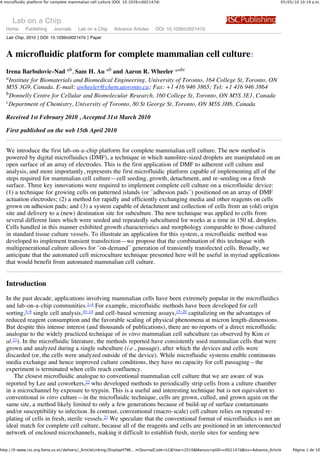
  • 2. A microfluidic platform for complete mammalian cell culture (DOI: 10.1039/c002147d) 05/05/10 10:19 a.m. generations of cells. Recently, digital microfluidics (DMF) has emerged as an alternative to the conventional format of enclosed microchannels. 24,25 As depicted in Fig. 1, digital microfluidic devices are formed from an array of electrodes which are used to manipulate discrete fluidic droplets. We recently reported the first application of DMF to cell-based analysis, generating dose–response toxicity data for cells suspended in 150 nL droplets.26 Under these conditions, cell vitality was shown to be unaffected by digital microfluidic manipulation. Other DMF/cell studies have followed,27–29 but we are not aware of any reports of the application of DMF to adherent cells (i.e., cells grown on surfaces). Fig. 1 Photo of a digital microfluidic platform developed for complete cell culture. The device comprised seven reservoirs (left), an array of actuation electrodes (2.22 mm ! 2.22 mm) used to manipulate 1 µL droplets (center), and a series of specially designated sites for cell culture in 150 nL droplets (right and inset). Here, we report the first application of digital microfluidics to adherent cell studies. The system is powered by several key innovations including a heterogeneous device format with dedicated cell culture sites, a passive reagent dispensing mechanism for seeding and feeding cells, and a new scheme for collection and delivery of cells in nanolitre droplets. This work represents the first true microfluidic analogue to macro-scale mammalian cell culture—cells are seeded, grown, and passaged onto fresh culture sites ad nauseam. We anticipate that the microfluidic technique presented here will be useful in myriad applications that would benefit from automated, miniaturized mammalian cell culture. Results and discussion Cell seeding and growth Adherent cells are typically cultured on substrates bearing a hydrophilic charged surface, which is required for cell attachment and spreading. For example, commercial cell culture flasks are formed from polystyrene which is treated with plasma oxidation to generate a charged surface. In some cases, this characteristic is enhanced by coating surfaces with extracellular matrix (ECM) proteins like fibronectin or collagen. Surfaces formed in this manner are more biomimetic than those formed by plasma oxidation and such techniques are required for certain cell types including some primary cells, 30 and for the maintenance of (feeder layer-free) undifferentiated embryonic stem cells. 31 In the current work, we used ECM protein coatings to make digital microfluidic devices amenable to adherent cell growth. Droplet actuation by digital microfluidics requires devices with hydrophobic surfaces. To make DMF devices compatible with both cell growth and droplet actuation, we developed a new strategy in which each device is globally coated with Teflon-AF (which features a contact angle for water of 115°), and is then locally modified with islands of ECM proteins, forming adhesion pads (Fig. 2a). The adhesion pads were formed by dispensing droplets containing fibronectin and allowing them to dry to the surface. For the purposes of imaging, devices were designed with transparent windows in the electrode array, such that cells growing on adhesion pads could be monitored by microscopy. This had the added advantage of isolating the cells from long-term exposure to electrical fields. Fig. 2b shows HeLa cells on an adhesion pad on a device immediately after seeding, and after 24 h of incubation in a 150 nL droplet of media. As shown, the cells shifted from round to spread morphology, as is observed for cells grown in multiwell plates (Fig. 2c). Similar effects were observed for many different cell lines, including CHO-K1, NIH-3T3 and INS-1 (not shown). During cell growth, evaporation was controlled by storing devices in humidified chambers in a tissue culture http://0-www.rsc.org.fama.us.es/delivery/_ArticleLinking/DisplayHTML…m?JournalCode=LC&Year=2010&ManuscriptID=c002147d&Iss=Advance_Article Página 2 de 10
  • 3. A microfluidic platform for complete mammalian cell culture (DOI: 10.1039/c002147d) 05/05/10 10:19 a.m. incubator. Fig. 2 Digital microfluidic device with adhesion pad for culturing adherent cells. (a) Top- and side-view schematics of a DMF device with an adhesion pad. (b) Pictures of HeLa cells on an adhesion pad immediately after seeding (left) and after 24 h (right). In the pictures, the top plate is not observed because it is transparent. (c) Picture of control HeLa cells after 24 h incubation in a well coated with fibronectin. The cells in droplets showed similar morphology and spreading characteristics to the cells grown in wells. Scale bars are 200 µm and 80 µm for the main panels and insets, respectively. Media exchange Cell seeding and growth is the first of many requirements for a complete cell culture platform; the next requirement is a method to remove old cell growth media and replace it with fresh media. In macro-scale tissue culture, this is accomplished by aspiration and pipetting, and care must be taken to dispense the new media quickly to avoid drying out the cells. Here, we introduce a new DMF-driven method for solution replacement for cell culture, which we have termed passive dispensing. In this method, solution removal and replacement are simultaneous processes; hence, cells are continuously hydrated, with no concern for cell drying or death between removal and replacement of media. Passive dispensing is depicted in Fig. 3a. A large source droplet of new media is driven across an adhesion pad bearing a smaller droplet of old media. As the large droplet crosses the adhesion pad, the old media are swept away, leaving a portion of the new media behind. We call this process passive dispensing because the act of splitting the small droplet (which remains on the pad) from the source droplet (which moves away from the pad) is driven by the difference in hydrophilicities of the surface rather than by actuation of electrodes. As depicted in Fig. 1S in the ESI , this method works both for delivery of an initial droplet to a dry contact pad as well as for exchanging fluid on a pad that already bears a droplet. In the work reported here, passive dispensing was used to generate 150 nL droplets on the adhesion pads, while active dispensing (i.e., charging adjacent electrodes to dispense a droplet from a reservoir by necking and splitting 32) was used to generate the 1 µL source droplets actuated on the array of electrodes. We note that passive dispensing is similar to what was developed independently by Yang et al. 33 for non-biological applications. http://0-www.rsc.org.fama.us.es/delivery/_ArticleLinking/DisplayHTML…m?JournalCode=LC&Year=2010&ManuscriptID=c002147d&Iss=Advance_Article Página 3 de 10
  • 4. A microfluidic platform for complete mammalian cell culture (DOI: 10.1039/c002147d) 05/05/10 10:19 a.m. Fig. 3 Passive dispensing for long-term cell culture. (a) Schematics depicting passive media exchange. A new source droplet sweeps the old media away from the culture site while simultaneously delivering fresh media to the cells. (b) Passive dispensing efficiency evaluated by exchanging droplets of 10 nM fluorescein with consecutive droplets of PBS. The fluorescence is reduced to 3% of its initial value after exchange with a single droplet, and is completely extinguished after exchange with three source droplets. (c) Pictures of HeLa cells on a device from day 2 to day 5 of culture, with passive exchange of culture medium every 24 h. Scale bars are 200 µm. In developing this technique, we hypothesized that the solution transfer efficiency of passive dispensing (i.e., the degree of exchange of new media for old) would be high, because the Reynold's number (Re) in such systems is low (Re < 100 in the device reported here). To test this hypothesis, we evaluated the persistence of fluorescence in droplets containing fluorescein that were replaced with droplets containing phosphate buffered saline (PBS). Fig. 3b shows the results of these experiments, which verify that the solution transfer efficiency is high. After a single passive exchange, less than 3% of the initial fluorescence persists in the droplet on the adhesion pad. And after three passive exchanges, the dye is completely removed. Thus, in the work described here, three passive exchanges were used to deliver new media and other reagents to cells growing on adhesion pads. Passive dispensing was used to facilitate all aspects of cell culture and growth, including the initial seeding of cells from suspension, and delivery of fresh media to cells growing on adhesion pads. Several different types of cells were cultured in nanolitre droplets for multiple days with media exchange every 24 h (Fig. 3c) with proliferation rates comparable to those observed in macro-scale culture. Cell subculture In addition to cell seeding and media exchange, a complete cell culture system must be able to facilitate cell harvesting and passaging (i.e. subculture), which comprises separation of a portion of cells from a source population and seeding on a fresh surface. In conventional cell culture, this process is tedious and time- consuming, involving multiple aspiration, dispensing, and centrifugation steps. As described in the introduction, despite intense interest in the lab-on-a-chip community for applications involving cells, 1–4 no methods have been reported that are analogous to conventional mammalian cell passaging. Here, we introduce the first microfluidic platform for mammalian cell subculture; the four steps in this process are outlined in Fig. 4. Cells were first grown in 150 nL droplets on primary (1°) adhesion pads (Fig. 4a) with regular media exchange via passive dispensing. In the second half of the exponential growth phase, cells were detached by passively exchanging the media with trypsin (Fig. 4b). After cells detached, a fresh droplet of cell culture media was translated over the 1° pad, blocking trypsin and collecting some of the cells. The droplet was translated to the secondary (2°) adhesion pad, where a second passive dispensing step delivered some of the cells in the droplet to the new site (Fig. 4c). In the devices described here, we observed that 2 to 3% of the 1° cells were delivered to the 2° adhesion pad per droplet; thus, on average four 1 µL droplets were used to collect and deliver 10% of the 1° cells to the 2° pad. Finally, the subculture of cells was grown on the 2° adhesion pad (Fig. 4d) before they were subcultured again. We applied this technique to several cell lines (CHO-K1, HeLa, and NIH-3T3) for up to 4 subcultures, which suggests that this process is broadly applicable to long-term cell subculture. http://0-www.rsc.org.fama.us.es/delivery/_ArticleLinking/DisplayHTML…m?JournalCode=LC&Year=2010&ManuscriptID=c002147d&Iss=Advance_Article Página 4 de 10
  • 5. A microfluidic platform for complete mammalian cell culture (DOI: 10.1039/c002147d) 05/05/10 10:19 a.m. Fig. 4 Schematic and pictures depicting subculture of CHO-K1 cells in droplets by digital microfluidics. (a) Monolayer of the first generation of cells on the primary (1°) adhesion pad. (b) Cells dissociating from the surface of the 1° pad after delivery of trypsin. (c) Trypsinized cells being harvested in a droplet of media containing serum, and then seeded on the secondary (2°) pad. (d) Monolayer of subcultured cells on the 2° pad after 72 h. Scale bars are 200 µm. In addition to the advantage of being automated, the new microfluidic cell subculture technique is faster and more straightforward than conventional macro-scale methods. These improvements are a result of the efficiency of passive dispensing. For example, the standard cell culture technique of washing cells with PBS before trypsinization can be omitted because passive dispensing efficiently removes the old solution (media) in addition to delivering the new one (trypsin) (see Fig. 3b). Likewise, after exposure to trypsin, separate blocking and washing steps are not needed in the microfluidic technique because as the first droplet of media efficiently replaces the trypsin, most of the cells remain on the surface of the device. This eliminates the requirement for centrifugation. To evaluate the differences between the new microfluidic method and conventional techniques, two cell lines, CHO-K1 and HeLa, were subcultured on microfluidic devices and in 96 well plates, and their proliferation was monitored for 6–8 days. As shown in Fig. 5, CHO-K1 cell proliferation was nearly identical in the micro- and macro-scale formats but HeLa cell proliferation exhibited differences. In the microfluidic technique, HeLa cells experienced a longer lag phase prior to entering exponential growth, and, once begun, the doubling time in wells (1.28 ± 0.09 days) was shorter than the doubling time in droplets (1.81 ± 0.27 days). http://0-www.rsc.org.fama.us.es/delivery/_ArticleLinking/DisplayHTML…m?JournalCode=LC&Year=2010&ManuscriptID=c002147d&Iss=Advance_Article Página 5 de 10
  • 6. A microfluidic platform for complete mammalian cell culture (DOI: 10.1039/c002147d) 05/05/10 10:19 a.m. Fig. 5 Growth curves for CHO-K1 and HeLa cells in macro-scale (wells, open circles) and micro-scale (droplets, closed circles) formats. (a) CHO-K1 cells seeded at 20 cells per mm2 were cultured for 3 days. Upon reaching a density of 200 cells per mm2 they were split and cultured in a new well/adhesion pad for another 3 days. (b) HeLa cells seeded at 100 cells per mm2 were cultured for 4 days, and then split and cultured for another 4 days. A one-day lag was observed in growth of cells in droplets whereas cells in wells almost immediately entered exponential growth phase. The exponential growth rates in the two systems were similar. While the data suggest that the proliferation rates in micro- and macro-scale formats are not identical, their similarities are striking, given the many differences in physical parameters. For example, the density of cells per unit area was kept constant in the micro- and macro-scale systems, which led to a volumetric density of cells at seeding in droplets ( 133 to 666 cells per µL) 20 times higher than that in wells ( 6 to 32 cells per µL). To compensate for this difference, media in microfluidic devices were exchanged more frequently than in wells (every 24 h vs. every 3–4 days), but the initial 20-fold difference in nutrients and cell growth factors (as well as differences in accumulated cell excretions) is a likely source of differing growth conditions. In the future, we may design microfluidic devices with volumetric densities closer to that of macro-scale systems; however, we note that this would sacrifice some of the advantages of miniaturization (reduced reagent use, etc.). Another potential source of variation between the macro- and micro-scale systems is the integrity of the ECM coating: fibronectin densities and adhesion characteristics may be slightly different on Teflon-AF (microfluidic device) relative to plasma-treated polystyrene (macro-scale). In on-going work, we are evaluating different kinds of local surface treatments for devices, including plasma oxidation and various types of ECM coatings. Regardless, both CHO-K1 and HeLa cells were observed to grow robustly, suggesting that the digital microfluidic environment is suitable for long-term culture and analysis. On-demand transfection As an example of an application of the new techniques reported here, we developed means to implement transient transfections in cells grown in the digital microfluidic system. Gene transfer and manipulation of http://0-www.rsc.org.fama.us.es/delivery/_ArticleLinking/DisplayHTML…m?JournalCode=LC&Year=2010&ManuscriptID=c002147d&Iss=Advance_Article Página 6 de 10
  • 7. A microfluidic platform for complete mammalian cell culture (DOI: 10.1039/c002147d) 05/05/10 10:19 a.m. eukaryotic genomes are used widely to develop understanding of gene functions and regulatory sequences, post-translational events, and for the development of gene therapy.34,35 Most transfections are performed transiently, such that DNA introduced into cells is not passed onto progeny; hence, each time a researcher wants to use such cells, a new batch must be generated. By integrating routine transfections with subculture on an automated digital microfluidic device, we propose that researchers will have access to transfected cells on-demand. To illustrate this concept, we performed transient transfection of CHO-K1 cells on microfluidic devices with an expression vector encoding green fluorescent protein (GFP). Prior to transfection, cells were grown and subcultured on the microfluidic platform. Fig. 6a (left) is a picture of CHO-K1 cells 24 h after transfection in a device (in a 150 nL droplet) while Fig. 6a (right) shows CHO-K1 cells similarly transfected in wells (100 µL). As shown in Fig. 6b, the transfection efficiency in the micro-system is comparable to that in wells. While cell transfection in microchannels has been established, 36,37 the work reported here is the first report of transfection by digital microfluidics, and more importantly, represents the first combination of automated multigenerational subculture techniques with transfection (i.e., transfection on-demand) in any microfluidic format. Fig. 6 On-demand transient transfection of CHO-K1 cells in droplets. (a) Pictures of CHO-K1 cells transfected to express GFP in a droplet (left) and in a well (right). Transfected cells appear green as a consequence of GFP emission. (b) Comparison of transfection efficiency in droplets and wells. Approximately 20% of cells were typically transfected in both systems (n = 3). Scale bars are 200 µm. Conclusion We present the first microfluidic analogue to in vitro mammalian cell culture. Development of this system required several key innovations, including techniques for growing cells on adhesion pads on an array of electrodes, a passive system for exchanging media on cells grown on devices, and a method for detachment, collection, and delivery of cells to fresh and sterile sites for subculture. Cells grown using this method exhibit growth characteristics comparable to those found in conventional tissue culture, and are compatible with on- http://0-www.rsc.org.fama.us.es/delivery/_ArticleLinking/DisplayHTML…m?JournalCode=LC&Year=2010&ManuscriptID=c002147d&Iss=Advance_Article Página 7 de 10
  • 8. A microfluidic platform for complete mammalian cell culture (DOI: 10.1039/c002147d) 05/05/10 10:19 a.m. chip genetic transformation. In addition, the new methods developed for media exchange can be used to deliver reagents and staining solutions to cells cultured on DMF devices for cell-based assays. We anticipate that these new methods may be useful for a wide range of applications in cell biology, tissue engineering, - omics, screening and beyond. Experimental Reagents and materials Unless otherwise indicated, general-use reagents were from Sigma-Aldrich (Oakville, ON) and cells and cell culture reagents were from American Type Culture Collection (ATCC, Manassas, VA). Details relating to device fabrication and operation can be found in the ESI . Macro-scale cell culture For macro-scale work, cell lines CHO-K1, NIH-3T3, HeLa, and INS-1 were maintained in cell culture flasks in a humidified incubator (5% CO 2 , 37 °C). The growth media included DMEM (NIH-3T3 and HeLa), F- 12/DMEM (50/50) (CHO-K1), and RPMI 1640 with 20 µM glucose (INS1). All media formulations were supplemented with 10% fetal bovine serum (Invitrogen Canada, Burlington, ON), and all but F-12/DMEM were supplemented with penicillin (100 IU mL "1) and streptomycin (100 µg mL "1). CHO-K1, NIH-3T3, and HeLa were subcultured every 3–4 days at 5 ! 105 cells per mm2 , and INS-1 was subcultured every 7 days at the same density. Prior to experiments, cells were harvested and resuspended in fresh media, and cell numbers and viability were quantified using a hemocytometer and trypan blue exclusion (Invitrogen Canada). All reagents, washing solutions and media contained 0.05% Pluronic F68 (w/v). Microfluidic cell culture For micro-scale work, CHO-K1, NIH-3T3, HeLa, and INS-1 cells were seeded on adhesion pads in 150 nL droplets of media containing 20–100 cells. For long-term experiments, the media were exchanged by passive dispensing (using three source droplets) every 24 h, and devices were stored in a humidified chamber in an incubator (5% CO 2 , 37 °C). For subculture experiments, cells were grown to a high density ( 200 to 300 cells) on a primary (1°) adhesion pad. The old media were then replaced with a 150 nL droplet containing trypsin (0.25% vol/vol) and EDTA (1 mM) by passive dispensing (using three source droplets), and cells were allowed to dissociate for 3–5 min. To harvest cells, a droplet of fresh media (1 µL) was translated over the 1° adhesion pad, collecting dissociated cells from the surface. The droplet was then driven to a secondary (2°) pad, where some of the cells were seeded in a 150 nL droplet by passive dispensing. The procedure was repeated four times such that the droplet on the 2° pad contained 20–100 cells. In some cases, the process was repeated for up to four generations of cells (on four adhesion pads). Cell numbers and densities per mm2 were determined using ImageJ (US National Institutes of Health, http://rsb.info.nih.gov/ij/). For comparison, some cells were grown and subcultured in 96 well plates (Corning Inc., USA). In these experiments, 50 µL of fibronectin (33 µg mL "1 in DI water) was dispensed into each well and then incubated in the tissue culture incubator (1 h). After aspirating and drying, wells were seeded with 20–100 cells per mm2 in 100 µL of media, and were monitored as described above (densities per mm2 were measured in three locations per well and then averaged). For subculture experiments, cells were rinsed in PBS, incubated in trypsin/EDTA (3–5 min), rinsed again in cell media, and then split into new fibronectin-coated wells at 10% of their original density (in 100 µL of media). All solution exchanges in macro-scale experiments were carried out by manual pipetting, and all cell culture experiments in micro- and macro-scale systems were repeated in at least four replicates. Cell transfection In transfection experiments, pCAG–GFP plasmid DNA38 (Addgene, Cambridge, MA) was extracted and purified from E. coli using GenElute HP Plasmid Miniprep Kits (Sigma-Aldrich, Oakville, ON). A stock solution for transfection was prepared by mixing 0.25 µg DNA and 1 µL Dreamfect cationic lipid (Oz Biosciences, Marseille, France) in 25 µL PBS. That mixture was allowed to complex for 20 minutes and was subsequently diluted 1 : 4 in CHO-K1 culture media for use in experiments (supplemented 0.05% w/v Pluronic F68). CHO-K1 cells were seeded in 150 nL droplets on DMF devices on a 1° adhesion pad, incubated until they reached high density, and then subcultured on a 2° adhesion pad, as described above. After incubating overnight, cell media were exchanged with 150 nL of transfection solution by passive dispensing, such that the cells were exposed to approximately 20 nL lipid reagent and 5 ng DNA. After incubating 3 h, the transfection solution was replaced with fresh media by passive dispensing, and the cells http://0-www.rsc.org.fama.us.es/delivery/_ArticleLinking/DisplayHTML…m?JournalCode=LC&Year=2010&ManuscriptID=c002147d&Iss=Advance_Article Página 8 de 10
  • 9. A microfluidic platform for complete mammalian cell culture (DOI: 10.1039/c002147d) 05/05/10 10:19 a.m. were incubated overnight. For comparison, cells were also seeded in 96 well plates and then transfected using the same reagents according to the manufacturer's protocol for the transfection of CHO-K1 cells. Acknowledgements We thank Evan Mills and Prof. Kevin Truong (Institute of Biomaterials and Biomedical Engineering, University of Toronto) for assistance with cell transfection and Connie Cepko for kindly providing plasmids. We also thank Dr Alessandro Datti (Mount Sinai Hospital, Toronto) for helpful discussions on automation of cell culture. We acknowledge the Natural Sciences and Engineering Council (NSERC), the Canada Foundation for Innovation (CFI), and the Canadian Institutes of Health Research (CIHR) for financial support. IBN and SHA thank NSERC for graduate fellowships, and ARW thanks the CRC for a Canada Research Chair. References 1 H. Andersson and A. van den Berg, Sens. Actuators, B, 2003, 92, 315–325 [Links]. 2 I. Barbulovic-Nad and A. R. Wheeler, in Encyclopedia of Microfluidics and Nanofluidics, ed. D. Q. Li, Springer, New York, 1st edn, 2008, pp. 209–216. 3 J. El-Ali, P. K. Sorger and K. F. Jensen, Nature, 2006, 442, 403–411 [Links]. 4 I. Meyvantsson and D. J. Beebe, Annu. Rev. Anal. Chem., 2008, 1, 423–449. 5 A. Y. Fu, C. Spence, A. Scherer, F. H. Arnold and S. R. Quake, Nat. Biotechnol., 1999, 17, 1109–1111 [Links]. 6 A. Wolff, I. R. Perch-Nielsen, U. D. Larsen, P. Friis, G. Goranovic, C. R. Poulsen, J. P. Kutter and P. Telleman, Lab Chip, 2003, 3, 22–27 [Links]. 7 J. Kruger, K. Singh, A. O'Neill, C. Jackson, A. Morrison and P. O'Brien, J. Micromech. Microeng., 2002, 12, 486–494 [Links]. 8 M. M. Wang, E. Tu, D. E. Raymond, J. M. Yang, H. C. Zhang, N. Hagen, B. Dees, E. M. Mercer, A. H. Forster, I. Kariv, P. J. Marchand and W. F. Butler, Nat. Biotechnol., 2005, 23, 83–87 [Links]. 9 X. B. Wang, J. Yang, Y. Huang, J. Vykoukal, F. F. Becker and P. R. C. Gascoyne, Anal. Chem., 2000, 72, 832–839 [Links]. 10 M. Khine, A. Lau, C. Ionescu-Zanetti, J. Seo and L. P. Lee, Lab Chip, 2005, 5, 38–43 [Links]. 11 S. Takayama, E. Ostuni, P. LeDuc, K. Naruse, D. E. Ingber and G. M. Whitesides, Nature, 2001, 411, 1016 [Links]. 12 A. R. Wheeler, W. Throndset, R. J. Whelan, A. M. Leach, R. N. Zare, Y.-H. Liau, K. Farrell, I. Manger and A. Daridon, Anal. Chem., 2003, 75, 3581–3586 [Links]. 13 D. Di Carlo, L. Y. Wu and L. P. Lee, Lab Chip, 2006, 6, 1445–1449 [Links]. 14 P. C. H. Li, L. de Camprieu, J. Cai and M. Sangar, Lab Chip, 2004, 4, 174–180 [Links]. 15 T. Thorsen, S. J. Maerkl and S. R. Quake, Science, 2002, 298, 580–584 [Links]. 16 E. Leclerc, Y. Sakai and T. Fujii, Biotechnol. Prog., 2004, 20, 750–755 [Links]. 17 N. L. Jeon, H. Baskaran, S. K. W. Dertinger, G. M. Whitesides, L. Van De Water and M. Toner, Nat. Biotechnol., 2002, 20, 826–830 [Links]. 18 H. M. Yu, C. M. Alexander and D. J. Beebe, Lab Chip, 2007, 7, 388–391 [Links]. 19 K. R. King, S. H. Wang, D. Irimia, A. Jayaraman, M. Toner and M. L. Yarmush, Lab Chip, 2007, 7, 77– 85 [Links]. 20 L. Kim, M. D. Vahey, H. Y. Lee and J. Voldman, Lab Chip, 2006, 6, 394–406 [Links]. 21 L. Kim, Y. C. Toh, J. Voldman and H. Yu, Lab Chip, 2007, 7, 681–694 [Links]. 22 P. J. Hung, P. J. Lee, P. Sabounchi, R. Lin and L. P. Lee, Biotechnol. Bioeng., 2005, 89, 1–8 [Links]. 23 R. I. Freshney, Culture of Animal Cells: A Manual of Basic Technique, Wiley-Liss, Hoboken, N.J., 5th edn, 2005. 24 M. Abdelgawad and A. R. Wheeler, Adv. Mater., 2009, 21, 920–925 [Links]. 25 A. R. Wheeler, Science, 2008, 322, 539–540 [Links]. 26 I. Barbulovic-Nad, H. Yang, P. S. Park and A. R. Wheeler, Lab Chip, 2008, 8, 519–526 [Links]. 27 G. J. Shah, A. T. Ohta, E. P. Y. Chiou, M. C. Wu and C. J. Kim, Lab Chip, 2009, 9, 1732–1739 [Links]. 28 S. K. Fan, P. W. Huang, T. T. Wang and Y. H. Peng, Lab Chip, 2008, 8, 1325–1331 [Links]. http://0-www.rsc.org.fama.us.es/delivery/_ArticleLinking/DisplayHTML…m?JournalCode=LC&Year=2010&ManuscriptID=c002147d&Iss=Advance_Article Página 9 de 10
  • 10. A microfluidic platform for complete mammalian cell culture (DOI: 10.1039/c002147d) 05/05/10 10:19 a.m. 29 J. Zhou, I. Lu, K. Byrapogu, D. M. Wootton, P. I. Lelkes and R. Fair, Virtual and Physical Prototyping, 2007, 2, 217–223. 30 R. I. Freshney, in Animal Cell Culture: A Practical Approach, ed. J. R. W. Masters, Oxford University Press, Oxford, New York, 2nd edn, 2002. 31 C. H. Xu, M. S. Inokuma, J. Denham, K. Golds, P. Kundu, J. D. Gold and M. K. Carpenter, Nat. Biotechnol., 2001, 19, 971–974 [Links]. 32 S. K. Cho, H. J. Moon and C. J. Kim, J. Microelectromech. Syst., 2003, 12, 70–80 [Links]. 33 T. H. Chen, C. M. Su, H. C. Chih and C. T. Yang, Proceedings of the 5th International Conference on Nanochannels, Microchannels, and Minichannels, 2007, pp. 147–153. 34 A. Colosimo, K. K. Goncz, A. R. Holmes, K. Kunzelmann, G. Novelli, R. W. Malone, M. J. Bennett and D. C. Gruenert, BioTechniques, 2000, 29, 314–318 [Links]. 35 F. Recillas-Targa, Mol. Biotechnol., 2006, 34, 337–354. 36 T. Houchin-Ray, K. J. Whittlesey and L. D. Shea, Mol. Ther., 2007, 15, 705–712 [Links]. 37 T. R. Sodunke, M. J. Bouchard and H. M. Noh, Biomed. Microdevices, 2008, 10, 393–402 [Links]. 38 T. Matsuda and C. L. Cepko, Proc. Natl. Acad. Sci. U. S. A., 2004, 101, 16–22 [Links]. Footnote Electronic supplementary information (ESI) available: Experimental details on device fabrication and operation, including Supplementary figure 1S, which depicts passive dispensing. See DOI: 10.1039/c002147d This journal is © The Royal Society of Chemistry 2010 http://0-www.rsc.org.fama.us.es/delivery/_ArticleLinking/DisplayHTML…?JournalCode=LC&Year=2010&ManuscriptID=c002147d&Iss=Advance_Article Página 10 de 10