The elbow joint in concern of diagnostic imaging .pptx 1
•Transferir como PPTX, PDF•
10 gostaram•4,332 visualizações

Regarding the Diagnostic imaging what we concern in the elbow joint

Recomendados
Recomendados
Mais conteúdo relacionado
Mais procurados
Mais procurados (20)
Destaque
Destaque (20)
Semelhante a The elbow joint in concern of diagnostic imaging .pptx 1
Semelhante a The elbow joint in concern of diagnostic imaging .pptx 1 (20)
Mais de DR Laith
Mais de DR Laith (20)
Último
Último (20)
The elbow joint in concern of diagnostic imaging .pptx 1
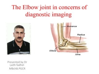
- 1. The Elbow joint in concerns of diagnostic imaging Presented by Dr Laith fadhel MBchB.PGCR
- 2. X-Ray ) bones and articulation
- 3. Ossification centers and bone age Ryan –page 254 /Sutton –page 1847
- 5. Anatomy and relation study When you study the anatomy of the elbow, it is good to use the inside-out approach. First study the bones and then continue with the ligaments and the tendons and then the surrounding structures.
- 6. MRI technique • Scan planes • Just like in the shoulder you need to be sure to get the imaging planes correctly in a standardized way. Use the axis of the epicondyles on a axial localizer to plan the coronal scan. The Sagittal images are scanned perpendicular to the coronal scan. • In this way you get very persistent images and you will get used to the normal anatomy.
- 7. Imaging sequences T1 In every joint that is studied you should have at least one T1-sequence not only to look at the anatomy, but also as a back up for looking at the marrow. Of course the T2-fatsat images will show marrow abnormalities, but T1 can be helpful in telling us what is really going on. T1 is certainly used in MR-arthrography. T2-fatsat T2 will show us most of the pathology, whether it is in the bone marrow, ligaments or muscle because of the high water content. It can also be used to image cartilage. Gradient echo With gradient echo we can use 3-D thin sections to image the cartilage and the ligaments. In the MR-protocol we do T1 and T2-fatsat in all three imaging planes. Sometimes STIR is used.
- 8. Tendon attachments Common flexor tendon Attaches at the medial epicondyle Ulnar collateral ligament or UCL Starts at the undersurface of the medial epicondyle and runs down to the sublime tubercle, which is the medial side of the coronoid process. Common extensor tendon Originates at the lateral epicondyle. Lateral collateral ligament Originates just underneath the attachment of the common extensor tendon. Lateral ulnar collateral ligament This is a somewhat confusing term for a tendon that also originates just underneath the common extensor tendon. It swings down behind the radial head and attaches at the area of the ulna that is called the supinator crest - see lateral view. Biceps tendon Attaches on the radial tuberosity. Brachialis tendon Attaches on the coronoid process. Annular ligament Attaches on the volar side of the sigmoid notch of the ulna and runs around the radial head and attaches on the dorsal side of the sigmoid notch. Attachment sites
- 9. Medical epicondyle avulsion fracture A fat-suppressed T2 weighted coronal image in a 15 year old baseball pitcher reveals an avulsion fracture (arrow) of the medial epicondyle apophysis,
- 10. Ulnar Collateral ligament If you look at the medial epicondyle you will notice the posterior bundle as a thin structure (blue arrow). The posterior bundle forms the floor of the cubital tunnel. A retinaculum covers the cubital tunnel. Notice that the anterior bundle is much thicker (white arrow). As we go distally we'll see that they merge together to attach to the sublime tubercle It is normal to see some high signal in the proximal part (arrow).
- 11. UCL tear Remember that the UCL should attach very tightly on the sublime tubercle. In this case it doesn't, so even on these two images you can tell that there is a complete tear. Notice that there is some marrow edema in the sublime tubercle. The mechanism of injury to the UCL is usually chronic tensile forces, which create micro tears. This is seen in pitchers and other overhead throwingathletes. A tear can also occur in a fall on the outstretched hand. Most commonly there is a complete tear, but sometimes there is a partial tear which can be very hard to see. That is why in these athletes MR- arthrogram is usually performed. This is a 18 year old baseball pitcher with medial elbow pain. A partial tear is seen creating a 'T-sign‘ Notice that the anterior bundle is intact and firmly attaches to the sublime tubercle (yellow arrow). On the next two images there is some soft tissue edema and more abnormal signal posteriorly (red arrow). So we suspect pathology of the posterior bundle.
- 12. UCL tear On the axial image we nicely see the anterior bundle is o.k. (red arrow). There is only some edema next to it. However the posterior bundle is not o.k. This is partial tearing. We see this occasionally in throwing athletes, where the anterior bundle is intact and their elbow is not unstable. They somehow have torn their posterior bundle, which causes pain. They do not need surgery, but it still may keep them out of the game for quite a while. Now here is the last case. This is a 38 year old male who has been weight-lifting for 20 years. He complains of intermittent elbow pain and popping. Notice that the UCL is abnormal with some areas of very high signal indicating a partial tear. On the lateral side there is subchondral edema and cartilage. This is arthrosis secondary to chronic instability due to the chronic partial tearing.
- 13. Lateral Collateral Ligament When you look for the radial collateral ligament, first try to identify the common extensor tendon, because right underneath it you will find the radial collateral ligament (yellow arrow). As you go more posteriorly you will see the LUCL - the lateral ulnar collateral ligament, which sweeps behind the radial head (white arrows). The annular ligament is usually difficult to differentiate from the RCL, but sometimes it can be identified on a Sagittal MRartrogram.
- 14. Muscles tendons The common flexor tendon originates at the medial epicondyle. On a T1W-images the tendon should have a low signal intensity (red arrow). The common extensor tendon originates at the lateral epicondyle. On a T1W-images the tendon should have a low signal intensity (yellow arrow).
- 15. Muscles tendons through the axial images of the biceps tendon from the musculotendinous junction to the attachment on the radial tuberositas.
- 16. Muscles tendons The Brachialis originates from the lower half of the front of the hummers, near the insertion of the deltoid muscle. It lies deeper than the biceps brachii, and is a synergist that assists the biceps in flexing the elbow. The thick tendon inserts on the anterior surface of the coronoid process of the ulna. On a Sagittal view, when you compare the Brachialis tendon (yellow arrows) with the biceps tendon (red arrows), notice that the Brachialis is almost all muscle. It only has a very short tendon distally
- 17. Nerves
- 18. Soft Tissue Masses & bursitis On MR a mass was seen just above the medial epicondyle, where the epitrochlear lymph nodes live. The mass is very heterogeneous as is the enhancement. Based on the MRfindings you still have to call this mass indeterminate. The final diagnosis was cat scratch disease based on high Bartonella henselae titers.
- 19. Soft Tissue Masses & bursitis Here images of a 26 year old female who also came with a mass in the peritrochlear region. It looks quite homogeneous and cystic. Continue with the post-Gd image. Notice the inhomogeneous enhancement on the MRI and prominent internal vascularity on the Sagittal ultrasound image. So this was not a cystic mass. Again this was diagnosed as indeterminate. The final diagnosis at biopsy was Lymphoma
- 20. Soft Tissue Masses & bursitis Olecranon Brachioradilus bursitis