SlideShare uma empresa Scribd logo
1 de 52
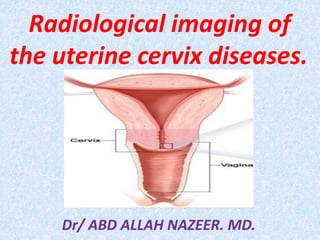
Radiological imaging of
the uterine cervix diseases.
Dr/ ABD ALLAH NAZEER. MD.
The uterine cervix is composed of the portio, which protrudes into the vagina,
and the supravaginal part of the cervix.
Squamous cells cover the epithelial surface of the portio continuing from the
vagina. With age, they grow back to cover the columnar cells of the endocervical
gland. This transitional area is called the squamocolumnar junction (SCJ)
Carcinoma of the cervix develops almost exclusively within the transformation
zone that extends between the original SCJ and the physiologic (SCJ).
Anatomy.
Radiological imaging.
Chest X-Rays: X-ray often can show whether cancer has spread to the lungs.
CT Scan: CT is the imaging modality that is most commonly used in clinical
practice to evaluate the extent of spread of cervical cancer. The oral, rectal,
or intravenous administration of contrast material is necessary for optimal
CT evaluation (unless a contraindication exists). The intravenous injection
of contrast material is particularly useful in increasing the conspicuity of the
cervical tumor and in facilitating the evaluation of the parametria and the
pelvic sidewalls
US has been used to evaluate the size and local regional extent of the
tumor. In the early stage of cervical carcinoma, the primary lesion is
difficult to depict with any imaging modality, including transvaginal
US and TRUS.
Eventually, with disease progression, the tumor mass may appear as a
hypoechoic or isoechoic region with undefined margins, or the disease
may be manifested as an enlarged cervix with heterogeneous
echogenicity (see the images below). In general, the intracervical cancer
is not clearly distinguished from the surrounding normal cervical stroma.
MRI appearance of cervical carcinoma.
Cervical carcinoma has a long T1 and a long T2 that render this
disease relatively hyperintense on proton-density–weighted and
T2-weighted images, in which the signal intensity of the tumor is
approximately similar to that of the endometrium. The
hyperintense appearance on T2-weighted images accounts for the
essentially consistent conspicuity of the primary tumor against the
background of the hypointense normal cervical stroma.
Positron emission tomography (PET) with use of
fluorodeoxyglucose (FDG) has some value relative to conventional
imaging methods for the detection of nodal metastatic disease and
recurrent cervical cancer, possibly being effective in the evaluation
of cases of locally advanced cervical cancer in which CT findings are
negative or equivocal, and possibly being predictive of survival
outcome.
Nonneoplastic Diseases.
Cervical Pregnancy
The prevalence of cervical pregnancy ranges from 1 in 1,000 to 1 in
24,000 of all pregnancies . Recently, the occurrence has been increasing,
possibly due to the increased number of induced abortions. The exact
cause is still unknown. Reported risk factors include multiparity, prior
surgical manipulation of the cervix or endometrial cavity, cervical or
uterine leiomyomas, atrophic endometrium, and septate uterus.
The cervix is made up of two different types of epithelium: squamous
epithelium and glandular epithelium. The cause of cervical inflammation
depends on the affected epithelium. The same microorganisms as those
that cause vaginitis can affect the endocervical squamous epithelium.
Trichomonas vaginalis, Candida albicans, and herpes simplex virus can
cause inflammation of the ectocervix. Conversely, Neisseria gonorrhoeae
and Chlamydia trachomatis infect only the glandular epithelium and are
major causes of mucopurulent endocervicitis .
Nabothian Cyst.
Nabothian cysts are common retention cysts of the uterine cervix. They are
formed as a result of the healing process of chronic cervicitis . During chronic
inflammation, the squamous epithelium proliferates, covering the columnar
epithelium of the endocervical glands. After that, the mucus secreted by the
columnar epithelium (now covered by the squamous epithelium) cannot be
evacuated and forms a retention cyst .
Cervical Polyps
Cervical polyps are one of the most common causes of intermenstrual vaginal
bleeding. Most patients are perimenopausal, especially in the 5th decade of
life. The polyps are usually pedunculated, with a slender pedicle of varying
length, but some are sessile .
Endometriosis.
Endometriosis is a common disease that affects the uterine body. Endometriosis
seldom affects the uterine cervix. Internal endometriosis is called adenomyosis; in
this condition, lesions form an ill-defined hypointense area continuing to the
junctional zone with some hyperintense dots on T2-weighted images. However,
adenomyosis can form a uterus-like polypoid mass growing into the endocervical
canal . It has a cavity surrounded by endometrial mucosa and smooth muscle
layers resembling myometrium.
Cervical endometriotic cysts.
Cervical Ectopic Pregnancy Ultrasound.
Cervical pregnancy.
Uterine cervicitis with multiple uterine body fibroids.
Nabothian Cysts.
Nabothian cysts.
Cervical polyp.
Carcinoma of the cervix is the third most common malignancy of
the female reproductive tract and the second most common
malignancy in women 15 to 34 years of age, with a peak incidence
at 45 to 55 years.
Risk factors include low socioeconomic class, black race, early
sexual activity, multiple sexual partners, multiparity, and infection
with herpes simplex virus type 2. Because of extensive
Papanicolaou smear screening, the incidence of invasive cervical
cancer continues to decline. In the United States, there are an
estimated 15,700 new cases diagnosed every year.
The vast majority are squamous cell carcinomas(80-80%). Other
histologies include adenocarcinoma(10-20%), small cell carcinoma,
adenoid cystic carcinoma, sarcoma, and lymphoma(5-10%).
CERVICAL CANCER.
Epithelial Neoplasms:
Signs and symptoms.
Vaginal bleeding.
Menstrual bleeding is longer and heavier than usual.
Bleeding after menopause or increased vaginal discharge.
Bleeding following intercourse or pelvic examination.
Pain during intercourse.
Other symptoms that may occur include:
Unusual vaginal discharge
Pain in the pelvic area
Excessive tiredness
Swollen or painful legs
Lower back pain.
Adenoma Malignum
Adenoma malignum (also known as minimal deviation
adenocarcinoma) is a special subtype of mucinous adenocarcinoma of
the cervix. Its prevalence is about 3% of all cervical adenocarcinomas.
The most common initial symptom is a watery discharge. The prognosis
of this tumor has been reported to be unfavorable , as it disseminates
into the peritoneal cavity even in the early stage of the disease and its
response to radiation or chemotherapy is poor. Therefore, its
deceptively benign histologic appearance occasionally leads to an
incorrect diagnosis.
Malignant Melanoma
Malignant melanoma of the female genital tract accounts for 1%–5%
of all melanoma cases . It usually occurs in the vaginal mucosa and
occasionally involves the uterine cervix. Malignant melanoma arising in
the uterine cervix is extremely rare, with only about 30 cases reported
in the literature.
Nonepithelial Neoplasms.
Malignant Lymphoma.
Malignant lymphoma frequently infiltrates the uterus in advanced disease.
However, it rarely involves the uterine cervix as the initial manifestation . Its
frequency in Western countries was reported to be 0.008% of primary cervical
tumors and 2% of extranodal lymphomas in women . The common presenting
symptoms are vaginal bleeding, perineal discomfort, and vaginal discharge .
Cervical lymphomas are treated with chemotherapy alone or in combination
with irradiation or surgery.
Cervical Leiomyoma.
As about 90% of uterine leiomyomas occur in the uterine body, cervical
leiomyoma is relatively rare. Its prevalence is reported to be less than 10%
of all leiomyomas of the uterus . Clinical symptoms of cervical leiomyomas,
including hypermenorrhea, dysmenorrhea, or abdominal distention, are
identical to those of leiomyomas in the uterine body. They occasionally form
polypoid tumors and protrude into the cervical canal or even the vagina when
they grow in the submucosal region. Because they are located along the birth
canal, they occasionally cause maternal dystocia.
Cervical carcinoma with exophytic growth.
Cervical carcinoma with endophytic growth.
Stage Ib cervical carcinoma.
Cervical carcinoma stage IB
Stage IB cervical carcinoma.
Stage IIa cervical carcinoma.
IIB cervical carcinoma.
Stage IIB cervical carcinoma.
Stage IIb cervical carcinoma
Stage IIIa cervical carcinoma.
Stage IIIb cervical carcinoma.
Stage IVa cervical carcinoma.
Advanced cervical carcinoma.
Stage IVb cervical carcinoma with metastasis at the para-aortic lymph nodes.
IVB cervical carcinoma with pelvic LN , ureteric and uterine involvement.
Stage IVb cervical carcinoma.
Local recurrence after radiation therapy.
Adenoma malignum.
Adenoma malignum in a patient with Peutz-Jeghers syndrome.
Adenoma malignum
Atypical carcinoid tumor of the uterine cervix
Malignant melanoma of the vagina with direct invasion of the cervix
Malignant lymphoma of the uterine cervix
Two cases of Lymphoma of the cervix.
Cervical tumor PET.
Metastasis to the uterine cervix is a complication of breast cancer
Conclusions.
MR imaging is an essential modality for diagnosing cervical
lesions because the signal intensity or configuration of the lesion
demonstrated on MR images reflects the pathologic findings.
Although sagittal T1-weighted and T2-weighted images and
oblique axial T2-weighted images perpendicular to the uterine
axis are sufficient for staging cervical carcinoma in most cases,
only a detailed reading based on the pelvic anatomy and the
pathologic features of the tumor can allow an accurate staging
diagnosis. However, some tumors or tumor-like lesions can show
similar MR imaging findings, such as adenocarcinoma, adenoma
malignum, and florid endocervical hyperplasia. Therefore, a
diagnosis should be made based on clinical manifestations in
conjunction with imaging findings.
Thank You.

Mais conteúdo relacionado

Mais procurados

Imaging of Malignant Liver Lesions
Imaging of Malignant Liver LesionsImaging of Malignant Liver Lesions
Imaging of Malignant Liver LesionsSahil Chaudhry
 
Presentation1.pptx, radiological imaging of uterine lesions.
Presentation1.pptx, radiological imaging of uterine lesions.Presentation1.pptx, radiological imaging of uterine lesions.
Presentation1.pptx, radiological imaging of uterine lesions.Abdellah Nazeer
 
Radioanatomy of mediastinum and approach to mediastinal masses
Radioanatomy of mediastinum and approach to mediastinal massesRadioanatomy of mediastinum and approach to mediastinal masses
Radioanatomy of mediastinum and approach to mediastinal massesAkankshaMalviya3
 
Presentation1, radiological imaging of placenta accreta.
Presentation1, radiological imaging of placenta accreta.Presentation1, radiological imaging of placenta accreta.
Presentation1, radiological imaging of placenta accreta.Abdellah Nazeer
 
Radiological imaging of mediastinal masses
Radiological imaging of mediastinal massesRadiological imaging of mediastinal masses
Radiological imaging of mediastinal massesPankaj Kaira
 
Ultrasound of breast
Ultrasound of  breastUltrasound of  breast
Ultrasound of breastLALIT KARKI
 
Presentation1, mri imaging of the prostate
Presentation1, mri imaging of the prostatePresentation1, mri imaging of the prostate
Presentation1, mri imaging of the prostateAbdellah Nazeer
 
Radiology Spots PPT- 2 by Dr Chandni Wadhwani
Radiology Spots PPT- 2 by Dr Chandni WadhwaniRadiology Spots PPT- 2 by Dr Chandni Wadhwani
Radiology Spots PPT- 2 by Dr Chandni WadhwaniChandni Wadhwani
 
BREAST IMAGING MRI ppt DR ABHIJIT R SINGH
BREAST IMAGING MRI ppt DR ABHIJIT R SINGHBREAST IMAGING MRI ppt DR ABHIJIT R SINGH
BREAST IMAGING MRI ppt DR ABHIJIT R SINGHDrABHIJITRSINGH
 
Ultrasound of Vascular anomalies by Oscar M. Navarro
Ultrasound of Vascular anomalies by Oscar M. NavarroUltrasound of Vascular anomalies by Oscar M. Navarro
Ultrasound of Vascular anomalies by Oscar M. NavarroDr. Naveed Quetta
 
Imaging Of Peritoneal Pathology
Imaging Of Peritoneal Pathology  Imaging Of Peritoneal Pathology
Imaging Of Peritoneal Pathology Sakher Alkhaderi
 
Breast ultrasound
Breast ultrasoundBreast ultrasound
Breast ultrasoundairwave12
 
IMAGING OF CYSTIC PANCREATIC TUMOURS
IMAGING OF CYSTIC PANCREATIC TUMOURSIMAGING OF CYSTIC PANCREATIC TUMOURS
IMAGING OF CYSTIC PANCREATIC TUMOURSDr I Gurubharath .
 
Presentation2.pptx, radiological imaging of the rectal diseases.
Presentation2.pptx, radiological imaging of the rectal diseases.Presentation2.pptx, radiological imaging of the rectal diseases.
Presentation2.pptx, radiological imaging of the rectal diseases.Abdellah Nazeer
 
Ectopic pregnancy Radiology
Ectopic pregnancy RadiologyEctopic pregnancy Radiology
Ectopic pregnancy RadiologySajan Paul
 

Mais procurados (20)

Imaging of Malignant Liver Lesions
Imaging of Malignant Liver LesionsImaging of Malignant Liver Lesions
Imaging of Malignant Liver Lesions
 
Presentation1.pptx, radiological imaging of uterine lesions.
Presentation1.pptx, radiological imaging of uterine lesions.Presentation1.pptx, radiological imaging of uterine lesions.
Presentation1.pptx, radiological imaging of uterine lesions.
 
Radioanatomy of mediastinum and approach to mediastinal masses
Radioanatomy of mediastinum and approach to mediastinal massesRadioanatomy of mediastinum and approach to mediastinal masses
Radioanatomy of mediastinum and approach to mediastinal masses
 
Imaging of the scrotum
Imaging of the scrotumImaging of the scrotum
Imaging of the scrotum
 
MRI carcinoma Uterus and Cervix
MRI carcinoma Uterus and  CervixMRI carcinoma Uterus and  Cervix
MRI carcinoma Uterus and Cervix
 
Presentation1, radiological imaging of placenta accreta.
Presentation1, radiological imaging of placenta accreta.Presentation1, radiological imaging of placenta accreta.
Presentation1, radiological imaging of placenta accreta.
 
Radiological imaging of mediastinal masses
Radiological imaging of mediastinal massesRadiological imaging of mediastinal masses
Radiological imaging of mediastinal masses
 
Imaging of placenta
Imaging of placentaImaging of placenta
Imaging of placenta
 
Ultrasound of breast
Ultrasound of  breastUltrasound of  breast
Ultrasound of breast
 
Presentation1, mri imaging of the prostate
Presentation1, mri imaging of the prostatePresentation1, mri imaging of the prostate
Presentation1, mri imaging of the prostate
 
Radiology Spots PPT- 2 by Dr Chandni Wadhwani
Radiology Spots PPT- 2 by Dr Chandni WadhwaniRadiology Spots PPT- 2 by Dr Chandni Wadhwani
Radiology Spots PPT- 2 by Dr Chandni Wadhwani
 
BREAST IMAGING MRI ppt DR ABHIJIT R SINGH
BREAST IMAGING MRI ppt DR ABHIJIT R SINGHBREAST IMAGING MRI ppt DR ABHIJIT R SINGH
BREAST IMAGING MRI ppt DR ABHIJIT R SINGH
 
Ultrasound of Vascular anomalies by Oscar M. Navarro
Ultrasound of Vascular anomalies by Oscar M. NavarroUltrasound of Vascular anomalies by Oscar M. Navarro
Ultrasound of Vascular anomalies by Oscar M. Navarro
 
Imaging Of Peritoneal Pathology
Imaging Of Peritoneal Pathology  Imaging Of Peritoneal Pathology
Imaging Of Peritoneal Pathology
 
Breast ultrasound
Breast ultrasoundBreast ultrasound
Breast ultrasound
 
IMAGING OF CYSTIC PANCREATIC TUMOURS
IMAGING OF CYSTIC PANCREATIC TUMOURSIMAGING OF CYSTIC PANCREATIC TUMOURS
IMAGING OF CYSTIC PANCREATIC TUMOURS
 
Presentation2.pptx, radiological imaging of the rectal diseases.
Presentation2.pptx, radiological imaging of the rectal diseases.Presentation2.pptx, radiological imaging of the rectal diseases.
Presentation2.pptx, radiological imaging of the rectal diseases.
 
Radiology Spotters
Radiology Spotters Radiology Spotters
Radiology Spotters
 
Ectopic pregnancy Radiology
Ectopic pregnancy RadiologyEctopic pregnancy Radiology
Ectopic pregnancy Radiology
 
Liver lesions SYMPOSIUM RADIOLOGY
Liver lesions SYMPOSIUM RADIOLOGYLiver lesions SYMPOSIUM RADIOLOGY
Liver lesions SYMPOSIUM RADIOLOGY
 

Destaque

Imaging of female reproductive system RV
Imaging of female reproductive system  RVImaging of female reproductive system  RV
Imaging of female reproductive system RVRoshan Valentine
 
Contouring Guidelines for Gynecological Malignancy
Contouring Guidelines for Gynecological MalignancyContouring Guidelines for Gynecological Malignancy
Contouring Guidelines for Gynecological MalignancyJyotirup Goswami
 
Contouring guidelines Cervix IMRT
Contouring guidelines Cervix IMRTContouring guidelines Cervix IMRT
Contouring guidelines Cervix IMRTDebarshi Lahiri
 
Radiotherapy in carcinoma cervix
Radiotherapy in carcinoma cervixRadiotherapy in carcinoma cervix
Radiotherapy in carcinoma cervixDebarshi Lahiri
 
Presentation1.pptx, radiological anatomy of the abdomen and pelvis.
Presentation1.pptx, radiological anatomy of the abdomen and pelvis.Presentation1.pptx, radiological anatomy of the abdomen and pelvis.
Presentation1.pptx, radiological anatomy of the abdomen and pelvis.Abdellah Nazeer
 
Cancer Cervix- NACT vs RT+CCT
Cancer Cervix- NACT vs RT+CCTCancer Cervix- NACT vs RT+CCT
Cancer Cervix- NACT vs RT+CCTSheh Rawat
 
Cancer de cervix grethel garcia
Cancer de cervix grethel garciaCancer de cervix grethel garcia
Cancer de cervix grethel garciaGrethel Gc
 
Fertility preservation in Cancer Cervix
Fertility preservation in Cancer CervixFertility preservation in Cancer Cervix
Fertility preservation in Cancer CervixSujoy Dasgupta
 
Premalignantcx lecture (1)
Premalignantcx lecture (1)Premalignantcx lecture (1)
Premalignantcx lecture (1)s_thunga
 
Small bowel obstruction cases - Julie Cornish
Small bowel obstruction cases - Julie CornishSmall bowel obstruction cases - Julie Cornish
Small bowel obstruction cases - Julie Cornishwelshbarbers
 
Current controversies in cervical cancer management (2014)
Current controversies in cervical cancer management (2014)Current controversies in cervical cancer management (2014)
Current controversies in cervical cancer management (2014)Jyotirup Goswami
 
46 magnetic resonance imaging of the female pelvis
46 magnetic resonance imaging of the female pelvis46 magnetic resonance imaging of the female pelvis
46 magnetic resonance imaging of the female pelvisDr. Muhammad Bin Zulfiqar
 
Female genital tract anatomy
Female genital tract anatomy Female genital tract anatomy
Female genital tract anatomy Tariq Mohammed
 
Guidelines for rational use of blood
Guidelines for rational use of bloodGuidelines for rational use of blood
Guidelines for rational use of bloodGuvera Vasireddy
 

Destaque (20)

Radiation for Cervix Cancer
Radiation for Cervix CancerRadiation for Cervix Cancer
Radiation for Cervix Cancer
 
Imaging of female reproductive system RV
Imaging of female reproductive system  RVImaging of female reproductive system  RV
Imaging of female reproductive system RV
 
Contouring Guidelines for Gynecological Malignancy
Contouring Guidelines for Gynecological MalignancyContouring Guidelines for Gynecological Malignancy
Contouring Guidelines for Gynecological Malignancy
 
Contouring guidelines Cervix IMRT
Contouring guidelines Cervix IMRTContouring guidelines Cervix IMRT
Contouring guidelines Cervix IMRT
 
Radiotherapy in carcinoma cervix
Radiotherapy in carcinoma cervixRadiotherapy in carcinoma cervix
Radiotherapy in carcinoma cervix
 
Cervical Cancer
Cervical CancerCervical Cancer
Cervical Cancer
 
Presentation1.pptx, radiological anatomy of the abdomen and pelvis.
Presentation1.pptx, radiological anatomy of the abdomen and pelvis.Presentation1.pptx, radiological anatomy of the abdomen and pelvis.
Presentation1.pptx, radiological anatomy of the abdomen and pelvis.
 
Cancer Cervix- NACT vs RT+CCT
Cancer Cervix- NACT vs RT+CCTCancer Cervix- NACT vs RT+CCT
Cancer Cervix- NACT vs RT+CCT
 
Notes ca cervix pdf
Notes ca cervix pdfNotes ca cervix pdf
Notes ca cervix pdf
 
Cancer de cervix grethel garcia
Cancer de cervix grethel garciaCancer de cervix grethel garcia
Cancer de cervix grethel garcia
 
Fertility preservation in Cancer Cervix
Fertility preservation in Cancer CervixFertility preservation in Cancer Cervix
Fertility preservation in Cancer Cervix
 
Immunizations In Women 2014
Immunizations In Women 2014Immunizations In Women 2014
Immunizations In Women 2014
 
CERVICAL CANCER
CERVICAL CANCERCERVICAL CANCER
CERVICAL CANCER
 
Premalignantcx lecture (1)
Premalignantcx lecture (1)Premalignantcx lecture (1)
Premalignantcx lecture (1)
 
Small bowel obstruction cases - Julie Cornish
Small bowel obstruction cases - Julie CornishSmall bowel obstruction cases - Julie Cornish
Small bowel obstruction cases - Julie Cornish
 
Current controversies in cervical cancer management (2014)
Current controversies in cervical cancer management (2014)Current controversies in cervical cancer management (2014)
Current controversies in cervical cancer management (2014)
 
V. Kesic - Cervical cancer - State of the art
V. Kesic - Cervical cancer - State of the art V. Kesic - Cervical cancer - State of the art
V. Kesic - Cervical cancer - State of the art
 
46 magnetic resonance imaging of the female pelvis
46 magnetic resonance imaging of the female pelvis46 magnetic resonance imaging of the female pelvis
46 magnetic resonance imaging of the female pelvis
 
Female genital tract anatomy
Female genital tract anatomy Female genital tract anatomy
Female genital tract anatomy
 
Guidelines for rational use of blood
Guidelines for rational use of bloodGuidelines for rational use of blood
Guidelines for rational use of blood
 

Semelhante a Presentation1.pptx, radiological imaging of uterine cervix diseases.

Breast cancer. TNM as a logical model in Cancer Diagnosis
Breast cancer. TNM as a logical model in Cancer DiagnosisBreast cancer. TNM as a logical model in Cancer Diagnosis
Breast cancer. TNM as a logical model in Cancer DiagnosisEneutron
 
Cancer of the Female Genital Tract
Cancer of the Female Genital TractCancer of the Female Genital Tract
Cancer of the Female Genital TractEneutron
 
Benign & precancerous tumors of female genitale organs
Benign & precancerous tumors of female genitale organsBenign & precancerous tumors of female genitale organs
Benign & precancerous tumors of female genitale organsRuslan Migorianu
 
PREMALIGNANT AND MALIGNANT LESIONS OF THE VAGINA.docx
PREMALIGNANT AND MALIGNANT LESIONS OF THE VAGINA.docxPREMALIGNANT AND MALIGNANT LESIONS OF THE VAGINA.docx
PREMALIGNANT AND MALIGNANT LESIONS OF THE VAGINA.docxRay Victor
 
4 u1.0-b978-1-4160-6643-9..00193-4..docpdf
4 u1.0-b978-1-4160-6643-9..00193-4..docpdf4 u1.0-b978-1-4160-6643-9..00193-4..docpdf
4 u1.0-b978-1-4160-6643-9..00193-4..docpdfLoveis1able Khumpuangdee
 
gynaecology.Carcinoma of the endometrium.(dr.rojan)
gynaecology.Carcinoma of the endometrium.(dr.rojan)gynaecology.Carcinoma of the endometrium.(dr.rojan)
gynaecology.Carcinoma of the endometrium.(dr.rojan)student
 
Endometril carcinoma
Endometril carcinoma Endometril carcinoma
Endometril carcinoma Alaa Badawi
 
Pathology of cervix &uterus
Pathology of cervix &uterusPathology of cervix &uterus
Pathology of cervix &uterusayeayetun08
 
vulva and vaginal cancers by Basira.pptx
vulva and vaginal cancers by Basira.pptxvulva and vaginal cancers by Basira.pptx
vulva and vaginal cancers by Basira.pptxAzadov1
 
Ovarian Cancer[1].ppt
Ovarian Cancer[1].pptOvarian Cancer[1].ppt
Ovarian Cancer[1].pptDeveshAhir
 
Breast Carcinoma pakistan.pptx
Breast Carcinoma pakistan.pptxBreast Carcinoma pakistan.pptx
Breast Carcinoma pakistan.pptxJawad Awan
 
Fwd: Benign Breast Disease Mr. Evoy
Fwd: Benign Breast Disease Mr. EvoyFwd: Benign Breast Disease Mr. Evoy
Fwd: Benign Breast Disease Mr. EvoyJeku Jacob
 

Semelhante a Presentation1.pptx, radiological imaging of uterine cervix diseases. (20)

Breast cancer. TNM as a logical model in Cancer Diagnosis
Breast cancer. TNM as a logical model in Cancer DiagnosisBreast cancer. TNM as a logical model in Cancer Diagnosis
Breast cancer. TNM as a logical model in Cancer Diagnosis
 
Cancer of the Female Genital Tract
Cancer of the Female Genital TractCancer of the Female Genital Tract
Cancer of the Female Genital Tract
 
E uterus
E uterusE uterus
E uterus
 
Breast cancer
Breast cancerBreast cancer
Breast cancer
 
Benign & precancerous tumors of female genitale organs
Benign & precancerous tumors of female genitale organsBenign & precancerous tumors of female genitale organs
Benign & precancerous tumors of female genitale organs
 
PREMALIGNANT AND MALIGNANT LESIONS OF THE VAGINA.docx
PREMALIGNANT AND MALIGNANT LESIONS OF THE VAGINA.docxPREMALIGNANT AND MALIGNANT LESIONS OF THE VAGINA.docx
PREMALIGNANT AND MALIGNANT LESIONS OF THE VAGINA.docx
 
4 u1.0-b978-1-4160-6643-9..00193-4..docpdf
4 u1.0-b978-1-4160-6643-9..00193-4..docpdf4 u1.0-b978-1-4160-6643-9..00193-4..docpdf
4 u1.0-b978-1-4160-6643-9..00193-4..docpdf
 
Carcinoma vagina dr.kiran
Carcinoma vagina  dr.kiranCarcinoma vagina  dr.kiran
Carcinoma vagina dr.kiran
 
Carcinoma vagina
Carcinoma vaginaCarcinoma vagina
Carcinoma vagina
 
Endometrium part 1 2018
Endometrium part 1 2018Endometrium part 1 2018
Endometrium part 1 2018
 
gynaecology.Carcinoma of the endometrium.(dr.rojan)
gynaecology.Carcinoma of the endometrium.(dr.rojan)gynaecology.Carcinoma of the endometrium.(dr.rojan)
gynaecology.Carcinoma of the endometrium.(dr.rojan)
 
Endometril carcinoma
Endometril carcinoma Endometril carcinoma
Endometril carcinoma
 
Pathology of cervix &uterus
Pathology of cervix &uterusPathology of cervix &uterus
Pathology of cervix &uterus
 
document.pptx
document.pptxdocument.pptx
document.pptx
 
15
1515
15
 
vulva and vaginal cancers by Basira.pptx
vulva and vaginal cancers by Basira.pptxvulva and vaginal cancers by Basira.pptx
vulva and vaginal cancers by Basira.pptx
 
Ovarian Tumors 2.pptx gynecology lecture
Ovarian Tumors 2.pptx gynecology lectureOvarian Tumors 2.pptx gynecology lecture
Ovarian Tumors 2.pptx gynecology lecture
 
Ovarian Cancer[1].ppt
Ovarian Cancer[1].pptOvarian Cancer[1].ppt
Ovarian Cancer[1].ppt
 
Breast Carcinoma pakistan.pptx
Breast Carcinoma pakistan.pptxBreast Carcinoma pakistan.pptx
Breast Carcinoma pakistan.pptx
 
Fwd: Benign Breast Disease Mr. Evoy
Fwd: Benign Breast Disease Mr. EvoyFwd: Benign Breast Disease Mr. Evoy
Fwd: Benign Breast Disease Mr. Evoy
 

Mais de Abdellah Nazeer

Muculoskeletal Pediatic Imaging..pptx
Muculoskeletal Pediatic Imaging..pptxMuculoskeletal Pediatic Imaging..pptx
Muculoskeletal Pediatic Imaging..pptxAbdellah Nazeer
 
Presentation1, Ultrasound of the bowel loops and the lymph nodes..pptx
Presentation1, Ultrasound of the bowel loops and the lymph nodes..pptxPresentation1, Ultrasound of the bowel loops and the lymph nodes..pptx
Presentation1, Ultrasound of the bowel loops and the lymph nodes..pptxAbdellah Nazeer
 
Presentation1 Short cases MD..pptx
Presentation1 Short cases MD..pptxPresentation1 Short cases MD..pptx
Presentation1 Short cases MD..pptxAbdellah Nazeer
 
Presentation1, MD MCQ Cases..pptx
Presentation1, MD MCQ Cases..pptxPresentation1, MD MCQ Cases..pptx
Presentation1, MD MCQ Cases..pptxAbdellah Nazeer
 
Presentation1, Short Cases Quiz..pptx
Presentation1, Short Cases Quiz..pptxPresentation1, Short Cases Quiz..pptx
Presentation1, Short Cases Quiz..pptxAbdellah Nazeer
 
Presentation1, radiological imaging of lateral hindfoot impingement.
Presentation1, radiological imaging of lateral hindfoot impingement.Presentation1, radiological imaging of lateral hindfoot impingement.
Presentation1, radiological imaging of lateral hindfoot impingement.Abdellah Nazeer
 
Presentation2, radiological anatomy of the liver and spleen.
Presentation2, radiological anatomy of the liver and spleen.Presentation2, radiological anatomy of the liver and spleen.
Presentation2, radiological anatomy of the liver and spleen.Abdellah Nazeer
 
Presentation1, artifacts and pitfalls of the wrist and elbow joints.
Presentation1, artifacts and pitfalls of the wrist and elbow joints.Presentation1, artifacts and pitfalls of the wrist and elbow joints.
Presentation1, artifacts and pitfalls of the wrist and elbow joints.Abdellah Nazeer
 
Presentation1, artifact and pitfalls of the knee, hip and ankle joints.
Presentation1, artifact and pitfalls of the knee, hip and ankle joints.Presentation1, artifact and pitfalls of the knee, hip and ankle joints.
Presentation1, artifact and pitfalls of the knee, hip and ankle joints.Abdellah Nazeer
 
Presentation1, radiological imaging of artifact and pitfalls in shoulder join...
Presentation1, radiological imaging of artifact and pitfalls in shoulder join...Presentation1, radiological imaging of artifact and pitfalls in shoulder join...
Presentation1, radiological imaging of artifact and pitfalls in shoulder join...Abdellah Nazeer
 
Presentation1, radiological imaging of internal abdominal hernia.
Presentation1, radiological imaging of internal abdominal hernia.Presentation1, radiological imaging of internal abdominal hernia.
Presentation1, radiological imaging of internal abdominal hernia.Abdellah Nazeer
 
Presentation11, radiological imaging of ovarian torsion.
Presentation11, radiological imaging of ovarian torsion.Presentation11, radiological imaging of ovarian torsion.
Presentation11, radiological imaging of ovarian torsion.Abdellah Nazeer
 
Presentation1, musculoskeletal anatomy.
Presentation1, musculoskeletal anatomy.Presentation1, musculoskeletal anatomy.
Presentation1, musculoskeletal anatomy.Abdellah Nazeer
 
Presentation1, new mri techniques in the diagnosis and monitoring of multiple...
Presentation1, new mri techniques in the diagnosis and monitoring of multiple...Presentation1, new mri techniques in the diagnosis and monitoring of multiple...
Presentation1, new mri techniques in the diagnosis and monitoring of multiple...Abdellah Nazeer
 
Presentation1, radiological application of diffusion weighted mri in neck mas...
Presentation1, radiological application of diffusion weighted mri in neck mas...Presentation1, radiological application of diffusion weighted mri in neck mas...
Presentation1, radiological application of diffusion weighted mri in neck mas...Abdellah Nazeer
 
Presentation1, radiological application of diffusion weighted images in breas...
Presentation1, radiological application of diffusion weighted images in breas...Presentation1, radiological application of diffusion weighted images in breas...
Presentation1, radiological application of diffusion weighted images in breas...Abdellah Nazeer
 
Presentation1, radiological application of diffusion weighted images in abdom...
Presentation1, radiological application of diffusion weighted images in abdom...Presentation1, radiological application of diffusion weighted images in abdom...
Presentation1, radiological application of diffusion weighted images in abdom...Abdellah Nazeer
 
Presentation1, radiological application of diffusion weighted imges in neuror...
Presentation1, radiological application of diffusion weighted imges in neuror...Presentation1, radiological application of diffusion weighted imges in neuror...
Presentation1, radiological application of diffusion weighted imges in neuror...Abdellah Nazeer
 
Presentation1, mr physics.
Presentation1, mr physics.Presentation1, mr physics.
Presentation1, mr physics.Abdellah Nazeer
 
Presentation1. ct physics.
Presentation1. ct physics.Presentation1. ct physics.
Presentation1. ct physics.Abdellah Nazeer
 

Mais de Abdellah Nazeer (20)

Muculoskeletal Pediatic Imaging..pptx
Muculoskeletal Pediatic Imaging..pptxMuculoskeletal Pediatic Imaging..pptx
Muculoskeletal Pediatic Imaging..pptx
 
Presentation1, Ultrasound of the bowel loops and the lymph nodes..pptx
Presentation1, Ultrasound of the bowel loops and the lymph nodes..pptxPresentation1, Ultrasound of the bowel loops and the lymph nodes..pptx
Presentation1, Ultrasound of the bowel loops and the lymph nodes..pptx
 
Presentation1 Short cases MD..pptx
Presentation1 Short cases MD..pptxPresentation1 Short cases MD..pptx
Presentation1 Short cases MD..pptx
 
Presentation1, MD MCQ Cases..pptx
Presentation1, MD MCQ Cases..pptxPresentation1, MD MCQ Cases..pptx
Presentation1, MD MCQ Cases..pptx
 
Presentation1, Short Cases Quiz..pptx
Presentation1, Short Cases Quiz..pptxPresentation1, Short Cases Quiz..pptx
Presentation1, Short Cases Quiz..pptx
 
Presentation1, radiological imaging of lateral hindfoot impingement.
Presentation1, radiological imaging of lateral hindfoot impingement.Presentation1, radiological imaging of lateral hindfoot impingement.
Presentation1, radiological imaging of lateral hindfoot impingement.
 
Presentation2, radiological anatomy of the liver and spleen.
Presentation2, radiological anatomy of the liver and spleen.Presentation2, radiological anatomy of the liver and spleen.
Presentation2, radiological anatomy of the liver and spleen.
 
Presentation1, artifacts and pitfalls of the wrist and elbow joints.
Presentation1, artifacts and pitfalls of the wrist and elbow joints.Presentation1, artifacts and pitfalls of the wrist and elbow joints.
Presentation1, artifacts and pitfalls of the wrist and elbow joints.
 
Presentation1, artifact and pitfalls of the knee, hip and ankle joints.
Presentation1, artifact and pitfalls of the knee, hip and ankle joints.Presentation1, artifact and pitfalls of the knee, hip and ankle joints.
Presentation1, artifact and pitfalls of the knee, hip and ankle joints.
 
Presentation1, radiological imaging of artifact and pitfalls in shoulder join...
Presentation1, radiological imaging of artifact and pitfalls in shoulder join...Presentation1, radiological imaging of artifact and pitfalls in shoulder join...
Presentation1, radiological imaging of artifact and pitfalls in shoulder join...
 
Presentation1, radiological imaging of internal abdominal hernia.
Presentation1, radiological imaging of internal abdominal hernia.Presentation1, radiological imaging of internal abdominal hernia.
Presentation1, radiological imaging of internal abdominal hernia.
 
Presentation11, radiological imaging of ovarian torsion.
Presentation11, radiological imaging of ovarian torsion.Presentation11, radiological imaging of ovarian torsion.
Presentation11, radiological imaging of ovarian torsion.
 
Presentation1, musculoskeletal anatomy.
Presentation1, musculoskeletal anatomy.Presentation1, musculoskeletal anatomy.
Presentation1, musculoskeletal anatomy.
 
Presentation1, new mri techniques in the diagnosis and monitoring of multiple...
Presentation1, new mri techniques in the diagnosis and monitoring of multiple...Presentation1, new mri techniques in the diagnosis and monitoring of multiple...
Presentation1, new mri techniques in the diagnosis and monitoring of multiple...
 
Presentation1, radiological application of diffusion weighted mri in neck mas...
Presentation1, radiological application of diffusion weighted mri in neck mas...Presentation1, radiological application of diffusion weighted mri in neck mas...
Presentation1, radiological application of diffusion weighted mri in neck mas...
 
Presentation1, radiological application of diffusion weighted images in breas...
Presentation1, radiological application of diffusion weighted images in breas...Presentation1, radiological application of diffusion weighted images in breas...
Presentation1, radiological application of diffusion weighted images in breas...
 
Presentation1, radiological application of diffusion weighted images in abdom...
Presentation1, radiological application of diffusion weighted images in abdom...Presentation1, radiological application of diffusion weighted images in abdom...
Presentation1, radiological application of diffusion weighted images in abdom...
 
Presentation1, radiological application of diffusion weighted imges in neuror...
Presentation1, radiological application of diffusion weighted imges in neuror...Presentation1, radiological application of diffusion weighted imges in neuror...
Presentation1, radiological application of diffusion weighted imges in neuror...
 
Presentation1, mr physics.
Presentation1, mr physics.Presentation1, mr physics.
Presentation1, mr physics.
 
Presentation1. ct physics.
Presentation1. ct physics.Presentation1. ct physics.
Presentation1. ct physics.
 

Último

VIP Call Girls Indore Kirti 💚😋 9256729539 🚀 Indore Escorts
VIP Call Girls Indore Kirti 💚😋  9256729539 🚀 Indore EscortsVIP Call Girls Indore Kirti 💚😋  9256729539 🚀 Indore Escorts
VIP Call Girls Indore Kirti 💚😋 9256729539 🚀 Indore Escortsaditipandeya
 
VIP Russian Call Girls in Varanasi Samaira 8250192130 Independent Escort Serv...
VIP Russian Call Girls in Varanasi Samaira 8250192130 Independent Escort Serv...VIP Russian Call Girls in Varanasi Samaira 8250192130 Independent Escort Serv...
VIP Russian Call Girls in Varanasi Samaira 8250192130 Independent Escort Serv...Neha Kaur
 
(👑VVIP ISHAAN ) Russian Call Girls Service Navi Mumbai🖕9920874524🖕Independent...
(👑VVIP ISHAAN ) Russian Call Girls Service Navi Mumbai🖕9920874524🖕Independent...(👑VVIP ISHAAN ) Russian Call Girls Service Navi Mumbai🖕9920874524🖕Independent...
(👑VVIP ISHAAN ) Russian Call Girls Service Navi Mumbai🖕9920874524🖕Independent...Taniya Sharma
 
Call Girls Bhubaneswar Just Call 9907093804 Top Class Call Girl Service Avail...
Call Girls Bhubaneswar Just Call 9907093804 Top Class Call Girl Service Avail...Call Girls Bhubaneswar Just Call 9907093804 Top Class Call Girl Service Avail...
Call Girls Bhubaneswar Just Call 9907093804 Top Class Call Girl Service Avail...Dipal Arora
 
Night 7k to 12k Chennai City Center Call Girls 👉👉 7427069034⭐⭐ 100% Genuine E...
Night 7k to 12k Chennai City Center Call Girls 👉👉 7427069034⭐⭐ 100% Genuine E...Night 7k to 12k Chennai City Center Call Girls 👉👉 7427069034⭐⭐ 100% Genuine E...
Night 7k to 12k Chennai City Center Call Girls 👉👉 7427069034⭐⭐ 100% Genuine E...hotbabesbook
 
Top Rated Bangalore Call Girls Mg Road ⟟ 8250192130 ⟟ Call Me For Genuine Sex...
Top Rated Bangalore Call Girls Mg Road ⟟ 8250192130 ⟟ Call Me For Genuine Sex...Top Rated Bangalore Call Girls Mg Road ⟟ 8250192130 ⟟ Call Me For Genuine Sex...
Top Rated Bangalore Call Girls Mg Road ⟟ 8250192130 ⟟ Call Me For Genuine Sex...narwatsonia7
 
Vip Call Girls Anna Salai Chennai 👉 8250192130 ❣️💯 Top Class Girls Available
Vip Call Girls Anna Salai Chennai 👉 8250192130 ❣️💯 Top Class Girls AvailableVip Call Girls Anna Salai Chennai 👉 8250192130 ❣️💯 Top Class Girls Available
Vip Call Girls Anna Salai Chennai 👉 8250192130 ❣️💯 Top Class Girls AvailableNehru place Escorts
 
The Most Attractive Hyderabad Call Girls Kothapet 𖠋 6297143586 𖠋 Will You Mis...
The Most Attractive Hyderabad Call Girls Kothapet 𖠋 6297143586 𖠋 Will You Mis...The Most Attractive Hyderabad Call Girls Kothapet 𖠋 6297143586 𖠋 Will You Mis...
The Most Attractive Hyderabad Call Girls Kothapet 𖠋 6297143586 𖠋 Will You Mis...chandars293
 
Artifacts in Nuclear Medicine with Identifying and resolving artifacts.
Artifacts in Nuclear Medicine with Identifying and resolving artifacts.Artifacts in Nuclear Medicine with Identifying and resolving artifacts.
Artifacts in Nuclear Medicine with Identifying and resolving artifacts.MiadAlsulami
 
Call Girls Nagpur Just Call 9907093804 Top Class Call Girl Service Available
Call Girls Nagpur Just Call 9907093804 Top Class Call Girl Service AvailableCall Girls Nagpur Just Call 9907093804 Top Class Call Girl Service Available
Call Girls Nagpur Just Call 9907093804 Top Class Call Girl Service AvailableDipal Arora
 
Call Girls Cuttack Just Call 9907093804 Top Class Call Girl Service Available
Call Girls Cuttack Just Call 9907093804 Top Class Call Girl Service AvailableCall Girls Cuttack Just Call 9907093804 Top Class Call Girl Service Available
Call Girls Cuttack Just Call 9907093804 Top Class Call Girl Service AvailableDipal Arora
 
Premium Call Girls Cottonpet Whatsapp 7001035870 Independent Escort Service
Premium Call Girls Cottonpet Whatsapp 7001035870 Independent Escort ServicePremium Call Girls Cottonpet Whatsapp 7001035870 Independent Escort Service
Premium Call Girls Cottonpet Whatsapp 7001035870 Independent Escort Servicevidya singh
 
Russian Escorts Girls Nehru Place ZINATHI 🔝9711199012 ☪ 24/7 Call Girls Delhi
Russian Escorts Girls  Nehru Place ZINATHI 🔝9711199012 ☪ 24/7 Call Girls DelhiRussian Escorts Girls  Nehru Place ZINATHI 🔝9711199012 ☪ 24/7 Call Girls Delhi
Russian Escorts Girls Nehru Place ZINATHI 🔝9711199012 ☪ 24/7 Call Girls DelhiAlinaDevecerski
 
Night 7k to 12k Navi Mumbai Call Girl Photo 👉 BOOK NOW 9833363713 👈 ♀️ night ...
Night 7k to 12k Navi Mumbai Call Girl Photo 👉 BOOK NOW 9833363713 👈 ♀️ night ...Night 7k to 12k Navi Mumbai Call Girl Photo 👉 BOOK NOW 9833363713 👈 ♀️ night ...
Night 7k to 12k Navi Mumbai Call Girl Photo 👉 BOOK NOW 9833363713 👈 ♀️ night ...aartirawatdelhi
 
♛VVIP Hyderabad Call Girls Chintalkunta🖕7001035870🖕Riya Kappor Top Call Girl ...
♛VVIP Hyderabad Call Girls Chintalkunta🖕7001035870🖕Riya Kappor Top Call Girl ...♛VVIP Hyderabad Call Girls Chintalkunta🖕7001035870🖕Riya Kappor Top Call Girl ...
♛VVIP Hyderabad Call Girls Chintalkunta🖕7001035870🖕Riya Kappor Top Call Girl ...astropune
 
Call Girl Coimbatore Prisha☎️ 8250192130 Independent Escort Service Coimbatore
Call Girl Coimbatore Prisha☎️  8250192130 Independent Escort Service CoimbatoreCall Girl Coimbatore Prisha☎️  8250192130 Independent Escort Service Coimbatore
Call Girl Coimbatore Prisha☎️ 8250192130 Independent Escort Service Coimbatorenarwatsonia7
 
Bangalore Call Girls Hebbal Kempapura Number 7001035870 Meetin With Bangalor...
Bangalore Call Girls Hebbal Kempapura Number 7001035870  Meetin With Bangalor...Bangalore Call Girls Hebbal Kempapura Number 7001035870  Meetin With Bangalor...
Bangalore Call Girls Hebbal Kempapura Number 7001035870 Meetin With Bangalor...narwatsonia7
 
💎VVIP Kolkata Call Girls Parganas🩱7001035870🩱Independent Girl ( Ac Rooms Avai...
💎VVIP Kolkata Call Girls Parganas🩱7001035870🩱Independent Girl ( Ac Rooms Avai...💎VVIP Kolkata Call Girls Parganas🩱7001035870🩱Independent Girl ( Ac Rooms Avai...
💎VVIP Kolkata Call Girls Parganas🩱7001035870🩱Independent Girl ( Ac Rooms Avai...Taniya Sharma
 
Kesar Bagh Call Girl Price 9548273370 , Lucknow Call Girls Service
Kesar Bagh Call Girl Price 9548273370 , Lucknow Call Girls ServiceKesar Bagh Call Girl Price 9548273370 , Lucknow Call Girls Service
Kesar Bagh Call Girl Price 9548273370 , Lucknow Call Girls Servicemakika9823
 

Último (20)

VIP Call Girls Indore Kirti 💚😋 9256729539 🚀 Indore Escorts
VIP Call Girls Indore Kirti 💚😋  9256729539 🚀 Indore EscortsVIP Call Girls Indore Kirti 💚😋  9256729539 🚀 Indore Escorts
VIP Call Girls Indore Kirti 💚😋 9256729539 🚀 Indore Escorts
 
VIP Russian Call Girls in Varanasi Samaira 8250192130 Independent Escort Serv...
VIP Russian Call Girls in Varanasi Samaira 8250192130 Independent Escort Serv...VIP Russian Call Girls in Varanasi Samaira 8250192130 Independent Escort Serv...
VIP Russian Call Girls in Varanasi Samaira 8250192130 Independent Escort Serv...
 
(👑VVIP ISHAAN ) Russian Call Girls Service Navi Mumbai🖕9920874524🖕Independent...
(👑VVIP ISHAAN ) Russian Call Girls Service Navi Mumbai🖕9920874524🖕Independent...(👑VVIP ISHAAN ) Russian Call Girls Service Navi Mumbai🖕9920874524🖕Independent...
(👑VVIP ISHAAN ) Russian Call Girls Service Navi Mumbai🖕9920874524🖕Independent...
 
Call Girls Bhubaneswar Just Call 9907093804 Top Class Call Girl Service Avail...
Call Girls Bhubaneswar Just Call 9907093804 Top Class Call Girl Service Avail...Call Girls Bhubaneswar Just Call 9907093804 Top Class Call Girl Service Avail...
Call Girls Bhubaneswar Just Call 9907093804 Top Class Call Girl Service Avail...
 
Night 7k to 12k Chennai City Center Call Girls 👉👉 7427069034⭐⭐ 100% Genuine E...
Night 7k to 12k Chennai City Center Call Girls 👉👉 7427069034⭐⭐ 100% Genuine E...Night 7k to 12k Chennai City Center Call Girls 👉👉 7427069034⭐⭐ 100% Genuine E...
Night 7k to 12k Chennai City Center Call Girls 👉👉 7427069034⭐⭐ 100% Genuine E...
 
Top Rated Bangalore Call Girls Mg Road ⟟ 8250192130 ⟟ Call Me For Genuine Sex...
Top Rated Bangalore Call Girls Mg Road ⟟ 8250192130 ⟟ Call Me For Genuine Sex...Top Rated Bangalore Call Girls Mg Road ⟟ 8250192130 ⟟ Call Me For Genuine Sex...
Top Rated Bangalore Call Girls Mg Road ⟟ 8250192130 ⟟ Call Me For Genuine Sex...
 
Vip Call Girls Anna Salai Chennai 👉 8250192130 ❣️💯 Top Class Girls Available
Vip Call Girls Anna Salai Chennai 👉 8250192130 ❣️💯 Top Class Girls AvailableVip Call Girls Anna Salai Chennai 👉 8250192130 ❣️💯 Top Class Girls Available
Vip Call Girls Anna Salai Chennai 👉 8250192130 ❣️💯 Top Class Girls Available
 
The Most Attractive Hyderabad Call Girls Kothapet 𖠋 6297143586 𖠋 Will You Mis...
The Most Attractive Hyderabad Call Girls Kothapet 𖠋 6297143586 𖠋 Will You Mis...The Most Attractive Hyderabad Call Girls Kothapet 𖠋 6297143586 𖠋 Will You Mis...
The Most Attractive Hyderabad Call Girls Kothapet 𖠋 6297143586 𖠋 Will You Mis...
 
Artifacts in Nuclear Medicine with Identifying and resolving artifacts.
Artifacts in Nuclear Medicine with Identifying and resolving artifacts.Artifacts in Nuclear Medicine with Identifying and resolving artifacts.
Artifacts in Nuclear Medicine with Identifying and resolving artifacts.
 
Call Girls Nagpur Just Call 9907093804 Top Class Call Girl Service Available
Call Girls Nagpur Just Call 9907093804 Top Class Call Girl Service AvailableCall Girls Nagpur Just Call 9907093804 Top Class Call Girl Service Available
Call Girls Nagpur Just Call 9907093804 Top Class Call Girl Service Available
 
Call Girls Cuttack Just Call 9907093804 Top Class Call Girl Service Available
Call Girls Cuttack Just Call 9907093804 Top Class Call Girl Service AvailableCall Girls Cuttack Just Call 9907093804 Top Class Call Girl Service Available
Call Girls Cuttack Just Call 9907093804 Top Class Call Girl Service Available
 
Premium Call Girls Cottonpet Whatsapp 7001035870 Independent Escort Service
Premium Call Girls Cottonpet Whatsapp 7001035870 Independent Escort ServicePremium Call Girls Cottonpet Whatsapp 7001035870 Independent Escort Service
Premium Call Girls Cottonpet Whatsapp 7001035870 Independent Escort Service
 
Russian Escorts Girls Nehru Place ZINATHI 🔝9711199012 ☪ 24/7 Call Girls Delhi
Russian Escorts Girls  Nehru Place ZINATHI 🔝9711199012 ☪ 24/7 Call Girls DelhiRussian Escorts Girls  Nehru Place ZINATHI 🔝9711199012 ☪ 24/7 Call Girls Delhi
Russian Escorts Girls Nehru Place ZINATHI 🔝9711199012 ☪ 24/7 Call Girls Delhi
 
Night 7k to 12k Navi Mumbai Call Girl Photo 👉 BOOK NOW 9833363713 👈 ♀️ night ...
Night 7k to 12k Navi Mumbai Call Girl Photo 👉 BOOK NOW 9833363713 👈 ♀️ night ...Night 7k to 12k Navi Mumbai Call Girl Photo 👉 BOOK NOW 9833363713 👈 ♀️ night ...
Night 7k to 12k Navi Mumbai Call Girl Photo 👉 BOOK NOW 9833363713 👈 ♀️ night ...
 
Russian Call Girls in Delhi Tanvi ➡️ 9711199012 💋📞 Independent Escort Service...
Russian Call Girls in Delhi Tanvi ➡️ 9711199012 💋📞 Independent Escort Service...Russian Call Girls in Delhi Tanvi ➡️ 9711199012 💋📞 Independent Escort Service...
Russian Call Girls in Delhi Tanvi ➡️ 9711199012 💋📞 Independent Escort Service...
 
♛VVIP Hyderabad Call Girls Chintalkunta🖕7001035870🖕Riya Kappor Top Call Girl ...
♛VVIP Hyderabad Call Girls Chintalkunta🖕7001035870🖕Riya Kappor Top Call Girl ...♛VVIP Hyderabad Call Girls Chintalkunta🖕7001035870🖕Riya Kappor Top Call Girl ...
♛VVIP Hyderabad Call Girls Chintalkunta🖕7001035870🖕Riya Kappor Top Call Girl ...
 
Call Girl Coimbatore Prisha☎️ 8250192130 Independent Escort Service Coimbatore
Call Girl Coimbatore Prisha☎️  8250192130 Independent Escort Service CoimbatoreCall Girl Coimbatore Prisha☎️  8250192130 Independent Escort Service Coimbatore
Call Girl Coimbatore Prisha☎️ 8250192130 Independent Escort Service Coimbatore
 
Bangalore Call Girls Hebbal Kempapura Number 7001035870 Meetin With Bangalor...
Bangalore Call Girls Hebbal Kempapura Number 7001035870  Meetin With Bangalor...Bangalore Call Girls Hebbal Kempapura Number 7001035870  Meetin With Bangalor...
Bangalore Call Girls Hebbal Kempapura Number 7001035870 Meetin With Bangalor...
 
💎VVIP Kolkata Call Girls Parganas🩱7001035870🩱Independent Girl ( Ac Rooms Avai...
💎VVIP Kolkata Call Girls Parganas🩱7001035870🩱Independent Girl ( Ac Rooms Avai...💎VVIP Kolkata Call Girls Parganas🩱7001035870🩱Independent Girl ( Ac Rooms Avai...
💎VVIP Kolkata Call Girls Parganas🩱7001035870🩱Independent Girl ( Ac Rooms Avai...
 
Kesar Bagh Call Girl Price 9548273370 , Lucknow Call Girls Service
Kesar Bagh Call Girl Price 9548273370 , Lucknow Call Girls ServiceKesar Bagh Call Girl Price 9548273370 , Lucknow Call Girls Service
Kesar Bagh Call Girl Price 9548273370 , Lucknow Call Girls Service
 

Presentation1.pptx, radiological imaging of uterine cervix diseases.

  • 1. Radiological imaging of the uterine cervix diseases. Dr/ ABD ALLAH NAZEER. MD.
  • 2. The uterine cervix is composed of the portio, which protrudes into the vagina, and the supravaginal part of the cervix. Squamous cells cover the epithelial surface of the portio continuing from the vagina. With age, they grow back to cover the columnar cells of the endocervical gland. This transitional area is called the squamocolumnar junction (SCJ) Carcinoma of the cervix develops almost exclusively within the transformation zone that extends between the original SCJ and the physiologic (SCJ). Anatomy.
  • 3. Radiological imaging. Chest X-Rays: X-ray often can show whether cancer has spread to the lungs. CT Scan: CT is the imaging modality that is most commonly used in clinical practice to evaluate the extent of spread of cervical cancer. The oral, rectal, or intravenous administration of contrast material is necessary for optimal CT evaluation (unless a contraindication exists). The intravenous injection of contrast material is particularly useful in increasing the conspicuity of the cervical tumor and in facilitating the evaluation of the parametria and the pelvic sidewalls US has been used to evaluate the size and local regional extent of the tumor. In the early stage of cervical carcinoma, the primary lesion is difficult to depict with any imaging modality, including transvaginal US and TRUS. Eventually, with disease progression, the tumor mass may appear as a hypoechoic or isoechoic region with undefined margins, or the disease may be manifested as an enlarged cervix with heterogeneous echogenicity (see the images below). In general, the intracervical cancer is not clearly distinguished from the surrounding normal cervical stroma.
  • 4. MRI appearance of cervical carcinoma. Cervical carcinoma has a long T1 and a long T2 that render this disease relatively hyperintense on proton-density–weighted and T2-weighted images, in which the signal intensity of the tumor is approximately similar to that of the endometrium. The hyperintense appearance on T2-weighted images accounts for the essentially consistent conspicuity of the primary tumor against the background of the hypointense normal cervical stroma. Positron emission tomography (PET) with use of fluorodeoxyglucose (FDG) has some value relative to conventional imaging methods for the detection of nodal metastatic disease and recurrent cervical cancer, possibly being effective in the evaluation of cases of locally advanced cervical cancer in which CT findings are negative or equivocal, and possibly being predictive of survival outcome.
  • 5. Nonneoplastic Diseases. Cervical Pregnancy The prevalence of cervical pregnancy ranges from 1 in 1,000 to 1 in 24,000 of all pregnancies . Recently, the occurrence has been increasing, possibly due to the increased number of induced abortions. The exact cause is still unknown. Reported risk factors include multiparity, prior surgical manipulation of the cervix or endometrial cavity, cervical or uterine leiomyomas, atrophic endometrium, and septate uterus. The cervix is made up of two different types of epithelium: squamous epithelium and glandular epithelium. The cause of cervical inflammation depends on the affected epithelium. The same microorganisms as those that cause vaginitis can affect the endocervical squamous epithelium. Trichomonas vaginalis, Candida albicans, and herpes simplex virus can cause inflammation of the ectocervix. Conversely, Neisseria gonorrhoeae and Chlamydia trachomatis infect only the glandular epithelium and are major causes of mucopurulent endocervicitis .
  • 6. Nabothian Cyst. Nabothian cysts are common retention cysts of the uterine cervix. They are formed as a result of the healing process of chronic cervicitis . During chronic inflammation, the squamous epithelium proliferates, covering the columnar epithelium of the endocervical glands. After that, the mucus secreted by the columnar epithelium (now covered by the squamous epithelium) cannot be evacuated and forms a retention cyst . Cervical Polyps Cervical polyps are one of the most common causes of intermenstrual vaginal bleeding. Most patients are perimenopausal, especially in the 5th decade of life. The polyps are usually pedunculated, with a slender pedicle of varying length, but some are sessile . Endometriosis. Endometriosis is a common disease that affects the uterine body. Endometriosis seldom affects the uterine cervix. Internal endometriosis is called adenomyosis; in this condition, lesions form an ill-defined hypointense area continuing to the junctional zone with some hyperintense dots on T2-weighted images. However, adenomyosis can form a uterus-like polypoid mass growing into the endocervical canal . It has a cavity surrounded by endometrial mucosa and smooth muscle layers resembling myometrium.
  • 10. Uterine cervicitis with multiple uterine body fibroids.
  • 14. Carcinoma of the cervix is the third most common malignancy of the female reproductive tract and the second most common malignancy in women 15 to 34 years of age, with a peak incidence at 45 to 55 years. Risk factors include low socioeconomic class, black race, early sexual activity, multiple sexual partners, multiparity, and infection with herpes simplex virus type 2. Because of extensive Papanicolaou smear screening, the incidence of invasive cervical cancer continues to decline. In the United States, there are an estimated 15,700 new cases diagnosed every year. The vast majority are squamous cell carcinomas(80-80%). Other histologies include adenocarcinoma(10-20%), small cell carcinoma, adenoid cystic carcinoma, sarcoma, and lymphoma(5-10%). CERVICAL CANCER. Epithelial Neoplasms:
  • 15. Signs and symptoms. Vaginal bleeding. Menstrual bleeding is longer and heavier than usual. Bleeding after menopause or increased vaginal discharge. Bleeding following intercourse or pelvic examination. Pain during intercourse. Other symptoms that may occur include: Unusual vaginal discharge Pain in the pelvic area Excessive tiredness Swollen or painful legs Lower back pain.
  • 16.
  • 17. Adenoma Malignum Adenoma malignum (also known as minimal deviation adenocarcinoma) is a special subtype of mucinous adenocarcinoma of the cervix. Its prevalence is about 3% of all cervical adenocarcinomas. The most common initial symptom is a watery discharge. The prognosis of this tumor has been reported to be unfavorable , as it disseminates into the peritoneal cavity even in the early stage of the disease and its response to radiation or chemotherapy is poor. Therefore, its deceptively benign histologic appearance occasionally leads to an incorrect diagnosis. Malignant Melanoma Malignant melanoma of the female genital tract accounts for 1%–5% of all melanoma cases . It usually occurs in the vaginal mucosa and occasionally involves the uterine cervix. Malignant melanoma arising in the uterine cervix is extremely rare, with only about 30 cases reported in the literature.
  • 18. Nonepithelial Neoplasms. Malignant Lymphoma. Malignant lymphoma frequently infiltrates the uterus in advanced disease. However, it rarely involves the uterine cervix as the initial manifestation . Its frequency in Western countries was reported to be 0.008% of primary cervical tumors and 2% of extranodal lymphomas in women . The common presenting symptoms are vaginal bleeding, perineal discomfort, and vaginal discharge . Cervical lymphomas are treated with chemotherapy alone or in combination with irradiation or surgery. Cervical Leiomyoma. As about 90% of uterine leiomyomas occur in the uterine body, cervical leiomyoma is relatively rare. Its prevalence is reported to be less than 10% of all leiomyomas of the uterus . Clinical symptoms of cervical leiomyomas, including hypermenorrhea, dysmenorrhea, or abdominal distention, are identical to those of leiomyomas in the uterine body. They occasionally form polypoid tumors and protrude into the cervical canal or even the vagina when they grow in the submucosal region. Because they are located along the birth canal, they occasionally cause maternal dystocia.
  • 19. Cervical carcinoma with exophytic growth.
  • 20. Cervical carcinoma with endophytic growth.
  • 21. Stage Ib cervical carcinoma.
  • 23. Stage IB cervical carcinoma.
  • 24. Stage IIa cervical carcinoma.
  • 26. Stage IIB cervical carcinoma.
  • 27. Stage IIb cervical carcinoma
  • 28. Stage IIIa cervical carcinoma.
  • 29. Stage IIIb cervical carcinoma.
  • 30. Stage IVa cervical carcinoma.
  • 32. Stage IVb cervical carcinoma with metastasis at the para-aortic lymph nodes.
  • 33. IVB cervical carcinoma with pelvic LN , ureteric and uterine involvement.
  • 34. Stage IVb cervical carcinoma.
  • 35. Local recurrence after radiation therapy.
  • 37. Adenoma malignum in a patient with Peutz-Jeghers syndrome.
  • 39. Atypical carcinoid tumor of the uterine cervix
  • 40. Malignant melanoma of the vagina with direct invasion of the cervix
  • 41. Malignant lymphoma of the uterine cervix
  • 42. Two cases of Lymphoma of the cervix.
  • 43.
  • 44.
  • 46.
  • 47.
  • 48.
  • 49.
  • 50. Metastasis to the uterine cervix is a complication of breast cancer
  • 51. Conclusions. MR imaging is an essential modality for diagnosing cervical lesions because the signal intensity or configuration of the lesion demonstrated on MR images reflects the pathologic findings. Although sagittal T1-weighted and T2-weighted images and oblique axial T2-weighted images perpendicular to the uterine axis are sufficient for staging cervical carcinoma in most cases, only a detailed reading based on the pelvic anatomy and the pathologic features of the tumor can allow an accurate staging diagnosis. However, some tumors or tumor-like lesions can show similar MR imaging findings, such as adenocarcinoma, adenoma malignum, and florid endocervical hyperplasia. Therefore, a diagnosis should be made based on clinical manifestations in conjunction with imaging findings.