SlideShare a Scribd company logo
1 of 68
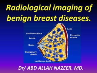
Radiological imaging of
benign breast diseases.
Dr/ ABD ALLAH NAZEER. MD.
Imaging modalities.
Benign breast diseases.
Ductal ectasia with inspissated secretion inside.
Fibroadenoma
a) Raw dynamic contrast-enhanced MR image on lesion, which exhibits high
signal intensity. The mass-like enhancement area is marked by purple arrow
and the lesion; (b) Raw Diffusion-weighted MR image (b = 800 s/mm2); (c)
Calculated ADC map from (b). Lesion area exhibits with light green (pointed
in purple arrow), implying a high ADC value. ADC measured in this lesion is
1.91×10⁻³ s/mm2. doi:10.1371/journal.pone.0087387.g002
a: Dynamic contrast MR demonstrates a breast lesion with rim enhancement. b:
Plateau (type 2) enhancement pattern. Signal intensity values were obtained from the
area of greatest enhancement. c: Spectroscopy detected no Cho signal (SNR 1.7) at 3.2
ppm in representative spectrum and magnified (50) region in the lesion. Histological
analysis of the tissue was benign breast tissue.(Fibroadenoma).
Type I enhancement curve – Fibroadenoma.
Proton MRI and MRSI in a 38-year-old patient (#7) with a fibroadenoma. a: Post-GdDTPA
T1-weighted MR image. b: MRSI of water, Cho, and lipids. c: Unmagnified spectra from the
lesion, demonstrating water and lipid peaks, and a weak Cho signal (SNR ! 4). d: Magnified
($50) spectrum from a voxel in the lesion demonstrating the weak Cho resonance.
A 37-year-old woman with fibroadenoma. Sagittal fat-saturated
postcontrast MRI image (a) shows a well-defined mass lesion with dark
internal septa, indicative of a fibroadenoma. MRS (b) shows a Cho peak at
3.28 ppm, possibly representing GPC, mI, and taurine instead of Cho and PC.
Cystosarcoma phyllodes of the breast (CSPB).
Fibrocystic disease of both breast.
Fibrocystic disease of the breast.
Fibrocystic disease of the left breast.
Cysts are fluid-filled, round or ovoid structures that are
found in as many as one third of women between 35 and 50
years old. Although most are subclinical “microcysts,” in about
20%–25% of cases, palpable (gross) cystic change, which
generally presents as a simple cyst, is encountered . Cysts
cannot reliably be distinguished from solid masses by clinical
breast examination or mammography; in these cases,
ultrasonography and fine needle aspiration (FNA) cytology,
which are highly accurate, are used.
Cysts are derived from the terminal duct lobular unit. In most
cysts, the epithelial lining is either flattened or totally absent.
In only a small number of cysts, an apocrine epithelial lining is
observed. Because gross cysts are not associated with an
increased risk of carcinoma development, the current
consensus on the management of gross cysts is routine follow-
up of the patient, without further therapy.
Simple cyst. Nonenhancing
Idiopathic granulomatous mastitis
Bilateral Lupus Mastitis.
Epidermal inclusion cyst with smooth margins.
Breast MRI shows homogeneous enhancement of perilobular
hemangioma (left), with a type 2 T-SI curve (right)
Hydatid Cyst of the Breast.
Thank You.

More Related Content

What's hot

Presentation1.pptx, radiological imaging of uterine lesions.
Presentation1.pptx, radiological imaging of uterine lesions.Presentation1.pptx, radiological imaging of uterine lesions.
Presentation1.pptx, radiological imaging of uterine lesions.
Abdellah Nazeer
 
Presentation1.pptx, ultrasound examination of the uterus and ovaries.
Presentation1.pptx, ultrasound examination of the uterus and ovaries.Presentation1.pptx, ultrasound examination of the uterus and ovaries.
Presentation1.pptx, ultrasound examination of the uterus and ovaries.
Abdellah Nazeer
 
Presentation1.pptx, radilogical imaging of ovarian lesions.
Presentation1.pptx, radilogical imaging of ovarian lesions.Presentation1.pptx, radilogical imaging of ovarian lesions.
Presentation1.pptx, radilogical imaging of ovarian lesions.
Abdellah Nazeer
 

What's hot (20)

Presentation1.pptx, radiological imaging of uterine lesions.
Presentation1.pptx, radiological imaging of uterine lesions.Presentation1.pptx, radiological imaging of uterine lesions.
Presentation1.pptx, radiological imaging of uterine lesions.
 
BIRADS, Breast Ultrasound, mamography
BIRADS, Breast Ultrasound, mamographyBIRADS, Breast Ultrasound, mamography
BIRADS, Breast Ultrasound, mamography
 
Breast imaging modalities
Breast  imaging modalitiesBreast  imaging modalities
Breast imaging modalities
 
Imaging of breast pathologies
Imaging of breast pathologiesImaging of breast pathologies
Imaging of breast pathologies
 
Breast lesion by ultrasound
Breast lesion by ultrasoundBreast lesion by ultrasound
Breast lesion by ultrasound
 
MRI carcinoma Uterus and Cervix
MRI carcinoma Uterus and  CervixMRI carcinoma Uterus and  Cervix
MRI carcinoma Uterus and Cervix
 
Sono Mammography part 2- Dr Chandni Wadhwani
Sono Mammography part 2- Dr Chandni WadhwaniSono Mammography part 2- Dr Chandni Wadhwani
Sono Mammography part 2- Dr Chandni Wadhwani
 
Breast imaging power point
Breast imaging power pointBreast imaging power point
Breast imaging power point
 
Bi Rads Breast
Bi Rads BreastBi Rads Breast
Bi Rads Breast
 
Radiological approach for malignant breast lesions
Radiological approach for malignant breast lesionsRadiological approach for malignant breast lesions
Radiological approach for malignant breast lesions
 
Sono Mammography part 1- Dr Chandni Wadhwani
Sono Mammography part 1- Dr Chandni WadhwaniSono Mammography part 1- Dr Chandni Wadhwani
Sono Mammography part 1- Dr Chandni Wadhwani
 
Imaging of placenta
Imaging of placentaImaging of placenta
Imaging of placenta
 
BI-RADS (MAMMOGRAPHY)
BI-RADS (MAMMOGRAPHY)BI-RADS (MAMMOGRAPHY)
BI-RADS (MAMMOGRAPHY)
 
Presentation1.pptx, ultrasound examination of the uterus and ovaries.
Presentation1.pptx, ultrasound examination of the uterus and ovaries.Presentation1.pptx, ultrasound examination of the uterus and ovaries.
Presentation1.pptx, ultrasound examination of the uterus and ovaries.
 
Birads mamo n usg dr sumit
Birads mamo n usg dr sumitBirads mamo n usg dr sumit
Birads mamo n usg dr sumit
 
Breast imaging
Breast imagingBreast imaging
Breast imaging
 
Ultrasound breast mass
Ultrasound breast massUltrasound breast mass
Ultrasound breast mass
 
Retroperitoneal masses radiology
Retroperitoneal masses radiologyRetroperitoneal masses radiology
Retroperitoneal masses radiology
 
Presentation1.pptx, radilogical imaging of ovarian lesions.
Presentation1.pptx, radilogical imaging of ovarian lesions.Presentation1.pptx, radilogical imaging of ovarian lesions.
Presentation1.pptx, radilogical imaging of ovarian lesions.
 
Imaging breast mammogram
Imaging breast mammogramImaging breast mammogram
Imaging breast mammogram
 

Viewers also liked

Presentation1.pptx, radiological imaging of dementia.
Presentation1.pptx, radiological imaging of dementia.Presentation1.pptx, radiological imaging of dementia.
Presentation1.pptx, radiological imaging of dementia.
Abdellah Nazeer
 
Liliane ollivier : Breast MR Imaging in Women with High Genetic Risk
Liliane ollivier : Breast MR Imaging in Women with High Genetic RiskLiliane ollivier : Breast MR Imaging in Women with High Genetic Risk
Liliane ollivier : Breast MR Imaging in Women with High Genetic Risk
breastcancerupdatecongress
 
Breast imaging pitffals mri p taourel
Breast imaging pitffals mri p taourelBreast imaging pitffals mri p taourel
Breast imaging pitffals mri p taourel
JFIM
 
Presentation1.pptx, radiological imaging of aids diseases
Presentation1.pptx, radiological imaging of aids diseasesPresentation1.pptx, radiological imaging of aids diseases
Presentation1.pptx, radiological imaging of aids diseases
Abdellah Nazeer
 
Prostate presentation
Prostate presentationProstate presentation
Prostate presentation
Matthew Buck
 
Breast imaging breast mri s taieb
Breast imaging breast mri s taiebBreast imaging breast mri s taieb
Breast imaging breast mri s taieb
JFIM
 

Viewers also liked (20)

Presentation1.pptx, radiological imaging of dementia.
Presentation1.pptx, radiological imaging of dementia.Presentation1.pptx, radiological imaging of dementia.
Presentation1.pptx, radiological imaging of dementia.
 
Approach to a patient with breast lump
Approach to a patient with breast lumpApproach to a patient with breast lump
Approach to a patient with breast lump
 
Benign breast disease
Benign breast diseaseBenign breast disease
Benign breast disease
 
Presentation1, radiological imaging of ear microcia.
Presentation1, radiological imaging of ear microcia.Presentation1, radiological imaging of ear microcia.
Presentation1, radiological imaging of ear microcia.
 
Translational Research in Functional Optical Brain Imaging
Translational Research in Functional Optical  Brain ImagingTranslational Research in Functional Optical  Brain Imaging
Translational Research in Functional Optical Brain Imaging
 
NIR imaging
NIR imaging NIR imaging
NIR imaging
 
Liliane ollivier : Breast MR Imaging in Women with High Genetic Risk
Liliane ollivier : Breast MR Imaging in Women with High Genetic RiskLiliane ollivier : Breast MR Imaging in Women with High Genetic Risk
Liliane ollivier : Breast MR Imaging in Women with High Genetic Risk
 
Onaral IITL_Brainstorm25feb10v4
Onaral IITL_Brainstorm25feb10v4Onaral IITL_Brainstorm25feb10v4
Onaral IITL_Brainstorm25feb10v4
 
NIR Three dimensional imaging of breast model using f-DOT
NIR Three dimensional imaging of breast model using f-DOT  NIR Three dimensional imaging of breast model using f-DOT
NIR Three dimensional imaging of breast model using f-DOT
 
Emerging new imaging_techniques_2014
Emerging new imaging_techniques_2014Emerging new imaging_techniques_2014
Emerging new imaging_techniques_2014
 
2014 CFTCC Annual Symposium: A New Wearable Diffuse Optical Spectroscopic Ima...
2014 CFTCC Annual Symposium: A New Wearable Diffuse Optical Spectroscopic Ima...2014 CFTCC Annual Symposium: A New Wearable Diffuse Optical Spectroscopic Ima...
2014 CFTCC Annual Symposium: A New Wearable Diffuse Optical Spectroscopic Ima...
 
Breast imaging pitffals mri p taourel
Breast imaging pitffals mri p taourelBreast imaging pitffals mri p taourel
Breast imaging pitffals mri p taourel
 
Whole brain optical imaging
Whole brain optical imagingWhole brain optical imaging
Whole brain optical imaging
 
Breast Carcinoma
Breast CarcinomaBreast Carcinoma
Breast Carcinoma
 
1.breast cancer screening(backup slide-1)
1.breast cancer screening(backup slide-1)1.breast cancer screening(backup slide-1)
1.breast cancer screening(backup slide-1)
 
Presentation1.pptx, radiological imaging of aids diseases
Presentation1.pptx, radiological imaging of aids diseasesPresentation1.pptx, radiological imaging of aids diseases
Presentation1.pptx, radiological imaging of aids diseases
 
Prostate presentation
Prostate presentationProstate presentation
Prostate presentation
 
Breast imaging breast mri s taieb
Breast imaging breast mri s taiebBreast imaging breast mri s taieb
Breast imaging breast mri s taieb
 
Mr Mammography New
Mr Mammography NewMr Mammography New
Mr Mammography New
 
Breast cancer video 1
Breast cancer video 1Breast cancer video 1
Breast cancer video 1
 

Similar to Presentation1.pptx, radiological imaging of beign breast diseases

DWI borderline / malignant epithelial ovarian tumors
DWI borderline / malignant epithelial ovarian tumorsDWI borderline / malignant epithelial ovarian tumors
DWI borderline / malignant epithelial ovarian tumors
Naglaa Mahmoud
 
The study of different presentations of breast lumps in radiographic. acta me...
The study of different presentations of breast lumps in radiographic. acta me...The study of different presentations of breast lumps in radiographic. acta me...
The study of different presentations of breast lumps in radiographic. acta me...
Sanjeev kumar Jain
 
Presentation1.pptx, perfusiona and specroscopy imaging in brain tumour.
Presentation1.pptx, perfusiona and specroscopy imaging in brain tumour.Presentation1.pptx, perfusiona and specroscopy imaging in brain tumour.
Presentation1.pptx, perfusiona and specroscopy imaging in brain tumour.
Abdellah Nazeer
 
Imaging modalities
Imaging modalitiesImaging modalities
Imaging modalities
Rad Tech
 
Presentation1, radiological application of diffusion weighted images in breas...
Presentation1, radiological application of diffusion weighted images in breas...Presentation1, radiological application of diffusion weighted images in breas...
Presentation1, radiological application of diffusion weighted images in breas...
Abdellah Nazeer
 

Similar to Presentation1.pptx, radiological imaging of beign breast diseases (20)

DWI borderline / malignant epithelial ovarian tumors
DWI borderline / malignant epithelial ovarian tumorsDWI borderline / malignant epithelial ovarian tumors
DWI borderline / malignant epithelial ovarian tumors
 
The study of different presentations of breast lumps in radiographic. acta me...
The study of different presentations of breast lumps in radiographic. acta me...The study of different presentations of breast lumps in radiographic. acta me...
The study of different presentations of breast lumps in radiographic. acta me...
 
Mammogram and BI-RADS classification .pptx
Mammogram and BI-RADS classification .pptxMammogram and BI-RADS classification .pptx
Mammogram and BI-RADS classification .pptx
 
USG OF PELVIC MASSES BY DR ABHIJIT R SINGH
USG OF PELVIC MASSES BY DR ABHIJIT R SINGHUSG OF PELVIC MASSES BY DR ABHIJIT R SINGH
USG OF PELVIC MASSES BY DR ABHIJIT R SINGH
 
Breast Cancer by dr Isa Basuki
Breast Cancer by dr Isa BasukiBreast Cancer by dr Isa Basuki
Breast Cancer by dr Isa Basuki
 
Investigations of breast cancer
Investigations of breast cancerInvestigations of breast cancer
Investigations of breast cancer
 
Mammography
MammographyMammography
Mammography
 
Mammography -A ppt bt J K PATIL, Prof,dept of radiology
Mammography -A ppt bt J K PATIL, Prof,dept of radiologyMammography -A ppt bt J K PATIL, Prof,dept of radiology
Mammography -A ppt bt J K PATIL, Prof,dept of radiology
 
Presentation1.pptx, perfusiona and specroscopy imaging in brain tumour.
Presentation1.pptx, perfusiona and specroscopy imaging in brain tumour.Presentation1.pptx, perfusiona and specroscopy imaging in brain tumour.
Presentation1.pptx, perfusiona and specroscopy imaging in brain tumour.
 
Imaging modalities
Imaging modalitiesImaging modalities
Imaging modalities
 
medulloblastoma
medulloblastomamedulloblastoma
medulloblastoma
 
A rare case of primary rectal 2012
A rare case of primary rectal 2012A rare case of primary rectal 2012
A rare case of primary rectal 2012
 
Dr. masciotra case of the day n.1 - breast cancer
Dr. masciotra  case of the day n.1 - breast cancerDr. masciotra  case of the day n.1 - breast cancer
Dr. masciotra case of the day n.1 - breast cancer
 
23204937
2320493723204937
23204937
 
Recent advances in Mammography
Recent advances in MammographyRecent advances in Mammography
Recent advances in Mammography
 
recentadvancesinmammography-150410103808-conversion-gate01.pdf
recentadvancesinmammography-150410103808-conversion-gate01.pdfrecentadvancesinmammography-150410103808-conversion-gate01.pdf
recentadvancesinmammography-150410103808-conversion-gate01.pdf
 
D. antonio pio masciotra breast cancer seen on chest ct
D. antonio pio masciotra   breast cancer seen on chest ctD. antonio pio masciotra   breast cancer seen on chest ct
D. antonio pio masciotra breast cancer seen on chest ct
 
Presentation1, radiological application of diffusion weighted images in breas...
Presentation1, radiological application of diffusion weighted images in breas...Presentation1, radiological application of diffusion weighted images in breas...
Presentation1, radiological application of diffusion weighted images in breas...
 
3 d ultrasound in gynecology presentation
3 d ultrasound in gynecology presentation3 d ultrasound in gynecology presentation
3 d ultrasound in gynecology presentation
 
Presentation1, radiological imaging of anal carcinoma.
Presentation1, radiological imaging of anal carcinoma.Presentation1, radiological imaging of anal carcinoma.
Presentation1, radiological imaging of anal carcinoma.
 

More from Abdellah Nazeer

Presentation1, radiological imaging of lateral hindfoot impingement.
Presentation1, radiological imaging of lateral hindfoot impingement.Presentation1, radiological imaging of lateral hindfoot impingement.
Presentation1, radiological imaging of lateral hindfoot impingement.
Abdellah Nazeer
 
Presentation2, radiological anatomy of the liver and spleen.
Presentation2, radiological anatomy of the liver and spleen.Presentation2, radiological anatomy of the liver and spleen.
Presentation2, radiological anatomy of the liver and spleen.
Abdellah Nazeer
 
Presentation1, artifacts and pitfalls of the wrist and elbow joints.
Presentation1, artifacts and pitfalls of the wrist and elbow joints.Presentation1, artifacts and pitfalls of the wrist and elbow joints.
Presentation1, artifacts and pitfalls of the wrist and elbow joints.
Abdellah Nazeer
 
Presentation1, artifact and pitfalls of the knee, hip and ankle joints.
Presentation1, artifact and pitfalls of the knee, hip and ankle joints.Presentation1, artifact and pitfalls of the knee, hip and ankle joints.
Presentation1, artifact and pitfalls of the knee, hip and ankle joints.
Abdellah Nazeer
 
Presentation1, radiological imaging of artifact and pitfalls in shoulder join...
Presentation1, radiological imaging of artifact and pitfalls in shoulder join...Presentation1, radiological imaging of artifact and pitfalls in shoulder join...
Presentation1, radiological imaging of artifact and pitfalls in shoulder join...
Abdellah Nazeer
 
Presentation1, radiological imaging of internal abdominal hernia.
Presentation1, radiological imaging of internal abdominal hernia.Presentation1, radiological imaging of internal abdominal hernia.
Presentation1, radiological imaging of internal abdominal hernia.
Abdellah Nazeer
 
Presentation11, radiological imaging of ovarian torsion.
Presentation11, radiological imaging of ovarian torsion.Presentation11, radiological imaging of ovarian torsion.
Presentation11, radiological imaging of ovarian torsion.
Abdellah Nazeer
 
Presentation1, new mri techniques in the diagnosis and monitoring of multiple...
Presentation1, new mri techniques in the diagnosis and monitoring of multiple...Presentation1, new mri techniques in the diagnosis and monitoring of multiple...
Presentation1, new mri techniques in the diagnosis and monitoring of multiple...
Abdellah Nazeer
 
Presentation1, radiological application of diffusion weighted mri in neck mas...
Presentation1, radiological application of diffusion weighted mri in neck mas...Presentation1, radiological application of diffusion weighted mri in neck mas...
Presentation1, radiological application of diffusion weighted mri in neck mas...
Abdellah Nazeer
 
Presentation1, radiological application of diffusion weighted images in abdom...
Presentation1, radiological application of diffusion weighted images in abdom...Presentation1, radiological application of diffusion weighted images in abdom...
Presentation1, radiological application of diffusion weighted images in abdom...
Abdellah Nazeer
 
Presentation1, radiological application of diffusion weighted imges in neuror...
Presentation1, radiological application of diffusion weighted imges in neuror...Presentation1, radiological application of diffusion weighted imges in neuror...
Presentation1, radiological application of diffusion weighted imges in neuror...
Abdellah Nazeer
 

More from Abdellah Nazeer (20)

Muculoskeletal Pediatic Imaging..pptx
Muculoskeletal Pediatic Imaging..pptxMuculoskeletal Pediatic Imaging..pptx
Muculoskeletal Pediatic Imaging..pptx
 
Presentation1, Ultrasound of the bowel loops and the lymph nodes..pptx
Presentation1, Ultrasound of the bowel loops and the lymph nodes..pptxPresentation1, Ultrasound of the bowel loops and the lymph nodes..pptx
Presentation1, Ultrasound of the bowel loops and the lymph nodes..pptx
 
Presentation1 Short cases MD..pptx
Presentation1 Short cases MD..pptxPresentation1 Short cases MD..pptx
Presentation1 Short cases MD..pptx
 
Presentation1, MD MCQ Cases..pptx
Presentation1, MD MCQ Cases..pptxPresentation1, MD MCQ Cases..pptx
Presentation1, MD MCQ Cases..pptx
 
Presentation1, Short Cases Quiz..pptx
Presentation1, Short Cases Quiz..pptxPresentation1, Short Cases Quiz..pptx
Presentation1, Short Cases Quiz..pptx
 
Presentation1, radiological imaging of lateral hindfoot impingement.
Presentation1, radiological imaging of lateral hindfoot impingement.Presentation1, radiological imaging of lateral hindfoot impingement.
Presentation1, radiological imaging of lateral hindfoot impingement.
 
Presentation2, radiological anatomy of the liver and spleen.
Presentation2, radiological anatomy of the liver and spleen.Presentation2, radiological anatomy of the liver and spleen.
Presentation2, radiological anatomy of the liver and spleen.
 
Presentation1, artifacts and pitfalls of the wrist and elbow joints.
Presentation1, artifacts and pitfalls of the wrist and elbow joints.Presentation1, artifacts and pitfalls of the wrist and elbow joints.
Presentation1, artifacts and pitfalls of the wrist and elbow joints.
 
Presentation1, artifact and pitfalls of the knee, hip and ankle joints.
Presentation1, artifact and pitfalls of the knee, hip and ankle joints.Presentation1, artifact and pitfalls of the knee, hip and ankle joints.
Presentation1, artifact and pitfalls of the knee, hip and ankle joints.
 
Presentation1, radiological imaging of artifact and pitfalls in shoulder join...
Presentation1, radiological imaging of artifact and pitfalls in shoulder join...Presentation1, radiological imaging of artifact and pitfalls in shoulder join...
Presentation1, radiological imaging of artifact and pitfalls in shoulder join...
 
Presentation1, radiological imaging of internal abdominal hernia.
Presentation1, radiological imaging of internal abdominal hernia.Presentation1, radiological imaging of internal abdominal hernia.
Presentation1, radiological imaging of internal abdominal hernia.
 
Presentation11, radiological imaging of ovarian torsion.
Presentation11, radiological imaging of ovarian torsion.Presentation11, radiological imaging of ovarian torsion.
Presentation11, radiological imaging of ovarian torsion.
 
Presentation1, musculoskeletal anatomy.
Presentation1, musculoskeletal anatomy.Presentation1, musculoskeletal anatomy.
Presentation1, musculoskeletal anatomy.
 
Presentation1, new mri techniques in the diagnosis and monitoring of multiple...
Presentation1, new mri techniques in the diagnosis and monitoring of multiple...Presentation1, new mri techniques in the diagnosis and monitoring of multiple...
Presentation1, new mri techniques in the diagnosis and monitoring of multiple...
 
Presentation1, radiological application of diffusion weighted mri in neck mas...
Presentation1, radiological application of diffusion weighted mri in neck mas...Presentation1, radiological application of diffusion weighted mri in neck mas...
Presentation1, radiological application of diffusion weighted mri in neck mas...
 
Presentation1, radiological application of diffusion weighted images in abdom...
Presentation1, radiological application of diffusion weighted images in abdom...Presentation1, radiological application of diffusion weighted images in abdom...
Presentation1, radiological application of diffusion weighted images in abdom...
 
Presentation1, radiological application of diffusion weighted imges in neuror...
Presentation1, radiological application of diffusion weighted imges in neuror...Presentation1, radiological application of diffusion weighted imges in neuror...
Presentation1, radiological application of diffusion weighted imges in neuror...
 
Presentation1, mr physics.
Presentation1, mr physics.Presentation1, mr physics.
Presentation1, mr physics.
 
Presentation1. ct physics.
Presentation1. ct physics.Presentation1. ct physics.
Presentation1. ct physics.
 
Presentation1, ultrasound physics.
Presentation1, ultrasound physics.Presentation1, ultrasound physics.
Presentation1, ultrasound physics.
 

Presentation1.pptx, radiological imaging of beign breast diseases

  • 1. Radiological imaging of benign breast diseases. Dr/ ABD ALLAH NAZEER. MD.
  • 2.
  • 3.
  • 4.
  • 6.
  • 7.
  • 8.
  • 9.
  • 10.
  • 11.
  • 12.
  • 14.
  • 15.
  • 16.
  • 17.
  • 18.
  • 19.
  • 20.
  • 21.
  • 22.
  • 23.
  • 24.
  • 25.
  • 26.
  • 27. Ductal ectasia with inspissated secretion inside.
  • 28.
  • 29.
  • 30.
  • 31.
  • 32.
  • 33.
  • 35.
  • 36. a) Raw dynamic contrast-enhanced MR image on lesion, which exhibits high signal intensity. The mass-like enhancement area is marked by purple arrow and the lesion; (b) Raw Diffusion-weighted MR image (b = 800 s/mm2); (c) Calculated ADC map from (b). Lesion area exhibits with light green (pointed in purple arrow), implying a high ADC value. ADC measured in this lesion is 1.91×10⁻³ s/mm2. doi:10.1371/journal.pone.0087387.g002
  • 37. a: Dynamic contrast MR demonstrates a breast lesion with rim enhancement. b: Plateau (type 2) enhancement pattern. Signal intensity values were obtained from the area of greatest enhancement. c: Spectroscopy detected no Cho signal (SNR 1.7) at 3.2 ppm in representative spectrum and magnified (50) region in the lesion. Histological analysis of the tissue was benign breast tissue.(Fibroadenoma).
  • 38. Type I enhancement curve – Fibroadenoma.
  • 39.
  • 40. Proton MRI and MRSI in a 38-year-old patient (#7) with a fibroadenoma. a: Post-GdDTPA T1-weighted MR image. b: MRSI of water, Cho, and lipids. c: Unmagnified spectra from the lesion, demonstrating water and lipid peaks, and a weak Cho signal (SNR ! 4). d: Magnified ($50) spectrum from a voxel in the lesion demonstrating the weak Cho resonance.
  • 41. A 37-year-old woman with fibroadenoma. Sagittal fat-saturated postcontrast MRI image (a) shows a well-defined mass lesion with dark internal septa, indicative of a fibroadenoma. MRS (b) shows a Cho peak at 3.28 ppm, possibly representing GPC, mI, and taurine instead of Cho and PC.
  • 42.
  • 43.
  • 44. Cystosarcoma phyllodes of the breast (CSPB).
  • 45.
  • 46.
  • 47.
  • 48. Fibrocystic disease of both breast.
  • 49. Fibrocystic disease of the breast.
  • 50. Fibrocystic disease of the left breast.
  • 51. Cysts are fluid-filled, round or ovoid structures that are found in as many as one third of women between 35 and 50 years old. Although most are subclinical “microcysts,” in about 20%–25% of cases, palpable (gross) cystic change, which generally presents as a simple cyst, is encountered . Cysts cannot reliably be distinguished from solid masses by clinical breast examination or mammography; in these cases, ultrasonography and fine needle aspiration (FNA) cytology, which are highly accurate, are used. Cysts are derived from the terminal duct lobular unit. In most cysts, the epithelial lining is either flattened or totally absent. In only a small number of cysts, an apocrine epithelial lining is observed. Because gross cysts are not associated with an increased risk of carcinoma development, the current consensus on the management of gross cysts is routine follow- up of the patient, without further therapy.
  • 53.
  • 54.
  • 57.
  • 58.
  • 59.
  • 60.
  • 61.
  • 62.
  • 63. Epidermal inclusion cyst with smooth margins.
  • 64. Breast MRI shows homogeneous enhancement of perilobular hemangioma (left), with a type 2 T-SI curve (right)
  • 65. Hydatid Cyst of the Breast.
  • 66.
  • 67.