1. Earn
4 CE credits
This course was
written for dentists,
dental hygienists,
and assistants.
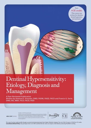
Dentinal Hypersensitivity:
Etiology, Diagnosis and
Management
A Peer-Reviewed Publication
Written by Howard E. Strassler, DMD, FADM, FAGD, FACD and Francis G. Serio,
DMD, MS, MBA, FICD, FACD, FADI
PennWell is an ADA CERP recognized provider
ADA CERP is a service of the American Dental Association to assist dental professionals in identifying
quality providers of continuing dental education. ADA CERP does not approve or endorse individual
courses or instructors, nor does it imply acceptance of credit hours by boards of dentistry.
Concerns of complaints about a CE provider may be directed to the provider or to ADA CERP at
www.ada.org/goto/cerp.
This course has been made possible through an unrestricted educational grant from Colgate-Palmolive Company. The cost of this CE course is $59.00 for 4 CE credits.
Cancellation/Refund Policy: Any participant who is not 100% satisfied with this course can request a full refund by contacting PennWell in writing.
2. Educational Objectives
The overall goal of this course is to provide dental professionals with information on the etiology, diagnosis and
treatment of dentinal hypersensitivity. Upon completion of
this course, the participant will be able to do the following:
1. Know the incidence of dentinal hypersensitivity and
risk factors for this condition
2. Know the anatomical and physiological features, and
the accepted theory, associated with dentinal hypersensitivity
3. Understand the need for screening and diagnosis by
exclusion for dentinal hypersensitivity
4. Know the treatment options available for dentinal
hypersensitivity and considerations in selecting these.
Abstract
Dentinal hypersensitivity has been referred to as one of the
most painful and chronic dental conditions, with a reported
prevalence of between 4% and 57% in the general population
and a higher prevalence in periodontal patients. It may also
occur as a result of, or during, dental treatment. Clinicians
must screen for dentinal hypersensitivity and diagnose by
exclusion, determine appropriate treatment, and provide
treatment and preventive recommendations. Consideration
should also be given to treating dentinal hypersensitivity
associated with dental treatment. Traditional treatments
have included adhesive resins, fluoride varnishes, HEMA,
iontophoresis, gingival grafts and desensitizing dentifrices.
Other technologies include the use of bioglass particles,
ACP, as well as 8% arginine and calcium carbonate paste.
Introduction
During routine dental examinations, our patients frequently inquire about dentinal hypersensitivity that was one
episode or is chronic and recurring due to a given action,
e.g., drinking cold beverages, eating hot foods, breathing
in and out. This common complaint is defined as dentinal
hypersensitivity, but it is also known as root sensitivity,
or just sensitivity. Patients describe this phenomenon as
sharp, short-lasting tooth pain, irrespective of the stimulus.1 Holland et al. described dentinal hypersensitivity as
“characterized by short, sharp pain arising from exposed
dentin in response to stimuli typically thermal, evaporative,
tactile, osmotic or chemical and which cannot be ascribed
to any other form of dental defect or pathology.”2
The prevalence of dentinal hypersensitivity has been
reported to be between 4% and 57% in the general population.3-10 Among periodontal patients, its frequency is considerably higher (60%–98%).11,12 This hypersensitivity may
be due to cementum removal during root instrumentation.
Dentinal hypersensitivity has been described as generally
occurring in patients 30 to 40 years old,13 but it can occur in
patients significantly younger or older. Women may be affected more often than men.14 Dentinal hypersensitivity af2
fects incisors, canines, premolars and molars, with canines
and premolars reported to be affected most often.15,16
Patients with dentinal hypersensitivity may not specifically seek treatment, because they do not view it as a
significant dental health problem, but will mention it at a
routine dental appointment.17 At other times, patients will
seek treatment recommendations from their dental professionals. Some patients are concerned whenever there is
dental pain,18 and for some the first time they experience
dentinal hypersensitivity creates fear that there is something more serious occurring. The authors of this course
have had patients report sensitivity who believe that it may
be a toothache that requires immediate attention so that the
pain does not get worse. Patients can identify areas of dentinal hypersensitivity before a clinical exam is performed.
This may be chronic, or unpredictable and cause intermittent discomfort that is difficult to pinpoint.19,20 Other patients cannot distinguish between dentinal sensitivity and
gingival sensitivity. Patients may also experience dentinal
hypersensitivity as a result of treatment such as scaling and
root planing or during routine and normal actions associated with treatment, such as when a tooth is dried using an
air spray or scratched with the tip of an explorer. Dental
treatment can also exacerbate pre-existing sensitivity.
Dentinal hypersensitivity has all the criteria to be
considered a true pain syndrome.21 It is important to distinguish sensitivity pain, that of short duration, from pain
of longer duration not treatable with desensitizing agents.
A painful response that lingers or that wakens the person
from a sound sleep may be the result of pulpal inflammation. A diagnosis by the dentist is necessary to establish
a cause and effect, and a diagnosis by exclusion must be
made for dentinal hypersensitivity, ruling out other conditions requiring different treatment. After the diagnosis
of dentinal hypersensitivity has been made, depending
on the etiology, recommendations can be made for effective treatment. Calvo noted in 1884: “There is great need
of a medicament, which while lessening the sensitivity of
dentin, will not impair the vitality of the pulp.”22 Recommendations can include in-office, at-home professionally
dispensed or over-the counter treatments.23-26 Regardless of
which treatment recommendations are made and provided,
it is important to follow up with the patient to evaluate the
therapeutic results.
Etiology and Physiology of Dentinal
Hypersensitivity
Dentinal hypersensitivity can have multiple etiologies. It
is important that the patient’s medical and social history,
lifestyle, medications and supplements being taken, diet
and food habits, and oral hygiene be thoroughly reviewed.
Before making a diagnosis of dentinal hypersensitivity,
other oral conditions must be ruled out, including occlusal
trauma, caries, defective restorations, fractured or cracked
www.ineedce.com
3. teeth, potential reversible or irreversible pulpal pathology,
or gingival conditions.14,24 For instance, pain during chewing may be due to a fractured and mobile restoration that
is rubbing against the dentin or diagnostic for a cracked
tooth.27
Dentin is sensitive due to its anatomy and physiology. It
is a porous, mineralized connective tissue with an organic
matrix of collagenous proteins and an inorganic component, hydroxyapatite. Dentinal tubules are micro-canals
that radiate outward through the dentin from the pulp
cavity to the dentinal surface, with different configurations
and diameters in different teeth. For human dentin, one
square millimeter can contain 30,000 tubules, depending
on depth. Each tubule contains a Tomes fiber (cytoplastic
cell process) and an odontoblast that communicates with
the pulp. Within the dentinal tubules there are two types
of nerve fibers, myelinated (A-fibers) and unmyelinated
(C-fibers).28 The A-fibers are responsible for the sensation
of dentinal hypersensitivity, perceived as pain in response
to all stimuli.
The most widely accepted mechanism of dentinal
sensitivity is the hydrodynamic theory, first described by
Brännström.29,30 In this model, the aspiration of odontoblasts into the dentinal tubules, as an immediate effect
of physical stimuli applied to exposed dentin, results in
the outward flow of the tubular contents (dentinal fluids)
through capillary action (Figure 1). The changes to the
dentinal surface lead to stimulation of the A-type nerve
fibers surrounding the odontoblasts. For there to be a
stimulus response, the tubules must be open at both the
dentinal interface and within the pulp. Absi and coworkers
reported that nonsensitive teeth were not responsive to any
physical stimuli; sensitive teeth had up to eight times the
number of open dentinal tubules per surface area compared
to nonresponsive teeth.31 Another theory is an alteration in
pulpal sensory nerve activity.32 The treatment of exposed,
open dentinal tubules is based upon the physiology of the
stimulus response.
Figure 1. The hydrodynamic theory
www.ineedce.com
Location of Dentinal Hypersensitivity –
Patients at Risk
Why are some root surfaces hypersensitive and others
are not?
Exposed root surfaces due to gingival recession are a
major predisposing factor to dentinal root hypersensitivity (Figure 2).33 According to a recent report of adults over
the age of 60, almost 32% had root caries or a restored root
surface.34 Since root caries are an indication of periodontal
attachment loss and subsequent recession, this defines the
population of adults over 60 with an at-risk of recession in
at least one or more teeth as at least 30%. Another study
concluded that at least 22% of the adult population between
30 and 90 years of age will have evidence of recession in
one or more teeth of 3 mm or more.35 Gingival recession
is more common as patients age and in patients with better
oral hygiene.14,36 Common causes include inadequate attached gingiva, prominent roots with a thin alveolar housing or bony dehiscence, toothbrush abrasion, periodontal
surgery, factitial habits (e.g., picking at cervical area of the
tooth with a fingernail), excessive tooth cleaning, excessive
flossing, loss of gingival attachment due to specific pathologies, and iatrogenic loss of attachment during restorative
procedures.33,37
Figure 2. Gingival recession with exposed root surfaces
Exposed lingual root surfaces
Dentinal hypersensitivity can also occur as a result of a routine dental cleaning, or be exacerbated during scaling and
root planing or routine dental prophylaxis and polishing due
to pre-existing dentin-root hypersensitivity. Patients who
have had or are having periodontal therapy are at risk;12 the
prevalence of root sensitivity has been reported as 9%–23%
before and 54%–55% after periodontal therapy. An increase
in the intensity of root sensitivity occurred one to three weeks
following therapy, after which it slowly decreased. An assessment found that all patients experienced increased discomfort
and dentinal hypersensitivity after periodontal treatment,
including scaling and root planing.37 Fear of pain and discomfort during subgingival instrumentation has been reported to
deter 10% of the population from seeking treatment.38 Once
the root surfaces are exposed, the cementum/dentin is more
susceptible to caries and loss of tooth substance due to erosion, abrasion and abfraction (Figure 3).39-42 Postprocedural
3
4. sensitivity can also be a result of etching beyond restoration
margins, leaving dentinal tubules open, or of finishing and
polishing a restoration that extends to the root surfaces,
which can also leave dentinal tubules open. Root surfaces on
teeth adjacent to a tooth being extracted can be abraded and
scarred with the use of dental elevators during the extraction
procedure. Resective periodontal surgical procedures may
also leave roots exposed. Enamel loss with exposed dentin due
to attrition and tooth wear due to bruxism, occlusal habits and
other forms of parafunctional activity can also contribute to
the etiology of dentinal hypersensitivity (Figure 4).41
Figure 3. Gingival recession with associated noncarious cervical lesions
Biofilm deposits on root surfaces may also increase hypersensitivity. The opening of dentinal tubules can also occur
due to poor oral hygiene techniques leaving bacterial plaque/
biofilm on root surfaces, with the acidic by-products of the
biofilm opening the dentinal tubules. Conversely, overzealous oral hygiene techniques can cause continued dentinal
tubule exposure. Root surfaces exposed to the physical action
of toothbrushing with and without toothpaste can be predisposing factors in removing the smear layer, leaving a tooth
hypersensitive.13,45 Exposure of the oral cavity to acids, e.g.,
ingestion of acidic foods and beverages46-48 or ingestion of
chlorinated pool water,49 as well as bulimia and gastrointestinal reflux disease can also contribute to the opening of the end
of the dentinal tubules (Figure 6).50 Brushing immediately
after ingesting acidic foods or beverages should be avoided.51
Figure 6. Erosion of the maxillary anterior teeth in a bulemic
patient due to stomach acid
Figure 4. Enamel loss with exposed dentin due to attrition
Screening and Diagnosis of Dentinal
Hypersensitivity
In normal function, the tubules sclerose and become plugged,
and when dentin is cut or abraded the mineralized matrix
produces debris that spreads over the dentin surface to form a
smear layer.43,44 This occurs to both enamel and dentin,44 but the
loss of this smear layer, the unplugging of the dentinal tubules,
contributes to dentinal hypersensitivity (Figure 5).
Figure 5. Scanning electron micrograph demonstrating open
dentinal tubules
4
Dentists and dental hygienists unfortunately do not all routinely include screening for dentinal hypersensitivity.25 In
1995, a random sample of Dutch dentists completed a survey on the prevalence, conditions and treatment of cervical
hypersensitivity of their patients.52 A similar questionnaire
was administered to U.K. dentists in 2002.53 For both groups,
the results revealed discrepancies in screening, perceptions
and knowledge of treatment. A separate study administered
a questionnaire by mail to 5,000 dentists and 3,000 dental
hygienists in Canada and revealed that fewer than half of the
respondents considered a differential diagnosis for dentinal
hypersensitivity, even though it is by definition a diagnosis
of exclusion.25 Many misidentified the etiology: 64% of the
dentists and 77% of the hygienists incorrectly cited bruxism
and malocclusion as triggers for dentinal hypersensitivity,
while only 7% of dentists and 5% of dental hygienists correctly
identified erosion as a primary cause and 17% of dentists
and 48% of hygienists were unable to identify the accepted
theory of hypersensitivity. Only half of the respondents had
the confidence to manage a patient’s pain and to consider the
modification of predisposing factors to control a patient’s
pain. This survey also demonstrated a lack of understanding
of desensitizing toothpastes – most dentists (56%) and dental hygienists (68%) believed these helped prevent dentinal
www.ineedce.com
5. hypersensitivity, while 31% and 16%, respectively, did not
believe that desensitizing toothpastes provided relief from
dentinal hypersensitivity.
Dental professionals need to fully understand the etiology and treatment of dentinal hypersensitivity, to screen for
it and to diagnose it by exclusion. It is also worth noting that
patients with unresolved hypersensitivity over many years
provide the dental professional with varied behavioral and
postural clues, some of which are easily recognized. These
include avoidance of routine dental exams, necessary treatment and follow-up care, reluctance to schedule planned
treatment or follow-up care, insistence on the use of local
anesthesia for even the most minor of dental treatments,
tense facial muscles, tooth clenching, a rigid torso, holding
hands tightly on the arm rest, crossed arms, an awkward
head position and an inability to follow routine instructions
for head and body positioning.19
As part of any screening for dentinal hypersensitivity, the
clinician should assess whether there is a localized or generalized problem. In addition, for patients with identified isolated
and generalized dentinal hypersensitivity, a routine dental
cleaning can be anxiety provoking.38 Consideration should
be given to dentinal hypersensitivity associated with dental
treatment – during treatment and postoperatively. While the
focus of controlling pain for many dental professionals during periodontal scaling and root planing and routine dental
cleanings has been the use of local and topical anesthetic
agents,37,54,55 we should also give thought to providing our
patients with treatments to relieve postprocedural dentinal
hypersensitivity.19,26,56,57
Treatment and Prevention of Dentinal
Hypersensitivity
Once the diagnosis of dentinal hypersensitivity has been
made and the etiologic factors identified, treatment and prevention should be primary goals,19,58,59 and a treatment plan
can be developed and implemented. Once a tooth or teeth are
predisposed to dentinal hypersensitivity, they will need to be
re-evaluated for continued treatment. The patient should be
shown correct brushing techniques to prevent further loss of
dentin that would contribute to dentinal hypersensitivity;
improper toothbrushing has also been associated with dentinal hypersensitivity.1 It has been shown that both a manual
and a power brush used with a desensititizing toothpaste are
almost equivalent in effectiveness.60 If there are changes and
behavior modifications or treatments that can be made, these
should be discussed with the patient. Drisko summarized
preventive recommendations (Table 1).61
Treatment of Dentinal Hypersensitivity
Two major groups of products are used to treat dentinal
hypersensitivity: those that block and occlude dentinal tubules, and those that interfere with the transmission of neural impulses. Localized dentinal hypersensitivity can usually
www.ineedce.com
be treated in-office. For generalized conditions where there is
significant recession on multiple teeth, an at-home treatment
regimen may be a better choice.
Table 1. Preventive Recommendations for
Dentinal Hypersensitivity
Suggestions for patients:
Avoid using large amounts of dentifrice or reapplying it during
brushing.
Avoid medium- or hard-bristle toothbrushes.
Avoid brushing teeth immediately after ingesting acidic foods.
Avoid overbrushing with excessive pressure or for an extended
period of time.
Avoid excessive flossing or improper use of other interproximal
cleaning devices.
Avoid “picking” or scratching at the gumline or using toothpicks
inappropriately.
Suggestions for professionals:
Avoid overinstrumenting the root surfaces during scaling and
root planing, particularly in the cervical area of the tooth.
Avoid overpolishing exposed dentin during stain removal.
Avoid violating the biologic width during restoration placement,
as this may cause recession.
Avoid burning the gingival tissues during in-office bleaching,
and advise patients to be careful when using home bleaching
products.
Professional in-office treatments
In-office desensitizing agents work by occluding and sealing the dentin tubules.62,63 When treating patients with an
in-office treatment, American Dental Association treatment
codes can be noted for insurance reimbursement (Table 2).
Table 2. In-office desensitizing codes
Miscellaneous services
D9910 Application of desensitizing medicament
Includes in-office treatment of root sensitivity. Typically reported on a “per visit” basis for application of topical fluoride or
other desensitizing agents. This code is not used for bases, liners
or adhesives used under restorations.
D9911 Application of desensitizing resin for cervical and/or root
surface
Typically reported on a “per tooth” basis for application of adhesive resins. This code is not used for bases, liners or adhesives
used under restorations.
A recent novel approach is a technology based on arginine, a natural product, and calcium carbonate. This technology was introduced as a result of the need to provide patients
with a treatment regimen to reduce and treat postprocedural
dentinal hypersensitivity after dental cleanings. In 2002,
Kleinberg et al. reported on the development of this novel
desensitizing technology based upon the role that saliva
plays in naturally reducing dentinal hypersensitivity. Saliva
5
6. provides calcium and phosphate, which over time will occlude and block open dentinal tubules from external stimuli
associated with dentinal hypersensitivity.19,56 Reduced salivary flow, hyposalivation and xerostomia are risk factors for
caries and tooth demineralization and may exacerbate dentinal hypersensitivity. While hyposalivation may be due to
medical conditions and aging, it is also a side effect of more
than 500 prescription and over-the-counter medications.64
The mechanism providing for the clinical effectiveness
of this technology utilizes arginine, an amino acid; bicarbonate, a pH buffer; and calcium carbonate, a source of calcium.
This technology, originally introduced as Sensistat® (Ortek
Therapeutics, Roslyn Heights, NY), effectively relieves
dentinal hypersensitivity.56 The technology is proposed to
block dentinal hypersensitivity pain by occluding dentinal
tubules by using arginine, which is positively charged at
physiologic pH of 6.5-7.5, to bind to the negatively charged
dentin surface, and helps attract a calcium-rich layer from
the saliva to infiltrate and block the dentinal tubules. An
in-office product based upon this technology (ProClude®)
was used for the management of tooth sensitivity during
professional dental cleanings. Early studies on this technology demonstrated instant relief from discomfort that lasted
28 days after a single application and reported a 71.7% reduction in sensitivity measured by air-blast and an 84.2%
reduction by the “scratch” test immediately following application.56 The same technology was used in a toothpaste
(DenClude®).
In 2007, Colgate-Palmolive Company acquired the
rights to the technology, now known as Pro-Argin™ technology, and has introduced Colgate® Sensitive Pro-Relief™ Desensitizing Paste (Figure 7). This is applied in-office using a
prophylaxis cup on a prophy angle. The recommendation is
that the paste be applied using a low speed handpiece with
a moderate amount of pressure to burnish the paste into the
exposed tubules, optimizing their occlusion. This product
can be used before or after dental procedures.
Figure 7. Colgate® Sensitive Pro-Relief™ Desensitizing Paste
In clinical trials, this product has been found to provide immediate and lasting relief of hypersensitivity for four weeks
when it is applied in patients immediately after dental scaling, as the final polishing step during a professional cleaning
procedure.57 A second study demonstrated its effectiveness
in relieving dentinal hypersensitivity when applied prior to
dental prophylaxis, with a significant reduction in dentinal
hypersensitivity demonstrated postprocedurally.65 Based
6
on these results, application of the paste pre-procedurally
would reduce patient discomfort during scaling and root
planing and thereby enable thorough treatment without
causing patients pain. An evaluation of this desensitizing
paste containing 8% arginine and calcium carbonate on dentin and enamel, as well as on restorative materials, found no
significant effect on surface roughness.66 In investigating the
mechanism of action of arginine and calcium carbonate paste
using scanning electron microscopy, confocal laser scanning
microscopy and atomic force microscopy, Petrou et al. found
that the technology totally occluded the dentinal tubules rapidly. This was the result of the formation of a deposit on the
surface and plugs in the dentinal tubules that contained high
amounts of phosphate, calcium and carbonate. In addition, it
was determined through hydraulic conductance testing that
these deposits significantly reduced the flow of dentinal fluid
in the tubules.67
Figure 8. Occlusion of dentinal tubules by the Pro-Argin™ technology
SEM of untreated dentin surface with
exposed tubules
SEM of dentin surface showing occlusion of dentin tubules after application of Colgate® Sensitive Pro-Relief TM
Desensitizing Paste
In-office paint-on surface treatments are a popular approach to treating root hypersensitivity, and are especially
effective for localized dentinal hypersensitivity (single teeth).
These products generally occlude and seal the dentin tubules. A variety of products has been reported to effectively
reduce dentinal hypersensitivity, including resin-based
materials.68-71 5% sodium fluoride varnish (Duraphat®,
Colgate-Palmolive, New York, NY) painted over exposed
root surfaces has been shown to be an effective treatment for
dentinal hypersensitivity.62 An aqueous solution of glutaraldehyde and hydroxyethylmethacrylate (HEMA) (Gluma
Desensitizer, Heraeus-Kulzer; Calm-It™, Dentsply-Caulk)
has been reported to be an effective desensitizing agent for
up to nine months.71,72 The mechanism for tubule occlusion appears to be due to the glutaraldehyde.73 The use of
oxalates has also been shown to be effective, with the oxalate
precipitating and occluding the open dentinal tubules.74 In
addition, while there have not been any controlled studies on
its effectiveness, anecdotal evidence suggests that burnishing
a 0.5% solution of prednisolone onto exposed sensitive root
surfaces may mitigate intractable hypersensitivity.
Other treatment options include gingival grafts, adhesive resins, lasers and topically applied agents. Gingival
grafts should be considered, in particular when the recession
is progressive, there are aesthetic concerns or the sensitivity
is unresponsive to more conservative treatment.75 When
the exposed sensitive root surface has surface loss due to
www.ineedce.com
7. abrasion, erosion and/or abfraction leaving a notching of
the root, consideration should be given to placing either
an adhesive composite resin or glass ionomer restoration,76
which would both restore the tooth to full contour and seal
the dentinal tubules. Lasers have been used successfully to
seal open dentinal tubules either alone or with surface treatments.77-79 Iontophoresis can also be used, a technique that
utilizes a low galvanic current to accelerate ionic exchanges
and precipitation of insoluble calcium with fluoride gels to
occlude the open tubules.80
Figure 9. In-office paint-on surface treatments
Recommendations for use and technique are product specific.
The clinician needs to understand the in-office desensitizing
agents to select one that is appropriate for the patient.
Professionally dispensed self-applied
treatments
A professionally prescribed at-home treatment has been
introduced (SootheRxTM, 3M/ESPE Preventive Care) that
contains a calcium sodium phosphosilicate bioactive glass
(NovaMin®). This has been shown in vitro to seal and clog
open dentinal tubules and to be effective for sensitivity
relief after 6 weeks of home use.81,82 Amorphous calcium
phosphate and casein phosphopeptide-amorphous calcium
phosphate products (Relief ACP, Discus Dental; MI Paste™,
GC America) can also be used for desensitization by brushing them on the teeth, including before and after mouthguard or in-office bleaching. ACP has also been found to
be effective for control of bleaching sensitivity when incorporated into bleaching gels.83-85 The use of ProClude®, the
precursor to Colgate® Sensitive Pro-Relief™ Desensitizing
Paste, was also reported to decrease sensitivity when used
before bleaching.86
is a therapeutic claim and the toothpaste must contain an
active ingredient that is recognized by the FDA as being an
effective desensitizer at that concentration. For anything not
recognized by the FDA as a desensitizing ingredient, a new
drug application is required. The most popular desensitizing
ingredient in toothpastes is potassium nitrate. According to
the FDA monograph, for a potassium nitrate toothpaste
to claim to be desensitizing, it must contain 5% potassium
nitrate87 (Sensodyne®, GlaxoSmithKline; Colgate® Sensitive and Colgate® Sensitive Enamel Protect™, ColgatePalmolive; Crest® Sensitivity, Procter & Gamble).The
mode of action involves penetration of the potassium ions
through the tubules to the A-fibers of the nerves, decreasing
the excitability of these nerves.88-90 Many clinical trials have
provided evidence of a reduction in tooth sensitivity with
toothpastes containing potassium nitrate.91-94 These toothpastes may take up to two weeks to show any effectiveness.
For best results, the toothpaste should be used twice a day as
part of the person’s oral care regimen.
In recent years, vital bleaching has become very popular, with transient tooth sensitivity as a primary reported side
effect with an incidence of 7% to 75%.95-99 For many patients,
this is a barrier to continuing treatment, and 5% potassium
nitrate desensitizing toothpaste has been recommended for
patients undergoing bleaching.100,101 Two effective strategies
using a 5% potassium nitrate desensitizing toothpaste are
brushing with it for two weeks prior to initiating bleaching
and having the patient place it into his or her bleaching tray
and wear the tray for 30 minutes a day one week prior to the
initiation of bleaching.100,101
Conclusion
As part of the routine dental examination and during every
recall appointment, dental professionals should include
in their patient questions queries about whether there
are any sensitive teeth. Patients with dentinal hypersensitivity should be evaluated based upon risk factors and a
proper diagnosis made, after which a treatment plan can be
outlined for the patient. In most circumstances, the least
invasive, most cost-effective treatment is the use of an effective desensitizing toothpaste. Depending on the severity of
dentinal hypersensitivity, clinical management may include
both in-office and self-applied at-home therapies, including
recent and novel technologies that have been introduced.
References
1
2
Self-applied over-the-counter treatments
Over-the-counter (OTC) treatments for sensitive teeth
can be the most cost-effective means to relieve sensitivity,
and many people make the decision to self-medicate with
desensitizing toothpastes. The claim of desensitizing teeth
www.ineedce.com
3
4
5
Dababneh RH, Khouri AT, Addy M. Dentine hypersensitivity –
an enigma? A review of terminology, mechanisms, aetiology and
management. Brit Dent J. 1999; 187:606-11.
Holland GR, Narhi MN, Addy M, Gangarosa L, Orchardson R.
Guidelines for the design and conduct of clinical trials on dentine
hypersensitivity. J Clin Periodontol. 1997; 24:808-13.
Rees JS. The prevalence of dentine hypersensitivity in general
dental practice in the UK. J Clin Periodontol. 2000; 27:860-5.
Irwin CR, McCusker P. Prevalence of dentine hypersensitivity in a
general dental population. J Ir Dent Assoc. 1997; 43:7-9.
Clayton DR, McCarthy D, et al. A study of the prevalence and
7
8. 6
7
8
9
10
11
12
13
14
15
16
17
18
19
20
21
22
23
24
25
26
27
28
29
30
31
32
33
8
distribution of dentine sensitivity in a population of 17-58-yearolds serving on an RAF base in the Midlands. J Oral Rehabil.
2002; 29:14-23.
Al-Sabbagh M, Andreanna S, Ciancio SG. Dentinal hypersensitivity:
review of aetiology, differential diagnosis, prevalence and
mechanism. J Int Acad Periodontol. 2004; 6(1):8-12.
Fischer C, Fischer RG, Wennberg A. Prevalence and distribution of
cervical dentine hypersensitivity in a population in Rio de Janeiro.
Brazil J Dent. 1992; 20:272-76.
Liu HC, Lan WH, Hsieh CC. Prevalence and distribution of
cervical dentin hypersensitivity in a population in Taipei, Taiwan.
J Endod. 1998; 24:45-7.
Taani DQ, Awartani F. Prevalence and distribution of dentin
hypersensitivity and plaque in a dental hospital population.
Quintessence Int. 2001; 32:372-6.
Rees JS, Addy M. A cross-sectional study of dentine hypersensitivity.
J Clin Periodontol. 2002; 29:997-1003.
Chabanski MB, Gillam DG, Bulman JS, et al. Prevalence of cervical
dentine sensitivity in a population of patients referred to a specialist
periodontology department. J Clin Periodontol. 1996; 23:989-92.
von Troil B, Needleman E, Sanz M. A systematic review of the
prevalence of root sensitivity following periodontal therapy. J Clin
Periodontol. 2002; 29(Suppl) 3:173-7.
Addy M. Dentine hypersensitivity: Definition, prevalence,
distribution and aetiology. In Addy M, Embery G, Edgar WM,
Orchardson R, eds. Tooth wear and sensitivity: Clinical advances
in restorative dentistry. London, Martin Dunitz; 2000:239-48.
Addy M. Dentine hypersensitivity: New perspectives on an old
problem. Int Dent J. 2002; 52:375-6.
Orchardson R, Collins WJ. Clinical features of hypersensitive teeth.
Br Dent J. 1987; 162:253-6.
Addy M, Mostafa P, Newcombe RG. Dentine hypersensitivity:
The distribution of recession, sensitivity and plaque. J Dent. 1987;
15:242-8.
Gillam DG, Seo HS, Bulman JS, Newman HN. Perceptions of
dentine hypersensitivity in a general practice population. J Oral
Rehabil. 1999; 26:710-4.
Strassler HE, Gerhardt DE. Troubleshooting everyday restorative
emergencies. Dent Clin North Am. 1993; 37(3):353-65.
Panagakos F, Schiff T, Guignon A. Dentin hypersensitivity:
Effective treatment with an in-office desensitizing paste containing
8% arginine and calcium carbonate. Am J Dent. 2009; 22(Special
Issue): 3A-7A.
Strassler HE, Serio F. Managing dentin hypersensitivity. Inside
Dentistry 2008; 4(7):73-8.
Curro FA. Tooth hypersensitivity in spectrum of pain. Dent Clin
North Am. 1990; 34:429-37.
Calvo P. Treatment of sensitive dentine. Dent Cosmos. 1884;13941.
Orchardson R, Gillam GC. Managing dentin hypersensitivity. J
Am Dent Assoc. 2006; 137:990-8.
Pashley DH, Tay FR, Haywood VB, Collins MC, Drisko CL.
Dentin hypersensitivity: Consensus-based recommendations for
the diagnosis and management of dentin hypersensitivity. Inside
Dentistry. 2008; 4(Special Issue): I-35.
Canadian Advisory Board on Dentin Hypersensitivity. Consensusbased recommendations for the diagnosis and management of
dentin hypersensitivity. J Can Dent Assoc. 2003; 69:221-6.
Idle M. The differential diagnosis of sensitive teeth. Dent Update.
1998; 25:462-6.
Ailor JE Jr. Managing incomplete tooth fractures. J Am Dent Assoc.
2000; 131:1158-74.
Johnson DC. Innervation of the dentin, predentin and pulp. J
Dent Res. 1985; 64(Special Issue):555-63.
Brännström M. Dentin sensitivity and aspiration of odontoblasts. J
Am Dent Assoc. 1963; 66:366-70.
Cummins D. Dentin hypersensitivity: From diagnosis to a
breakthrough therapy for everyday sensitivity relief. J Clin Dent.
2009; 20(Special Issue):1-9.
Absi EG, Addy M, Adams D. Dentine hypersensitivity: A study
of the patency of dentinal tubules in sensitive and non-sensitive
cervical dentine. J Clin Periodontol. 1987; 14(5):280-4.
Kim S. Hypersensitive teeth: Desensitization of pulpal nerves. J
Endod. 1986; 12:482-5.
Jacobsen PL, Bruce G. Clinical dental hypersensitivity:
Understanding the causes and prescribing a treatment. J Contemp
Dent Pract. 2001; 2(1):1-8.
34 Beltran-Aguilar ED, Barker LK, Canto MT, Dye BA, et al. Centers
for Disease Control and Prevention (CDC). Surveillance for dental
caries, dental sealants, tooth retention, edentulism and enamel
fluorosis – United States, 1988-1994 and 1999-2002. MMWR
Surveill Summ 2005; 54(3):1-43.
35 Holland GR, Narhi MN, Addy M, et al. Gingival recession, gingival
bleeding and dental calculus in adults 30 years of age and older in
the United States, 1988-1994. J Periodontol. 1999; 70:30-43.
36 Tugnait A, Clerehugh V. Gingival recession – its significance and
management. J Dent. 2001; 29:381-94.
37 Canakci CF, Canakci V. Pain experienced by patients undergoing
different periodontal therapies. J Am Dent Assoc. 2007; 138:156373.
38 Kumar PS, Leblebicioglu B. Pain control during nonsurgical
periodontal therapy. Compend Contin Educ Dent. 2007; 28:666-70.
39 Piotrowski BT, Gillette WB, Hancock EB. Examining the prevalence
and characteristics of abfractionlike cervical lesions in a population
of U.S. veterans. J Am Dent Assoc. 2001; 132:1694-701.
40 Braem M, Lambrechts P, Vanderle G. Stress induced cervical
lesions. J Prosthet Dent. 1992; 67:718-22.
41 Smith GBN, Knight JK. A comparison of patterns of tooth wear
with the etiologic factors. Br Dent J. 1984; 157:16-19.
42 Grippo JO. Abfraction: A new classification of hard tissue lesions of
teeth. J Esthet Dent. 1991; 3:14-19.
43 Eik JD, Wilko RA, Anderson CH, Sorensen SE. Scanning electron
microscopy of cut tooth surfaces and identification of debris by use
of electron microprobe. J Dent Res. 1970; 49:1359-68.
44 Pashley DH. Smear layer: biologic considerations. Oper Dent
Suppl. 1984; 3:13-29.
45 Addy M. Tooth brushing, tooth wear and dentine sensitivity – are
they associated? Int Dent J. 2005; 55(4 Suppl 1):261-7.
46 Corrêa FOB, Sampaio JEC, Júnior CR, Orrico SRP. Influence of
natural fruit juices in removing the smear layer from root surfaces –
an in vitro study. J Can Dent Assoc. 2004; 70:697-702.
47 Rees JS, Loyn T, Rowe W, Kunst Q, et al. The ability of fruit teas to
remove the smear layer: An in vitro study of tubule patency. J Dent.
2005; 34:67-76.
48 Mongiorgi R, Sauro S, Bernardi F, et al. Dentinal hypersensitivity
induced by acid drinks: An innovative phytocomplexes based
treatment. J Dent Res. 2006;85(Spec Issue A):Abstract no. 2063.
49 Geurtsen W. Rapid general dental erosion by gas-chlorinated
swimming pool water. Review of the literature and case report. Am
J Dent. 2000; 13:291-3.
50 Carlaio RG, Grassi RF, Losacco T, et al. Gastroesophageal reflux
disease and dental erosion: A case report and review of the literature.
Clin Ter. 2007; 158:349-53.
51 Lussi A, Hellwig E. Risk assessment and preventive measures.
Monogr Oral Sci. 2006;20:190-9.
52 Schuurs AH, Wesselink PR, Eijkman MA, Duivenvoorden HJ.
Dentists’ views on cervical hypersensitivity and their knowledge of
its treatment. Endod Dent Traumatol. 1995;11(5):240-4.
53 Gillam DG, Bulman JS, Eijkman MA, Newman HN. Dentists’
perceptions of dentine hypersensitivity and knowledge of its
treatment. J Oral Rehabil. 2002; 29:219-25.
54 Gunsolley JC. The need for pain control during scaling and root
planning. Compend Contin Educ Dent. 2005; 26(2 Suppl 1):3-5.
55 Friskopp J, Nilsson M, Isacsson G. The anesthetic onset and
duration of a new lidocaine/prilocaine gel intra-pocket anesthetic
(Oraqix) for periodontal scaling/root planing. J Clin Periodontol.
2001; 28:453-8.
56 Kleinberg I. Sensistat: A new saliva-based composition for simple
and effective treatment of dentinal sensitivity pain. Dent Today.
2002; 21:42-7.
57 Schiff T, Delgado E, Zhang YP, et al. Clinical evaluation of the
efficacy of an in-office desensitizing paste containing 8% arginine
and calcium carbonate in providing instant and lasting relief of
dentin hypersensitivity. Am J Dent. 2009; 22 (Spec Issue):8A-15A.
58 Orchardson R, Gangarosa LP, Holland GR, et al. Dentine
hypersensitivity – into the 21st century. Arch Oral Biol. 1994; 39
(Suppl):113S-9S.
59 Vieira AHM, Santiago SL. Management of dentinal hypersensitivity.
Gen Dent. 2009; 57:120-6.
60 Sengupta K, Lawrence HP, Limeback H, et al. Comparison of
power and manual toothbrushes in dentine sensitivity. J Dent Res.
(Spec Issue A). 2005; 84: Abstr. 942.
61 Drisko CH. Dentine hypersensitivity - dental hygiene and periodontal
considerations. Int Dent J. 2002;52:385-393
www.ineedce.com
9. 62 Gaffar A. Treating hypersensitivity with fluoride varnishes.
Compend Contin Educ Dent. 1988; 19:1088-97.
63 Al-Sabbagh M, Brown A, Thomas MV. In-office treatment of
dentinal hypersensitivity. Dent Clin North Am. 2009; 53(1):47-60.
64 Wynn RL, Meiller TF, Crossley HL. Lexi-Comp’s Drug Information
Handbook for Dentistry. Lexi-Comp, 2008.
65 Hamlin D, Phelan Williams E, Delgado E, et al. Clinical evaluation
of the efficacy of a desensitizing paste containing 8% arginine and
calcium carbonate for the in-office relief of dentin hypersensitivity
associated with dental prophylaxis. Am J Dent. 2009; 22:16A-20A.
66 Garcia-Godoy F, Garcia-Godoy A, Garcia-Godoy C. Effect of a
desensitizing paste containing 8% arginine and calcium carbonate
on the surface roughness of dental materials and human enamel.
Am J Dent. 2009; 22(Special Issue):21A-3A.
67 Petrou I, Heu R, Stranick M, et al. A breakthrough therapy for
dentin hypersensitivity:dental products containing 8% arginine and
calcium carbonate work to deliver effective relief of sensitive teeth.
J Clin Dent. 2009;20(Spec Iss):23-31.
68 Duran I, Sengun A. The long-term effectiveness of five current
desensitizing products on cervical dentine sensitivity. J Oral
Rehabil. 2004; 31:351-6.
69 Dondi dall’Orologio G, Lorenzi R, et al. Dentin desensitizing effects
of Gluma Alternative, Health-Dent Desensitizer, and Scotchbond
Multi-Purpose. Am J Dent. 1999; 12:103-6.
70 Pamir T, Dalgar H, Onal B. Clinical evaluation of three desensitizing
agents in relieving dentin sensitivity. Oper Dent. 2007; 32:544-8.
71 Kakaboura A, Rahiotis C, Thomaidis S, Doukoudakis S. Clinical
effectiveness of two agents on the treatment of tooth cervical
hypersensitivity. Am J Dent. 2005; 18:291-5.
72 Schüpback P, Lutz F, Finger WJ. Closing of dentinal tubules by
Gluma desensitizer. Eur J Oral Sci. 1997; 105:414-21.
73 Yiu CK, Hiraishi N, Chersoni S, Breschi L, et al. Single bottle
adhesives behave as permeable membranes after polymerisation.
II. Differential permeability reduction with an oxalate desensitiser.
J Dent. 2006; 34:106-16.
74 Crispin BJ. Dentin sensitivity and the clinical evaluation of a unique
dual-action dentin desensitizer. Contemp Esthet Restor Pract.
2001; 8(3)(Suppl):3-7.
75 Fombellida Cortazar F, Sanz Dominguez JR, et al. A novel surgical
approach to marginal soft tissue recessions: Two-year results of 11
case studies. Pract Proceed Aesthet Dent. 2002; 14:749-54.
76 Starr GB. Class 5 restorations. In Summitt JB, Robbins JW, Schwartz
RS, eds. Fundamentals of Operative Dentistry: A Contemporary
Approach. 2nd edition. Quintessence Books, Chicago. p. 386-400.
77 Schwarz F, Arweiler N, Georg T, Reich E. Desensitizing effects
of an Er:YAG laser on hypersensitive dentine. J Clin Periodontol.
2002; 29:211-5.
78 Gelskey SC, White JM, Pruthi VK. The effectiveness of the Nd:YAG
laser in the treatment of dentin hypersensitivity. J Can Dent Assoc.
1993; 59:377-86.
79 Lee B, Chang C, Chen W, Lan W, et al. In vitro study of dentin
hypersensitivity treated by Nd:YAP laser and bioglass. Dent Mater.
2005;21(6):511-9.
80 Gangarosa L Sr. Iontophoretic application of fluoride in tray
techniques for desensitizing multiple teeth. J Am Dent Assoc. 1981;
95:50-2.
81 Gillam DG, Tang JY, Mordan NJ, Newman HN. The effects of a
novel bioglass dentifrice on dentine sensitivity: A scanning electron
microscopy investigation. J Oral Rehabil. 2002; 29:305-13.
82 Du MQ, Tai BJ, Jiang H, et al. Efficacy of dentifrice containing
bioactive glass (NovaMin) on dentine hypersensitivity. J Dent Res.
2004; 83(Special Issue A):Abstract 1546.
83 Giniger M, Macdonald J, Siemba S, et al. The clinical performance
of professionally dispensed bleaching gel with added amorphous
calcium phosphate. J Am Dent Assoc. 2005; 136:383-92.
84 Matis B, Cochran MA, Ekert GJ, Matis JL. In vivo study of two
carbamide peroxide gels with different desensitizing agents. Oper
Dent. 2007; 32:549-55.
85 Geiger S, Matalon S, Blashalg J, et al. The clinical effect of
amorphous calcium phosphate (ACP) on root surface sensitivity.
Oper Dent. 2003; 28:496-500.
86 Rosen B. A successful approach to whitening without dentinal
sensitivity. Dent Today. 2005; 24(12):62-4.
87 Federal Register, Vol. 57 No. 91, May 11, 1992; 20114-5.
88 Markowitz K, Bilotto G, Kim S. Decreasing intradental nerve
activity in the cat with potassium and divalent cations. Archives of
Oral Biol. 1991; 36:1-7.
www.ineedce.com
89 Peacock JM, Orchardson R. Effects of potassium ions on action
potential conduction in A- and C-fibers of rat spinal nerves. J Dent
Res. 1995; 74:634-41.
90 Markowitz K, Kim S. The role of selected cations in the
desensitization of intradental nerves. Proc Finn Dent Soc. 1992;
88(Suppl) 1:39-54.
91 Sowinski J, Avad F, Petrone M, et al. Comparative investigations
of the desensitizing efficacy of a new dentifrice. J Clin Periodontol.
2001; 28:1032-6.
92 Avad F, Berta R, DeVizio W, et al. Comparative efficacy of two
dentifrices containing 5% potassium nitrate on dentinal sensitivity:
A twelve-week clinical study. J Clin Dent. 1994; 5 (Spec):97-101.
93 Conforti N, Battista GW, Petrone DM, et al. Comparative
investigation of the desensitizing efficacy of a new dentrifice: A 14-day
clinical trial. Compend Contin Educ Dent. 2000; 27(Suppl):17-22.
94 Schiff T, Zhang YP, DeVizio W, et al. A randomized clinical trial
of the desensitizing efficacy of three dentifrices. Compend Contin
Educ Dent. 2000; 27(Suppl):4-10.
95 Haywood VB. Treating sensitivity during tooth whitening.
Compend Contin Educ Dent. 2005; 26(Suppl):11-20.
96 Haywood VB, Leonard RH, Nelson CF, et al. Effectiveness, side
effects and long-term status of nightguard vital bleaching. J Am
Dent Assoc. 1994; 125:1219-26.
97 Swift EJ, May KN, Wilder AD, et al. Six-month clinical evaluation
of a tooth whitening systems using an innovative experimental
design. J Esthet Dent. 1997; 9:265-74.
98 Matis BA, Cochran MA, et al. The efficacy and safety of a 10%
carbamide peroxide bleaching gel. Quintess Int. 1994; 29:555-63.
99 Leonard RH, Bentley C, Eagle JC, et al. Nightguard vital bleaching:
A long-term study on efficacy, shade retention, side effects, and
patients’ perceptions. J Esthet Restor Dent. 2001;13:357-69.
100 Haywood VB, Cordero R, Wright K, et al. Brushing with a potassium
nitrate dentifrice to reduce bleaching sensitivity. J Clin Dent. 2005;
16:17-22.
101 Leonard RH Jr., Smith LR, Garland GE, Caplan DJ. Desensitizing
agent efficacy during whitening in an at-risk population. J Esthet
Restor Dent. 2004; 16:49-55.
Author Profile
Dr. Howard Strassler is professor and director of operative dentistry at the University of Maryland Dental School in the Departments of Endodontics, Prosthodontics, and Operative Dentistry. He
is a fellow in the Academy of Dental Materials and the Academy of
General Dentistry, a member of the American Dental Association,
the Academy of Operative Dentistry, and the International Association for Dental Research. Dr. Strassler has published more than 400
articles in the field of restorative dentistry and innovations in dental
practice, coauthored seven chapters in texts, and lectured nationally
and internationally. Dr. Strassler has a general practice in Baltimore,
Maryland, limited to restorative dentistry and aesthetics.
Dr. Francis Serio is professor and chairman of the department of
periodontics and preventive sciences at the University of Mississippi School of Dentistry, and a Diplomate of the American Board
of Periodontology. Dr. Serio completed his undergraduate studies
at The Johns Hopkins University and received his DMD from the
University of Pennsylvania. He earned his MS and certificate in
Periodontics at the University of Maryland and his MBA from
Millsaps College. Dr. Serio has presented over 120 lectures and continuing education courses in the U.S. and internationally, and has
written or coauthored over 35 scientific articles and four books.
Disclaimer
The authors of this course have no commercial ties with the sponsor
or provider of the unrestricted educational grant for this course.
Reader Feedback
We encourage your comments on this or any PennWell course.
For your convenience, an online feedback form is available at www.
ineedce.com.
9
10. Online Completion
Use this page to review the questions and answers. Return to www.ineedce.com and sign in. If you have not previously purchased the program select it from the “Online Courses” listing and complete the
online purchase. Once purchased the exam will be added to your Archives page where a Take Exam link will be provided. Click on the “Take Exam” link, complete all the program questions and submit your
answers. An immediate grade report will be provided and upon receiving a passing grade your “Verification Form” will be provided immediately for viewing and/or printing. Verification Forms can be viewed
and/or printed anytime in the future by returning to the site, sign in and return to your Archives Page.
Questions
1. Dentinal hypersensitivity has been
referred to as one of the most painful and
least successfully treated chronic dental
conditions.
a. True
b. False
2. The prevalence of dentinal hypersensitivity has been reported to be between
____________ in the general population,
and among periodontal patients,
its frequency is considerably higher
___________.
a.
b.
c.
d.
4% and 37%; 40%–68%
4% and 57%; 40%–68%
4% and 37%; 60%–98%
4% and 57%; 60%–98%
3. The canines and molars are reported
to be affected most often by dentinal
hypersensitivity.
a. True
b. False
4. Patients may experience dentinal
hypersensitivity ___________.
a. episodically in response to stimuli
b. during routine and normal actions associated with
dental treatment
c. postoperatively after dental treatment such as
scaling and root planing
d. all of the above
5. Conditions that need to be ruled out
before making a diagnosis of dentinal hypersensitivity include but are not limited
to ___________.
a.
b.
c.
d.
occlusal trauma
caries and fractured or cracked teeth
potential reversible or irreversible pulpal pathology
all of the above
6. For human dentin, one square millimeter
of dentin can contain 30,000 tubules,
depending on depth.
a. True
b. False
7. The most widely accepted mechanism of
dentin sensitivity is the ___________.
a.
b.
c.
d.
hydrostatic theory
pulpal sensory nerve activity theory
hydrodynamic theory
none of the above
8. Exposed root surfaces due to gingival
recession are ___________ predisposing
factor to dentinal root hypersensitivity.
a.
b.
c.
d.
a minor
a major
the only
none of the above
9. Fear of pain and discomfort during
subgingival instrumentation has been
reported to deter ___________ of the
population from seeking treatment.
a.
b.
c.
d.
5%
10%
15%
20%
10. Enamel loss with exposed dentin due to
attrition and tooth wear due to bruxism,
occlusal habits and other forms of
parafunctional activity can contribute to
the etiology of dentinal hypersensitivity.
a. True
b. False
11. Loss of the ___________ contributes to
dentinal hypersensitivity.
a.
b.
c.
d.
10
intaglia
smear layer
myelinated and nonmyelinated nerve fibers
all of the above
12. Biofilm deposits on root surfaces may
increase hypersensitivity.
a. True
b. False
13. One survey of dentists and dental
hygienists found that fewer than half of
respondents considered a differential
diagnosis for dentinal hypersensitivity,
even though it is by definition a diagnosis
of exclusion.
a. True
b. False
14. Patients with unresolved hypersensitivity
over many years provide the dental professional with varied ___________ clues.
a.
b.
c.
d.
behavioral and censorial
postural and censorial
postural and behavioral
none of the above
15. As part of any screening for dentinal
hypersensitivity, the clinician should
assess whether there is a localized or
generalized problem.
a. True
b. False
16. If a tooth or teeth are predisposed to
dentin hypersensitivity, they can be
definitively treated once and for all and
with no need for the problem to be a
future consideration.
a. True
b. False
17. Avoiding brushing teeth immediately
after the ingestion of acidic foods is a
___________ for dentinal hypersensitivity.
a. treatment recommendation
b. preventive recommendation
c. requirement only if the patient uses a hard-bristled
brush
d. problem
18. The two major groups of products used
to treat dentin hypersensitivity are
c. binding to the negatively charged dentin surface
and helping to attract a calcium-rich layer from the
saliva to infiltrate and block the dentin tubules
d. none of the above
22. In-office paint-on surface treatments
are a popular approach to treating root
hypersensitivity.
a. True
b. False
23. An aqueous solution of glutaraldehyde
and HEMA has been reported to be an
effective desensitizing agent for up to
___________.
a.
b.
c.
d.
three months
six months
nine months
one year
24. Several controlled studies have demonstrated the effectiveness of burnishing
a 0.5% solution of prednisolone onto
exposed sensitive root surfaces to mitigate
intractable hypersensitivity.
a. True
b. False
25. When the exposed sensitive root surface
has surface loss due to abrasion, erosion
and/or abfraction leaving a notching of
the root, consideration should be given to
placing ___________.
a. a temporary restoration
b. an adhesive composite resin or a glass ionomer
restoration
c. a luting cement or a liner
d. none of the above
26. The clinician needs to understand the
in-office desensitizing agents to select one
that is appropriate for the patient.
a. True
b. False
a. those that remove dentinal tubules, and those that
enhance transmission of neural impulses
b. those that occlude and block dentinal tubules, and
those that enhance transmission of neural impulses
c. those that occlude and block dentinal tubules, and
those that interfere with the transmission of neural
impulses
d. none of the above
27. Calcium sodium phosphosilicate bioactive glass, as well as amorphous calcium
phosphate, has been found to be effective
in treating dentinal hypersensitivity.
19. When treating patients with an in-office
professional treatment, the American
Dental Association treatment codes that
can be noted for insurance reimbursement
are ___________.
28. According to an FDA monograph,
for a potassium nitrate toothpaste to
claim to be desensitizing, it must contain
___________.
20. Eight percent arginine and calcium
carbonate paste has been shown to
occlude the dentin tubules and to provide
significant relief for patients postoperatively after scaling and root planing and
oral prophylaxis.
29. Using 5% potassium nitrate desensitizing
toothpaste and brushing with it for two
weeks prior to initiating bleaching is effective in reducing dentinal hypersensitivity
associated with bleaching.
21. Arginine provides relief from hypersensitivity by ___________.
30. Depending on the severity of the
condition, clinical management of
dentinal hypersensitivity may include
both in-office and self-applied at-home
therapies.
a.
b.
c.
d.
D9910 and D9920
D8810 and D9910
D9910 and D9911
none of the above
a. True
b. False
a. binding to the negatively charged oral mucosa and
helping to attract a fluoride-rich layer to infiltrate
and block the dentin tubules
b. binding to the positively charged dentin surface
and helping to attract a calcium-rich layer from the
saliva to infiltrate and block the dentin tubules
a. True
b. False
a.
b.
c.
d.
3% potassium nitrate
5% potassium nitrate
7% potassium nitrate
10% potassium nitrate
a. True
b. False
a. True
b. False
www.ineedce.com