
Recomendados
Mais conteúdo relacionado
Destaque
Destaque (17)
Semelhante a Seminario di gruppo in aula- P.Valeria;C.Paolo; A.Simona ( Coordinatrice Prof.ssa Albanese Ninfa)
Semelhante a Seminario di gruppo in aula- P.Valeria;C.Paolo; A.Simona ( Coordinatrice Prof.ssa Albanese Ninfa) (20)
Último
Último (20)
Seminario di gruppo in aula- P.Valeria;C.Paolo; A.Simona ( Coordinatrice Prof.ssa Albanese Ninfa)
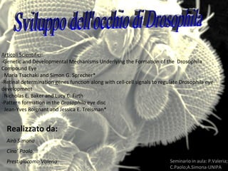
- 1. Realizzato da: Airò Simona Cina’ Paolo Prestigiacomo Valeria Articoli ScientificiArticoli Scientifici: -Genetic and Developmental Mechanisms Underlying the Formation of the Drosophila Compound Eye Maria Tsachaki and Simon G. Sprecher* -Retinal determination genes function along with cell-cell signals to regulate Drosophila eye development Nicholas E. Baker and Lucy C. Firth -Pattern formation in the Drosophila eye disc Jean-Yves Roignant and Jessica E. Treisman* Seminario in aula: P.Valeria; C.Paolo;A.Simona-UNIPA
- 2. Fig. 1. A: Picture of the compound eye of Drosophila. The compound eye consists of 750–800 individual subunits, the ‘‘ommatidia.’’ B: Confocal image of a larval eye-antennal imaginal disc. The disc is subdivided into the ‘‘antennal part’’ (anterior, to the left of the image) and the ‘‘eye’’ part (posterior, to the right of the image). The cells already determined as neurons express the neuronal marker Elav (red). The cells that are specified as photoreceptors express the photoreceptor-specific marker Chaoptin (Chp, green). All the cell nuclei are stained with DAPI (blue). MF, Morphogenetic furrow. Fig. 2. A: The genes comprising the Retinal Determination Network (RDN). The dotted lines represent physical interactions that have not been confirmed in vivo. The genes that control cell proliferation are shown in green and those that control retinal formation in blue. The genes for which the relation with the other members has not been determined by genetic interactions are shown in orange (for details see the text). B: Adult fly head exhibiting complete absence of eyes, caused by mutation in the RDN gene eyes absent (eya). C: Pupa showing ectopic eye formation after misexpression of eyeless (ey) using the dpp-Gal4 driver line. The arrowheads point towards ectopic Seminario in aula: P.Valeria C.Paolo;A.Simona-UNIPA
- 3. Il Solco Morfogenetico (MF) si forma al margine posteriore del disco imaginale, e progredisce in avanti verso la parte anteriore dell’epitelio. IL MOVIMENTO DEL SOLCO DIPENDE DA HEDGEHOG E DA DECAPENTAPLEGIC Seminario in aula: P.Valeria; C.Paolo;A.Simona-UNIPA
- 4. Seminario in aula: P.Valeria C.Paolo;A.Simona-UNIPA HEDGEHOG è espresso dalle cellule posteriori al MF ed induce l’espressione di DECAPENTAPLEGIC nel solco. Figure 2. Hh induces long-range and short-range secondary signals that control the precise position of the MF Hh acts over a short range to induce the expression of Dpp, which diffuses over a long range to turn off hth and turn on hairy, establishing a preproneural domain (PPN). Hh and Dpp also induce the expression of Delta, a transmembrane ligand that acts on adjacent cells to turn off hairy and allow ato expression, initiating photoreceptor differentiation. Roignant and Treisman Page 17 Int J
- 5. Gli ommatidi sono formati da 8 PRs: 6 esterni e 2 interni •I PRs esterni sono omologhi dei bastoncelli nei vertebrati •I PRs interni sono gli omologhi dei coni nei vertebrati Il PR R7 esprime la rodopsina UV-sensibile Il PR R8 esprime Rh5 blu sensibile o Rh6 verde sensibile Seminario in aula: P.Valeria; C.Paolo;A.Simona-UNIPA
- 6. Fig. 6. A: Schematic representation of ‘‘pale’’ and ‘‘yellow’’ ommatidia. Pale ommatidia express Rh3 in R7 and Rh5 in R8 and yellow ommatidia express Rh4 in R7 and Rh6 in R8. B: Confocal image of a compound eye, showing the mosaic of pale and yellow ommatidia. The pale R8 express Rh5 (red) and the yellow Rh6 (green). The cytoplasm of all the ommatidia is stained with phalloidin that binds to F- actin (blue). C: The choice between pale and yellow ommatidia. A part of the R7 starts to express spineless (ss) stochastically, which defines these R7 as yellow. Subsequently, ss sends a signal to the overlying R8 to express warts, which promotes rh6 expression. The R8 that do not turn on warts expression express melt, which activates rh5 expression. warts and melt suppress each other’s expression. D: Overview of the subtype specification of R7 and R8 PRs. Seminario in aula: P.Valeria; C.Paolo;A.Simona-UNIPA
- 7. La differenziazione tra ommatidi Yellow e Pale è mediato da R7 che esprime SPINGLESS (SS) Gli ommatidi Yellow sono il 70% mentre gli ommatidi Pale sono il 30% SS induce il destino Yellow, ORTHODENTICLE (OTD) il destino Pale Seminario in aula: P.Valeria; C.Paolo;A.Simona-UNIPA
- 8. I precursori cellulari pluripotenti che risiedono nel disco imaginale dell’occhio sono trasformati in cellule altamente specializzate dentro il MF. Parecchie cascate di segnalazione partecipano in questo processo. come ad es. EGFR e Notch che regolano il segnale di Hh Seminario in aula: P.Valeria; C.Paolo;A.Simona-UNIPA
- 9. Principali protagonisti nello sviluppo dell’occhio Geni effettori differenziamento Fattori trascrizionali attivati da unno stimolo esterno Geni Retinal Determination ( Geni regolatori master) Sviluppo spazio-temporale specifico Seminario in aula: P.Valeria; C.Paolo;A.Simona-UNIPA
- 10. Geni RD Eyless, sine oculis, eyes absent, teashirt. Pax 6 omologo di eyless. Seminario in aula: P.Valeria; C.Paolo;A.Simona-UNIPA
- 11. Attivazione della trascrizione di hh Seminario in aula: P.Valeria; C.Paolo;A.Simona-UNIPA
- 12. Organizzazione dei cluster all’interno di ciascuna colonna Figure 3. Inhibitory signals control cluster spacing and R8 specification (A) shows that spacing of intermediate groups is controlled by inhibitory signals from clusters in the preceding column, including Sca and a second factor downstream of EGFR signaling. (B) shows the process of restriction of Ato expression within the MF from a continuous stripe of cells to intermediate groups, 3-cell R8 equivalence groups, and single R8 cells. R8 selection requires local lateral inhibition mediated by Notch and Sca. Seminario in aula: P.Valeria; C.Paolo;A.Simona-UNIPA
- 13. Figure 4. Sequential recruitment of ommatidial cells is controlled by the short-range ligands Spi and Dl Spi promotes the differentiation of photoreceptors R2, 5, 3, 4, 1 and 6. It also induces the expression of Dl, which acts together with Spi and Boss to promote R7 differentiation, together with Spi to promote cone cell differentiation, and alone to promote primary pigment cell differentiation. Cells are added sequentially because Spi and Dl can only act over a short distance. La sequenziale differenziazione all’interno di ciascun gruppo dipende dal raggio di segnalazione Seminario in aula: P.Valeria; C.Paolo;A.Simona-UNIPA
- 14. Figure 5. Hh and Spi signals are transported down the photoreceptor axons to organize the target region Hh promotes the final division of lamina precursor cells (LPC) posterior to the lamina furrow (LF), and through Sim, their association with photoreceptor axons. Hh also induces the LPCs to express Dac, which activates expression of the EGFR, allowing them to respond to Spi. Cells that respond to Hh by expressing Dac and EGFR are colored yellow. Spi then promotes LPC differentiation adjacent to retinal axons. Cells that respond to Spi by expressing Elav are colored red. Release of the same signals from the cell body and the axons coordinates morphogenesis of the eye and the lamina. I fotorecerettori coordinano la differenziazione delle loro cellule bersaglio nelle lamina Seminario in aula: P.Valeria; C.Paolo;A.Simona-UNIPA
- 15. Conclusioni • Ruolo fondamentale dei cicli di autoregolazione positivi • Intervallo di tempo nella distribuzione dei segnali • Uso degli stessi segnali in occhio e cervello Seminario in aula: P.Valeria; C.Paolo;A.Simona-UNIPA