Differential diagnosis for Oral White Lesions
•
32 gostaram•15,408 visualizações

Oral Diagnosis II Forth Year

Recomendados
Mais conteúdo relacionado
Mais procurados
Mais procurados (20)
Destaque
Destaque (20)
Semelhante a Differential diagnosis for Oral White Lesions
Semelhante a Differential diagnosis for Oral White Lesions (20)
Mais de IAU Dent
Mais de IAU Dent (20)
Último
Último (20)
Differential diagnosis for Oral White Lesions
- 1. Dr Wael M Swelam 11/15/2008 1 By j ` fjtxÄ `É{tÅxw fãxÄtÅ Saturday, November 15, 2008 Diagnostic Sequence History Physical Examination Chief Complaint of the present Condition Radiographic Exam Differential Diagnosis Working Diagnosis 15 November 2008 2Differential Diagnosis of White oral lesions Examination set Li h i / i i “i i ”Lighting/vision “inspection” Palpation Wooden blade Therapeutic response Toluidine blue, Cytology &/or Biopsy, y gy / p y 15 November 2008 3Differential Diagnosis of White oral lesions 15 November 2008 Differential Diagnosis of White oral lesions 4
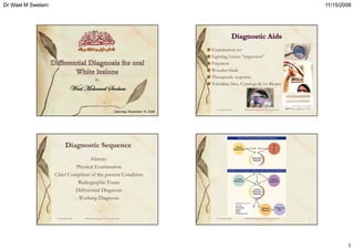
- 2. Dr Wael M Swelam 11/15/2008 2 Any history of trauma? Painful? Smoking / drinking habits Site, high risk area of mouth Removable +/- Note mucosal ulceration and erythema Homogeneous or not?g Skin conditions associated Palpate regional lymph nodes If palpable, fixed+/- 15 November 2008 5Differential Diagnosis of White oral lesions 15 November 2008 6Differential Diagnosis of White oral lesions Bilateral symmetry Asymptomatic Independent finding Remains unchanged VariationVariation of Healthyof Healthy ii Abnormality Remains unchanged Older patient tissuetissue By location & tissue affected White color of mucosa Dark color of mucosa Ulcers, Vesicles, or Bullae Tissue enlargement 15 November 2008 7Differential Diagnosis of White oral lesions No enlargement No loss of surface integrity No radiographic finding No enlargement No loss of surface integrity No radiographic finding , , Pain No enlargement No radiographic finding g Mucosal changes are mainly secondary No radiographic finding KERATOTIC NECROTIC VESICLES/BULLAE 15 November 2008 8Differential Diagnosis of White oral lesions
- 3. Dr Wael M Swelam 11/15/2008 3 •• ChemicalChemical – Tobacco products Eth n l– Ethanol – Alcohol-based mouth rinses – Acidic foods and beverages •• ThermalThermal – Smoking – Hot & cold foods and beverages •• MechanicalMechanical – Occlusal trauma Sharp edges of prostheses or teeth– Sharp edges of prostheses or teeth •• Actinic radiation & Ultraviolet lightActinic radiation & Ultraviolet light •• Microorganism relatedMicroorganism related – Syphilis – Presence of Candida albicans – Presence of viruses 15 November 2008 9Differential Diagnosis of White oral lesions S f S b i h li l 15 November 2008 Differential Diagnosis of White oral lesions 10 Epithelial Thickening Surface Material Subepithelial Change Oral White lesions (cannot be rubbed off) * Leukodema * Linea Alba * Leukoplakia *Nicotine Stomatitis * Lichen Planus (Can scrape off with Tongue blade or gauze) • Surface debris (food accretion) • Necrosis (sloughing) Lichen Planus *White hairy tongue *Candidiasis *White Sponge Nevus * Papilloma *Squamous Cell Carcinoma *Verrucous carcinoma • Thrush (Acute Pseudomembranous Candidiasis) 15 November 2008 11Differential Diagnosis of White oral lesions • Homogeneous type • Speckled type • White and red patches • Verrucous typeSpeckled type Verrucous type 15 November 2008 12Differential Diagnosis of White oral lesions
- 4. Dr Wael M Swelam 11/15/2008 4 Homogenous Sharp borders Area of friction Apparent cause Heterogenous Vague borders Any Location May enlarge Tobacco Rough, shaggy Vague borders Midline of tongue Symmetrical Often stained Homogenous Sharp borders Lower lip Older age Loss of vermilion border Dimished host resistance 15 November 2008 13 Improve with cause removal Older age Cofactors Homogenous gray Red spots Palate Pipe smoking Wrinkled velvety Mucobuccal Fold Smokeless tobacco Some material rubs off Poor oral hygiene improves Sun exposure Static/progressive Chronic Bilateral Buccal mucosa Dimished host resistance Chronic Bilateral Buccal mucosa Poor hygiene Areas atrophic/rub off Painful R i h At risk AIDS/HIV +ve Rough, thickened patches Lacy striae +/-Disappear when 15 November 2008 Regress with treatment Often coexist g , p Bilateral, Lateral surface of tongue Asymptomatic Persists/progress y erosions +/- skin lesions Changes but seldome regress stretched Mildly wrinkeled Milky appearance Congenital/Familial Dramatic appearance 14 Raised component Red component T b d l h h l• Tobacco and alchohol use • Nonsmoker and unknown etiology of lesion • Microscopic atypia AIDS related15 November 2008 15Differential Diagnosis of White oral lesions Curdy, white, diffuse Vague borders Poor oral hygiene Diminished host resistance Progress with treatment Ragged, delicate Focal Sharp borders Cause effect Heals Chemical Burn Pseudomembranous candidiasis Delicate, Grainy Pseudo-membranous May be cause related Ulcer Buccal mucosa Multiple Bilateral Asymptomatic Static Adults •Hx of trauma •Asymptomatic •Static • Hx Betel nut • Limitation of tissue movement • Progress Fordyce’s granules Scar Submucous fibrosis 15 November 2008 16
- 5. Dr Wael M Swelam 11/15/2008 5 • Epithelial or food debris • Chemical trauma (aspirin can ca se epithelial necrosis• Chemical trauma (aspirin can cause epithelial necrosis, sloughing, ulceration) • Pseudomembranous Candidiasis 15 November 2008 17Differential Diagnosis of White oral lesions 15 November 2008 18Differential Diagnosis of White oral lesions 15 November 2008 19Differential Diagnosis of White oral lesions 15 November 2008 20Differential Diagnosis of White oral lesions
- 6. Dr Wael M Swelam 11/15/2008 6 •Chemical burns•Chemical burns •Gangrenous stomatitis •Superficial bacterial infections •Traumatic ulcers •Necrotic lcers of s stemic dise se•Necrotic ulcers of systemic disease •Mucous patch of Treponema infections 15 November 2008 21Differential Diagnosis of White oral lesions 1. Patient history – If there are similar lesion in other members of the family and the patient is young aged (suggestive of hereditary lesion ex. WSN) – If this lesion is related to certain injurious agent (Traumatic, Thermal, Chemical ex. Frictional Keratosis ) – If the lesion appear after certain drug intake (like antibiotic for long period ex. Candidiasis) – If the lesion was related in site to certain habit (like check biting ex. Linea Alba Buccalis) 15 November 2008 22Differential Diagnosis of White oral lesions 2. Clinical manifestations – If the lesion is associated with skin lesion – The lesion is either unilateral or bilateral (bilateral lesion associated i h ki l i i i f Li h l )with skin lesion is suggestive of Lichen planus) – If this lesion have characteristic clinical shape (anatomizing striae “Wickham's striae” are characteristic for Lichen planus) – If the oral lesion is restricted to certain site (hairy Leukoplakia of HIV infection usually appear at lateral side of tongue) – Whether this site is changing or not (Geographic tongue is )migratory) – If the lesion disappear on its stretching (characteristic for leukodema) – Sponge texture with wrinkled morphology is suggestive of WSN15 November 2008 23Differential Diagnosis of White oral lesions 1. Congenital1. Congenital 2. Acquired 3. Infective 4. Dermatological 15 November 2008 24Differential Diagnosis of White oral lesions
- 7. Dr Wael M Swelam 11/15/2008 7 1. Congenital a. Fordyce Spots (ectopic sebaceous glands) b. White sponge naevus (defect of keratin - thickened, bilateral, often affect buccal mucosa) c. Dyskeratosis congenita (rare, potentially malignant) Differential Diagnosis of White oral lesions 15 November 2008 25 2. Acquired a)Traumatic b)Frictional keratosis cheek biting, sharp tooth, ill-fitting denture (often seen along occlusal line) c)Smoker’s keratosis (Nicotinic stomatitis) diffusely white palate with red dots showing dilated minor salivary glands d)Submucous fibrosisd)Submucous fibrosis Differential Diagnosis of White oral lesions 15 November 2008 26 3. Infective i. Candidal leukoplakia may be associated with an increased risk of malignant changechange ii. Syphilitic leukoplakia malignant potential high, rarely seen now – feature of tertiary syphilis, commonly dorsum of tongue iii. Oral hairy leukoplakia EBV causes, corrugated surface, almost always margins of tongue, often immunocompromised patients – especially HIV Differential Diagnosis of White oral lesions 15 November 2008 27 4. Dermatological a. Systemic lupus erythematosis - 20% of patients with SLE; - Symmetrically distributed erythematous areas, or white patches, - Difficult to distinguish from lichen planus, palate a common site b. Discoid lupus erythematosus Mostly affects buccal mucosa, alveolar ridge and vermillion of lip; central atrophic area with white striae radiating perpendicular to edge of lesion; can be indistinguishable from SLE c. Lichen planus and lichenoid drug reactions Very difficult to differentiate Differential Diagnosis of White oral lesions 15 November 2008 28
- 8. Dr Wael M Swelam 11/15/2008 8 15 November 2008 29Differential Diagnosis of White oral lesions 15 November 2008 30Differential Diagnosis of White oral lesions 15 November 2008 31Differential Diagnosis of White oral lesions 15 November 2008 32Differential Diagnosis of White oral lesions
- 9. Dr Wael M Swelam 11/15/2008 9 15 November 2008 33Differential Diagnosis of White oral lesions