SlideShare a Scribd company logo
1 of 90
Dr. Tareni Das, Scientist, Division of Pathology, ICAR-
IVRI, Izatnagar
Dr. Tareni Das, Scientist, Division of pathology
Importance of vascular system
 Delivery of nutrients
 Removal of waste
( Appx. 60% of lean body weight is water, intracellular-
40% and extracellular(15% interstitial+ 5% plasma
water)
Dr. Tareni Das, Scientist, Division of pathology
Circulatory system
 Blood
 Central pump- heart
 Blood distribution- arterial
 Collection- venous
 Exchange of nutrients- microcirculation ( arteriole,
metaarteriole, capillary, post capillary venule)
 ( continuous capillary-lung, brain,muscle, bone,
thymus, fenestrared capillary- villi, choroid plexus,
discontinuous capillary- liver, spleen)
 Lymphatics- draining fluid from extravascular spaces
into vascular system
Dr. Tareni Das, Scientist, Division of pathology
Derangements of fluid balance
 Hypermia
 Congestion
 Haemorrhage
 Thrombosis
 Embolism
 infarction
 oedema
 shock
Dr. Tareni Das, Scientist, Division of pathology
Hypermia
 Active engorgement of vascular beds with normal/
decreased outflow of blood
 Always acute
 Occurs due to increased metabolic activity of tissue in
order to supply additional nutrients/ oxygen
Acute General Hyperaemia:
Systemic disease ( pasteurellosis, eryspelas: due to rapid
heart beat)
Renal disease: increased fluid retention
Dr. Tareni Das, Scientist, Division of pathology
.
 Acute Local Active Hyperaemia
 Most common type
 Aetiology:
 Neurogenic mechanisms: stomach/ intestine after
meal,
 lactating mammary gland
 genital tract during oestrus
 organs of locomotion such as muscles during exercise
 Inflammation
 Gross pathology: arteries engorged with oxygenated
blood, bright red colour, warm, heavy
 Difficult to detect after death
Dr. Tareni Das, Scientist, Division of pathology
Congestion
 Passive engorgement due to
decreased outflow with a normal /
increased inflow
 Acute general congestion:
 Heart : myocardium degeneration,
necrosis and infarction
 Lung: pneumonia, pulmonary
thrombosis and embolism,
hydropericardium,
haemopericardium, or
pyopericardium, hydrothorax,
haemothorax, and pyothorax
 Euthansia: Relaxation of smooth
muscle
Dr. Tareni Das, Scientist, Division of pathology
. Gross lesions: cyanosis- deoxygenated blood, swollen
(oedema), cooler
 Chronic General congestion: persists for long time
results in permanent alteration( atrophy, fibrosis)
 Heart: stenosis of a valvular opening- tricuspid- liver,
mitral- lung; valvular insufficiency- blood is forced
backward through the leaky valve that obstructs the
incoming flow; myocardial failure; anomalies of the
heart, constrictive lesions
 Lung: obliteration of the capillary bed; compression of
major pulmonary vessels by tumours, cysts, or
abscesses
Dr. Tareni Das, Scientist, Division of pathology
Gross lesions
 Liver is affected in right-sided heart failure
 nutmeg liver‘
 cardiac cirrhosis
Dr. Tareni Das, Scientist, Division of pathology
.
Lungs are affected in the left-sided heart failure
heart-failure cells
brown induration of the lung
Dr. Tareni Das, Scientist, Division of pathology
. Acute Local congestion
vein compression due to: malposition of the viscera
and external pressure
obstruction persists- death of cell
Partial obstruction- atrophy and fibrosis
•Chronic Local congestion;
external pressure upon a vein
obstruction within a vein
Dr. Tareni Das, Scientist, Division of pathology
.
 HYPOSTATIC CONGESTION
 Accumulation of blood in the ventral portions of the body due to the
influence of gravity.
 Observed in cardiac injury; and in recumbent, restrained, or inactive
animals
 common in organs such as the lungs that have poorly supported
capillaries
 Agonal congestion and postmortem congestion
 Pathology:
 Veins in the ventral portion of an organ are distended with blood
 Causes Pneumonia in the case of lungs, and gangrene of the intestine
 Oedema, haemorrhage, inflammation, and necrosis are the
complicating changes
 In tissue sections, the affected veins will contain erythrocytes, on it will
be a layer of leukocytes, and above this a zone of plasma
 Its location is used in medico-Iegal cases to indicate the position of the
body at the time of death
Dr. Tareni Das, Scientist, Division of pathology
.
Dr. Tareni Das, Scientist, Division of pathology
HAEMORRHAGE
 Extravascular loss of blood/escape of blood from a vessel
 Two types:
 (1) haemorrhage by rhexis, when there is rupture or break of a
blood vessel
 (2) haemorrhage by diapedesis, when blood leaves through an
apparently intact vascular wall
 Aetiology:
 physiological causes
 Trauma
 Bacterial disease
 viral diseases
 Parasites
 Fungal- guttural pouch mycosis
 necrosis and destruction of vessel wall
 neoplasms
Dr. Tareni Das, Scientist, Division of pathology
.
 Toxic chemical agents: P, As, Uremia
 Type III hypersensitivity- immunocomplex
 Ehler –Danlos syndrome- developemental collagen
defect
 Increased vascular fragility
 Thrombocytopaenia- decreased production and
increased destruction
 Defective platelet function-
Bernard-soulier syndrome( GPIb),
Glanzmann thrombosthenia-GPIIb and GPIIIa,
Storage pool disease- deficient release of granule,
Chediak-higashi syndrome- defective platelet release
of ADP
Dr. Tareni Das, Scientist, Division of pathology
NSAID- aspirin- inhibit cyclooxygenase- decrease
thromboxane production
Uremia
 Abnormalities in clotting factors (haemophilia A, von
Willebrand's disease, DIC, vit-K def)
 Passive hyperaemia
Dr. Tareni Das, Scientist, Division of pathology
Classification of haemorrhages
 Source: The words cardiac, arterial, venous, and
capillary haemorrhages indicate the source of blood
 Size and shape: Petechial haemorrhages-1-2 mm
 Purpura-3-5 mm
 Ecchymotic haemorrhages-1 to 2 cm
 Haematoma or haematocyst- enclosed in a tissue
cavity
 Suffusions- Diffuse, flat, irregular areas
 Extravasations- large areas
 Linear haemorrhages- crest of the fold in intestine
 Agonal haemorrhages-Petechiae or ecchymoses
associated with death struggle
Dr. Tareni Das, Scientist, Division of pathology
.
 Location wise:
 Perivascular, perirenal, subserous, subcutaneous,
parenchymatous, subcapsular
 Haemothorax, haemopericardium,
haemoperitoneum, haemometra and haemarthrosis
 Epitasis , haemoptysis, haematemesis , enterorrhagia,
metrorrhagia , haematuria, melena, haematocele
haemosalpinx and apoplexy
 Microscopically:
 RBCs outside BV- Fresh hemorrhage- RBCs intact
 Then degraded- phagocytosed by macrophages-
Hb(red blue colour)-bilirubin( blue green colour)-
haemosiderin (Golden brown colour) detected by
Prussian blue reaction
Dr. Tareni Das, Scientist, Division of pathology
.
Dr. Tareni Das, Scientist, Division of pathology
Significance
 Hypovolemic shock when loss is >20%.
 Haemorrhage into brain, pericardial sac and
respiratory tract-very serious and may be fatal
Dr. Tareni Das, Scientist, Division of pathology
Thrombosis
 Characterized by formation of in appropriate
thrombus of fibrin and/or platelets along with other
blood elements on the wall of blood/ lymphatic vessel,
heart.
 Thrombosis is, basically, a pathological extension of
the normal haemostatic mechanism, and depends on
three components: (i) the vascular wall, (ii) platelets,
and (iii) the blood clotting system
Dr. Tareni Das, Scientist, Division of pathology
Endothelium
 Anti thrombotic properties
 Inhibition of platelet aggregation: Prostacyclin (PGI2)
and Nitric oxide (NO)
 Binding and inhibition of thrombin: Heparin-like
molecules and Thrombomodulin
 Fibrinolysis: Tissue plasminogen activator (t-PA)
 Prothrombotic Properties
 Favour platelet adhesion and aggregation: von
Willebrand factor
 Favour coagulation:Inhibitors of Plasminogen
activators
Dr. Tareni Das, Scientist, Division of pathology
Platelets
 Platelet activation occurs in contact with ECM
especially collagen:
 (1) Adhesion and shape change,

Dr. Tareni Das, Scientist, Division of pathology
. (2) Secretion (release reaction)
 Alpha granules contain fibrinogen, fibronectin,
platelet factor 4 (an antiheparin), factor V and VIII,
platelet-derived growth factor (PDGF), transforming
growth factor alpha(TGF-alpha), and P-selectin.
 Dense bodies contain adenine nucleotides (ADP and
 ATP), ionized calcium (Ca++ ions), histamine,
serotonin, and epinephrine
 (3) Aggregation: ADP, thromboxane A2
Dr. Tareni Das, Scientist, Division of pathology
Coagulation system
Dr. Tareni Das, Scientist, Division of pathology
Aetiology of thrombosis
(1)Endothelialinjury;
mechanical forces,
 Parasites: strongyle larvae
 Bacteria:Erysipelothrix,Stre
ptococcus,andCorynebacter
ium
 Virus: swine fever
 Arteriosclerotic lesions and
tumours
(2) Alterations in normal
blood flow: stasis and
turbulence
Dr. Tareni Das, Scientist, Division of pathology
 (3) Blood hyper coagulability:
Increased levels of fibrinogen, prothrombin and
factors VIIa, VIlla, and Xa.
 Increased numbers (or stickiness) of platelets
 Decreased levels of inhibitors such as antithrombin
Ill, protein C, and fibrinolysins
 ( Injury, burns, suppuration, cancer, late pregnancy
and delivery)
Dr. Tareni Das, Scientist, Division of pathology
Pathogenesis
 Platelets recognize ·the sites of endothelial injury and adhere to
the exposed subendothelial collagen and activated.
 Platelets secrete granule products (e.g., ADP) and synthesize
thromboxane
 Platelets also expose phospholipids complexes important in the
activation of intrinsic coagulation pathway,
 Tissue factor released from injured or activated endothelial cells
activates extrinsic coagulation pathway,
 ADP released from platelets stimulates formation of a reversible
primary haemostatic plug of aggregated platelets,
 Soon the primary plug is converted into a larger irreversible
secondary plug by ADP, thrombin, and TXA2
 Deposition of fibrin (derived from platelets and plasma
fibrinogen) within and around the aggregated platelets stabilizes
the mass (thrombus) and attaches it firmly to site of origin.
Dr. Tareni Das, Scientist, Division of pathology
.
Dr. Tareni Das, Scientist, Division of pathology
.
 Location of thrombi: arch of the aorta, bifurcation of a
vessel, valves- turbulence, vein- stasis
Dr. Tareni Das, Scientist, Division of pathology
Samavati and Uhal, 2020
Classification
 Cardiac thrombi:
Valvular (Streptococcus pyogenes or Erysipelothrix
rhusiopathiae. Corynebacterium pyogenes in cattle and
Streptococcus equi in horses)
Mural: left auricle(Clostridium chauvoei)
 Arterial thrombi: Strongylus vulgaris
 Venous thrombi:
Human: leg vein
Animals: nasal vascular sinuses of the cow and horse,
the veins of the broad ligament of the cow, and in the
scrotal plexus of the horse.
 Capillary thrombi
 Lymphatic thrombi
Dr. Tareni Das, Scientist, Division of pathology
. Mural thrombi
 Valvular thrombi
 Lateral thrombi
 Occluding thrombi
 Saddle thrombi
 Canalized thrombi
 Septic thrombi
 Parasitic thrombi
 Aseptic thrombi
 pale or white thrombus
 red thrombus
 laminated thrombus
Dr. Tareni Das, Scientist, Division of pathology
.
Dr. Tareni Das, Scientist, Division of pathology
Fate of the Thrombus
 Propagation
 Emboli formation
 Abscessation
 Dissolution
 Organization
 Calcification
.
Dr. Tareni Das, Scientist, Division of pathology
Significance
 Emboli
 Infarction
 Oedema
 Aneurysm
Dr. Tareni Das, Scientist, Division of pathology
.
 Difference between venous and arterial thrombi and
PM clot??/
Dr. Tareni Das, Scientist, Division of pathology
DIC
 Extremely large number of fibrin thrombi within the
small blood vessels, including capillaries and sinusoids
 Humans: sepsis, obstetric complications, malignancy,
and major trauma
 Animals: severe systemic infections, septic (endotoxic)
shock, neoplastic diseases, extensive trauma or bums,
and following extensive surgery, swine fever, ICH, and
BT, Sarcocystosis
 Consumption coagulopathy
Dr. Tareni Das, Scientist, Division of pathology
Embolism
 An embolus (plural emboli) is any foreign body
floating in the blood.
 Intravascular solid, liquid, or gaseous mass
Location of emboli: artery or a capillary
 Domestic animals- arteries, humans- venous
embolism, pulmonary embolism- common
 Paradoxical embolism-congenital inter-auricular or
interventricular defect
Dr. Tareni Das, Scientist, Division of pathology
Aetiology
 Thrombi
 Bacteria
 Parasites: Dirofilaria(Pulm.
Artery), Schistosome spp, strongyle,
hook worm, ascarid, trypanosoma
spp
 Neoplasms
 Fibrin
 Fat emboli- human, G. pig,
chicken-soft fat
 Clumps of normal body
cells- amniotic fluid
 Air or gas emboli-Caisson
disease
 Broken needles
 Hair introduced in
venipuncture
 Atheromatous debris
Dr. Tareni Das, Scientist, Division of pathology
Significance
 Large / multiple emboli- block large size / more no. of
BV
 Infection spread- septic emboli
 New tumor growth- Tumor emboli
 Infarction- heart, spleen, kidney-no collateral
circulation
Dr. Tareni Das, Scientist, Division of pathology
INFARCTION
 An infarct is an area of ischemic necrosis caused by
occlusion of either the arterial supply, or rarely the
venous drainage in a particular tissue
 To stuff or to fill fully
Dr. Tareni Das, Scientist, Division of pathology
Aetiology:
 Within the lumen of the vessel (Thrombi and emboli)
 In the wall of the artery (atherosclerotic plaque/ergot)
 In the periarterial tissue (expanding tumours,
abscesses, cysts, inflammatory fibrous adhesions,
ligatures and tourniquets)
Dr. Tareni Das, Scientist, Division of pathology
Types of Infarcts
 Depends upon (1) the solidity of the organ involved
 (ii) the type of vascular occlusion
 Types:
 Pale, white or anaemic, and red or haemorrhagic
 Septic or bland
Dr. Tareni Das, Scientist, Division of pathology
PATHOGENESIS
 Obstruction of the arterial blood supply
 Transient ischaemia- Dialation of capillary
 Engorged with blood from anatomising capillary
 Red infaracts- haemorrhagr by diapedesis( due to
hypoxic endothelial damage)
 RBCs homogenised- degeneration of the area
 Coagulative necrosis(centre to periphery)
 Zone of inflammation due to irritation
 Granulation tissue- scar tissue- distoration of organ
Dr. Tareni Das, Scientist, Division of pathology
.
Solid organ
few haemorhage
haemolysis
Hb diffuses out/
transformed to
haemosiderin
pale infaracts
In spongy organ
more RBCs
all are not haemolysed
infaracts not become pale
Dr. Tareni Das, Scientist, Division of pathology
FACTORS AFFECTING INFARCTION
 The nature of blood supply
 Rate of development of occlusion
 Vulnerability to hypoxaemia
 Oxygen content of blood
Dr. Tareni Das, Scientist, Division of pathology
Appearance
 Wedge shaped
 Red infaracts- Bulged
 Pale infaracts-depressed surface
 Scar tissue- contaction and puckering of the organ
Dr. Tareni Das, Scientist, Division of pathology
Cont…
Kidney Spleen
 pale or anaemic  Red types in animals
Dr. Tareni Das, Scientist, Division of pathology
Cont..
Lung ( red infaracts) .
 Pig- swine fever
 cattle, sheep, and pigs -
Pasteurellamultocida
Dr. Tareni Das, Scientist, Division of pathology
Cont..
Liver Intestine (red type)
 Clostridium haemolyticum
infection in cattle
 Migrating strongyle larvae
in horse of anterior
mesenteric artery
Dr. Tareni Das, Scientist, Division of pathology
Heart
 Rare in animals- pale/ red
 Common in human- due to atherosclerosis
Dr. Tareni Das, Scientist, Division of pathology
Significance
Depends upon location and size
 Death of tissue
 Organization
 Shock
 Death of the individual
Dr. Tareni Das, Scientist, Division of pathology
Edema
 It is the accumulation of excess interstitial fluid due to
changes in distribution of fluid between interstitium
and plasma.
 Oedema fluid/ transudate- non inflammatory with
low protein and low sp. Gravity(1.012).
Dr. Tareni Das, Scientist, Division of pathology
Formation of tissue fluid
Dr. Tareni Das, Scientist, Division of pathology
Causes of edema
1. Increased vascular permeability
 CAUSE
 Vascular leakage associated with inflammation
Viruses(influenza, arteri virus, canine adeno virus)
Bacteria(Clostridia, E. coli, Eryspelas )
Rickettsia( Cowdria, Ehrlichia, Rickettsia)
• Neovascularization
• Anaphylaxis
• Type-III hypersensitivity
• Toxin-endotoxin, paraquat
• Clotting abnormality-DIC
Dr. Tareni Das, Scientist, Division of pathology
MECHANISM
 In inflammation and immunological stimuli-
Histamine, bradykinin, leukotriene, substance –p:
cause endothelial contraction and increased inter
endothelial gaps
 IL-1, TNF, INF-γ: Endothelial cell retraction due to
cytoskeletal arrangement- increased inter endothelial
gaps
Dr. Tareni Das, Scientist, Division of pathology
2. Increased intravascular hydrostatic pressure
 Portal hypertension (right side heart failure, hepatic
fibrosis
 Pulmonary hyper tension (left side heart failure, high
altutude disease)
 Local congestion
 Iatrogenic fluid overload
Dr. Tareni Das, Scientist, Division of pathology
MECHANISM
 (45 mm Hg- 30 mm of Hg)- (30 mm of Hg-20 mm Hg)
 =15 mm of Hg-10 mm Hg
 = 5 mm Hg
 As most cause of congestion is impaied cardiac
function- cardiac oedema
Dr. Tareni Das, Scientist, Division of pathology
Decreased intravascular osmotic pressure
 Cause
 Decreased albumin production- severe hepatic disease
malnutrition- nutritional odema
• Excessive albumin loss- protein loosing enteropathy
• Parasitic oedema- haemonchus, trichostrongyle
• Renal disease- protein loosing enteropathy- renal
oedema
• Severe burn
• Water intoxication
Dr. Tareni Das, Scientist, Division of pathology
.
Dr. Tareni Das, Scientist, Division of pathology
MECHANISM
 (45-20) mm Hg- (20-15) mm Hg
 =(25-5) mm Hg
 =20 mm Hg
 This type of edema is severe
Dr. Tareni Das, Scientist, Division of pathology
Lymphatic obstruction
 Cause
 Lymphatic obstruction/ compression- inflammatory/
neoplastic mass, filaria, Demodex
 Congenital lymphatic aplasia/ hypoplasia
 Lympangitis- paraTB, Sporotrichosis, Epizootic
lymphangitis
Dr. Tareni Das, Scientist, Division of pathology
MECHANISM
 (45-25) mm Hg- (25-15) mm Hg
 =(20-10) mm Hg
 =10 mm Hg
Dr. Tareni Das, Scientist, Division of pathology
Gross change
 Swollen, increased in weight, cold
 Colour less than normal
 No pain
 Fullness and turgidity of affected area
 Doughy
 Pits on pressure
 fibrosis
Dr. Tareni Das, Scientist, Division of pathology
Microscopic lesion
 Space-enlarged between adjacent cells
 Pink fluid
 Atrophy
 Fibrosis
Dr. Tareni Das, Scientist, Division of pathology
Significance
 Brain, lung- oedema fatal
 S/C tissue oedema- impair wound healing
Dr. Tareni Das, Scientist, Division of pathology
Shock
 It is a circulatory dyshomeostasis associated with loss of
circulatory blood volume, reduced cardiac output and
inappropriate peripheral vascular resistance.
 Cause :
 Severe haemorrhage
 Diarrhoea
 Burns
 Tissue trauma
 Endotoxemia
 Extensive myocardial infarction
 Massive pulmonary embolism
 Cold, exhaustion, depression,
 General anaesthesia
Dr. Tareni Das, Scientist, Division of pathology
Types
 Cardiogenic
 Hypovolemic
 Blood maldistribution-a. Septic shock
b. Anaphylactic shock
c. Neurogenic shock
Dr. Tareni Das, Scientist, Division of pathology
CARDIOGENIC SHOCK
 Cardiac shock results from myocardial pump failure.
CAUSE:
 myocardial damage
 Haemopericardium
 Outflow obstruction (pulmonary embolism/ stenosis)
 Compensatory mechanism- sympathetic stimulation
of heart
 Unsuccessful compensation-stagnation of blood and
progressive tissue hypoperfusion
Dr. Tareni Das, Scientist, Division of pathology
Hypovolemic shock
Hypovolaemia means abnormally decreased volume of
circulating blood in the body which leads to decreased
blood pressure and tissue hypoperfusion.
Cause:
 loss of blood or plasma volume
 caused by haemorrhage, fluid loss from extensive skin
bums,
 Exudation from large traumatic wounds, diarrhoea,
and vomiting.
Compensatory mechanism: peripheral vasoconstriction
and fluid movement into plasma and maintain blood
flow to brain, heart, kidney
Dr. Tareni Das, Scientist, Division of pathology
Pathogenesis
 Reduced perfusion
 Cellular hypoxia
 Anaerobic glycolysis
 Metabolic lactic acidosis
 Lowering of pH in the tissues reduces the vasomotor
response
 Arterioles dilate and blood begins to pool in the
microcirculation.
 Anoxic injury to endothelium and fluid loss to tissue
 Haemo concentration
 Decreased cardiac output
 Again tissue hypoxia and cell death
Dr. Tareni Das, Scientist, Division of pathology
Blood maldistribution
 Characterized by decreased peripheral vascular
resistance and pooling of blood in peripheral tissue
caused by vasodilation.
 Types
 Anaphylactic shock
 Neurologic shock
 Septic shock
Dr. Tareni Das, Scientist, Division of pathology
Anaphylactic shock
 Type -1 hypersensitivity
 The interaction of inciting substance( allergens, drug,
vaccine) with IgE bound to mast cell
 Mast cell degranulation and histamine release
 Vasodilation, IVP, hypotension, tissue perfusion
Dr. Tareni Das, Scientist, Division of pathology
Neurogenic shock
 Trauma to brain
 Electrocution
 Fear
 Emotional stress
 Autonomic discharge- peripheral vasodilation, pooling
of blood and tissue hypoperfusion
Dr. Tareni Das, Scientist, Division of pathology
Septic shock
 Vasodilation is caused by vascular/ inflammatory
mediators
 Cause
 LPS
 Peptidoglycan
 Lipoteichoic Acid
Dr. Tareni Das, Scientist, Division of pathology
PATHOGENESIS
 Free LPS attaches to a circulating LPS binding protein,
CD4 and TLR4.
 LPS prevent synthesis of anticoagulants( TFPI and
thrombomodulin) from Endothelial cell
 Induce release of IL-1, TNF, IL-6, IL-8 from monocytes
and macrophages
 activate complements- C3a, C5a
 Activate XII and related intrinsic coagulation system,
 This response increases the local acute inflammatory
response and improves clearance of the infection at
low conc. of LPS
Dr. Tareni Das, Scientist, Division of pathology
.
Dr. Tareni Das, Scientist, Division of pathology
Cont..
 Higher concentration of LPS induce more IL-1, TNF and
other cytokine and induced fever, increased acute-phase
reactants
 TF expression and extrinsic coagulation system activated
 Increased expression of endothelial leukocyte adhesion
molecule
 IL-1 induce more PAF release and TPAI which causes
platelet aggregation, thrombosis, IVP
 production of nitric oxide, PGI2 increased
 Vasodilation
 Neutrophil activation and adhesion to endothelium also
increased
 which finally leads to decreased cardiac output, low
peripheral resistance and DIC leading to septic shock
Dr. Tareni Das, Scientist, Division of pathology
.
Dr. Tareni Das, Scientist, Division of pathology
Stages of shock
 Non progressive
 Neurohumoral mechanisms maintain cardiac output and
blood pressure
 Net effect is tachycardia (rapid heart beats), peripheral
vasoconstriction, and renal conservation of fluid
 Left atrial volume receptor and hypothalamic
osmoreceptor-regulate BP through ADH activation and
renin-angiotensin-aldosterone system
 ADH and Angiotensin-II cause vasoconstriction
 Decresed blood pressure in shock also causes increased
movement of fluid from interstitium to plasma to increase
plasma volume
Dr. Tareni Das, Scientist, Division of pathology
Cont…
 Progressive stage
 If underlying causes are not corrected shock passes to
the 'progressive phase' during which there is
widespread tissue hypoxia
 anaerobic glycolysis
 Metabolic lactic acidosis
 Increased adenosine, K, increased co2, local hypoxia-
vasodilation and pooling of blood
 endothelial cells at risk of developing anoxic injury
with subsequent DIC
 The stage is characterised by tachypnoea and oliguria
Dr. Tareni Das, Scientist, Division of pathology
Cont…
 Irreversible stage
 Multiple organ failure due to ischaemic (hypoxic) cell
death
Dr. Tareni Das, Scientist, Division of pathology
Clinical signs
 Depressed, lethargic
 Body temperature is subnormal and skin cold
 Fall in blood pressure
 Tachycardia
 Weak pulse
 Hyperventillation
 Oligouria
Dr. Tareni Das, Scientist, Division of pathology
LESIONS
 Acute general passive hyperaemia: Diffuse cyanotic colour
of organs with oedema, haemorrhage, thrombosis
 Heart - subepicardial and subendocardial haemorrhages
and necrosis, myofibril coagulation due to extensive
contraction of sarcomere
 Brain- cerebral oedema, ischaemia of neuron
 Kidneys - acute tubular necrosis
 Lung- pulmonary congestion, oedema, haemorrhage,
necrosis, fibrin exudation and hyaline membrane
formation
 Liver-passive congestion and centrilobular necrosis
 Intestine- congestion, necrosis, haemorrhage, oedema
 Adrenal : early phase intense yellow, later due to
exhaustion- yellow color lost
Dr. Tareni Das, Scientist, Division of pathology
Significance
 May reverse
 Death occurs if the blood volume cannot be restored
Dr. Tareni Das, Scientist, Division of pathology

More Related Content

Similar to Dr. TARENI DAS; HAEMODYNAMIC DISORDERS.pptx

Pulmonary embolism1
Pulmonary embolism1Pulmonary embolism1
Pulmonary embolism1GAMANDEEP
 
Shock and Blood transfusion
Shock and Blood transfusionShock and Blood transfusion
Shock and Blood transfusionAmar Yahia
 
Congenital heart disease
Congenital heart diseaseCongenital heart disease
Congenital heart diseasesurendra sharma
 
03 msu disease of the vessels hajhamad m
03 msu disease of the vessels hajhamad m03 msu disease of the vessels hajhamad m
03 msu disease of the vessels hajhamad mMohammed M. H. Hajhamad
 
Congenital heart disease
Congenital heart diseaseCongenital heart disease
Congenital heart diseaseSurendra Sharma
 
Venous Thromboembolism
Venous ThromboembolismVenous Thromboembolism
Venous ThromboembolismTsegaye Melaku
 
Cirrhosis and Portal Hypertension
Cirrhosis and Portal HypertensionCirrhosis and Portal Hypertension
Cirrhosis and Portal Hypertensionfracpractice
 
Pulmonary Thromboembolism | Jindal Chest Clinic
Pulmonary Thromboembolism | Jindal Chest ClinicPulmonary Thromboembolism | Jindal Chest Clinic
Pulmonary Thromboembolism | Jindal Chest ClinicJindal Chest Clinic
 
Pulmonary embolism
Pulmonary embolismPulmonary embolism
Pulmonary embolismdrdoaagad
 
Management of aorto arteritis
Management of aorto arteritisManagement of aorto arteritis
Management of aorto arteritisIndia CTVS
 
Congenital Heart Disease & Surgical Management
Congenital Heart Disease & Surgical ManagementCongenital Heart Disease & Surgical Management
Congenital Heart Disease & Surgical ManagementRakesh Sharma
 
Anaesthesia for turp dr.amir
Anaesthesia for turp dr.amirAnaesthesia for turp dr.amir
Anaesthesia for turp dr.amirAmirShams11
 
Diseases and disorders a nursing therapeutics manual
  Diseases and disorders a nursing therapeutics manual  Diseases and disorders a nursing therapeutics manual
Diseases and disorders a nursing therapeutics manualSturgo863
 
Hemodynamics - Hyperemia
Hemodynamics - HyperemiaHemodynamics - Hyperemia
Hemodynamics - HyperemiaJyothi Reshma S
 
Hypertrophic cardiomyopathy
Hypertrophic cardiomyopathyHypertrophic cardiomyopathy
Hypertrophic cardiomyopathyDr.Deepika T
 
ANEURYSMS , TYPES AND THERE MANAGEMENT.pptx
ANEURYSMS , TYPES  AND  THERE MANAGEMENT.pptxANEURYSMS , TYPES  AND  THERE MANAGEMENT.pptx
ANEURYSMS , TYPES AND THERE MANAGEMENT.pptxBipul Thakur
 

Similar to Dr. TARENI DAS; HAEMODYNAMIC DISORDERS.pptx (20)

Pulmonary embolism1
Pulmonary embolism1Pulmonary embolism1
Pulmonary embolism1
 
Cardiac tamponade Toufiqur Rahman
Cardiac tamponade Toufiqur RahmanCardiac tamponade Toufiqur Rahman
Cardiac tamponade Toufiqur Rahman
 
8
88
8
 
Shock and Blood transfusion
Shock and Blood transfusionShock and Blood transfusion
Shock and Blood transfusion
 
Congenital heart disease
Congenital heart diseaseCongenital heart disease
Congenital heart disease
 
Hemodynamic disorders
Hemodynamic disorders Hemodynamic disorders
Hemodynamic disorders
 
HemodynamicsLecture2010.pdfanyasin mohammed
HemodynamicsLecture2010.pdfanyasin mohammedHemodynamicsLecture2010.pdfanyasin mohammed
HemodynamicsLecture2010.pdfanyasin mohammed
 
03 msu disease of the vessels hajhamad m
03 msu disease of the vessels hajhamad m03 msu disease of the vessels hajhamad m
03 msu disease of the vessels hajhamad m
 
Congenital heart disease
Congenital heart diseaseCongenital heart disease
Congenital heart disease
 
Venous Thromboembolism
Venous ThromboembolismVenous Thromboembolism
Venous Thromboembolism
 
Cirrhosis and Portal Hypertension
Cirrhosis and Portal HypertensionCirrhosis and Portal Hypertension
Cirrhosis and Portal Hypertension
 
Pulmonary Thromboembolism | Jindal Chest Clinic
Pulmonary Thromboembolism | Jindal Chest ClinicPulmonary Thromboembolism | Jindal Chest Clinic
Pulmonary Thromboembolism | Jindal Chest Clinic
 
Pulmonary embolism
Pulmonary embolismPulmonary embolism
Pulmonary embolism
 
Management of aorto arteritis
Management of aorto arteritisManagement of aorto arteritis
Management of aorto arteritis
 
Congenital Heart Disease & Surgical Management
Congenital Heart Disease & Surgical ManagementCongenital Heart Disease & Surgical Management
Congenital Heart Disease & Surgical Management
 
Anaesthesia for turp dr.amir
Anaesthesia for turp dr.amirAnaesthesia for turp dr.amir
Anaesthesia for turp dr.amir
 
Diseases and disorders a nursing therapeutics manual
  Diseases and disorders a nursing therapeutics manual  Diseases and disorders a nursing therapeutics manual
Diseases and disorders a nursing therapeutics manual
 
Hemodynamics - Hyperemia
Hemodynamics - HyperemiaHemodynamics - Hyperemia
Hemodynamics - Hyperemia
 
Hypertrophic cardiomyopathy
Hypertrophic cardiomyopathyHypertrophic cardiomyopathy
Hypertrophic cardiomyopathy
 
ANEURYSMS , TYPES AND THERE MANAGEMENT.pptx
ANEURYSMS , TYPES  AND  THERE MANAGEMENT.pptxANEURYSMS , TYPES  AND  THERE MANAGEMENT.pptx
ANEURYSMS , TYPES AND THERE MANAGEMENT.pptx
 

Recently uploaded

linearity concept of significance, standard deviation, chi square test, stude...
linearity concept of significance, standard deviation, chi square test, stude...linearity concept of significance, standard deviation, chi square test, stude...
linearity concept of significance, standard deviation, chi square test, stude...KavyasriPuttamreddy
 
Factors Affecting child behavior in Pediatric Dentistry
Factors Affecting child behavior in Pediatric DentistryFactors Affecting child behavior in Pediatric Dentistry
Factors Affecting child behavior in Pediatric DentistryDr Simran Deepak Vangani
 
Denture base resins materials and its mechanism of action
Denture base resins materials and its mechanism of actionDenture base resins materials and its mechanism of action
Denture base resins materials and its mechanism of actionDr.shiva sai vemula
 
World Hypertension Day 17th may 2024 ppt
World Hypertension Day 17th may 2024 pptWorld Hypertension Day 17th may 2024 ppt
World Hypertension Day 17th may 2024 pptdesktoppc
 
Introducing VarSeq Dx as a Medical Device in the European Union
Introducing VarSeq Dx as a Medical Device in the European UnionIntroducing VarSeq Dx as a Medical Device in the European Union
Introducing VarSeq Dx as a Medical Device in the European UnionGolden Helix
 
TEST BANK for The Nursing Assistant Acute, Subacute, and Long-Term Care, 6th ...
TEST BANK for The Nursing Assistant Acute, Subacute, and Long-Term Care, 6th ...TEST BANK for The Nursing Assistant Acute, Subacute, and Long-Term Care, 6th ...
TEST BANK for The Nursing Assistant Acute, Subacute, and Long-Term Care, 6th ...marcuskenyatta275
 
Retinal consideration in cataract surgery
Retinal consideration in cataract surgeryRetinal consideration in cataract surgery
Retinal consideration in cataract surgeryKafrELShiekh University
 
Hemodialysis: Chapter 1, Physiological Principles of Hemodialysis - Dr.Gawad
Hemodialysis: Chapter 1, Physiological Principles of Hemodialysis - Dr.GawadHemodialysis: Chapter 1, Physiological Principles of Hemodialysis - Dr.Gawad
Hemodialysis: Chapter 1, Physiological Principles of Hemodialysis - Dr.GawadNephroTube - Dr.Gawad
 
Anuman- An inference for helpful in diagnosis and treatment
Anuman- An inference for helpful in diagnosis and treatmentAnuman- An inference for helpful in diagnosis and treatment
Anuman- An inference for helpful in diagnosis and treatmentabdeli bhadarva
 
Cardiac Impulse: Rhythmical Excitation and Conduction in the Heart
Cardiac Impulse: Rhythmical Excitation and Conduction in the HeartCardiac Impulse: Rhythmical Excitation and Conduction in the Heart
Cardiac Impulse: Rhythmical Excitation and Conduction in the HeartMedicoseAcademics
 
Vaccines: A Powerful and Cost-Effective Tool Protecting Americans Against Dis...
Vaccines: A Powerful and Cost-Effective Tool Protecting Americans Against Dis...Vaccines: A Powerful and Cost-Effective Tool Protecting Americans Against Dis...
Vaccines: A Powerful and Cost-Effective Tool Protecting Americans Against Dis...PhRMA
 
TEST BANK For Timby's Introductory Medical-Surgical Nursing, 13th Edition by ...
TEST BANK For Timby's Introductory Medical-Surgical Nursing, 13th Edition by ...TEST BANK For Timby's Introductory Medical-Surgical Nursing, 13th Edition by ...
TEST BANK For Timby's Introductory Medical-Surgical Nursing, 13th Edition by ...marcuskenyatta275
 
5CL-ADB powder supplier 5cl adb 5cladba 5cl raw materials vendor on sale now
5CL-ADB powder supplier 5cl adb 5cladba 5cl raw materials vendor on sale now5CL-ADB powder supplier 5cl adb 5cladba 5cl raw materials vendor on sale now
5CL-ADB powder supplier 5cl adb 5cladba 5cl raw materials vendor on sale nowSherrylee83
 
Why invest into infodemic management in health emergencies
Why invest into infodemic management in health emergenciesWhy invest into infodemic management in health emergencies
Why invest into infodemic management in health emergenciesTina Purnat
 
Tips and tricks to pass the cardiovascular station for PACES exam
Tips and tricks to pass the cardiovascular station for PACES examTips and tricks to pass the cardiovascular station for PACES exam
Tips and tricks to pass the cardiovascular station for PACES examJunhao Koh
 
Muscle Energy Technique (MET) with variant and techniques.
Muscle Energy Technique (MET) with variant and techniques.Muscle Energy Technique (MET) with variant and techniques.
Muscle Energy Technique (MET) with variant and techniques.Anjali Parmar
 
Video capsule endoscopy (VCE ) in children
Video capsule endoscopy (VCE ) in childrenVideo capsule endoscopy (VCE ) in children
Video capsule endoscopy (VCE ) in childrenRaju678948
 
In-service education (Nursing Mangement)
In-service education (Nursing Mangement)In-service education (Nursing Mangement)
In-service education (Nursing Mangement)Monika Kanwar
 
Is Rheumatoid Arthritis a Metabolic Disorder.pptx
Is Rheumatoid Arthritis a Metabolic Disorder.pptxIs Rheumatoid Arthritis a Metabolic Disorder.pptx
Is Rheumatoid Arthritis a Metabolic Disorder.pptxSamar Tharwat
 
Cervical screening – taking care of your health flipchart (Vietnamese)
Cervical screening – taking care of your health flipchart (Vietnamese)Cervical screening – taking care of your health flipchart (Vietnamese)
Cervical screening – taking care of your health flipchart (Vietnamese)Cancer Institute NSW
 

Recently uploaded (20)

linearity concept of significance, standard deviation, chi square test, stude...
linearity concept of significance, standard deviation, chi square test, stude...linearity concept of significance, standard deviation, chi square test, stude...
linearity concept of significance, standard deviation, chi square test, stude...
 
Factors Affecting child behavior in Pediatric Dentistry
Factors Affecting child behavior in Pediatric DentistryFactors Affecting child behavior in Pediatric Dentistry
Factors Affecting child behavior in Pediatric Dentistry
 
Denture base resins materials and its mechanism of action
Denture base resins materials and its mechanism of actionDenture base resins materials and its mechanism of action
Denture base resins materials and its mechanism of action
 
World Hypertension Day 17th may 2024 ppt
World Hypertension Day 17th may 2024 pptWorld Hypertension Day 17th may 2024 ppt
World Hypertension Day 17th may 2024 ppt
 
Introducing VarSeq Dx as a Medical Device in the European Union
Introducing VarSeq Dx as a Medical Device in the European UnionIntroducing VarSeq Dx as a Medical Device in the European Union
Introducing VarSeq Dx as a Medical Device in the European Union
 
TEST BANK for The Nursing Assistant Acute, Subacute, and Long-Term Care, 6th ...
TEST BANK for The Nursing Assistant Acute, Subacute, and Long-Term Care, 6th ...TEST BANK for The Nursing Assistant Acute, Subacute, and Long-Term Care, 6th ...
TEST BANK for The Nursing Assistant Acute, Subacute, and Long-Term Care, 6th ...
 
Retinal consideration in cataract surgery
Retinal consideration in cataract surgeryRetinal consideration in cataract surgery
Retinal consideration in cataract surgery
 
Hemodialysis: Chapter 1, Physiological Principles of Hemodialysis - Dr.Gawad
Hemodialysis: Chapter 1, Physiological Principles of Hemodialysis - Dr.GawadHemodialysis: Chapter 1, Physiological Principles of Hemodialysis - Dr.Gawad
Hemodialysis: Chapter 1, Physiological Principles of Hemodialysis - Dr.Gawad
 
Anuman- An inference for helpful in diagnosis and treatment
Anuman- An inference for helpful in diagnosis and treatmentAnuman- An inference for helpful in diagnosis and treatment
Anuman- An inference for helpful in diagnosis and treatment
 
Cardiac Impulse: Rhythmical Excitation and Conduction in the Heart
Cardiac Impulse: Rhythmical Excitation and Conduction in the HeartCardiac Impulse: Rhythmical Excitation and Conduction in the Heart
Cardiac Impulse: Rhythmical Excitation and Conduction in the Heart
 
Vaccines: A Powerful and Cost-Effective Tool Protecting Americans Against Dis...
Vaccines: A Powerful and Cost-Effective Tool Protecting Americans Against Dis...Vaccines: A Powerful and Cost-Effective Tool Protecting Americans Against Dis...
Vaccines: A Powerful and Cost-Effective Tool Protecting Americans Against Dis...
 
TEST BANK For Timby's Introductory Medical-Surgical Nursing, 13th Edition by ...
TEST BANK For Timby's Introductory Medical-Surgical Nursing, 13th Edition by ...TEST BANK For Timby's Introductory Medical-Surgical Nursing, 13th Edition by ...
TEST BANK For Timby's Introductory Medical-Surgical Nursing, 13th Edition by ...
 
5CL-ADB powder supplier 5cl adb 5cladba 5cl raw materials vendor on sale now
5CL-ADB powder supplier 5cl adb 5cladba 5cl raw materials vendor on sale now5CL-ADB powder supplier 5cl adb 5cladba 5cl raw materials vendor on sale now
5CL-ADB powder supplier 5cl adb 5cladba 5cl raw materials vendor on sale now
 
Why invest into infodemic management in health emergencies
Why invest into infodemic management in health emergenciesWhy invest into infodemic management in health emergencies
Why invest into infodemic management in health emergencies
 
Tips and tricks to pass the cardiovascular station for PACES exam
Tips and tricks to pass the cardiovascular station for PACES examTips and tricks to pass the cardiovascular station for PACES exam
Tips and tricks to pass the cardiovascular station for PACES exam
 
Muscle Energy Technique (MET) with variant and techniques.
Muscle Energy Technique (MET) with variant and techniques.Muscle Energy Technique (MET) with variant and techniques.
Muscle Energy Technique (MET) with variant and techniques.
 
Video capsule endoscopy (VCE ) in children
Video capsule endoscopy (VCE ) in childrenVideo capsule endoscopy (VCE ) in children
Video capsule endoscopy (VCE ) in children
 
In-service education (Nursing Mangement)
In-service education (Nursing Mangement)In-service education (Nursing Mangement)
In-service education (Nursing Mangement)
 
Is Rheumatoid Arthritis a Metabolic Disorder.pptx
Is Rheumatoid Arthritis a Metabolic Disorder.pptxIs Rheumatoid Arthritis a Metabolic Disorder.pptx
Is Rheumatoid Arthritis a Metabolic Disorder.pptx
 
Cervical screening – taking care of your health flipchart (Vietnamese)
Cervical screening – taking care of your health flipchart (Vietnamese)Cervical screening – taking care of your health flipchart (Vietnamese)
Cervical screening – taking care of your health flipchart (Vietnamese)
 

Dr. TARENI DAS; HAEMODYNAMIC DISORDERS.pptx

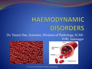
  • 1. Dr. Tareni Das, Scientist, Division of Pathology, ICAR- IVRI, Izatnagar Dr. Tareni Das, Scientist, Division of pathology
  • 2. Importance of vascular system  Delivery of nutrients  Removal of waste ( Appx. 60% of lean body weight is water, intracellular- 40% and extracellular(15% interstitial+ 5% plasma water) Dr. Tareni Das, Scientist, Division of pathology
  • 3. Circulatory system  Blood  Central pump- heart  Blood distribution- arterial  Collection- venous  Exchange of nutrients- microcirculation ( arteriole, metaarteriole, capillary, post capillary venule)  ( continuous capillary-lung, brain,muscle, bone, thymus, fenestrared capillary- villi, choroid plexus, discontinuous capillary- liver, spleen)  Lymphatics- draining fluid from extravascular spaces into vascular system Dr. Tareni Das, Scientist, Division of pathology
  • 4. Dr. Tareni Das, Scientist, Division of pathology
  • 5. Derangements of fluid balance  Hypermia  Congestion  Haemorrhage  Thrombosis  Embolism  infarction  oedema  shock Dr. Tareni Das, Scientist, Division of pathology
  • 6. Hypermia  Active engorgement of vascular beds with normal/ decreased outflow of blood  Always acute  Occurs due to increased metabolic activity of tissue in order to supply additional nutrients/ oxygen Acute General Hyperaemia: Systemic disease ( pasteurellosis, eryspelas: due to rapid heart beat) Renal disease: increased fluid retention Dr. Tareni Das, Scientist, Division of pathology
  • 7. .  Acute Local Active Hyperaemia  Most common type  Aetiology:  Neurogenic mechanisms: stomach/ intestine after meal,  lactating mammary gland  genital tract during oestrus  organs of locomotion such as muscles during exercise  Inflammation  Gross pathology: arteries engorged with oxygenated blood, bright red colour, warm, heavy  Difficult to detect after death Dr. Tareni Das, Scientist, Division of pathology
  • 8. Congestion  Passive engorgement due to decreased outflow with a normal / increased inflow  Acute general congestion:  Heart : myocardium degeneration, necrosis and infarction  Lung: pneumonia, pulmonary thrombosis and embolism, hydropericardium, haemopericardium, or pyopericardium, hydrothorax, haemothorax, and pyothorax  Euthansia: Relaxation of smooth muscle Dr. Tareni Das, Scientist, Division of pathology
  • 9. . Gross lesions: cyanosis- deoxygenated blood, swollen (oedema), cooler  Chronic General congestion: persists for long time results in permanent alteration( atrophy, fibrosis)  Heart: stenosis of a valvular opening- tricuspid- liver, mitral- lung; valvular insufficiency- blood is forced backward through the leaky valve that obstructs the incoming flow; myocardial failure; anomalies of the heart, constrictive lesions  Lung: obliteration of the capillary bed; compression of major pulmonary vessels by tumours, cysts, or abscesses Dr. Tareni Das, Scientist, Division of pathology
  • 10. Gross lesions  Liver is affected in right-sided heart failure  nutmeg liver‘  cardiac cirrhosis Dr. Tareni Das, Scientist, Division of pathology
  • 11. . Lungs are affected in the left-sided heart failure heart-failure cells brown induration of the lung Dr. Tareni Das, Scientist, Division of pathology
  • 12. . Acute Local congestion vein compression due to: malposition of the viscera and external pressure obstruction persists- death of cell Partial obstruction- atrophy and fibrosis •Chronic Local congestion; external pressure upon a vein obstruction within a vein Dr. Tareni Das, Scientist, Division of pathology
  • 13. .  HYPOSTATIC CONGESTION  Accumulation of blood in the ventral portions of the body due to the influence of gravity.  Observed in cardiac injury; and in recumbent, restrained, or inactive animals  common in organs such as the lungs that have poorly supported capillaries  Agonal congestion and postmortem congestion  Pathology:  Veins in the ventral portion of an organ are distended with blood  Causes Pneumonia in the case of lungs, and gangrene of the intestine  Oedema, haemorrhage, inflammation, and necrosis are the complicating changes  In tissue sections, the affected veins will contain erythrocytes, on it will be a layer of leukocytes, and above this a zone of plasma  Its location is used in medico-Iegal cases to indicate the position of the body at the time of death Dr. Tareni Das, Scientist, Division of pathology
  • 14. . Dr. Tareni Das, Scientist, Division of pathology
  • 15. HAEMORRHAGE  Extravascular loss of blood/escape of blood from a vessel  Two types:  (1) haemorrhage by rhexis, when there is rupture or break of a blood vessel  (2) haemorrhage by diapedesis, when blood leaves through an apparently intact vascular wall  Aetiology:  physiological causes  Trauma  Bacterial disease  viral diseases  Parasites  Fungal- guttural pouch mycosis  necrosis and destruction of vessel wall  neoplasms Dr. Tareni Das, Scientist, Division of pathology
  • 16. .  Toxic chemical agents: P, As, Uremia  Type III hypersensitivity- immunocomplex  Ehler –Danlos syndrome- developemental collagen defect  Increased vascular fragility  Thrombocytopaenia- decreased production and increased destruction  Defective platelet function- Bernard-soulier syndrome( GPIb), Glanzmann thrombosthenia-GPIIb and GPIIIa, Storage pool disease- deficient release of granule, Chediak-higashi syndrome- defective platelet release of ADP Dr. Tareni Das, Scientist, Division of pathology
  • 17. NSAID- aspirin- inhibit cyclooxygenase- decrease thromboxane production Uremia  Abnormalities in clotting factors (haemophilia A, von Willebrand's disease, DIC, vit-K def)  Passive hyperaemia Dr. Tareni Das, Scientist, Division of pathology
  • 18. Classification of haemorrhages  Source: The words cardiac, arterial, venous, and capillary haemorrhages indicate the source of blood  Size and shape: Petechial haemorrhages-1-2 mm  Purpura-3-5 mm  Ecchymotic haemorrhages-1 to 2 cm  Haematoma or haematocyst- enclosed in a tissue cavity  Suffusions- Diffuse, flat, irregular areas  Extravasations- large areas  Linear haemorrhages- crest of the fold in intestine  Agonal haemorrhages-Petechiae or ecchymoses associated with death struggle Dr. Tareni Das, Scientist, Division of pathology
  • 19. .  Location wise:  Perivascular, perirenal, subserous, subcutaneous, parenchymatous, subcapsular  Haemothorax, haemopericardium, haemoperitoneum, haemometra and haemarthrosis  Epitasis , haemoptysis, haematemesis , enterorrhagia, metrorrhagia , haematuria, melena, haematocele haemosalpinx and apoplexy  Microscopically:  RBCs outside BV- Fresh hemorrhage- RBCs intact  Then degraded- phagocytosed by macrophages- Hb(red blue colour)-bilirubin( blue green colour)- haemosiderin (Golden brown colour) detected by Prussian blue reaction Dr. Tareni Das, Scientist, Division of pathology
  • 20. . Dr. Tareni Das, Scientist, Division of pathology
  • 21. Significance  Hypovolemic shock when loss is >20%.  Haemorrhage into brain, pericardial sac and respiratory tract-very serious and may be fatal Dr. Tareni Das, Scientist, Division of pathology
  • 22. Thrombosis  Characterized by formation of in appropriate thrombus of fibrin and/or platelets along with other blood elements on the wall of blood/ lymphatic vessel, heart.  Thrombosis is, basically, a pathological extension of the normal haemostatic mechanism, and depends on three components: (i) the vascular wall, (ii) platelets, and (iii) the blood clotting system Dr. Tareni Das, Scientist, Division of pathology
  • 23. Endothelium  Anti thrombotic properties  Inhibition of platelet aggregation: Prostacyclin (PGI2) and Nitric oxide (NO)  Binding and inhibition of thrombin: Heparin-like molecules and Thrombomodulin  Fibrinolysis: Tissue plasminogen activator (t-PA)  Prothrombotic Properties  Favour platelet adhesion and aggregation: von Willebrand factor  Favour coagulation:Inhibitors of Plasminogen activators Dr. Tareni Das, Scientist, Division of pathology
  • 24. Platelets  Platelet activation occurs in contact with ECM especially collagen:  (1) Adhesion and shape change,  Dr. Tareni Das, Scientist, Division of pathology
  • 25. . (2) Secretion (release reaction)  Alpha granules contain fibrinogen, fibronectin, platelet factor 4 (an antiheparin), factor V and VIII, platelet-derived growth factor (PDGF), transforming growth factor alpha(TGF-alpha), and P-selectin.  Dense bodies contain adenine nucleotides (ADP and  ATP), ionized calcium (Ca++ ions), histamine, serotonin, and epinephrine  (3) Aggregation: ADP, thromboxane A2 Dr. Tareni Das, Scientist, Division of pathology
  • 26. Coagulation system Dr. Tareni Das, Scientist, Division of pathology
  • 27. Aetiology of thrombosis (1)Endothelialinjury; mechanical forces,  Parasites: strongyle larvae  Bacteria:Erysipelothrix,Stre ptococcus,andCorynebacter ium  Virus: swine fever  Arteriosclerotic lesions and tumours (2) Alterations in normal blood flow: stasis and turbulence Dr. Tareni Das, Scientist, Division of pathology
  • 28.  (3) Blood hyper coagulability: Increased levels of fibrinogen, prothrombin and factors VIIa, VIlla, and Xa.  Increased numbers (or stickiness) of platelets  Decreased levels of inhibitors such as antithrombin Ill, protein C, and fibrinolysins  ( Injury, burns, suppuration, cancer, late pregnancy and delivery) Dr. Tareni Das, Scientist, Division of pathology
  • 29. Pathogenesis  Platelets recognize ·the sites of endothelial injury and adhere to the exposed subendothelial collagen and activated.  Platelets secrete granule products (e.g., ADP) and synthesize thromboxane  Platelets also expose phospholipids complexes important in the activation of intrinsic coagulation pathway,  Tissue factor released from injured or activated endothelial cells activates extrinsic coagulation pathway,  ADP released from platelets stimulates formation of a reversible primary haemostatic plug of aggregated platelets,  Soon the primary plug is converted into a larger irreversible secondary plug by ADP, thrombin, and TXA2  Deposition of fibrin (derived from platelets and plasma fibrinogen) within and around the aggregated platelets stabilizes the mass (thrombus) and attaches it firmly to site of origin. Dr. Tareni Das, Scientist, Division of pathology
  • 30. . Dr. Tareni Das, Scientist, Division of pathology
  • 31. .  Location of thrombi: arch of the aorta, bifurcation of a vessel, valves- turbulence, vein- stasis Dr. Tareni Das, Scientist, Division of pathology
  • 32. Dr. Tareni Das, Scientist, Division of pathology Samavati and Uhal, 2020
  • 33. Classification  Cardiac thrombi: Valvular (Streptococcus pyogenes or Erysipelothrix rhusiopathiae. Corynebacterium pyogenes in cattle and Streptococcus equi in horses) Mural: left auricle(Clostridium chauvoei)  Arterial thrombi: Strongylus vulgaris  Venous thrombi: Human: leg vein Animals: nasal vascular sinuses of the cow and horse, the veins of the broad ligament of the cow, and in the scrotal plexus of the horse.  Capillary thrombi  Lymphatic thrombi Dr. Tareni Das, Scientist, Division of pathology
  • 34. . Mural thrombi  Valvular thrombi  Lateral thrombi  Occluding thrombi  Saddle thrombi  Canalized thrombi  Septic thrombi  Parasitic thrombi  Aseptic thrombi  pale or white thrombus  red thrombus  laminated thrombus Dr. Tareni Das, Scientist, Division of pathology
  • 35. . Dr. Tareni Das, Scientist, Division of pathology
  • 36. Dr. Tareni Das, Scientist, Division of pathology
  • 37. Dr. Tareni Das, Scientist, Division of pathology
  • 38. Fate of the Thrombus  Propagation  Emboli formation  Abscessation  Dissolution  Organization  Calcification . Dr. Tareni Das, Scientist, Division of pathology
  • 39. Significance  Emboli  Infarction  Oedema  Aneurysm Dr. Tareni Das, Scientist, Division of pathology
  • 40. .  Difference between venous and arterial thrombi and PM clot??/ Dr. Tareni Das, Scientist, Division of pathology
  • 41. DIC  Extremely large number of fibrin thrombi within the small blood vessels, including capillaries and sinusoids  Humans: sepsis, obstetric complications, malignancy, and major trauma  Animals: severe systemic infections, septic (endotoxic) shock, neoplastic diseases, extensive trauma or bums, and following extensive surgery, swine fever, ICH, and BT, Sarcocystosis  Consumption coagulopathy Dr. Tareni Das, Scientist, Division of pathology
  • 42. Embolism  An embolus (plural emboli) is any foreign body floating in the blood.  Intravascular solid, liquid, or gaseous mass Location of emboli: artery or a capillary  Domestic animals- arteries, humans- venous embolism, pulmonary embolism- common  Paradoxical embolism-congenital inter-auricular or interventricular defect Dr. Tareni Das, Scientist, Division of pathology
  • 43. Aetiology  Thrombi  Bacteria  Parasites: Dirofilaria(Pulm. Artery), Schistosome spp, strongyle, hook worm, ascarid, trypanosoma spp  Neoplasms  Fibrin  Fat emboli- human, G. pig, chicken-soft fat  Clumps of normal body cells- amniotic fluid  Air or gas emboli-Caisson disease  Broken needles  Hair introduced in venipuncture  Atheromatous debris Dr. Tareni Das, Scientist, Division of pathology
  • 44. Dr. Tareni Das, Scientist, Division of pathology
  • 45. Significance  Large / multiple emboli- block large size / more no. of BV  Infection spread- septic emboli  New tumor growth- Tumor emboli  Infarction- heart, spleen, kidney-no collateral circulation Dr. Tareni Das, Scientist, Division of pathology
  • 46. INFARCTION  An infarct is an area of ischemic necrosis caused by occlusion of either the arterial supply, or rarely the venous drainage in a particular tissue  To stuff or to fill fully Dr. Tareni Das, Scientist, Division of pathology
  • 47. Aetiology:  Within the lumen of the vessel (Thrombi and emboli)  In the wall of the artery (atherosclerotic plaque/ergot)  In the periarterial tissue (expanding tumours, abscesses, cysts, inflammatory fibrous adhesions, ligatures and tourniquets) Dr. Tareni Das, Scientist, Division of pathology
  • 48. Types of Infarcts  Depends upon (1) the solidity of the organ involved  (ii) the type of vascular occlusion  Types:  Pale, white or anaemic, and red or haemorrhagic  Septic or bland Dr. Tareni Das, Scientist, Division of pathology
  • 49. PATHOGENESIS  Obstruction of the arterial blood supply  Transient ischaemia- Dialation of capillary  Engorged with blood from anatomising capillary  Red infaracts- haemorrhagr by diapedesis( due to hypoxic endothelial damage)  RBCs homogenised- degeneration of the area  Coagulative necrosis(centre to periphery)  Zone of inflammation due to irritation  Granulation tissue- scar tissue- distoration of organ Dr. Tareni Das, Scientist, Division of pathology
  • 50. . Solid organ few haemorhage haemolysis Hb diffuses out/ transformed to haemosiderin pale infaracts In spongy organ more RBCs all are not haemolysed infaracts not become pale Dr. Tareni Das, Scientist, Division of pathology
  • 51. FACTORS AFFECTING INFARCTION  The nature of blood supply  Rate of development of occlusion  Vulnerability to hypoxaemia  Oxygen content of blood Dr. Tareni Das, Scientist, Division of pathology
  • 52. Appearance  Wedge shaped  Red infaracts- Bulged  Pale infaracts-depressed surface  Scar tissue- contaction and puckering of the organ Dr. Tareni Das, Scientist, Division of pathology
  • 53. Cont… Kidney Spleen  pale or anaemic  Red types in animals Dr. Tareni Das, Scientist, Division of pathology
  • 54. Cont.. Lung ( red infaracts) .  Pig- swine fever  cattle, sheep, and pigs - Pasteurellamultocida Dr. Tareni Das, Scientist, Division of pathology
  • 55. Cont.. Liver Intestine (red type)  Clostridium haemolyticum infection in cattle  Migrating strongyle larvae in horse of anterior mesenteric artery Dr. Tareni Das, Scientist, Division of pathology
  • 56. Heart  Rare in animals- pale/ red  Common in human- due to atherosclerosis Dr. Tareni Das, Scientist, Division of pathology
  • 57. Significance Depends upon location and size  Death of tissue  Organization  Shock  Death of the individual Dr. Tareni Das, Scientist, Division of pathology
  • 58. Edema  It is the accumulation of excess interstitial fluid due to changes in distribution of fluid between interstitium and plasma.  Oedema fluid/ transudate- non inflammatory with low protein and low sp. Gravity(1.012). Dr. Tareni Das, Scientist, Division of pathology
  • 59. Formation of tissue fluid Dr. Tareni Das, Scientist, Division of pathology
  • 60. Causes of edema 1. Increased vascular permeability  CAUSE  Vascular leakage associated with inflammation Viruses(influenza, arteri virus, canine adeno virus) Bacteria(Clostridia, E. coli, Eryspelas ) Rickettsia( Cowdria, Ehrlichia, Rickettsia) • Neovascularization • Anaphylaxis • Type-III hypersensitivity • Toxin-endotoxin, paraquat • Clotting abnormality-DIC Dr. Tareni Das, Scientist, Division of pathology
  • 61. MECHANISM  In inflammation and immunological stimuli- Histamine, bradykinin, leukotriene, substance –p: cause endothelial contraction and increased inter endothelial gaps  IL-1, TNF, INF-γ: Endothelial cell retraction due to cytoskeletal arrangement- increased inter endothelial gaps Dr. Tareni Das, Scientist, Division of pathology
  • 62. 2. Increased intravascular hydrostatic pressure  Portal hypertension (right side heart failure, hepatic fibrosis  Pulmonary hyper tension (left side heart failure, high altutude disease)  Local congestion  Iatrogenic fluid overload Dr. Tareni Das, Scientist, Division of pathology
  • 63. MECHANISM  (45 mm Hg- 30 mm of Hg)- (30 mm of Hg-20 mm Hg)  =15 mm of Hg-10 mm Hg  = 5 mm Hg  As most cause of congestion is impaied cardiac function- cardiac oedema Dr. Tareni Das, Scientist, Division of pathology
  • 64. Decreased intravascular osmotic pressure  Cause  Decreased albumin production- severe hepatic disease malnutrition- nutritional odema • Excessive albumin loss- protein loosing enteropathy • Parasitic oedema- haemonchus, trichostrongyle • Renal disease- protein loosing enteropathy- renal oedema • Severe burn • Water intoxication Dr. Tareni Das, Scientist, Division of pathology
  • 65. . Dr. Tareni Das, Scientist, Division of pathology
  • 66. MECHANISM  (45-20) mm Hg- (20-15) mm Hg  =(25-5) mm Hg  =20 mm Hg  This type of edema is severe Dr. Tareni Das, Scientist, Division of pathology
  • 67. Lymphatic obstruction  Cause  Lymphatic obstruction/ compression- inflammatory/ neoplastic mass, filaria, Demodex  Congenital lymphatic aplasia/ hypoplasia  Lympangitis- paraTB, Sporotrichosis, Epizootic lymphangitis Dr. Tareni Das, Scientist, Division of pathology
  • 68. MECHANISM  (45-25) mm Hg- (25-15) mm Hg  =(20-10) mm Hg  =10 mm Hg Dr. Tareni Das, Scientist, Division of pathology
  • 69. Gross change  Swollen, increased in weight, cold  Colour less than normal  No pain  Fullness and turgidity of affected area  Doughy  Pits on pressure  fibrosis Dr. Tareni Das, Scientist, Division of pathology
  • 70. Microscopic lesion  Space-enlarged between adjacent cells  Pink fluid  Atrophy  Fibrosis Dr. Tareni Das, Scientist, Division of pathology
  • 71. Significance  Brain, lung- oedema fatal  S/C tissue oedema- impair wound healing Dr. Tareni Das, Scientist, Division of pathology
  • 72. Shock  It is a circulatory dyshomeostasis associated with loss of circulatory blood volume, reduced cardiac output and inappropriate peripheral vascular resistance.  Cause :  Severe haemorrhage  Diarrhoea  Burns  Tissue trauma  Endotoxemia  Extensive myocardial infarction  Massive pulmonary embolism  Cold, exhaustion, depression,  General anaesthesia Dr. Tareni Das, Scientist, Division of pathology
  • 73. Types  Cardiogenic  Hypovolemic  Blood maldistribution-a. Septic shock b. Anaphylactic shock c. Neurogenic shock Dr. Tareni Das, Scientist, Division of pathology
  • 74. CARDIOGENIC SHOCK  Cardiac shock results from myocardial pump failure. CAUSE:  myocardial damage  Haemopericardium  Outflow obstruction (pulmonary embolism/ stenosis)  Compensatory mechanism- sympathetic stimulation of heart  Unsuccessful compensation-stagnation of blood and progressive tissue hypoperfusion Dr. Tareni Das, Scientist, Division of pathology
  • 75. Hypovolemic shock Hypovolaemia means abnormally decreased volume of circulating blood in the body which leads to decreased blood pressure and tissue hypoperfusion. Cause:  loss of blood or plasma volume  caused by haemorrhage, fluid loss from extensive skin bums,  Exudation from large traumatic wounds, diarrhoea, and vomiting. Compensatory mechanism: peripheral vasoconstriction and fluid movement into plasma and maintain blood flow to brain, heart, kidney Dr. Tareni Das, Scientist, Division of pathology
  • 76. Pathogenesis  Reduced perfusion  Cellular hypoxia  Anaerobic glycolysis  Metabolic lactic acidosis  Lowering of pH in the tissues reduces the vasomotor response  Arterioles dilate and blood begins to pool in the microcirculation.  Anoxic injury to endothelium and fluid loss to tissue  Haemo concentration  Decreased cardiac output  Again tissue hypoxia and cell death Dr. Tareni Das, Scientist, Division of pathology
  • 77. Blood maldistribution  Characterized by decreased peripheral vascular resistance and pooling of blood in peripheral tissue caused by vasodilation.  Types  Anaphylactic shock  Neurologic shock  Septic shock Dr. Tareni Das, Scientist, Division of pathology
  • 78. Anaphylactic shock  Type -1 hypersensitivity  The interaction of inciting substance( allergens, drug, vaccine) with IgE bound to mast cell  Mast cell degranulation and histamine release  Vasodilation, IVP, hypotension, tissue perfusion Dr. Tareni Das, Scientist, Division of pathology
  • 79. Neurogenic shock  Trauma to brain  Electrocution  Fear  Emotional stress  Autonomic discharge- peripheral vasodilation, pooling of blood and tissue hypoperfusion Dr. Tareni Das, Scientist, Division of pathology
  • 80. Septic shock  Vasodilation is caused by vascular/ inflammatory mediators  Cause  LPS  Peptidoglycan  Lipoteichoic Acid Dr. Tareni Das, Scientist, Division of pathology
  • 81. PATHOGENESIS  Free LPS attaches to a circulating LPS binding protein, CD4 and TLR4.  LPS prevent synthesis of anticoagulants( TFPI and thrombomodulin) from Endothelial cell  Induce release of IL-1, TNF, IL-6, IL-8 from monocytes and macrophages  activate complements- C3a, C5a  Activate XII and related intrinsic coagulation system,  This response increases the local acute inflammatory response and improves clearance of the infection at low conc. of LPS Dr. Tareni Das, Scientist, Division of pathology
  • 82. . Dr. Tareni Das, Scientist, Division of pathology
  • 83. Cont..  Higher concentration of LPS induce more IL-1, TNF and other cytokine and induced fever, increased acute-phase reactants  TF expression and extrinsic coagulation system activated  Increased expression of endothelial leukocyte adhesion molecule  IL-1 induce more PAF release and TPAI which causes platelet aggregation, thrombosis, IVP  production of nitric oxide, PGI2 increased  Vasodilation  Neutrophil activation and adhesion to endothelium also increased  which finally leads to decreased cardiac output, low peripheral resistance and DIC leading to septic shock Dr. Tareni Das, Scientist, Division of pathology
  • 84. . Dr. Tareni Das, Scientist, Division of pathology
  • 85. Stages of shock  Non progressive  Neurohumoral mechanisms maintain cardiac output and blood pressure  Net effect is tachycardia (rapid heart beats), peripheral vasoconstriction, and renal conservation of fluid  Left atrial volume receptor and hypothalamic osmoreceptor-regulate BP through ADH activation and renin-angiotensin-aldosterone system  ADH and Angiotensin-II cause vasoconstriction  Decresed blood pressure in shock also causes increased movement of fluid from interstitium to plasma to increase plasma volume Dr. Tareni Das, Scientist, Division of pathology
  • 86. Cont…  Progressive stage  If underlying causes are not corrected shock passes to the 'progressive phase' during which there is widespread tissue hypoxia  anaerobic glycolysis  Metabolic lactic acidosis  Increased adenosine, K, increased co2, local hypoxia- vasodilation and pooling of blood  endothelial cells at risk of developing anoxic injury with subsequent DIC  The stage is characterised by tachypnoea and oliguria Dr. Tareni Das, Scientist, Division of pathology
  • 87. Cont…  Irreversible stage  Multiple organ failure due to ischaemic (hypoxic) cell death Dr. Tareni Das, Scientist, Division of pathology
  • 88. Clinical signs  Depressed, lethargic  Body temperature is subnormal and skin cold  Fall in blood pressure  Tachycardia  Weak pulse  Hyperventillation  Oligouria Dr. Tareni Das, Scientist, Division of pathology
  • 89. LESIONS  Acute general passive hyperaemia: Diffuse cyanotic colour of organs with oedema, haemorrhage, thrombosis  Heart - subepicardial and subendocardial haemorrhages and necrosis, myofibril coagulation due to extensive contraction of sarcomere  Brain- cerebral oedema, ischaemia of neuron  Kidneys - acute tubular necrosis  Lung- pulmonary congestion, oedema, haemorrhage, necrosis, fibrin exudation and hyaline membrane formation  Liver-passive congestion and centrilobular necrosis  Intestine- congestion, necrosis, haemorrhage, oedema  Adrenal : early phase intense yellow, later due to exhaustion- yellow color lost Dr. Tareni Das, Scientist, Division of pathology
  • 90. Significance  May reverse  Death occurs if the blood volume cannot be restored Dr. Tareni Das, Scientist, Division of pathology