SlideShare uma empresa Scribd logo
1 de 4
Baixar para ler offline
Combining Placido and OCT Imaging Technologies:
    Bringing Diagnostic Power to Corneal Analysis

                                  8 Shareef Mahdavi • SM2 Strategic • Pleasanton, CA 7


    The ability of ophthalmic clinicians to evaluate the cornea            a direct and magnified view of the cornea; keratometry,
has improved dramatically over the past quarter century. From              measuring the change in curvature in the central 3 mm
the era of manual keratometry, new methods of imaging and                  of the cornea; and contact pachymetry, showing cor-
analyzing corneal structure have emerged and have moved cor-               neal thickness at each point of applanation. By the mid
neal care from an analog to a digital world.                               1980s, published papers by Rowsey, et al showed how
    In the past 10 years, an explosion of technology has emerged           Placido rings could be used to construct the cornea, and
that at times can overwhelm the eye care practitioner. SM2                 in 1987 Stephen Klyce, PhD (along with Leo Maguire,
Strategic was asked by Carl Zeiss Meditec (Dublin, CA) to help             MD) introduced the color-coded contour map and scale
eye doctors understand the path of development that has taken              to standardize interpretation, which evolved and became
place and the tools that are now available to help with corneal            an ANSI Standard. The following year saw the intro-
analysis in everyday clinical practice. Three leading corneal spe-         duction of the first computerized corneal topographer
cialists as well as a pioneer in corneal imaging were interviewed:         (Computed Anatomy). The modern age of corneal imag-
Amin Ashrafzadeh, MD (Modesto, CA), Robert Cionni, MD (Salt                ing was officially underway.
Lake City, UT), Roger Steinert, MD (Irvine, CA) and Stephen                    Since that first device, innovation in both imaging
Klyce, PhD (Port Washington, NY). These doctors provided their             and interpretation has been spurred by clinical need
perspective on the technological milestones that have led to the           and the rise of refractive surgery. In the 1990s, corneal
current state-of-the-art in corneal imaging.                               topography proliferated and gave refractive surgeons
    This report summarizes those interviews and intends to pro-            the ability to measure the eye’s surface out to 10 mm
vide ophthalmologists with an appreciation of how rapidly the              (ie, beyond the optical zone) both before and after laser
field has changed, while offering guidance in terms of decision-           vision correction. Devices from Tomey, EyeSys, and
making with respect to future investment in corneal diagnostics.           Humphrey (acquired from ORC) proved most popular
                                                                           with clinicians. By the late 1990s, surgeons realized that
Technology Development                                                     they needed a better way to screen for corneal thinning
    The past quarter century has provided stellar innova-                  and determine which prospective LASIK patients were
tion in the realm of imaging and diagnosis of the cornea                   most at risk for ectasia and/or keratoconus.
(see Figure 1). For most of the 20th century, clinicians                       Product developers focused on new approaches to
had three basic tools available: the slit lamp, providing                  imaging. Orbtec's Orbscan device used a scanning


Figure 1: 25 Years of Corneal Analysis Development Timeline

              1987                Late 1980’s                                                2001               2005                     2009
              Klyce color scale   Ultrasonic                                                 Scheimpflug        Anterior                 Placido +
              to standardize      biomicroscopy                                              corneal            segment                  OCT
              interpretation                                                                 tomography         OCT                      (Zeiss)
                                                                                             (Oculus)           (Zeiss)


  1984                1987              Early 1990’s      Mid & Late 1990s       2000             2002                     2007
  First papers on     First             Commercial        Slit scanning          Placido +        Digital VHF              Placido +
  use of Placido      computerized      Placido devices   device                 wavefront        ultrasound               Scheimpflug
  to construct/       Placido + slit    (Tomey, EyeSys,   (Orbscan I)            (Nidek)          (Artemis 2)              (Ziemer)
  model cornea        device            Humphrey)         Slit scan + Placido
                      (Computed                           (Orbscan II)
                      Anatomy)


    1984                                 1990                                       2000                                                 2009
slit-beam to image the back surface of the cornea, giv-                                  measuring corneal thickness manually at a handful of
ing clinicians posterior measurements and the ability                                    well-spaced locations in the central cornea.
to calculate elevation from the front to the back of                                              While this device could be used in tandem with a
the cornea. It was limited to 4 diopters of elevation                                    corneal topographer to provide greater information, it
change and required higher-skilled technicians to pre-                                   initially lacked the interpretative power afforded by the
vent patient eye movement and get reliable readings. In                                  Holladay report. By 2007, corneal analysis software for
2000, Nidek’s OPD Scan combined Placido + wavefront                                      the ATLAS 9000 (known as Pathfinder II) gave clini-
measurement in a single device. The use of ultrasound                                    cians an anterior topographic screening module to help
(e.g., ultrasonic biomicroscopy) gave high quality images                                identify abnormal corneal conditions. And by 2009,
of corneal structure, but had fallen out of favor with                                   Zeiss linked both the ATLAS and Visante together, call-
clinicians due to the required contact via a probe or                                    ing the merged technologies Visante Omni. Each device
water bath. In contrast to Placido-based topographers,                                   now accessed the other’s data, first to ensure proper
Oculus' Pentacam tomographer used slits to generate                                      biometric registration of the eye, then to conduct pre-
cross-sections of the cornea and reconstruct them into a                                 cise analysis of corneal structure and changes over time.
singular image. There were now two different technolo-                                   Interpretation was greatly enhanced when Visante Omni
gies popular for clinical                                                                                                                     incorporated the tradi-
diagnosis, each deriving       Figure 2: Corneal Imaging Technologies and Applications                                                        tional Holladay report
its final answer from                                                                                                                         plus several new color-
different approaches:          	                                    GALILEI	       ORBSCAN	            PENTACAM	          VISANTE	OMNI        scale maps to help diag-
the Pentacam, using            	                                  Placido	+	       Placido	+					          	                 Placido									 nose, specifically early
Scheimpflug imaging,           TECHNOLOGY:	                      Scheimpflug	    Slit	Scanning	       Scheimpflug	            +	OCT           corneal pathology such
measured elevation             Anterior & Posterior                                                                                           as suspect keratoconus.
                               Topography
directly and then calcu-
                               Narrow angle evaluation	                   	             	                  	                                  Clinical Significance
lated corneal curvature,
                               Clear Corneal Wound
while Placido measured         Evaluation	                                	             	                                                         Many diagnostic
corneal curvature              Cataract Evaluation and                                                                                        innovations sought to
directly.                      IOL Placement Analysis	                    	             	                  	                                  improve upon Placido
    Surgeons were drawn        Evaluation of Pars Plana	                  	             	                  	                                  measurements and
to the Pentacam’s ease-        Corneal Transplant                                                                                             render them obsolete,
                               Planning & Follow-up	                      	             	                  	
of-use with patients                                                                                                                          yet hindsight reveals a
                               Iris Tumors	                               	             	                  	
and the interpretive                                                                                                                          different outcome. “We
                               Imaging Through Opaque/
software developed by          White Corneas	                             	             	                  	                                  have come full circle,”
Jack Holladay, MD; as          Phakic IOL Planning                                                                                            noted Roger Steinert,
a result, the Pentacam       	                           Full	Capability	     Limited	Capability	          No	Capability	      	              MD. “We now realize
sold briskly to refractive                                                                                                                    Placido-based technolo-
surgeons in the early 2000s. By 2005, Carl Zeiss had                                     gies as the most reliable way to analyze anterior corneal
developed an anterior segment OCT device to comple-                                      topography.” Cornea specialist Amin Ashrafzadeh, MD
ment their rapidly-adopted posterior segment version for                                 has been using corneal topography to plan and analyze
use in retina and glaucoma. The Visante OCT provided                                     surgical outcomes since 2001 and believes the Placido
extremely crisp images of corneal structures, allow-                                     disk “remains one of the most essential instruments for
ing both increased width of coverage (out to 16 mm) as                                   imaging the corneal surface.” He lamented, however,
well as depth of coverage (up to 6 mm). This advanced                                    that “up until several years ago, you still had a lot of
technology took multiple “Optical A-scan” images of                                      work to do in figuring out why you had an odd-looking
the cornea and then re-constructed these slices into a                                   cornea.”
“Optical B-scan” image, putting directly into the clinic                                          That burden changed, according to both Steinert
images never before available (except in part from histol-                               and Ashrafzadeh, when surgeons gained the ability to
ogy slides). The new “map” of the eye included a differ-                                 correlate topography with corneal thinning. Both had
ent pachymetric approach that automated pachymetry                                       attempted to use Scheimpflug – which had become com-
measurement with exponentially greater data points                                       monplace for routine refractive cases – to help with
(2,048). This eliminated the potential inaccuracy from                                   this more sophisticated analysis, but found that they
were too often led down       in reaching diagnosis,” remarked Dr. Steinert. Figure 2
“The real value of              the wrong path in reach-      shows the different capabilities of four popular corneal
Visante Omni is its             ing the correct diagnosis.    imaging devices. An earlier survey by SM2 Strategic of 32
                                “Anterior topography from     of the first users of Visante showed significantly greater
ability to evaluate             the Pentacam was pointing     usage than anticipated once they had the device in their
numerous data...                to a completely different     practice and had become familiar with its capabilities.
all in one place”               area for astigmatism, and
                                treating this made cylinder   Interpretation Becomes Critical
    Robert Cionni, MD           worse not better,” said Dr.       The sheer volume of data on the cornea now avail-
                                Steinert. “I went back and    able to clinicians places a premium on software analytics
checked the Placido images and found they were accurate;      that can reliably distill the images and data points so they
this triggered the realization that we needed to go back to   make sense in diagnosis. “The amount of information
Placido based topography.” While the ability to use a sin-    can be overwhelming,” according to Dr. Ashrafzadeh.
gle device was indeed convenient, Dr. Ashrafzadeh found       “The Holladay report helps tremendously but still has
that the Pentacam software would “fill in” missing data       deficiencies that cause me to evaluate the ring image over-
points improperly on slightly irregular surfaces. “I liked    laid atop the report.” The real value of Visante Omni,
the way that Scheimpflug image looked, but even looks         remarked Dr. Cionni, is it’s ability to evaluate numer-
can be deceiving.” Both remarked that the presence of         ous data such as topography, corneal aberrations, ICL
artifacts in Scheimpflug images of suspicious corneas         positioning, pachymetry, ectasia analysis, angle measure-
has been a troublesome aspect of that technology.             ments… all in one place. Indeed, clinicians should view
    This issue appears to have been solved through the        Visante Omni as a workstation with multiple capabilities
linking of Placido and OCT, and the applications extend       rather than a niche device.
beyond the cornea. Robert Cionni, MD has found that
the Omni allows him greater ability to evaluate aberra-       Early Detection Capabilities
tions relevant only to the cornea. “I get better astigmatic       Each surgeon interviewed agreed that the Pentacam
analysis with Atlas and my toric IOLs are now more pre-       itself is sufficient for the refractive surgeon who is mainly
dictable,” according to Dr. Cionni.                           concerned about corneal elevation. If you are dealing
    Evaluation of glaucoma patients has been greatly          only with normal corneas, they say, then the Pentacam
enhanced by Omni. “Pupil size and the resultant angle         is fine. The dividing line is drawn around earlier detec-
opening are not influenced by Visante's non-visible light,    tion of corneal abnormalities. Dr. Ash did a comparison
providing an exquisitely accurate measure of physiologic      study of 60 eyes, measuring them both on the Pentacam
angle opening,” stated Dr. Ashrafzadeh.                       and the Visante Omni. His findings were that on corneas
                                                              with no prior surgery and no opacities, there was good
Efficiency in the Clinic                                      agreement between the devices. But in patients with
    Some clinicians view it a disadvantage to have to         prior refractive surgery, significant differences emerge. In
use two devices rather than one as part of their corneal      this study, Visante showed much greater consistency in
screening. Dr. Ashrafzadeh finds it actually works to his     posterior surface findings compared to the variability in
advantage. “I can counsel the patient while I’m doing         readings from the Pentacam, which also tended to under-
their Omni readings.” He described how this interaction       detect post-LASIK ectasia. The findings on the Visante
allows him to educate the patient and discuss candidacy       Omni correlated much more closely to what he was see-
for refractive surgery, while also giving the patient “sig-                                    ing clinically.
nificant confidence in seeing the whole process in action.                                        This type of study illus-
Omni takes away a lot of the mystery by allowing me to        “At present, Visante trates the sentiment of Dr.
describe what I’m doing and what the images are telling       Omni is the only                 Steinert regarding a surgeon’s
me…in real time.”                                             technology that I                ability to trust the device
    Dr. Cionni added, “I am able to show a patient how                                         you are using: “You need
removing their astigmatism will improve their quality of      can truly rely upon confidence in your device
vision as well.”                                              for subtle changes.” if you are doing refractive
    “If you use Visante for other applications such as             Roger Steinert, MD          surgery to pick up the subtle
evaluating the angle, then you actually gain efficiency                                        things like Form Fruste
Keratoconus. At present, Visante                          shown in Figure 3. As patient loads increase with the
“You must use             Omni is the only technology                               aging population, this justification becomes even more
Placido for early         that I can truly rely upon for                            attractive and helps the ophthalmic practice harness the
                          subtle changes.”                                          power of these two innovative technologies.
detection of                 Stephen Klyce, PhD, widely
Keratoconus”              regarded as a pioneer in corneal                          Summary and Discussion
    Stephen Klyce, PhD imaging and analysis, believes                                   Ophthalmic diagnosis and treatment paradigms have
                          that Scheimpflug is not well                              evolved significantly in the past two decades. Better diag-
suited to screening the cornea, as it is 20x less sensi-                            nostics for the cornea have improved surgeons’ abilities to
tive than Placido to changes in the amount of corneal                               the point that most recognize they can perform refractive
curvature. “The use of posterior corneal data has not                               surgery on patients who were not considered candidates
been shown convincingly to be useful for early detection.                           ten years ago…and vice-versa. The negative publicity sur-
Anterior surface Placido is the standard; you must use                              rounding LASIK in the past several years can be attrib-
Placido for early detection of Keratoconus.”                                        uted in part to the lack of understanding of the cornea in
                                                                                    the initial period following US commercialization. What
Reimbursement                                                                       surgeons needed – and now have – is a clinical “safety
    Lastly, changes in reimbursement effective January                              net” as a result of technological innovation.
2011 puts into effect a new code for Anterior Segment                                   The power in diagnosis afforded by Visante Omni
OCT (92132, a Category 1 CPT code). Prior to this,                                  and its marriage of Placido and OCT technology raises
doctors were treated unevenly. Some were able to bill                               the bar for all ophthalmic surgeons in several respects.
and get reimbursed, while others’ claims were denied.                               First, increased understanding of the risks presented by
The presence of a new code – specifically for anterior                              abnormal corneas – and how to best mitigate those risks
segment OCT – will help achieve a consistency that is                               – puts a premium on being able to detect it earlier in its
better overall when one considers patient care standards                            development. Second, an aging population will bring an
on a national basis. Although it is tempting to bemoan                              increasing prevalence of all eye diseases, including those
the fact that the code now pays bilaterally (i.e., $36.35                           in the anterior segment (e.g., glaucoma). Taken together,
national average reimbursement for both eyes), the previ-                           these two issues put additional pressure on clinicians to
ous SM2 survey showed users billing an average of 18                                get the right diagnosis in an efficient manner. And just
uses per month out of 47 uses overall (including refrac-                            as the lines between refractive and cataract surgery are
tive evaluation). With a specific billing code in place                             blurring, technology such as Visante Omni is making it
to guide clinicians, one would expect the reimbursable                              possible for all ophthalmic clinicians to more accurately
usage to increase significantly. A pro-forma analysis is                            diagnose disease.


                                                             Figure 3: Return On Investment
   	                                             VISANTE		          VISANTE	omni	                                    KEY	ASSUMPTIONS
   Cost
   Cost Per Month	                          $708	–	$1,018	        $982	–	$1,382	             Monthly	lease	cost	includes	device	and	service	for	5	years.		 	
   	                                                    	                 	                  Range	reflects	value	of	trade-in,	and	new/refurbished	unit.
   Reimbursed Procedures                                	                 	                  	
   OCT (CPT	code	92132)	                          $647		               $647	                 18	reimbursed	procedures	per	month	(from	prior	SM2	survey	of		
   	                                                    	                 	                  Visante	users);	national	average	reimbursement	of	$36.35	bilateral
   Corneal Topography 	                             -	                 $700	                 20	submitted	claims	per	month	(25%	of	80	eyes	per	month)
   (CPT	code	92025)	                                	                    	                   $35	average	reimbursement
   Non-Reimbursed Procedures
   OCT	                                         $1,044	               $1,044	                29	per	month	(from	prior	SM2	survey	of	Visante	users);	$36	per		
   	                                                    	                 	                  eye	component	of	elective	fee,	built-in	to	refractive	fee	for	LASIK		
   	                                                    	                 	                  and/or	Premium	IOL's
   Total Return per Month                        $1,691               $2,391
   Return on Investment Ratio                 1.7x – 2.4x           1.7x – 2.4x


© Copyright 2011, SM2 Strategic. All Rights Reserved.

Mais conteúdo relacionado

Mais procurados

Optical coherence tomography
Optical coherence tomographyOptical coherence tomography
Optical coherence tomographySinan çalışkan
 
Recent advances in radiographic technique /certified fixed orthodontic course...
Recent advances in radiographic technique /certified fixed orthodontic course...Recent advances in radiographic technique /certified fixed orthodontic course...
Recent advances in radiographic technique /certified fixed orthodontic course...Indian dental academy
 
Recent advances in imaging techniques/ /certified fixed orthodontic courses b...
Recent advances in imaging techniques/ /certified fixed orthodontic courses b...Recent advances in imaging techniques/ /certified fixed orthodontic courses b...
Recent advances in imaging techniques/ /certified fixed orthodontic courses b...Indian dental academy
 
Optical Coherence Tomography - principle and uses in ophthalmology
Optical Coherence Tomography - principle and uses in ophthalmologyOptical Coherence Tomography - principle and uses in ophthalmology
Optical Coherence Tomography - principle and uses in ophthalmologytapan_jakkal
 
Advanced imaging as orthodontics Aids
Advanced imaging as orthodontics AidsAdvanced imaging as orthodontics Aids
Advanced imaging as orthodontics AidsIndian dental academy
 
ABC of Oct and octa
ABC of Oct and octaABC of Oct and octa
ABC of Oct and octaAjitThakur45
 
EyeWorld Asia Pacific Association of Ophthalmologists Meeting - 09192010
EyeWorld Asia Pacific Association of Ophthalmologists Meeting - 09192010EyeWorld Asia Pacific Association of Ophthalmologists Meeting - 09192010
EyeWorld Asia Pacific Association of Ophthalmologists Meeting - 09192010devknowhow
 
Recent advances in radiographic technique in orthodontics
Recent advances in radiographic technique in orthodonticsRecent advances in radiographic technique in orthodontics
Recent advances in radiographic technique in orthodonticsIndian dental academy
 
Short intro for retinal biomarkers of Alzheimer’s Disease
Short intro for retinal biomarkers of Alzheimer’s DiseaseShort intro for retinal biomarkers of Alzheimer’s Disease
Short intro for retinal biomarkers of Alzheimer’s DiseasePetteriTeikariPhD
 
pachymetry confocal microscopy cornea ophthalmology diagnostics
pachymetry confocal microscopy cornea ophthalmology diagnosticspachymetry confocal microscopy cornea ophthalmology diagnostics
pachymetry confocal microscopy cornea ophthalmology diagnosticsPaavan Kalra
 
Hyperspectral Retinal Imaging
Hyperspectral Retinal ImagingHyperspectral Retinal Imaging
Hyperspectral Retinal ImagingPetteriTeikariPhD
 
EYE-OCT.(OPTICAL COHERANCE TOMOGRAPHY)
EYE-OCT.(OPTICAL  COHERANCE TOMOGRAPHY)EYE-OCT.(OPTICAL  COHERANCE TOMOGRAPHY)
EYE-OCT.(OPTICAL COHERANCE TOMOGRAPHY)Sanjib Pashi
 
20120927 台科大 oct原理及在眼科的應用 (1)
20120927 台科大 oct原理及在眼科的應用 (1)20120927 台科大 oct原理及在眼科的應用 (1)
20120927 台科大 oct原理及在眼科的應用 (1)doc30845
 
Smartphone-powered Ophthalmic Diagnostics
Smartphone-powered Ophthalmic DiagnosticsSmartphone-powered Ophthalmic Diagnostics
Smartphone-powered Ophthalmic DiagnosticsPetteriTeikariPhD
 
Minimally Invasive Image Guided Therapy
Minimally Invasive Image Guided TherapyMinimally Invasive Image Guided Therapy
Minimally Invasive Image Guided TherapyKendall Waters
 
Operating an OCT & evaluating normal scan (Optom.Saptarshi Mehta).....
Operating an OCT & evaluating normal scan (Optom.Saptarshi Mehta).....Operating an OCT & evaluating normal scan (Optom.Saptarshi Mehta).....
Operating an OCT & evaluating normal scan (Optom.Saptarshi Mehta).....Saptarshivision
 

Mais procurados (20)

Optical coherence tomography
Optical coherence tomographyOptical coherence tomography
Optical coherence tomography
 
Recent advances in radiographic technique /certified fixed orthodontic course...
Recent advances in radiographic technique /certified fixed orthodontic course...Recent advances in radiographic technique /certified fixed orthodontic course...
Recent advances in radiographic technique /certified fixed orthodontic course...
 
Recent advances in imaging techniques/ /certified fixed orthodontic courses b...
Recent advances in imaging techniques/ /certified fixed orthodontic courses b...Recent advances in imaging techniques/ /certified fixed orthodontic courses b...
Recent advances in imaging techniques/ /certified fixed orthodontic courses b...
 
Optical Coherence Tomography - principle and uses in ophthalmology
Optical Coherence Tomography - principle and uses in ophthalmologyOptical Coherence Tomography - principle and uses in ophthalmology
Optical Coherence Tomography - principle and uses in ophthalmology
 
Advanced imaging as orthodontics Aids
Advanced imaging as orthodontics AidsAdvanced imaging as orthodontics Aids
Advanced imaging as orthodontics Aids
 
ABC of Oct and octa
ABC of Oct and octaABC of Oct and octa
ABC of Oct and octa
 
EyeWorld Asia Pacific Association of Ophthalmologists Meeting - 09192010
EyeWorld Asia Pacific Association of Ophthalmologists Meeting - 09192010EyeWorld Asia Pacific Association of Ophthalmologists Meeting - 09192010
EyeWorld Asia Pacific Association of Ophthalmologists Meeting - 09192010
 
Recent advances in radiographic technique in orthodontics
Recent advances in radiographic technique in orthodonticsRecent advances in radiographic technique in orthodontics
Recent advances in radiographic technique in orthodontics
 
Short intro for retinal biomarkers of Alzheimer’s Disease
Short intro for retinal biomarkers of Alzheimer’s DiseaseShort intro for retinal biomarkers of Alzheimer’s Disease
Short intro for retinal biomarkers of Alzheimer’s Disease
 
Recent advances in diagnostic aids
Recent advances in diagnostic aidsRecent advances in diagnostic aids
Recent advances in diagnostic aids
 
pachymetry confocal microscopy cornea ophthalmology diagnostics
pachymetry confocal microscopy cornea ophthalmology diagnosticspachymetry confocal microscopy cornea ophthalmology diagnostics
pachymetry confocal microscopy cornea ophthalmology diagnostics
 
Hyperspectral Retinal Imaging
Hyperspectral Retinal ImagingHyperspectral Retinal Imaging
Hyperspectral Retinal Imaging
 
Dental Diagnostic imaging
Dental Diagnostic imagingDental Diagnostic imaging
Dental Diagnostic imaging
 
EYE-OCT.(OPTICAL COHERANCE TOMOGRAPHY)
EYE-OCT.(OPTICAL  COHERANCE TOMOGRAPHY)EYE-OCT.(OPTICAL  COHERANCE TOMOGRAPHY)
EYE-OCT.(OPTICAL COHERANCE TOMOGRAPHY)
 
20120927 台科大 oct原理及在眼科的應用 (1)
20120927 台科大 oct原理及在眼科的應用 (1)20120927 台科大 oct原理及在眼科的應用 (1)
20120927 台科大 oct原理及在眼科的應用 (1)
 
NW2007 Ophthalmic OCT
NW2007 Ophthalmic OCT NW2007 Ophthalmic OCT
NW2007 Ophthalmic OCT
 
Smartphone-powered Ophthalmic Diagnostics
Smartphone-powered Ophthalmic DiagnosticsSmartphone-powered Ophthalmic Diagnostics
Smartphone-powered Ophthalmic Diagnostics
 
Corneal pachymetry by ben okeeffe
Corneal pachymetry by ben okeeffeCorneal pachymetry by ben okeeffe
Corneal pachymetry by ben okeeffe
 
Minimally Invasive Image Guided Therapy
Minimally Invasive Image Guided TherapyMinimally Invasive Image Guided Therapy
Minimally Invasive Image Guided Therapy
 
Operating an OCT & evaluating normal scan (Optom.Saptarshi Mehta).....
Operating an OCT & evaluating normal scan (Optom.Saptarshi Mehta).....Operating an OCT & evaluating normal scan (Optom.Saptarshi Mehta).....
Operating an OCT & evaluating normal scan (Optom.Saptarshi Mehta).....
 

Semelhante a Combining placido and oct imaging technologies bringing diagnostic power to corneal analysis

3D Tomgraphic features in dome shaped macula by swept source OCT
3D Tomgraphic features in dome shaped macula by swept source OCT3D Tomgraphic features in dome shaped macula by swept source OCT
3D Tomgraphic features in dome shaped macula by swept source OCTAbdallah Ellabban
 
Corneal topography by suraj
Corneal topography by surajCorneal topography by suraj
Corneal topography by surajSuraj Chhetri
 
OCT cornea dogs and cats
OCT cornea dogs and catsOCT cornea dogs and cats
OCT cornea dogs and catsFrank FAMOSE
 
Principles and practice of corneal astigmatic surgery
Principles and practice of corneal astigmatic surgeryPrinciples and practice of corneal astigmatic surgery
Principles and practice of corneal astigmatic surgeryLaurence Sullivan
 
Ocular Biometry- IOL calculation methods
Ocular Biometry- IOL calculation methodsOcular Biometry- IOL calculation methods
Ocular Biometry- IOL calculation methodsDrMadhumita Prasad
 
LASIK Surgery is Safe in the Long-Term
LASIK Surgery is Safe in the Long-TermLASIK Surgery is Safe in the Long-Term
LASIK Surgery is Safe in the Long-TermLondon Vision Clinic
 
A Tomografia e o Implante de Anel de Ferrara
A Tomografia e o Implante de Anel de FerraraA Tomografia e o Implante de Anel de Ferrara
A Tomografia e o Implante de Anel de FerraraFerrara Ophthalmics
 
PENETRATING KERATOPLASTY - Cmmmmopy.pptx
PENETRATING KERATOPLASTY - Cmmmmopy.pptxPENETRATING KERATOPLASTY - Cmmmmopy.pptx
PENETRATING KERATOPLASTY - Cmmmmopy.pptxHarshika Malik
 
Pentacam and Corneal topography
Pentacam and Corneal topographyPentacam and Corneal topography
Pentacam and Corneal topographyPriyanka Raj
 
OCTA Optical Coherence Tomography angiography
OCTA Optical Coherence Tomography angiographyOCTA Optical Coherence Tomography angiography
OCTA Optical Coherence Tomography angiographyMohammad Abusamak
 
Macular Choroidal Thickness and Volume in Eyes With Angioid Streaks Measured ...
Macular Choroidal Thickness and Volume in Eyes With Angioid Streaks Measured ...Macular Choroidal Thickness and Volume in Eyes With Angioid Streaks Measured ...
Macular Choroidal Thickness and Volume in Eyes With Angioid Streaks Measured ...Abdallah Ellabban
 
spectral domain OCT by Hala Fathi Hannot
spectral domain OCT by Hala Fathi Hannotspectral domain OCT by Hala Fathi Hannot
spectral domain OCT by Hala Fathi HannotHala Hannot
 

Semelhante a Combining placido and oct imaging technologies bringing diagnostic power to corneal analysis (20)

Corneal topography...ppt
Corneal topography...pptCorneal topography...ppt
Corneal topography...ppt
 
3D Tomgraphic features in dome shaped macula by swept source OCT
3D Tomgraphic features in dome shaped macula by swept source OCT3D Tomgraphic features in dome shaped macula by swept source OCT
3D Tomgraphic features in dome shaped macula by swept source OCT
 
SOLIX Essential Brochure
SOLIX Essential Brochure SOLIX Essential Brochure
SOLIX Essential Brochure
 
Pentacam demystified 2016
Pentacam demystified 2016Pentacam demystified 2016
Pentacam demystified 2016
 
Corneal topography by suraj
Corneal topography by surajCorneal topography by suraj
Corneal topography by suraj
 
oct-ujjval solanki
oct-ujjval solankioct-ujjval solanki
oct-ujjval solanki
 
OCT cornea dogs and cats
OCT cornea dogs and catsOCT cornea dogs and cats
OCT cornea dogs and cats
 
Principles and practice of corneal astigmatic surgery
Principles and practice of corneal astigmatic surgeryPrinciples and practice of corneal astigmatic surgery
Principles and practice of corneal astigmatic surgery
 
Solix brochure
Solix brochureSolix brochure
Solix brochure
 
Ocular Biometry- IOL calculation methods
Ocular Biometry- IOL calculation methodsOcular Biometry- IOL calculation methods
Ocular Biometry- IOL calculation methods
 
LASIK Surgery is Safe in the Long-Term
LASIK Surgery is Safe in the Long-TermLASIK Surgery is Safe in the Long-Term
LASIK Surgery is Safe in the Long-Term
 
A Tomografia e o Implante de Anel de Ferrara
A Tomografia e o Implante de Anel de FerraraA Tomografia e o Implante de Anel de Ferrara
A Tomografia e o Implante de Anel de Ferrara
 
Basic of cephalometric rupesh
Basic of cephalometric rupeshBasic of cephalometric rupesh
Basic of cephalometric rupesh
 
PENETRATING KERATOPLASTY - Cmmmmopy.pptx
PENETRATING KERATOPLASTY - Cmmmmopy.pptxPENETRATING KERATOPLASTY - Cmmmmopy.pptx
PENETRATING KERATOPLASTY - Cmmmmopy.pptx
 
Pentacam and Corneal topography
Pentacam and Corneal topographyPentacam and Corneal topography
Pentacam and Corneal topography
 
OCTA Optical Coherence Tomography angiography
OCTA Optical Coherence Tomography angiographyOCTA Optical Coherence Tomography angiography
OCTA Optical Coherence Tomography angiography
 
Macular Choroidal Thickness and Volume in Eyes With Angioid Streaks Measured ...
Macular Choroidal Thickness and Volume in Eyes With Angioid Streaks Measured ...Macular Choroidal Thickness and Volume in Eyes With Angioid Streaks Measured ...
Macular Choroidal Thickness and Volume in Eyes With Angioid Streaks Measured ...
 
spectral domain OCT by Hala Fathi Hannot
spectral domain OCT by Hala Fathi Hannotspectral domain OCT by Hala Fathi Hannot
spectral domain OCT by Hala Fathi Hannot
 
Eyestar 900 brochure
Eyestar 900 brochureEyestar 900 brochure
Eyestar 900 brochure
 
Dome shaped macula
Dome shaped macula Dome shaped macula
Dome shaped macula
 

Mais de SM2 Strategic

The EHR Jungle: Survival of the Fittest
The EHR Jungle: Survival of the FittestThe EHR Jungle: Survival of the Fittest
The EHR Jungle: Survival of the FittestSM2 Strategic
 
Head-to-Head Comparative Study of Two Optical Biometric Devices in Modern Cat...
Head-to-Head Comparative Study of Two Optical Biometric Devices in Modern Cat...Head-to-Head Comparative Study of Two Optical Biometric Devices in Modern Cat...
Head-to-Head Comparative Study of Two Optical Biometric Devices in Modern Cat...SM2 Strategic
 
When fear is the factor
When fear is the factorWhen fear is the factor
When fear is the factorSM2 Strategic
 
What if marketing were run like a clinical study
What if marketing were run like a clinical studyWhat if marketing were run like a clinical study
What if marketing were run like a clinical studySM2 Strategic
 
Wavefront guided custom cataract surgery
Wavefront guided custom cataract surgeryWavefront guided custom cataract surgery
Wavefront guided custom cataract surgerySM2 Strategic
 
Using real time feedback during cataract surgery to improve refractive outcomes
Using real time feedback during cataract surgery to improve refractive outcomesUsing real time feedback during cataract surgery to improve refractive outcomes
Using real time feedback during cataract surgery to improve refractive outcomesSM2 Strategic
 
Using real time feedback during cataract surgery to improve refractive outcom...
Using real time feedback during cataract surgery to improve refractive outcom...Using real time feedback during cataract surgery to improve refractive outcom...
Using real time feedback during cataract surgery to improve refractive outcom...SM2 Strategic
 
Turn your practice inside out
Turn your practice inside outTurn your practice inside out
Turn your practice inside outSM2 Strategic
 
Top 10 marketing mistakes by refractive surgeons
Top 10 marketing mistakes by refractive surgeonsTop 10 marketing mistakes by refractive surgeons
Top 10 marketing mistakes by refractive surgeonsSM2 Strategic
 
Tipping the market in your favor
Tipping the market in your favorTipping the market in your favor
Tipping the market in your favorSM2 Strategic
 
The whoops factor why refractive surgeons need to take a much closer look at ...
The whoops factor why refractive surgeons need to take a much closer look at ...The whoops factor why refractive surgeons need to take a much closer look at ...
The whoops factor why refractive surgeons need to take a much closer look at ...SM2 Strategic
 
The secret to success at starbucks
The secret to success at starbucksThe secret to success at starbucks
The secret to success at starbucksSM2 Strategic
 
The purpose of advertising
The purpose of advertisingThe purpose of advertising
The purpose of advertisingSM2 Strategic
 
The marketing challenges of customized ablation
The marketing challenges of customized ablationThe marketing challenges of customized ablation
The marketing challenges of customized ablationSM2 Strategic
 
The iol master and its role in modern cataract surgery
The iol master and its role in modern cataract surgeryThe iol master and its role in modern cataract surgery
The iol master and its role in modern cataract surgerySM2 Strategic
 
The experience is the marketing
The experience is the marketingThe experience is the marketing
The experience is the marketingSM2 Strategic
 

Mais de SM2 Strategic (20)

The EHR Jungle: Survival of the Fittest
The EHR Jungle: Survival of the FittestThe EHR Jungle: Survival of the Fittest
The EHR Jungle: Survival of the Fittest
 
Head-to-Head Comparative Study of Two Optical Biometric Devices in Modern Cat...
Head-to-Head Comparative Study of Two Optical Biometric Devices in Modern Cat...Head-to-Head Comparative Study of Two Optical Biometric Devices in Modern Cat...
Head-to-Head Comparative Study of Two Optical Biometric Devices in Modern Cat...
 
Head-to-Head Comparative Study of Two Optical Biometric Devices in Modern Cat...
Head-to-Head Comparative Study of Two Optical Biometric Devices in Modern Cat...Head-to-Head Comparative Study of Two Optical Biometric Devices in Modern Cat...
Head-to-Head Comparative Study of Two Optical Biometric Devices in Modern Cat...
 
When fear is the factor
When fear is the factorWhen fear is the factor
When fear is the factor
 
What if marketing were run like a clinical study
What if marketing were run like a clinical studyWhat if marketing were run like a clinical study
What if marketing were run like a clinical study
 
Wavefront guided custom cataract surgery
Wavefront guided custom cataract surgeryWavefront guided custom cataract surgery
Wavefront guided custom cataract surgery
 
Using real time feedback during cataract surgery to improve refractive outcomes
Using real time feedback during cataract surgery to improve refractive outcomesUsing real time feedback during cataract surgery to improve refractive outcomes
Using real time feedback during cataract surgery to improve refractive outcomes
 
Using real time feedback during cataract surgery to improve refractive outcom...
Using real time feedback during cataract surgery to improve refractive outcom...Using real time feedback during cataract surgery to improve refractive outcom...
Using real time feedback during cataract surgery to improve refractive outcom...
 
Turn your practice inside out
Turn your practice inside outTurn your practice inside out
Turn your practice inside out
 
Top 10 marketing mistakes by refractive surgeons
Top 10 marketing mistakes by refractive surgeonsTop 10 marketing mistakes by refractive surgeons
Top 10 marketing mistakes by refractive surgeons
 
Tipping the market in your favor
Tipping the market in your favorTipping the market in your favor
Tipping the market in your favor
 
The wii and mii
The wii and miiThe wii and mii
The wii and mii
 
The whoops factor why refractive surgeons need to take a much closer look at ...
The whoops factor why refractive surgeons need to take a much closer look at ...The whoops factor why refractive surgeons need to take a much closer look at ...
The whoops factor why refractive surgeons need to take a much closer look at ...
 
The secret to success at starbucks
The secret to success at starbucksThe secret to success at starbucks
The secret to success at starbucks
 
The purpose of advertising
The purpose of advertisingThe purpose of advertising
The purpose of advertising
 
The marketing challenges of customized ablation
The marketing challenges of customized ablationThe marketing challenges of customized ablation
The marketing challenges of customized ablation
 
The lasik farmer
The lasik farmerThe lasik farmer
The lasik farmer
 
The iol master and its role in modern cataract surgery
The iol master and its role in modern cataract surgeryThe iol master and its role in modern cataract surgery
The iol master and its role in modern cataract surgery
 
The experience is the marketing
The experience is the marketingThe experience is the marketing
The experience is the marketing
 
The buck stops here
The buck stops hereThe buck stops here
The buck stops here
 

Último

Premium Call Girls In Jaipur {8445551418} ❤️VVIP SEEMA Call Girl in Jaipur Ra...
Premium Call Girls In Jaipur {8445551418} ❤️VVIP SEEMA Call Girl in Jaipur Ra...Premium Call Girls In Jaipur {8445551418} ❤️VVIP SEEMA Call Girl in Jaipur Ra...
Premium Call Girls In Jaipur {8445551418} ❤️VVIP SEEMA Call Girl in Jaipur Ra...parulsinha
 
Call Girls Agra Just Call 8250077686 Top Class Call Girl Service Available
Call Girls Agra Just Call 8250077686 Top Class Call Girl Service AvailableCall Girls Agra Just Call 8250077686 Top Class Call Girl Service Available
Call Girls Agra Just Call 8250077686 Top Class Call Girl Service AvailableDipal Arora
 
Call Girls Service Jaipur {9521753030} ❤️VVIP RIDDHI Call Girl in Jaipur Raja...
Call Girls Service Jaipur {9521753030} ❤️VVIP RIDDHI Call Girl in Jaipur Raja...Call Girls Service Jaipur {9521753030} ❤️VVIP RIDDHI Call Girl in Jaipur Raja...
Call Girls Service Jaipur {9521753030} ❤️VVIP RIDDHI Call Girl in Jaipur Raja...Sheetaleventcompany
 
Best Rate (Patna ) Call Girls Patna ⟟ 8617370543 ⟟ High Class Call Girl In 5 ...
Best Rate (Patna ) Call Girls Patna ⟟ 8617370543 ⟟ High Class Call Girl In 5 ...Best Rate (Patna ) Call Girls Patna ⟟ 8617370543 ⟟ High Class Call Girl In 5 ...
Best Rate (Patna ) Call Girls Patna ⟟ 8617370543 ⟟ High Class Call Girl In 5 ...Dipal Arora
 
Call Girls Faridabad Just Call 9907093804 Top Class Call Girl Service Available
Call Girls Faridabad Just Call 9907093804 Top Class Call Girl Service AvailableCall Girls Faridabad Just Call 9907093804 Top Class Call Girl Service Available
Call Girls Faridabad Just Call 9907093804 Top Class Call Girl Service AvailableDipal Arora
 
Premium Bangalore Call Girls Jigani Dail 6378878445 Escort Service For Hot Ma...
Premium Bangalore Call Girls Jigani Dail 6378878445 Escort Service For Hot Ma...Premium Bangalore Call Girls Jigani Dail 6378878445 Escort Service For Hot Ma...
Premium Bangalore Call Girls Jigani Dail 6378878445 Escort Service For Hot Ma...tanya dube
 
Best Rate (Hyderabad) Call Girls Jahanuma ⟟ 8250192130 ⟟ High Class Call Girl...
Best Rate (Hyderabad) Call Girls Jahanuma ⟟ 8250192130 ⟟ High Class Call Girl...Best Rate (Hyderabad) Call Girls Jahanuma ⟟ 8250192130 ⟟ High Class Call Girl...
Best Rate (Hyderabad) Call Girls Jahanuma ⟟ 8250192130 ⟟ High Class Call Girl...astropune
 
Call Girls Jabalpur Just Call 8250077686 Top Class Call Girl Service Available
Call Girls Jabalpur Just Call 8250077686 Top Class Call Girl Service AvailableCall Girls Jabalpur Just Call 8250077686 Top Class Call Girl Service Available
Call Girls Jabalpur Just Call 8250077686 Top Class Call Girl Service AvailableDipal Arora
 
Call Girls Siliguri Just Call 8250077686 Top Class Call Girl Service Available
Call Girls Siliguri Just Call 8250077686 Top Class Call Girl Service AvailableCall Girls Siliguri Just Call 8250077686 Top Class Call Girl Service Available
Call Girls Siliguri Just Call 8250077686 Top Class Call Girl Service AvailableDipal Arora
 
Top Rated Bangalore Call Girls Richmond Circle ⟟ 9332606886 ⟟ Call Me For Ge...
Top Rated Bangalore Call Girls Richmond Circle ⟟  9332606886 ⟟ Call Me For Ge...Top Rated Bangalore Call Girls Richmond Circle ⟟  9332606886 ⟟ Call Me For Ge...
Top Rated Bangalore Call Girls Richmond Circle ⟟ 9332606886 ⟟ Call Me For Ge...narwatsonia7
 
Book Paid Powai Call Girls Mumbai 𖠋 9930245274 𖠋Low Budget Full Independent H...
Book Paid Powai Call Girls Mumbai 𖠋 9930245274 𖠋Low Budget Full Independent H...Book Paid Powai Call Girls Mumbai 𖠋 9930245274 𖠋Low Budget Full Independent H...
Book Paid Powai Call Girls Mumbai 𖠋 9930245274 𖠋Low Budget Full Independent H...Call Girls in Nagpur High Profile
 
Call Girls Tirupati Just Call 8250077686 Top Class Call Girl Service Available
Call Girls Tirupati Just Call 8250077686 Top Class Call Girl Service AvailableCall Girls Tirupati Just Call 8250077686 Top Class Call Girl Service Available
Call Girls Tirupati Just Call 8250077686 Top Class Call Girl Service AvailableDipal Arora
 
College Call Girls in Haridwar 9667172968 Short 4000 Night 10000 Best call gi...
College Call Girls in Haridwar 9667172968 Short 4000 Night 10000 Best call gi...College Call Girls in Haridwar 9667172968 Short 4000 Night 10000 Best call gi...
College Call Girls in Haridwar 9667172968 Short 4000 Night 10000 Best call gi...perfect solution
 
(👑VVIP ISHAAN ) Russian Call Girls Service Navi Mumbai🖕9920874524🖕Independent...
(👑VVIP ISHAAN ) Russian Call Girls Service Navi Mumbai🖕9920874524🖕Independent...(👑VVIP ISHAAN ) Russian Call Girls Service Navi Mumbai🖕9920874524🖕Independent...
(👑VVIP ISHAAN ) Russian Call Girls Service Navi Mumbai🖕9920874524🖕Independent...Taniya Sharma
 
Pondicherry Call Girls Book Now 9630942363 Top Class Pondicherry Escort Servi...
Pondicherry Call Girls Book Now 9630942363 Top Class Pondicherry Escort Servi...Pondicherry Call Girls Book Now 9630942363 Top Class Pondicherry Escort Servi...
Pondicherry Call Girls Book Now 9630942363 Top Class Pondicherry Escort Servi...Genuine Call Girls
 
Call Girls Ooty Just Call 8250077686 Top Class Call Girl Service Available
Call Girls Ooty Just Call 8250077686 Top Class Call Girl Service AvailableCall Girls Ooty Just Call 8250077686 Top Class Call Girl Service Available
Call Girls Ooty Just Call 8250077686 Top Class Call Girl Service AvailableDipal Arora
 
Night 7k to 12k Chennai City Center Call Girls 👉👉 7427069034⭐⭐ 100% Genuine E...
Night 7k to 12k Chennai City Center Call Girls 👉👉 7427069034⭐⭐ 100% Genuine E...Night 7k to 12k Chennai City Center Call Girls 👉👉 7427069034⭐⭐ 100% Genuine E...
Night 7k to 12k Chennai City Center Call Girls 👉👉 7427069034⭐⭐ 100% Genuine E...hotbabesbook
 
VIP Hyderabad Call Girls Bahadurpally 7877925207 ₹5000 To 25K With AC Room 💚😋
VIP Hyderabad Call Girls Bahadurpally 7877925207 ₹5000 To 25K With AC Room 💚😋VIP Hyderabad Call Girls Bahadurpally 7877925207 ₹5000 To 25K With AC Room 💚😋
VIP Hyderabad Call Girls Bahadurpally 7877925207 ₹5000 To 25K With AC Room 💚😋TANUJA PANDEY
 
Call Girls Coimbatore Just Call 9907093804 Top Class Call Girl Service Available
Call Girls Coimbatore Just Call 9907093804 Top Class Call Girl Service AvailableCall Girls Coimbatore Just Call 9907093804 Top Class Call Girl Service Available
Call Girls Coimbatore Just Call 9907093804 Top Class Call Girl Service AvailableDipal Arora
 

Último (20)

Premium Call Girls In Jaipur {8445551418} ❤️VVIP SEEMA Call Girl in Jaipur Ra...
Premium Call Girls In Jaipur {8445551418} ❤️VVIP SEEMA Call Girl in Jaipur Ra...Premium Call Girls In Jaipur {8445551418} ❤️VVIP SEEMA Call Girl in Jaipur Ra...
Premium Call Girls In Jaipur {8445551418} ❤️VVIP SEEMA Call Girl in Jaipur Ra...
 
Call Girls Agra Just Call 8250077686 Top Class Call Girl Service Available
Call Girls Agra Just Call 8250077686 Top Class Call Girl Service AvailableCall Girls Agra Just Call 8250077686 Top Class Call Girl Service Available
Call Girls Agra Just Call 8250077686 Top Class Call Girl Service Available
 
Call Girls Service Jaipur {9521753030} ❤️VVIP RIDDHI Call Girl in Jaipur Raja...
Call Girls Service Jaipur {9521753030} ❤️VVIP RIDDHI Call Girl in Jaipur Raja...Call Girls Service Jaipur {9521753030} ❤️VVIP RIDDHI Call Girl in Jaipur Raja...
Call Girls Service Jaipur {9521753030} ❤️VVIP RIDDHI Call Girl in Jaipur Raja...
 
Best Rate (Patna ) Call Girls Patna ⟟ 8617370543 ⟟ High Class Call Girl In 5 ...
Best Rate (Patna ) Call Girls Patna ⟟ 8617370543 ⟟ High Class Call Girl In 5 ...Best Rate (Patna ) Call Girls Patna ⟟ 8617370543 ⟟ High Class Call Girl In 5 ...
Best Rate (Patna ) Call Girls Patna ⟟ 8617370543 ⟟ High Class Call Girl In 5 ...
 
Call Girls Faridabad Just Call 9907093804 Top Class Call Girl Service Available
Call Girls Faridabad Just Call 9907093804 Top Class Call Girl Service AvailableCall Girls Faridabad Just Call 9907093804 Top Class Call Girl Service Available
Call Girls Faridabad Just Call 9907093804 Top Class Call Girl Service Available
 
Premium Bangalore Call Girls Jigani Dail 6378878445 Escort Service For Hot Ma...
Premium Bangalore Call Girls Jigani Dail 6378878445 Escort Service For Hot Ma...Premium Bangalore Call Girls Jigani Dail 6378878445 Escort Service For Hot Ma...
Premium Bangalore Call Girls Jigani Dail 6378878445 Escort Service For Hot Ma...
 
Best Rate (Hyderabad) Call Girls Jahanuma ⟟ 8250192130 ⟟ High Class Call Girl...
Best Rate (Hyderabad) Call Girls Jahanuma ⟟ 8250192130 ⟟ High Class Call Girl...Best Rate (Hyderabad) Call Girls Jahanuma ⟟ 8250192130 ⟟ High Class Call Girl...
Best Rate (Hyderabad) Call Girls Jahanuma ⟟ 8250192130 ⟟ High Class Call Girl...
 
Call Girls Jabalpur Just Call 8250077686 Top Class Call Girl Service Available
Call Girls Jabalpur Just Call 8250077686 Top Class Call Girl Service AvailableCall Girls Jabalpur Just Call 8250077686 Top Class Call Girl Service Available
Call Girls Jabalpur Just Call 8250077686 Top Class Call Girl Service Available
 
Call Girls Siliguri Just Call 8250077686 Top Class Call Girl Service Available
Call Girls Siliguri Just Call 8250077686 Top Class Call Girl Service AvailableCall Girls Siliguri Just Call 8250077686 Top Class Call Girl Service Available
Call Girls Siliguri Just Call 8250077686 Top Class Call Girl Service Available
 
Top Rated Bangalore Call Girls Richmond Circle ⟟ 9332606886 ⟟ Call Me For Ge...
Top Rated Bangalore Call Girls Richmond Circle ⟟  9332606886 ⟟ Call Me For Ge...Top Rated Bangalore Call Girls Richmond Circle ⟟  9332606886 ⟟ Call Me For Ge...
Top Rated Bangalore Call Girls Richmond Circle ⟟ 9332606886 ⟟ Call Me For Ge...
 
Book Paid Powai Call Girls Mumbai 𖠋 9930245274 𖠋Low Budget Full Independent H...
Book Paid Powai Call Girls Mumbai 𖠋 9930245274 𖠋Low Budget Full Independent H...Book Paid Powai Call Girls Mumbai 𖠋 9930245274 𖠋Low Budget Full Independent H...
Book Paid Powai Call Girls Mumbai 𖠋 9930245274 𖠋Low Budget Full Independent H...
 
Call Girls Tirupati Just Call 8250077686 Top Class Call Girl Service Available
Call Girls Tirupati Just Call 8250077686 Top Class Call Girl Service AvailableCall Girls Tirupati Just Call 8250077686 Top Class Call Girl Service Available
Call Girls Tirupati Just Call 8250077686 Top Class Call Girl Service Available
 
College Call Girls in Haridwar 9667172968 Short 4000 Night 10000 Best call gi...
College Call Girls in Haridwar 9667172968 Short 4000 Night 10000 Best call gi...College Call Girls in Haridwar 9667172968 Short 4000 Night 10000 Best call gi...
College Call Girls in Haridwar 9667172968 Short 4000 Night 10000 Best call gi...
 
Call Girls in Gagan Vihar (delhi) call me [🔝 9953056974 🔝] escort service 24X7
Call Girls in Gagan Vihar (delhi) call me [🔝  9953056974 🔝] escort service 24X7Call Girls in Gagan Vihar (delhi) call me [🔝  9953056974 🔝] escort service 24X7
Call Girls in Gagan Vihar (delhi) call me [🔝 9953056974 🔝] escort service 24X7
 
(👑VVIP ISHAAN ) Russian Call Girls Service Navi Mumbai🖕9920874524🖕Independent...
(👑VVIP ISHAAN ) Russian Call Girls Service Navi Mumbai🖕9920874524🖕Independent...(👑VVIP ISHAAN ) Russian Call Girls Service Navi Mumbai🖕9920874524🖕Independent...
(👑VVIP ISHAAN ) Russian Call Girls Service Navi Mumbai🖕9920874524🖕Independent...
 
Pondicherry Call Girls Book Now 9630942363 Top Class Pondicherry Escort Servi...
Pondicherry Call Girls Book Now 9630942363 Top Class Pondicherry Escort Servi...Pondicherry Call Girls Book Now 9630942363 Top Class Pondicherry Escort Servi...
Pondicherry Call Girls Book Now 9630942363 Top Class Pondicherry Escort Servi...
 
Call Girls Ooty Just Call 8250077686 Top Class Call Girl Service Available
Call Girls Ooty Just Call 8250077686 Top Class Call Girl Service AvailableCall Girls Ooty Just Call 8250077686 Top Class Call Girl Service Available
Call Girls Ooty Just Call 8250077686 Top Class Call Girl Service Available
 
Night 7k to 12k Chennai City Center Call Girls 👉👉 7427069034⭐⭐ 100% Genuine E...
Night 7k to 12k Chennai City Center Call Girls 👉👉 7427069034⭐⭐ 100% Genuine E...Night 7k to 12k Chennai City Center Call Girls 👉👉 7427069034⭐⭐ 100% Genuine E...
Night 7k to 12k Chennai City Center Call Girls 👉👉 7427069034⭐⭐ 100% Genuine E...
 
VIP Hyderabad Call Girls Bahadurpally 7877925207 ₹5000 To 25K With AC Room 💚😋
VIP Hyderabad Call Girls Bahadurpally 7877925207 ₹5000 To 25K With AC Room 💚😋VIP Hyderabad Call Girls Bahadurpally 7877925207 ₹5000 To 25K With AC Room 💚😋
VIP Hyderabad Call Girls Bahadurpally 7877925207 ₹5000 To 25K With AC Room 💚😋
 
Call Girls Coimbatore Just Call 9907093804 Top Class Call Girl Service Available
Call Girls Coimbatore Just Call 9907093804 Top Class Call Girl Service AvailableCall Girls Coimbatore Just Call 9907093804 Top Class Call Girl Service Available
Call Girls Coimbatore Just Call 9907093804 Top Class Call Girl Service Available
 

Combining placido and oct imaging technologies bringing diagnostic power to corneal analysis

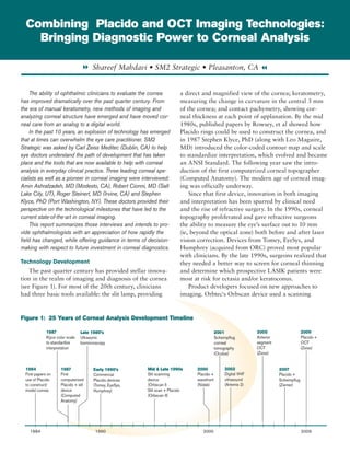
  • 1. Combining Placido and OCT Imaging Technologies: Bringing Diagnostic Power to Corneal Analysis 8 Shareef Mahdavi • SM2 Strategic • Pleasanton, CA 7 The ability of ophthalmic clinicians to evaluate the cornea a direct and magnified view of the cornea; keratometry, has improved dramatically over the past quarter century. From measuring the change in curvature in the central 3 mm the era of manual keratometry, new methods of imaging and of the cornea; and contact pachymetry, showing cor- analyzing corneal structure have emerged and have moved cor- neal thickness at each point of applanation. By the mid neal care from an analog to a digital world. 1980s, published papers by Rowsey, et al showed how In the past 10 years, an explosion of technology has emerged Placido rings could be used to construct the cornea, and that at times can overwhelm the eye care practitioner. SM2 in 1987 Stephen Klyce, PhD (along with Leo Maguire, Strategic was asked by Carl Zeiss Meditec (Dublin, CA) to help MD) introduced the color-coded contour map and scale eye doctors understand the path of development that has taken to standardize interpretation, which evolved and became place and the tools that are now available to help with corneal an ANSI Standard. The following year saw the intro- analysis in everyday clinical practice. Three leading corneal spe- duction of the first computerized corneal topographer cialists as well as a pioneer in corneal imaging were interviewed: (Computed Anatomy). The modern age of corneal imag- Amin Ashrafzadeh, MD (Modesto, CA), Robert Cionni, MD (Salt ing was officially underway. Lake City, UT), Roger Steinert, MD (Irvine, CA) and Stephen Since that first device, innovation in both imaging Klyce, PhD (Port Washington, NY). These doctors provided their and interpretation has been spurred by clinical need perspective on the technological milestones that have led to the and the rise of refractive surgery. In the 1990s, corneal current state-of-the-art in corneal imaging. topography proliferated and gave refractive surgeons This report summarizes those interviews and intends to pro- the ability to measure the eye’s surface out to 10 mm vide ophthalmologists with an appreciation of how rapidly the (ie, beyond the optical zone) both before and after laser field has changed, while offering guidance in terms of decision- vision correction. Devices from Tomey, EyeSys, and making with respect to future investment in corneal diagnostics. Humphrey (acquired from ORC) proved most popular with clinicians. By the late 1990s, surgeons realized that Technology Development they needed a better way to screen for corneal thinning The past quarter century has provided stellar innova- and determine which prospective LASIK patients were tion in the realm of imaging and diagnosis of the cornea most at risk for ectasia and/or keratoconus. (see Figure 1). For most of the 20th century, clinicians Product developers focused on new approaches to had three basic tools available: the slit lamp, providing imaging. Orbtec's Orbscan device used a scanning Figure 1: 25 Years of Corneal Analysis Development Timeline 1987 Late 1980’s 2001 2005 2009 Klyce color scale Ultrasonic Scheimpflug Anterior Placido + to standardize biomicroscopy corneal segment OCT interpretation tomography OCT (Zeiss) (Oculus) (Zeiss) 1984 1987 Early 1990’s Mid & Late 1990s 2000 2002 2007 First papers on First Commercial Slit scanning Placido + Digital VHF Placido + use of Placido computerized Placido devices device wavefront ultrasound Scheimpflug to construct/ Placido + slit (Tomey, EyeSys, (Orbscan I) (Nidek) (Artemis 2) (Ziemer) model cornea device Humphrey) Slit scan + Placido (Computed (Orbscan II) Anatomy) 1984 1990 2000 2009
  • 2. slit-beam to image the back surface of the cornea, giv- measuring corneal thickness manually at a handful of ing clinicians posterior measurements and the ability well-spaced locations in the central cornea. to calculate elevation from the front to the back of While this device could be used in tandem with a the cornea. It was limited to 4 diopters of elevation corneal topographer to provide greater information, it change and required higher-skilled technicians to pre- initially lacked the interpretative power afforded by the vent patient eye movement and get reliable readings. In Holladay report. By 2007, corneal analysis software for 2000, Nidek’s OPD Scan combined Placido + wavefront the ATLAS 9000 (known as Pathfinder II) gave clini- measurement in a single device. The use of ultrasound cians an anterior topographic screening module to help (e.g., ultrasonic biomicroscopy) gave high quality images identify abnormal corneal conditions. And by 2009, of corneal structure, but had fallen out of favor with Zeiss linked both the ATLAS and Visante together, call- clinicians due to the required contact via a probe or ing the merged technologies Visante Omni. Each device water bath. In contrast to Placido-based topographers, now accessed the other’s data, first to ensure proper Oculus' Pentacam tomographer used slits to generate biometric registration of the eye, then to conduct pre- cross-sections of the cornea and reconstruct them into a cise analysis of corneal structure and changes over time. singular image. There were now two different technolo- Interpretation was greatly enhanced when Visante Omni gies popular for clinical incorporated the tradi- diagnosis, each deriving Figure 2: Corneal Imaging Technologies and Applications tional Holladay report its final answer from plus several new color- different approaches: GALILEI ORBSCAN PENTACAM VISANTE OMNI scale maps to help diag- the Pentacam, using Placido + Placido + Placido nose, specifically early Scheimpflug imaging, TECHNOLOGY: Scheimpflug Slit Scanning Scheimpflug + OCT corneal pathology such measured elevation Anterior & Posterior as suspect keratoconus. Topography directly and then calcu- Narrow angle evaluation Clinical Significance lated corneal curvature, Clear Corneal Wound while Placido measured Evaluation Many diagnostic corneal curvature Cataract Evaluation and innovations sought to directly. IOL Placement Analysis improve upon Placido Surgeons were drawn Evaluation of Pars Plana measurements and to the Pentacam’s ease- Corneal Transplant render them obsolete, Planning & Follow-up of-use with patients yet hindsight reveals a Iris Tumors and the interpretive different outcome. “We Imaging Through Opaque/ software developed by White Corneas have come full circle,” Jack Holladay, MD; as Phakic IOL Planning noted Roger Steinert, a result, the Pentacam Full Capability Limited Capability No Capability MD. “We now realize sold briskly to refractive Placido-based technolo- surgeons in the early 2000s. By 2005, Carl Zeiss had gies as the most reliable way to analyze anterior corneal developed an anterior segment OCT device to comple- topography.” Cornea specialist Amin Ashrafzadeh, MD ment their rapidly-adopted posterior segment version for has been using corneal topography to plan and analyze use in retina and glaucoma. The Visante OCT provided surgical outcomes since 2001 and believes the Placido extremely crisp images of corneal structures, allow- disk “remains one of the most essential instruments for ing both increased width of coverage (out to 16 mm) as imaging the corneal surface.” He lamented, however, well as depth of coverage (up to 6 mm). This advanced that “up until several years ago, you still had a lot of technology took multiple “Optical A-scan” images of work to do in figuring out why you had an odd-looking the cornea and then re-constructed these slices into a cornea.” “Optical B-scan” image, putting directly into the clinic That burden changed, according to both Steinert images never before available (except in part from histol- and Ashrafzadeh, when surgeons gained the ability to ogy slides). The new “map” of the eye included a differ- correlate topography with corneal thinning. Both had ent pachymetric approach that automated pachymetry attempted to use Scheimpflug – which had become com- measurement with exponentially greater data points monplace for routine refractive cases – to help with (2,048). This eliminated the potential inaccuracy from this more sophisticated analysis, but found that they
  • 3. were too often led down in reaching diagnosis,” remarked Dr. Steinert. Figure 2 “The real value of the wrong path in reach- shows the different capabilities of four popular corneal Visante Omni is its ing the correct diagnosis. imaging devices. An earlier survey by SM2 Strategic of 32 “Anterior topography from of the first users of Visante showed significantly greater ability to evaluate the Pentacam was pointing usage than anticipated once they had the device in their numerous data... to a completely different practice and had become familiar with its capabilities. all in one place” area for astigmatism, and treating this made cylinder Interpretation Becomes Critical Robert Cionni, MD worse not better,” said Dr. The sheer volume of data on the cornea now avail- Steinert. “I went back and able to clinicians places a premium on software analytics checked the Placido images and found they were accurate; that can reliably distill the images and data points so they this triggered the realization that we needed to go back to make sense in diagnosis. “The amount of information Placido based topography.” While the ability to use a sin- can be overwhelming,” according to Dr. Ashrafzadeh. gle device was indeed convenient, Dr. Ashrafzadeh found “The Holladay report helps tremendously but still has that the Pentacam software would “fill in” missing data deficiencies that cause me to evaluate the ring image over- points improperly on slightly irregular surfaces. “I liked laid atop the report.” The real value of Visante Omni, the way that Scheimpflug image looked, but even looks remarked Dr. Cionni, is it’s ability to evaluate numer- can be deceiving.” Both remarked that the presence of ous data such as topography, corneal aberrations, ICL artifacts in Scheimpflug images of suspicious corneas positioning, pachymetry, ectasia analysis, angle measure- has been a troublesome aspect of that technology. ments… all in one place. Indeed, clinicians should view This issue appears to have been solved through the Visante Omni as a workstation with multiple capabilities linking of Placido and OCT, and the applications extend rather than a niche device. beyond the cornea. Robert Cionni, MD has found that the Omni allows him greater ability to evaluate aberra- Early Detection Capabilities tions relevant only to the cornea. “I get better astigmatic Each surgeon interviewed agreed that the Pentacam analysis with Atlas and my toric IOLs are now more pre- itself is sufficient for the refractive surgeon who is mainly dictable,” according to Dr. Cionni. concerned about corneal elevation. If you are dealing Evaluation of glaucoma patients has been greatly only with normal corneas, they say, then the Pentacam enhanced by Omni. “Pupil size and the resultant angle is fine. The dividing line is drawn around earlier detec- opening are not influenced by Visante's non-visible light, tion of corneal abnormalities. Dr. Ash did a comparison providing an exquisitely accurate measure of physiologic study of 60 eyes, measuring them both on the Pentacam angle opening,” stated Dr. Ashrafzadeh. and the Visante Omni. His findings were that on corneas with no prior surgery and no opacities, there was good Efficiency in the Clinic agreement between the devices. But in patients with Some clinicians view it a disadvantage to have to prior refractive surgery, significant differences emerge. In use two devices rather than one as part of their corneal this study, Visante showed much greater consistency in screening. Dr. Ashrafzadeh finds it actually works to his posterior surface findings compared to the variability in advantage. “I can counsel the patient while I’m doing readings from the Pentacam, which also tended to under- their Omni readings.” He described how this interaction detect post-LASIK ectasia. The findings on the Visante allows him to educate the patient and discuss candidacy Omni correlated much more closely to what he was see- for refractive surgery, while also giving the patient “sig- ing clinically. nificant confidence in seeing the whole process in action. This type of study illus- Omni takes away a lot of the mystery by allowing me to “At present, Visante trates the sentiment of Dr. describe what I’m doing and what the images are telling Omni is the only Steinert regarding a surgeon’s me…in real time.” technology that I ability to trust the device Dr. Cionni added, “I am able to show a patient how you are using: “You need removing their astigmatism will improve their quality of can truly rely upon confidence in your device vision as well.” for subtle changes.” if you are doing refractive “If you use Visante for other applications such as Roger Steinert, MD surgery to pick up the subtle evaluating the angle, then you actually gain efficiency things like Form Fruste
  • 4. Keratoconus. At present, Visante shown in Figure 3. As patient loads increase with the “You must use Omni is the only technology aging population, this justification becomes even more Placido for early that I can truly rely upon for attractive and helps the ophthalmic practice harness the subtle changes.” power of these two innovative technologies. detection of Stephen Klyce, PhD, widely Keratoconus” regarded as a pioneer in corneal Summary and Discussion Stephen Klyce, PhD imaging and analysis, believes Ophthalmic diagnosis and treatment paradigms have that Scheimpflug is not well evolved significantly in the past two decades. Better diag- suited to screening the cornea, as it is 20x less sensi- nostics for the cornea have improved surgeons’ abilities to tive than Placido to changes in the amount of corneal the point that most recognize they can perform refractive curvature. “The use of posterior corneal data has not surgery on patients who were not considered candidates been shown convincingly to be useful for early detection. ten years ago…and vice-versa. The negative publicity sur- Anterior surface Placido is the standard; you must use rounding LASIK in the past several years can be attrib- Placido for early detection of Keratoconus.” uted in part to the lack of understanding of the cornea in the initial period following US commercialization. What Reimbursement surgeons needed – and now have – is a clinical “safety Lastly, changes in reimbursement effective January net” as a result of technological innovation. 2011 puts into effect a new code for Anterior Segment The power in diagnosis afforded by Visante Omni OCT (92132, a Category 1 CPT code). Prior to this, and its marriage of Placido and OCT technology raises doctors were treated unevenly. Some were able to bill the bar for all ophthalmic surgeons in several respects. and get reimbursed, while others’ claims were denied. First, increased understanding of the risks presented by The presence of a new code – specifically for anterior abnormal corneas – and how to best mitigate those risks segment OCT – will help achieve a consistency that is – puts a premium on being able to detect it earlier in its better overall when one considers patient care standards development. Second, an aging population will bring an on a national basis. Although it is tempting to bemoan increasing prevalence of all eye diseases, including those the fact that the code now pays bilaterally (i.e., $36.35 in the anterior segment (e.g., glaucoma). Taken together, national average reimbursement for both eyes), the previ- these two issues put additional pressure on clinicians to ous SM2 survey showed users billing an average of 18 get the right diagnosis in an efficient manner. And just uses per month out of 47 uses overall (including refrac- as the lines between refractive and cataract surgery are tive evaluation). With a specific billing code in place blurring, technology such as Visante Omni is making it to guide clinicians, one would expect the reimbursable possible for all ophthalmic clinicians to more accurately usage to increase significantly. A pro-forma analysis is diagnose disease. Figure 3: Return On Investment VISANTE VISANTE omni KEY ASSUMPTIONS Cost Cost Per Month $708 – $1,018 $982 – $1,382 Monthly lease cost includes device and service for 5 years. Range reflects value of trade-in, and new/refurbished unit. Reimbursed Procedures OCT (CPT code 92132) $647 $647 18 reimbursed procedures per month (from prior SM2 survey of Visante users); national average reimbursement of $36.35 bilateral Corneal Topography - $700 20 submitted claims per month (25% of 80 eyes per month) (CPT code 92025) $35 average reimbursement Non-Reimbursed Procedures OCT $1,044 $1,044 29 per month (from prior SM2 survey of Visante users); $36 per eye component of elective fee, built-in to refractive fee for LASIK and/or Premium IOL's Total Return per Month $1,691 $2,391 Return on Investment Ratio 1.7x – 2.4x 1.7x – 2.4x © Copyright 2011, SM2 Strategic. All Rights Reserved.