2. vided further evidence that (per)chlorate reduction genes are
transferred horizontally, and several new genes were identified in
the PRI that could be involved in regulation of the metabolism
(11). Previous efforts to understand the regulation of perchlorate
reduction by environmental conditions have used the enzymatic
activity of whole cells or extracts, but little is known about the
genetic or transcriptional basis for these observations (13, 14).
While continued isolation of novel DPRB provides a diverse
collection of bacteria capable of this metabolism (15–17), under-
standing the basic tenets of perchlorate reduction is hindered by
the lack of a robust model system amenable to genetic manipula-
tion. To fill this role, we have selected the betaproteobacterium
Azospira suillum PS (formerly known as Dechlorosoma suillum)
(18). PS is a facultative anaerobe that is capable of respiring per-
chlorate and nitrate under anoxic conditions. It grows rapidly and
robustly on rich media, and its genome has been fully sequenced
and is publicly available (11, 19). Additionally, its perchlorate-
reducing physiology has been studied in the context of environ-
mental regulation (13), chemotaxis (20), and redox cycling of
electron shuttles (21, 22).
In this study, we set out to broaden our knowledge about the
molecular mechanisms and genetic factors involved in perchlor-
ate reduction. We performed a random transposon mutagenesis
of PS and identified 18 mutants unable to grow on perchlorate,
most of which contained transposon insertions in the PRI. To
further dissect this genomic island, we developed a method for
creating markerless deletions and constructed in-frame knockout
mutants for the 17 core genes in the PRI. We found that eight were
essential for perchlorate reduction, while the remaining nine
genes were either partially defective during growth on perchlorate
or had no obvious phenotype. As expected, pcrABCD and cld were
essential for perchlorate reduction, but so were genes for a re-
sponse regulator (pcrR), histidine kinase sensor (pcrS), and a PAS
domain-containing protein (pcrP), which form a putative regula-
tory system. Complementation of all eight genes using plasmid-
carried copies of the deleted gene resulted in partial or complete
restoration of growth via perchlorate reduction. This study ex-
pands the list of genes essential for perchlorate reduction but also
supplies the tools for further analysis of the genetics and regula-
tion of this metabolism.
RESULTS
Transposon mutagenesis of PS and screening for mutants.
Transposon mutagenesis and screening identify the genetic fac-
tors involved in a given phenotype in an unbiased manner. We
mutagenized PS using a plasmid-based MiniHimar transposon,
which was delivered using the conjugation-competent Esche-
richia coli strain WM3064 (23, 24). Mutants were isolated as sin-
gle, kanamycin-resistant colonies aerobically on a medium de-
signed to maximize the breadth of genes disrupted during
mutagenesis. This medium contained acetate, lactate, and pyru-
vate as carbon sources and was thus designated ALP media (see
Materials and Methods).
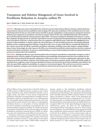
To screen the mutants for defects associated with perchlorate
reduction, we took advantage of the fact that PS grows rapidly in
the presence of both nitrate and perchlorate when transferred
from aerobic conditions (Fig. 1). Additionally, when both nitrate
and perchlorate are present under anaerobic conditions, nitrate is
used in preference to perchlorate by this organism, resulting in a
diauxic growth curve (13). The mutant library was screened for
growth under these conditions over a 30-h period. Individual mu-
tants that exhibited defects in either growth rate or final optical
density were restreaked and selected for further study. To identify
mutants with perchlorate-specific defects, each mutant was fur-
ther characterized for the ability to grow on nitrate, perchlorate,
nitrate/perchlorate, and oxygen. Many mutants that were selected
for additional characterization displayed wild-type growth char-
acteristics, suggesting that the initial screen had a high incidence
of false positives. Some mutants were pleiotropic, suffering defects
of various intensities in both growth rate and final optical density
under multiple conditions. In order to focus on mutants directly
involved in perchlorate reduction, we defined a subset of the mu-
tants as “perchlorate null” mutants. These mutants did not grow
at all on perchlorate but exhibited normal growth on nitrate alone,
as well as normal aerobic growth on both solid and liquid ALP
media. The full phenotypic characterizations of the transposon
mutants of interest are detailed in Table 1. In total, 18 perchlorate
null mutants and 18 pleiotropic mutants were isolated (Table 1).
Transposon mutagenesis highlights importance of genes in
the PRI. The genomic locations of transposon insertions were
identified using an arbitrary PCR-based method (Table 1). Many
pleiotropic mutations in genes predicted to be important for an-
aerobic respiration, including genes for molybdopterin biosyn-
thesis (moeA and mogA), molybdenum uptake (modC), nitrate
reduction/denitrification (napA, norD, and Dsui_1177), anaero-
bic heme biosynthesis (hemN), and ubiquinone biosynthesis
(ubiX), were identified. Additionally, three insertions in the rnf
operon resulted in wild-type growth on nitrate and oxygen but
very slow growth on perchlorate. Similarly, an insertion in a gene
from the relA/spoT family (Dsui_2816) was associated with a very
low growth rate on perchlorate.
While the pleiotropic mutants had insertions in genes located
throughout the chromosome, most (14 out of 18) of the perchlo-
rate null mutant insertions were localized to 8 genes in the PRI
(11). Insertions were found in previously identified structural
genes (pcrA, pcrB, pcrD, and cld) but also in several genes of un-
FIG 1 PS growth on nitrate and perchlorate. Wild-type PS was grown on
medium containing nitrate, perchlorate, and a mixture of nitrate and perchlo-
rate.
Melnyk et al.
2 ®
mbio.asm.org January/February 2014 Volume 5 Issue 1 e00769-13
mbio.asm.orgonJune2,2015-Publishedbymbio.asm.orgDownloadedfrom
4. known function. Three genes in the PRI identified in this screen
encode a putative histidine kinase signal transduction system
(HKS). We have named these genes pcrR, pcrS, and pcrP to reflect
their linkage with pcrA and the fact that homologs of some or all of
these genes can be identified in the PRI of other perchlorate re-
ducers, some of which are distantly related to PS (11). The gene
names pcrR, pcrS, and pcrP are intended to be suggestive of the
putative roles of each gene product in the HKS: the response reg-
ulator, the kinase sensor, and the PAS domain-containing protein.
PcrP and PcrS are predicted to be integral membrane proteins
with large regions in both the cytoplasm and periplasm. The cy-
toplasmic region of PcrS contains domains seen in many bacterial
histidine kinases: a HAMP linker domain (PF00672), a phospho-
acceptor domain (PF00512), and an ATPase domain (PF02518)
(25). PcrP contains two PAS domains from different subfamilies
in its cytoplasmic region (PF08448 and PF13188) (26). In contrast
to the cytoplasmic regions, which are composed of well-
characterized and common domains, the periplasmic regions of
PcrP and PcrS are not homologous to any domains in the COG or
Pfam databases. These regions are fairly large, consisting of ap-
proximately 250 and 270 amino acids in PcrS and PcrP, respec-
tively. BLAST-P searches of the PcrP domain against finished ge-
nomes identified several similar sequences, while a search using
the PcrS domain identified only the PcrS homolog from another
DPRB, Dechloromonas aromatica RCB. PcrR contains three do-
mains commonly found in response regulators: the receiver do-
main (PF00072), a 54 interaction domain (PF00158), and a helix-
turn-helix DNA-binding domain (PF02954) (27). Another
insertion in a perchlorate null mutant mapped to the cupin do-
main protein gene (Dsui_0153) in the PRI. Outside the PRI, three
genes also contained insertions that led to the perchlorate null
phenotype. Two separate mutants with insertions in Dsui_0704,
TABLE 1 (Continued)
Transposon
mutant ID
Locus tag
of insertion-
containing gene
Gene name and
gene product Phenotype
Growth
on nitrate
Growth on
perchlorate
Growth on
nitrate and
perchlorate
Aerobic
growth
A9G11 Dsui_0993 ubiX; 3-octaprenyl-
4-hydroxybenzoate
carboxylase
Pleiotropic ϩ ϩ ϩ ϩϩ
B7F10 Dsui_1054 trpB; tryptophan
synthase
Pleiotropic ϩϩ ϩ ϩ ϩϩ
A7B1 Dsui_1177 Heme b/c᎑containing
EbdC-like electron
transport protein
Pleiotropic 0 ϩ ϩ ϩϩ
B2H11 Dsui_1236 mogA; molybdopterin
adenylyltransferase
Pleiotropic ϩ 0 ϩ ϩϩ
B3E6 Dsui_1533 dsbB; disulfide bond
formation protein
Pleiotropic ϩ ϩϩ ϩ ϩϩ
A3C3 Dsui_1578 aceE; pyruvate
dehydrogenase
Pleiotropic ϩ ϩ ϩ ϩ
C1A4 Dsui_1647 hemN;
oxygen᎑independent
coproporphyrinogen
III oxidase
Pleiotropic ϩ ϩ ϩ ϩϩ
B9B5 Dsui_2152 moeA; molybdopterin
molybdotransferase
Pleiotropic ϩ ϩ ϩ ϩϩ
C4E9 Dsui_2816 (p)ppGpp synthetase,
RelA/SpoT family
Pleiotropic ϩϩ ϩ ϩ ϩϩ
C5B9 Upstream of
Dsui_2979
rnfA; Rnf electron
transport complex
alpha subunit
Pleiotropic ϩϩ ϩ ϩ ϩϩ
B9H11 Dsui_2979 rnfA; Rnf electron
transport complex
alpha subunit
Pleiotropic ϩ ϩ ϩ ϩϩ
B1F10 Dsui_2982 rnfD; Rnf electron
transport complex
delta subunit
Pleiotropic ϩϩ ϩ ϩ ϩϩ
B2D12 Dsui_3119 norD; nitric oxide
reductase activation
protein
Pleiotropic ϩ ϩϩ ϩ ϩϩ
A5B2 Dsui_2880 bioA;
adenosylmethionine-
8-amino-7-
oxononanoate
transaminase
No phenotype (control) ϩϩ ϩϩ ϩϩ ϩϩ
a Transposon mutants were scored “ϩϩ” if growth was identical to that of the wild type, “ϩ” if growth rate or final optical density was less than that of the wild type, or “0” if no
growth was observed.
b CoA, coenzyme A.
Melnyk et al.
4 ®
mbio.asm.org January/February 2014 Volume 5 Issue 1 e00769-13
mbio.asm.orgonJune2,2015-Publishedbymbio.asm.orgDownloadedfrom
5. which encodes the alternative 54-type sigma factor RpoN, were
isolated. The other two genes (Dsui_0128 and Dsui_1441) are of
unknown function.
Two diauxic phenotypes associated with “perchlorate null”
mutants. Perchlorate null mutants were defined as having no
growth on medium containing perchlorate, while being indistin-
guishable from wild-type PS on medium with nitrate. When the
18 perchlorate null mutants were grown on diauxic medium con-
taining nitrate and perchlorate, two different phenotypes were
observed. Most (15 out of 18) of the perchlorate null mutants had
similar growth curves on diauxic medium and nitrate-only me-
dium. The remaining three perchlorate null mutants had signifi-
cantly impaired growth on diauxic medium relative to that on
medium containing only nitrate. Interestingly, all three of these
mutants had transposon insertions in cld, the gene encoding chlo-
rite dismutase (Fig. 2). The other 15 perchlorate null mutants had
insertions in 10 different genes, including pcrA (Fig. 2). To high-
light the different diauxic growth phenotypes, pcrA::Himar and
cld::Himar transposon mutants were compared to a biotin biosyn-
thetic mutant (bioA::Himar) which had wild-type growth charac-
teristics on kanamycin-amended ALP medium, presumably due
to the presence of biotin in the vitamin supplement (Fig. 3A to C).
Neither the pcrA::Himar nor the cld::Himar mutants grew at all on
perchlorate (Fig. 3B), but the cld::Himar mutant grew to a much
lower optical density than the pcrA::Himar mutant on nitrate and
perchlorate (Fig. 3C).
Developing a genetic system in PS and systematic deletions
of genes in the PRI. To confirm that genes identified in the trans-
poson screen were essential for perchlorate reduction, we devel-
oped a system for making markerless chromosomal deletions in
PS. Several different suicide vectors did not undergo chromo-
somal integration when delivered via conjugation, but a suicide
vector derived from pNPTS138 (Dickon Alley, unpublished data)
was able to integrate into the chromosome. However, conjugation
proved to be an unpredictable method of vector delivery, since
conjugations would frequently fail and no chromosomal inser-
tions could be recovered. Reliable chromosomal insertions of vec-
tors derived from pNPTS138 were achieved using electroporation,
although efficiency of integration was low (5 to 10 insertions per
g plasmid DNA transformed). This is consistent with the previ-
ous observation that Azospira spp. are resistant to transformation
(28).
We used pNPTS138 to create deletions of the 17 genes in the
conserved core of the PRI. Because many genes in the PRI are in
operons (e.g., pcrABCD), we designed the deletion constructs to
create in-frame deletions of each gene that did not disturb the
ribosomal binding site or promoters of nearby genes. Once each
deletion was made, we characterized the strain’s ability to grow on
nitrate, perchlorate, and nitrate plus perchlorate. Of the eight
genes in the PRI that were defined as being perchlorate null mu-
tants during the transposon screen, seven (pcrABD, cld, and
pcrPSR) recapitulated the perchlorate null phenotype. Addition-
ally, the deletion of pcrC resulted in a perchlorate null phenotype,
although no insertions in pcrC were isolated during the trans-
poson screen. When Dsui_0153 was deleted, the resulting strain
exhibited no differences from the wild type under all conditions
tested, suggesting that the perchlorate null phenotype of
Dsui_0153::Himar was the result of a polar effect, possibly on the
adjacent pcrP gene.
In addition to Dsui_0153, deletion of six other genes in the PRI
also had no apparent phenotype. Five of these genes (Dsui_0154
to Dsui_0158) are situated at one end of the PRI and comprise two
oppositely oriented operons. The five genes contain a predicted
sigma factor/anti-sigma factor pair, genes homologous to yedYZ
from E. coli (29), and a gene encoding a hypothetical protein. The
sixth nonessential gene (Dsui_0141) is located at the opposite end
of the PRI and is a homolog of moaA, a gene involved in molyb-
dopterin biosynthesis (30). Two additional genes in the PRI,
Dsui_0143 and Dsui_0144, were not essential for perchlorate re-
duction, but both mutants suffered from slight yet significant
growth rate defects on perchlorate. Dsui_0144 is predicted to en-
code a tetraheme c-type cytochrome homologous to proteins such
as NapC, NirT, and CymA, all of which function as quinol dehy-
drogenases in electron transport chains involved in the reduction
of various electron acceptors (31). Dsui_0143 is predicted to be a
gene encoding a protein homologous to EbdC, the gamma sub-
unit of ethylbenzene dehydrogenase of Aromatoleum aromaticum,
which contains a heme b and forms a heterotrimer with the alpha
and beta subunits (32). However, the Dsui_0143 protein may con-
tain two additional heme c molecules, since it contains two puta-
tive heme c binding motifs (one CXXCH motif and an atypical
CWXXCH). These two motifs are conserved in the homologous
NirB protein, which is encoded in a cluster of genes involved in
nitrite reduction in Pseudomonas stutzeri (33). The growth pheno-
types for all 17 single deletions are summarized in Table 2.
Complementation of perchlorate reduction mutants. To
show that the eight genes from the PRI were independently essen-
tial for perchlorate reduction and the observed phenotypes were
FIG 2 Locations of transposon insertions in the PRI. The 17 genes in the PRI are displayed with the locations of transposon insertions that were identified in
the screen. The green pins indicate mutants that did not grow on perchlorate, while the red pins indicate mutants that did not grow on perchlorate or on the
diauxic medium. Genes are colored by functional group: green genes encode chlorite dismutase and perchlorate reductase components, royal-blue genes are the
PRI HKS, red genes are parts of the putative electron transport chain, orange genes are components of a putative oxidoreductase system, and light-blue genes are
part of a putative sigma factor/antisigma system.
Genetics of Perchlorate Reduction
January/February 2014 Volume 5 Issue 1 e00769-13 ®
mbio.asm.org 5
mbio.asm.orgonJune2,2015-Publishedbymbio.asm.orgDownloadedfrom
6. not the result of polar effects, we developed a system for comple-
mentation analysis. The broad-host-range plasmid pBBR1MCS2
was tested for stable replication in PS and was used as the back-
bone for all complementation vectors. The complementation vec-
tors were constructed in two groups. The first group contained the
genes pcrPSR, which are assumed to be transcribed independently
based on their alternating orientation (Fig. 2). When constructing
these vectors, we included the ~150-bp sequence upstream of the
start codon in order to retain the native promoter and regulation.
The second group contains five essential genes (pcrABCD and cld)
located in the same operon. To construct these vectors, we first
made a plasmid with the pcrA promoter (pRAM62) and then in-
corporated each of the five genes independently into pRAM62
with their endogenous ribosomal binding site. Each of the eight
perchlorate null deletion mutants received both the complemen-
tation vector and the empty pBBR1MCS2 vector. Under anaero-
bic conditions with both nitrate and perchlorate, all eight comple-
mented strains were able to grow on perchlorate, while the strains
with the empty vector recapitulated the defective growth curves
seen in the deletion mutants (see Fig. S1 to S3 in the supplemental
material). The complemented strains had variable lag times when
switching from nitrate to perchlorate and different growth rates
on perchlorate. The reasons for this are unknown but could be due
to differences in expression between the complemented mutants
and wild-type PS.
Phenotypic characterization of ⌬cld ⌬pcrA double mutant.
To further explore the discrepancy between the diauxic pheno-
types of ⌬cld and ⌬pcrA, we constructed a ⌬cld ⌬pcrA double
mutant by electroporating the cld deletion vector into the previ-
ously constructed ⌬pcrA strain. As previously described, the ⌬cld
strain exhibited normal growth on nitrate alone but was impaired
for growth on nitrate when perchlorate was also present (Fig. 4A
and B). We hypothesized that this was due to baseline expression
of pcrA and accumulation of chlorite in the absence of Cld activity.
As expected, the ⌬cld ⌬pcrA strain did not grow on perchlorate
but was able to grow normally on nitrate (Fig. 4A and C). How-
ever, in the presence of nitrate and perchlorate, the ⌬cld ⌬pcrA
mutant recovered the ability to grow on nitrate as did the ⌬pcrA
single deletion mutant (Fig. 4B).
DISCUSSION
Previous work identified the cld gene by sequencing a fragment of
a purified enzyme with chlorite dismutase activity (34). This se-
quence was used to amplify and sequence cld from Dechloromonas
agitata CKB and subsequently used to identify neighboring per-
chlorate reductases (6, 10). pcrA was directly mutagenized and
shown to be essential in Dechloromonas aromatica RCB; however,
this was done with an antibiotic cassette insertion and may have
resulted in polar effects on nearby genes (6). A forward genetics
approach confirmed that cld and pcrA are essential for perchlorate
reduction in Azospira suillum PS and also identified the impor-
tance of pcrB, pcrD, a new HKS, and rpoN. Markerless in-frame
deletion was used to confirm the insertions in the PRI and also led
to the identification of pcrC as an essential gene for perchlorate
reduction. These complementary genetic approaches advance our
understanding of the genetic factors involved in perchlorate re-
duction and establish Azospira suillum PS as a model perchlorate
reducer.
The majority of the genes identified as essential for perchlorate
reduction are located in the perchlorate reduction genomic island
(PRI), which contains 17 genes in PS that are conserved in Dechlo-
romonas aromatica RCB but not in other closely related organisms
incapable of perchlorate reduction (11). Our transposon screen
identified cld, pcrA, pcrB, and pcrD as essential for perchlorate
reduction, which we confirmed with deletion mutagenesis. Al-
FIG 3 Growth of pcrA::Himar and cld::Himar strains. The growth of strains
with transposon insertion mutations in cld, pcrA, and bioA (control) shows
distinct phenotypes on nitrate (A) or perchlorate (B) or under diauxic condi-
tions (C).
Melnyk et al.
6 ®
mbio.asm.org January/February 2014 Volume 5 Issue 1 e00769-13
mbio.asm.orgonJune2,2015-Publishedbymbio.asm.orgDownloadedfrom
7. though it was not identified in the transposon screen, pcrC was
also found to be essential for perchlorate reduction. pcrB encodes
the beta subunit of the perchlorate reductase, and the initial bio-
chemical characterization of perchlorate reductase from Azospira
sp. GR-1 identified a stable enzyme complex in an ␣33 confor-
mation (8). PcrB is likely required for the reductase complex sta-
bility and for delivering reducing equivalents from the inner
membrane electron transport chain to the active site of PcrA. PcrC
is a multiheme cytochrome c that was previously thought to be
part of the perchlorate reduction pathway in all DPRB (6), but a
recently isolated and described organism, Arcobacter sp. CAB, re-
duces perchlorate and does not have a gene homologous to pcrC in
its PRI or elsewhere in its genome (35). pcrD encodes a putative
chaperone that is homologous to both TorD and NarJ of E. coli
and has been predicted to be the chaperone for PcrA (6, 36, 37).
Many periplasmic molybdoenzymes of the DMSO reductase fam-
ily have dedicated chaperones which bind to the signal peptide
and prevent transport of the apoprotein via the Tat (twin-arginine
translocation pathway) until addition of the appropriate cofactor
(37, 38). The perchlorate null phenotype of the ⌬pcrD strain sug-
gests that PcrD may similarly be required for the attachment of
molybdopterin to PcrA and the proper translocation of the ho-
loenzyme.
pcrP, pcrR, and pcrS were identified both in the transposon
screen and by deletion mutagenesis as being essential for perchlo-
rate reduction. Together, their gene products comprise a histidine
kinase signal transduction system that is conserved in other DPRB
(11). The sensory inputs and physiological outputs of this system
are currently unknown, but the DNA-binding domain of PcrR
suggests that it likely binds to promoters to modulate transcrip-
tion. PcrR also contains a 54 interaction domain, which can re-
cruit 54-type sigma factors to promoters where the regulator is
bound and hydrolyze ATP to initiate transcription (39). PS con-
tains exactly one 54-type sigma factor, encoded by the rpoN gene
(Dsui_0704), which was one of only three genes outside the PRI
that resulted in a perchlorate null phenotype when mutated with a
transposon insertion. Additionally, there is a conserved promoter
region upstream of pcrA that contains an RpoN-binding motif
(11). Based on these observations, we hypothesize that at least one
of the outputs of this system is to upregulate transcription of the
pcrABCD operon in an RpoN-dependent manner. The presence
of two separate sensor proteins that are both essential (PcrP and
PcrS) hints at a complex regulatory mechanism.
The periplasmic domains of PcrP and PcrS are unique, and the
signals that modulate this system are unknown. Perchlorate, ni-
trate, or intermediates of both reductive pathways could be sensed
directly, as is the case for the E. coli NarX sensor kinase, which
autophosphorylates in response to binding nitrate at the
dimerization interface (40). An emerging paradigm for histidine
kinase “two-component” signal transduction is the presence of a
third auxiliary protein which can adjust the autophosphorylation
of the histidine kinase and/or phosphotransfer to the response
regulator (41). PcrP could be such an auxiliary protein, and it
could sense signals that originate either in the periplasm or in the
TABLE 2 Annotations of the 17 core PRI genes and phenotypic information based on deletion mutantsa
Locus tag Gene name Gene product Pfam domain(s)
Growth on
perchlorate
Growth on nitrate
and perchlorate
Dsui_0141 moaA Molybdenum cofactor
biosynthesis protein
PF04055, PF06463, PF13353 ϩϩ ϩϩ
Dsui_0143 Heme b/c-containing
EbdC-like electron
transport protein
PF09459 ϩ ϩϩ
Dsui_0144 Quinol dehydrogenase
tetraheme cytochrome c
(NapC-like)
PF03264 ϩ ϩϩ
Dsui_0145 cld Chlorite dismutase PF06778 0 0
Dsui_0146 pcrD Perchlorate reductase
cytoplasmic chaperone
PF02613 0 ϩ
Dsui_0147 pcrC Tetraheme cytochrome c PF13435 0 ϩ
Dsui_0148 pcrB Perchlorate reductase
beta subunit
PF13247 0 ϩ
Dsui_0149 pcrA Perchlorate reductase
alpha subunit
PF00384, PF01568 0 ϩ
Dsui_0150 pcrR PRI response regulator PF00072, PF00158, PF02954 0 0 ϩ
Dsui_0151 pcrS PRI sensor histidine
kinase
PF00512, PF00672, PF02518 0 0 ϩ
Dsui_0152 pcrP PRI PAS domain protein PF08448, PF13188 0 ϩ
Dsui_0153 Cupin domain protein PF07883 ϩϩ ϩϩ
Dsui_0154 nrsF Anti-sigma factor PF06532 ϩϩ ϩϩ
Dsui_0155 sigF ECF sigma factor PF04542, PF08281 ϩϩ ϩϩ
Dsui_0156 yedZ Cytochrome b membrane
electron transport protein
PF00033 ϩϩ ϩϩ
Dsui_0157 yedY Unknown molybdopterin
oxidoreductase
PF00174 ϩϩ ϩϩ
Dsui_0158 Hypothetical protein ϩϩ ϩϩ
a For growth on perchlorate, “ϩϩ” denotes wild-type growth on perchlorate, “ϩ” denotes growth at a lower rate than that of the wild type, and “0” denotes no growth. For growth
on nitrate and perchlorate, “ϩϩ” denotes wild-type growth, “ϩ” denotes growth only during the initial denitrification phase, and “0” denotes very little growth. All deletion
mutants grew identically to the wild type on nitrate alone.
Genetics of Perchlorate Reduction
January/February 2014 Volume 5 Issue 1 e00769-13 ®
mbio.asm.org 7
mbio.asm.orgonJune2,2015-Publishedbymbio.asm.orgDownloadedfrom
8. cytoplasm, perhaps via its two PAS domains. Interestingly, the
pcrPSR genes do not appear in the genome of Dechloromonas agi-
tata CKB, a DPRB that does not respire nitrate, unlike PS, suggest-
ing that the PRI HKS could play a role in nitrate-dependent re-
pression of perchlorate reduction (11, 13, 42). We have presented
a schematic for the basic functions of the PRI HKS, highlighting
the domain structure of PcrP and PcrS in the inner membrane, as
well as the putative function of PcrR in initiating transcription in
an RpoN-dependent manner (Fig. 5). Although we currently
know little about how the system functions, molecular analysis of
the signals and domains involved will be crucial in understanding
the evolution of the PRI.
Nine genes in the PRI were not required for perchlorate reduc-
tion (Dsui_0141, Dsui_0143-Dsui_0144, and Dsui_0153 to
Dsui_0158). All of these genes are conserved in at least one other
DPRB, and thus it was unexpected that so many (9 out of 17) had
no obvious role in perchlorate reduction (11). However, a similar
observation was made in a genetic study of the magnetosome
genomic island, where many of the conserved genes were not es-
sential for magnetosome formation (43). Possible reasons pro-
posed for this phenomenon were genetic redundancy, as well as
the inherent limitations of laboratory-imposed growth conditions
(43). It is reasonable to expect that both of these caveats are rele-
vant for genetic analysis of the PRI.
The perchlorate reductase receives electrons from the electron
transport chain via an unknown pathway. PS has a predicted
quinol dehydrogenase-type cytochrome c (Dsui_0144) encoded
in its PRI that was previously proposed to be a conduit from the
quinone pool to the reductase (6), but there was only a slight
impairment of growth on perchlorate when the gene was deleted
in PS. A similar phenotype was seen with the deletion of the pre-
dicted cytochrome b/c gene Dsui_0143. Because none of these
genes are absolutely essential for perchlorate reduction, we pro-
pose that the electron transport chain to PcrA is redundant. This
sort of multiplicity is seen on a larger scale in organisms such as
Geobacter spp., which utilize dozens of different cytochrome c
proteins to transport electrons across the cell envelope to multiple
electron acceptors (44). The perchlorate electron transport chain
may not be quite as complex, but additional genetic and biochem-
ical evidence is needed to resolve this pathway in PS. The limita-
tions of our experimental growth conditions may also occlude the
phenotypes of certain genes in the PRI. For example, Dsui_0154
and Dsui_0155 are predicted to encode an extracytoplasmic func-
tion sigma factor/anti-sigma factor system homologous to SigF
and NrsF of Caulobacter crescentus, which has been shown to play
a role in activating a response to reactive oxygen species and heavy
metal stress (45, 46). The phenotype of these knockouts in PS may
be apparent only under the appropriate oxidative stress condi-
tions, which are not met by our standard acceptor-limited condi-
tions in rich medium.
One possible source of oxidative stress associated with perchlo-
rate reduction could be an intermediate of the metabolism. Both
the transposon screen and deletion mutagenesis revealed that the
cld mutant had a different phenotype from the rest of the perchlo-
rate null mutants when grown on nitrate and perchlorate together
(Fig. 3). Interestingly, deletion of pcrA in the ⌬cld background
relieved the growth inhibition (Fig. 4). In the context of perchlo-
rate reduction, chlorite dismutase liberates molecular oxygen
from chlorite and sustains the microaerobic respiration that is
thought to accompany perchlorate reduction (5). However, Cld
can also function to detoxify chlorite, which has been proposed as
a role for Cld in organisms that cannot reduce perchlorate or
chlorate (47). Chlorite is a powerful oxidant that can result in cell
death even at parts-per-million concentrations (48). Previous ex-
periments with PS indicate that perchlorate is not reduced with
nitrate in large quantities, but if perchlorate reductase is expressed
at all under nitrate-reducing conditions in the absence of Cld, the
FIG 4 Growth characteristics of wild-type PS and the ⌬cld, ⌬pcrA, and ⌬cld
⌬pcrA strains. Growth on nitrate (A), under diauxic conditions (B), or on
perchlorate (C) shows that a ⌬cld ⌬pcrA double mutant grows better on mixed
electron acceptors than the ⌬cld and ⌬pcrA single mutants.
Melnyk et al.
8 ®
mbio.asm.org January/February 2014 Volume 5 Issue 1 e00769-13
mbio.asm.orgonJune2,2015-Publishedbymbio.asm.orgDownloadedfrom
9. micromolar amounts of chlorite that accumulate may result in the
inhibition of cell growth. A study performed with cell extracts of
the closely related DPRB Azospira sp. KJ grown on nitrate showed
some perchlorate reductase activity, suggesting that there could be
baseline expression of PcrA even under nitrate-reducing condi-
tions (14).
As previously proposed, perchlorate reduction is dependent on
genes in the horizontally transferred PRI but also on genes else-
where in the genome (11). Our transposon screen identified sev-
eral operons and pathways that are involved in anaerobic respira-
tion. Insertions in hemN (Dsui_1647) and ubiX (Dsui_0993)
resulted in pleiotropic mutants that were defective in growth on
both nitrate and perchlorate. HemN is an oxygen-independent
coproporphyrinogen III oxidase, and UbiX is a decarboxylase in-
volved in the biosynthesis of quinones (49, 50). Presumably, these
mutants are deficient in anaerobic heme and quinone biosynthe-
sis, which leads to a general growth defect under anaerobic con-
ditions. Perchlorate reductase requires a molybdopterin-bound
molybdenum to function, and several genes involved in molyb-
dopterin biosynthesis were identified in the transposon screen
(Table 1). These mutants were also wholly or partially defective
during denitrification, which is likely due to the nitrate reductase
utilizing the same cofactor. Additionally, the transposon screen
allowed the identification of several genes putatively involved in
denitrification, which has not been studied directly in PS. These
genes include napA, norD, and Dsui_1177. The gene product of
Dsui_1177 is a heme-containing protein which has been shown to
be upregulated under denitrifying conditions (21). Interestingly,
the napA and Dsui_1177 mutants also demonstrate some defects
in perchlorate reduction. The reason for this is currently un-
known.
Many other genes were identified in the transposon screen as
being partially defective in perchlorate reduction and not denitri-
fication, but their specific functions are difficult to predict. These
include the rnf operon, which was hit by transposons several times
in the mutagenesis and screen. The rnf operon encodes a
membrane-bound redox-active complex which couples the pro-
ton (or sodium) gradient of the inner membrane to the cellular
redox pools of NADϩ/NADH and ferredoxin (51). The activity of
the Rnf complex is reversible and has been shown to be important
in an array of different bacterial metabolisms, although its role in
perchlorate reduction is currently unknown. Other transposon
insertions are in genes that are not directly linked to cellular ener-
getics or are in genes that encode hypothetical proteins. Exploring
the roles of these genes will be essential to gaining a holistic un-
derstanding of perchlorate reduction in the context of global cel-
lular metabolism.
In this article, we set out to address a basic question about the
molecular biology of perchlorate reduction: which genes are es-
sential for this metabolism? Transposon mutagenesis and a sub-
sequent screen on medium containing nitrate and perchlorate
identified many mutants with insertions in the PRI, accentuating
FIG 5 A model of perchlorate reduction in Azospira suillum PS. Gene products from the PRI are depicted in a model that reflects our current understanding of
perchlorate reduction and its regulation in Azospira suillum PS. Several genes identified in the transposon screen as important for perchlorate reduction are also
shown (molybdopterin cofactor biosynthesis, denitrification, and the Rnf complex). Gene products from the PRI are colored to be consistent with the functional
group coloring in Fig. 2.
Genetics of Perchlorate Reduction
January/February 2014 Volume 5 Issue 1 e00769-13 ®
mbio.asm.org 9
mbio.asm.orgonJune2,2015-Publishedbymbio.asm.orgDownloadedfrom
10. its importance. To further characterize this region, we developed a
system for making markerless in-frame deletions and systemati-
cally knocked out each gene in the PRI. The results of these com-
plementary genetic approaches have increased our understanding
of the factors involved in perchlorate reduction (Fig. 5) but have
raised many new questions. The histidine kinase system in the PRI
is essential for perchlorate reduction, although we know little
about how it is activated or which genes it regulates. Specifically,
utilization of nitrate over perchlorate is still not understood. The
components that deliver electrons to PcrA are also unknown, as is
the role played by the nonessential genes in the PRI. Finally, the
evolution of the PRI is still obscure; how does the island integrate
with the host’s metabolism after it is acquired? Genetic analysis
using the deletion and complementation tools developed here will
be critical to answering these questions about the evolution and
function of the PRI in Azospira suillum PS and thus other DPRB.
MATERIALS AND METHODS
Bacterial strains and plasmids. Azospira suillum PS (ATCC BAA-33/
DSMZ 13638) was revived from a lab freezer stock and used as the wild-
type strain for all genetic manipulations (see Table S2 in the supplemental
material). Various E. coli strains were also used for cloning and conjuga-
tion purposes (see Table S2). Plasmids constructed or used in this study
are described in Table S3. Prior to all growth curves and genetic manipu-
lations, all strains were streaked out from master freezer stocks to get
single colonies.
Culture conditions and growth media. E. coli strains were grown in
LB medium. Kanamycin (Kan) (50 g/ml) was used for selection, and
diaminopimelic acid (DAP) (0.3 mM) was used as a supplement to culti-
vate the auxotrophic strain WM3064. For routine culturing, as well as
growth assays, wild-type and mutant strains of PS were grown in ALP
medium. One liter of ALP medium is composed of 0.49 g monobasic
sodium phosphate dihydrate, 0.97 g dibasic anhydrous sodium phos-
phate, 0.1 g potassium chloride, 0.25 g ammonium chloride, 0.82 g so-
dium acetate, 2.0 g yeast extract, 7.6 g of a 60% (wt/wt) sodium lactate
solution, 1.10 g sodium pyruvate, and 10 ml of both vitamin mix and
mineral mix as previously described (42). To make solid ALP medium for
plates, 15 g/liter agar was added. Kanamycin was used for selection of PS
mutants at a concentration of 50 g/ml. For anaerobic growth of PS, ALP
medium was supplemented with either 5 mM sodium nitrate, 2.5 mM
sodium perchlorate, or 5 mM of both sodium nitrate and sodium perchlo-
rate. No growth was observed when no electron acceptor was added, con-
firming that PS is unable to grow via fermentation of the carbon sources
present in ALP medium. All strains of E. coli and PS were cultivated at
37°C. Anaerobic growth curves of PS for the transposon screen and char-
acterization of strains was performed using a SpectraMax 340PC384 plate
reader in an anaerobic chamber (Coy Laboratory Products, Grass Lake,
MI).
Transposon mutagenesis via conjugation. The mariner transposon
suicide vector pMiniHimar RB1 was used to mutagenize PS (23).
WM3064 containing pMiniHimar RB1 (A. Arkin lab, University of Cali-
fornia [UC], Berkeley) was grown as overnight culture in 5 ml LB plus Kan
plus DAP. This culture was centrifuged and washed twice in ALP medium
to remove excess kanamycin. In parallel, a 5-ml culture of PS was grown
from a single colony overnight (optical density at 600 nm [OD600] of
~1.5) and centrifuged. The WM3064 and PS cell pellets were resuspended
in 50 l ALP medium and spotted onto an ALP plate supplemented with
DAP. The plate was dried by an open flame for 15 min on the benchtop to
ensure that the agar surface was dry and the mating reaction did not
spread. The plate was then incubated at 37°C for 6 h, after which the entire
spot was scraped off the plate and resuspended in 1 ml ALP medium. The
volume was divided, plated on 6 ALP-Kan plates, and incubated at 37°C.
After 36 to 48 h, ~300 to 500 whitish-pink opaque colonies formed on the
6 plates in total. The colonies were then transferred into 96-well plates
containing 300 l of ALP-Kan medium supplemented with 7.5% glycerol
as a cryoprotectant. The plates were then grown overnight aerobically on
a platform shaker at 37°C prior to screening and long-term storage at
Ϫ80°C.
Screeningthetransposonmutantlibrary.Thetransposonlibrarywas
screened prior to long-term storage. From each glycerol freezer stock
plate, 10 l was transferred into 300 l of ALP-Kan medium supple-
mented with 5 mM nitrate and perchlorate on the benchtop. Plates were
then transferred into the anaerobic chamber, where they were allowed to
equilibrate with the anaerobic atmosphere for 1 h. An initial optical den-
sity reading was taken, and the plates were placed in a GasPak box (BD)
with a palladium catalyst for maintenance of anaerobic conditions. The
box was removed from the glove bag and incubated at 37°C. Every 6 to 8 h
for 30 h, the box was moved back into the glove bag and the optical density
of the plates was measured. The growth curve data were plotted, and
mutants that had a defect in either growth rate or maximum optical den-
sity were identified. The “mutants of interest” were then streak purified to
isolate single colonies on ALP-Kan medium. The single colonies were
grown on ALP medium to make long-term freezer stocks.
Characterizationoftransposonmutantsdefectiveinperchloratere-
duction. Mutants of interest identified in the initial screen were subjected
to further phenotypic characterization. Each mutant was streaked out on
ALP-Kan plates from freezer stocks and then transferred into an overnight
culture of ALP-Kan liquid medium. The next morning, the optical densi-
ties of all strains were determined, and each culture was diluted back to an
OD600 of 1.0 to 1.2. A 50-l aliquot was used to inoculate 1.45 ml of
ALP-Kan medium supplemented with either nitrate (5 mM), perchlorate
(2.5 mM), or nitrate and perchlorate (both 5 mM). The concentration of
perchlorate under the perchlorate-only condition was lowered to avoid
unpredictably long lag periods seen with high perchlorate concentrations
(unpublished data). Each medium condition was distributed to 4 wells in
a 96-well plate such that 8 genotypes could be assayed simultaneously
using the SpectraMax plate reader in the anaerobic chamber. The charac-
terization growth curves were allowed to run for 48 h.
Mapping the locations of transposon mutants. Transposon mutants
of interest were mapped using a variation of arbitrarily primed PCR meth-
ods previously described (52, 53). Briefly, transposon mutants of interest
were streaked onto ALP-Kan plates, and single colonies were used as tem-
plates for colony PCR. The first round of PCR used a primer that annealed
to the end of the mariner transposon (HIMAR_EXT) (see Table S1 in the
supplemental material) and one of three arbitrary primers (PS_ARB4,
PS_ARB5, and PS_ARB6) which contain a conserved 20-mer at the 5= end,
followed by 10 random nucleotides and one of three most common pen-
tamers in the A. suillum PS genome. This first round of PCR used the
GoTaq Green master mix (Promega) and the following thermocycling
parameters: 10 min at 95°C; 35 cycles of 30 s at 95°C, 30 s at 38°C, and
2 min at 72°C; and 10 min at 72°C. The reaction mixture was treated with
ExoSAP-IT (Affymetrix) to remove the primers, and the product was used
as the template for a second round of PCR using a transposon primer
closer to the genomic insertion site (HIMAR_INT) and a primer consist-
ing of the conserved 20-mer at the 5= end of the arbitrary primers
(PS_ARB2). The second round of PCR used the following thermocycling
parameters: 5 min at 95°C; 35 cycles of 30 s at 95°C, 30 s at 55°C, and 2 min
at 72°C; and 10 min at 72°c. The second-round PCR product was purified
using the QIAquick PCR purification kit (Qiagen) and used as the tem-
plate for a sequencing reaction with the HIMAR_INT primer (UC Berke-
ley DNA Sequencing Facility). Sequencing reads were manually curated
and used as queries for searching the A. suillum genome with BLAST on
the NCBI server (54). Sequences were downloaded, and the exact inser-
tion site of the transposon in each mutant was determined.
Constructing suicide vectors and complementation vectors.
Genomic DNA from A. suillum was isolated using Trizol according to the
manufacturer’s directions (Invitrogen), and all PCRs were performed us-
ing Phusion DNA polymerase (Fisher Scientific). Specific primer pairs
were designed using the NCBI Primer-BLAST server to amplify products
Melnyk et al.
10 ®
mbio.asm.org January/February 2014 Volume 5 Issue 1 e00769-13
mbio.asm.orgonJune2,2015-Publishedbymbio.asm.orgDownloadedfrom
11. of 800 to 900 bp flanking the gene to be deleted (55). The internal primers
closest to the gene were designed to end between codons within the gene
so that the resulting deletion would be in-frame and have minimal polar
effects on nearby genes. For some genes, the internal primers had a 21-bp
“linker” at the 5= end that was used to assemble the flanking regions into a
1.6- to 1.8-kb insert using PCR. For the remaining genes, the internal
primers contained restriction sites that allowed assembly of the suicide
vector with a three-way ligation. Both methods used resulted in an in-
frame deletion allele of the gene of interest, thereby minimizing polar
effects on nearby genes. Sequences and brief descriptions of all primers are
listed in Table S1 in the supplemental material.
We used the vector pNPTS138 (Dickon Alley via Kathleen Ryan, UC,
Berkeley) as the suicide vector backbone for all of our deletion constructs
after testing several plasmids with no success (pAK31, pK19mobsacB, and
pSMV3). Deletion vectors were constructed by amplifying two regions
flanking the gene of interest and either performing an assembly PCR to
fuse the regions together or digesting the PCR product directly. In both
cases, the cut vector and digested PCR product(s) was digested with the
appropriate restriction enzymes, analyzed by electrophoresis on an aga-
rose gel, and extracted using the Qiaex II gel extraction kit (Qiagen). The
digested products were then ligated into the vector using T4 ligase (NEB).
The broad-host-range plasmid pBBR1MCS2 was used as the backbone for
the complementation vectors (56). For three genes (pcrP, pcrS, and pcrR),
pBBR1MCS2 and the amplified gene with its promoter were digested,
purified, and ligated as described above. For the pcrABCD and cld com-
plementation vectors, the pcrA promoter was first cloned into
pBBR1MCS2 to make pRAM62. The amplified genes were then cloned
downstream of this promoter as described above to generate these com-
plementation vectors. All ligation reactions were transformed into chem-
ically competent E. coli TOP10 cells and plated on selective medium. In-
dividual colonies were grown overnight in liquid broth, and plasmid DNA
was purified. Plasmids were checked for the presence of the correct insert
by digestion with appropriate restriction enzymes, followed by sequenc-
ing of the insert (UC, Berkeley DNA Sequencing Facility).
Electroporation of suicide vectors into PS and screening of dele-
tions. Electrocompetent A. suillum PS cells were prepared using a method
previously described for Zymomonas mobilis (57). One-hundred-
microliter aliquots of electrocompetent cells were thawed on ice and
mixed with 1 g of plasmid DNA and 0.8 l of TypeOne restriction
inhibitor (Epicentre Biotechnologies). The mixture was transferred into a
cuvette on ice and then electroporated at 1,750 V, 400 ⍀, and 25 F.
Immediately after electroporation, 300 l of ALP medium was added to
the cuvette, and the entire aliquot was transferred to a sterile 1.7-ml tube
for 6 h of recovery in a 37°C shaking incubator. After the recovery, the
aliquot was plated on 3 ALP-Kan plates and incubated at 37°C for 36 h.
Colonies that formed within 36 h were picked into 500 l of ALP-Kan
liquid medium and grown for 12 h to confirm integration of the suicide
vector into the chromosome. An aliquot of 50 l was then transferred into
5 ml of ALP medium and grown overnight. Dilutions of the overnight
culture were plated on ALP plates with 6% sucrose (ALP-Suc) to select
against the sacB allele present on the pNPTS138 backbone and incubated
at 37°C for 36 h. Colonies that appeared were patched onto ALP-Kan and
ALP-Suc and incubated at 37°C overnight. Colonies that were sensitive to
kanamycin and resistant to sucrose were used as a template for colony
PCR using primers flanking the gene to be deleted. The primers used for
each gene were the external primers used previously to amplify the flank-
ing regions of the deletion construct. The PCR products were analyzed
using gel electrophoresis on a 1% agarose gel, and colonies that yielded a
PCR product with the correctly sized deleted allele size were restreaked on
an ALP plate. Single colonies from the restreaked plate were checked once
again with colony PCR for the deleted allele and then picked into an
overnight ALP medium culture to make into freezer stocks.
SUPPLEMENTAL MATERIAL
Supplemental material for this article may be found at http://mbio.asm.org
/lookup/suppl/doi:10.1128/mBio.00769-13/-/DCSupplemental.
Figure S1, EPS file, 0.8 MB.
Figure S2, EPS file, 0.6 MB.
Figure S3, EPS file, 0.8 MB.
Table S1, PDF file, 0.1 MB.
Table S2, PDF file, 0.1 MB.
Table S3, PDF file, 0.1 MB.
ACKNOWLEDGMENTS
Work on (per)chlorate metabolism was supported by funding to J.D.C.
from the Energy Biosciences Institute.
We thank Arash Komeili and Kathleen Ryan for gifts of strains and
plasmids, as well as valuable advice regarding bacterial genetics and pro-
tocol development. We also thank members of the Coates lab, especially
Hans Carlson, for critical feedback and discussion regarding this research.
REFERENCES
1. Rajagopalan S, Anderson T, Cox S, Harvey G, Cheng Q, Jackson WA.
2009. Perchlorate in wet deposition across North America. Environ. Sci.
Technol. 43:616–622.
2. Kounaves SP, Stroble ST, Anderson RM, Moore Q, Catling DC, Doug-
las S, McKay CP, Ming DW, Smith PH, Tamppari LK, Zent AP. 2010.
Discovery of natural perchlorate in the Antarctic Dry Valleys and its global
implications. Environ. Sci. Technol. 44:2360–2364.
3. Parker DR. 2009. Perchlorate in the environment: the emerging emphasis
on natural occurrence. Environ. Chem. 6:10–27.
4. Coates JD, Michaelidou U, Bruce RA, O’Connor SM, Crespi JN, Achen-
bach LA. 1999. Ubiquity and diversity of dissimilatory (per)chlorate-
reducing bacteria. Appl. Environ. Microbiol. 65:5234–5241.
5. Coates JD, Achenbach LA. 2004. Microbial perchlorate reduction:
rocket-fueled metabolism. Nat. Rev. Microbiol. 2:569–580.
6. Bender KS, Shang C, Chakraborty R, Belchik SM, Coates JD, Achen-
bach LA. 2005. Identification, characterization, and classification of genes
encoding perchlorate reductase. J. Bacteriol. 187:5090–5096.
7. van Ginkel CG, Rikken GB, Kroon AGM, Kengen SWM. 1996. Purifi-
cation and characterization of chlorite dismutase: a novel oxygen-
generating enzyme. Arch. Microbiol. 166:321–326.
8. Kengen SW, Rikken GB, Hagen WR, van Ginkel CG, Stams AJ. 1999.
Purification and characterization of (per)chlorate reductase from the
chlorate-respiring strain GR-1. J. Bacteriol. 181:6706–6711.
9. Thorell HD, Karlsson J, Portelius E, Nilsson T. 2002. Cloning, charac-
terisation, and expression of a novel gene encoding chlorite dismutase
from Ideonella dechloratans. Biochim. Biophys. Acta 1577:445–451.
10. Bender KS, O’Connor SM, Chakraborty R, Coates JD, Achenbach LA.
2002. Sequencing and transcriptional analysis of the chlorite dismutase
gene of Dechloromonas agitata and its use as a metabolic probe. Appl.
Environ. Microbiol. 68:4820–4826.
11. Melnyk RA, Engelbrektson A, Clark IC, Carlson HK, Byrne-Bailey K,
Coates JD. 2011. Identification of a perchlorate reduction genomic island
with novel regulatory and metabolic genes. Appl. Environ. Microbiol. 77:
7401–7404.
12. Clark IC, Melnyk RA, Engelbrektson A, Coates JD. 2013. Structure and
evolution of chlorate reduction composite transposons. mBio 4(4):
e00379-13. http://dx.doi.org/10.1128/mBio.00379-13.
13. Chaudhuri SK, O’Connor SM, Gustavson RL, Achenbach LA, Coates
JD. 2002. Environmental factors that control microbial perchlorate re-
duction. Appl. Environ. Microbiol. 68:4425–4430.
14. Xu J, Trimble JJ, Steinberg L, Logan BE. 2004. Chlorate and nitrate
reduction pathways are separately induced in the perchlorate-respiring
bacterium Dechlorosoma sp. KJ and the chlorate-respiring bacterium
Pseudomonas sp. PDA. Water Res. 38:673–680.
15. Thrash JC, Pollock J, Torok T, Coates JD. 2010. Description of the novel
perchlorate-reducing bacteria Dechlorobacter hydrogenophilus gen.
nov., sp. nov. and Propionivibrio militaris, sp. nov. Appl. Microbiol. Bio-
technol. 86:335–343.
16. Thrash JC, Ahmadi S, Torok T, Coates JD. 2010. Magnetospirillum
bellicus sp. nov., a novel dissimilatory perchlorate-reducing alphaproteo-
bacterium isolated from a bioelectrical reactor. Appl. Environ. Microbiol.
76:4730–4737.
17. Vijaya Nadaraja A, Veetil PGP, Bhaskaran K. 2013. Perchlorate reduc-
tion by an isolated Serratia marcescens strain under high salt and extreme
pH. FEMS Microbiol. Lett. 339:117–121.
Genetics of Perchlorate Reduction
January/February 2014 Volume 5 Issue 1 e00769-13 ®
mbio.asm.org 11
mbio.asm.orgonJune2,2015-Publishedbymbio.asm.orgDownloadedfrom
12. 18. Achenbach LA, Michaelidou U, Bruce RA, Fryman J, Coates JD. 2001.
Dechloromonas agitata gen. nov., sp. nov. and Dechlorosoma suillum
gen. nov., sp. nov., two novel environmentally dominant (per)chlorate-
reducing bacteria and their phylogenetic position. Int. J. Syst. Evol. Mi-
crobiol. 51:527–533.
19. Byrne-Bailey KG, Coates JD. 2012. Complete genome sequence of the
anaerobic perchlorate-reducing bacterium Azospira suillum strain PS. J.
Bacteriol. 194:2767–2768.
20. Sun Y, Gustavson RL, Ali N, Weber KA, Westphal LL, Coates JD. 2009.
Behavioral response of dissimilatory perchlorate-reducing bacteria to dif-
ferent electron acceptors. Appl. Microbiol. Biotechnol. 84:955–963.
21. Clark IC, Carlson HK, Iavarone AT, Coates JD. 2012. Bioelectrical redox
cycling of anthraquinone-2,6-disulfonate coupled to perchlorate reduc-
tion. Energy Environ. Sci. 5:7970–7978.
22. Van Trump JI, Coates JD. 2009. Thermodynamic targeting of microbial
perchlorate reduction by selective electron donors. ISME J 3:466–476.
23. Bouhenni R, Gehrke A, Saffarini D. 2005. Identification of genes in-
volved in cytochrome c biogenesis in Shewanella oneidensis, using a mod-
ified mariner transposon. Appl. Environ. Microbiol. 71:4935–4937.
24. Larsen RA, Wilson MM, Guss AM, Metcalf WW. 2002. Genetic analysis
of pigment biosynthesis in Xanthobacter autotrophicus Py2 using a new,
highly efficient transposon mutagenesis system that is functional in a wide
variety of bacteria. Arch. Microbiol. 178:193–201.
25. Galperin MY, Nikolskaya AN, Koonin EV. 2001. Novel domains of the
prokaryotic two-component signal transduction systems. FEMS Micro-
biol. Lett. 203:11–21.
26. Möglich A, Ayers RA, Moffat K. 2009. Structure and signaling mecha-
nism of Per-ARNT-Sim domains. Structure 17:1282–1294.
27. Galperin MY. 2006. Structural classification of bacterial response
regulators: diversity of output domains and domain combinations. J. Bac-
teriol. 188:4169–4182.
28. Reinhold-Hurek B, Hurek T. 2006. The genera Azoarcus, Azovibrio,
Azospira and Azonexus, p 873–891. Springer Verlag, New York, NY.
29. Loschi L, Brokx SJ, Hills TL, Zhang G, Bertero MG, Lovering AL,
Weiner JH, Strynadka NC. 2004. Structural and biochemical identifica-
tion of a novel bacterial oxidoreductase. J. Biol. Chem. 279:50391–50400.
30. Schwarz G, Mendel RR, Ribbe MW. 2009. Molybdenum cofactors,
enzymes and pathways. Nature 460:839–847.
31. Simon J, Kern M. 2008. Quinone-reactive proteins devoid of haem b
form widespread membrane-bound electron transport modules in bacte-
rial respiration. Biochem. Soc. Trans. 36:1011–1016.
32. Kloer DP, Hagel C, Heider J, Schulz GE. 2006. Crystal structure of
ethylbenzene dehydrogenase from Aromatoleum aromaticum. Structure
14:1377–1388.
33. Jüngst A, Wakabayashi S, Matsubara H, Zumft WG. 1991. The nirSTBM
region coding for cytochrome CD1-dependent nitrite respiration of Pseu-
domonas stutzeri consists of a cluster of mono-, di-, and tetraheme pro-
teins. FEBS Lett. 279:205–209.
34. Stenklo K, Thorell HD, Bergius H, Aasa R, Nilsson T. 2001. Chlorite
dismutase from Ideonella dechloratans. J. Biol. Inorg. Chem. 6:601–607.
35. Carlström CI, Wang O, Melnyk RA, Bauer S, Lee J, Engelbrektson A,
Coates JD. 2013. Physiological and genetic description of dissimilatory
perchlorate reduction by the novel marine bacterium Arcobacter sp. strain
CAB. mBio 4(3):e00217-13. http://dx.doi.org/10.1128/mBio.00217-13.
36. Blasco F, Dos Santos JP, Magalon A, Frixon C, Guigliarelli B, Santini
CL, Giordano G. 1998. NarJ is a specific chaperone required for molyb-
denum cofactor assembly in nitrate reductase A of Escherichia coli. Mol.
Microbiol. 28:435–447.
37. Jack RL, Buchanan G, Dubini A, Hatzixanthis K, Palmer T, Sargent F.
2004. Coordinating assembly and export of complex bacterial proteins.
EMBO J. 23:3962–3972.
38. Maillard J, Spronk CA, Buchanan G, Lyall V, Richardson DJ, Palmer T,
Vuister GW, Sargent F. 2007. Structural diversity in twin-arginine signal
peptide-binding proteins. Proc. Natl. Acad. Sci. U. S. A. 104:15641–15646.
39. Bush M, Dixon R. 2012. The role of bacterial enhancer binding proteins
as specialized activators of 54-dependent transcription. Microbiol. Mol.
Biol. Rev. 76:497–529.
40. Lee AI, Delgado A, Gunsalus RP. 1999. Signal-dependent phosphoryla-
tion of the membrane-bound NarX two-component sensor-transmitter
protein of Escherichia coli: nitrate elicits a superior anion ligand response
compared to nitrite. J. Bacteriol. 181:5309–5316.
41. Buelow DR, Raivio TL. 2010. Three (and more) component regulatory
systems—auxiliary regulators of bacterial histidine kinases. Mol. Micro-
biol. 75:547–566.
42. Bruce RA, Achenbach LA, Coates JD. 1999. Reduction of (per)chlorate
by a novel organism isolated from paper mill waste. Environ. Microbiol.
1:319–329.
43. Murat D, Quinlan A, Vali H, Komeili A. 2010. Comprehensive genetic
dissection of the magnetosome gene island reveals the step-wise assembly
of a prokaryotic organelle. Proc. Natl. Acad. Sci. U. S. A. 107:5593–5598.
44. Shi L, Squier TC, Zachara JM, Fredrickson JK. 2007. Respiration of
metal (hydr)oxides by Shewanella and Geobacter: a key role for multih-
aem c-type cytochromes. Mol. Microbiol. 65:12–20.
45. Alvarez-Martinez CE, Baldini RL, Gomes SL. 2006. A Caulobacter cres-
centus extracytoplasmic function sigma factor mediating the response to
oxidative stress in stationary phase. J. Bacteriol. 188:1835–1846.
46. Kohler C, Lourenço RF, Avelar GM, Gomes SL. 2012. Extracytoplasmic
function (ECF) sigma factor F is involved in Caulobacter crescentus
response to heavy metal stress. BMC Microbiol. 12:210. http://dx.doi.org/
10.1186/1471-2180-12-210.
47. Mlynek G, Sjöblom B, Kostan J, Füreder S, Maixner F, Gysel K,
Furtmüller PG, Obinger C, Wagner M, Daims H, Djinovic´-Carugo K.
2011. Unexpected diversity of chlorite dismutases: a catalytically efficient
dimeric enzyme from Nitrobacter winogradskyi. J. Bacteriol. 193:
2408–2417.
48. Kwolek-Mirek M, Bartosz G, Spickett CM. 2011. Sensitivity of
antioxidant-deficient yeast to hypochlorite and chlorite. Yeast 28:
595–609.
49. Gulmezian M, Hyman KR, Marbois BN, Clarke CF, Javor GT. 2007.
The role of UbiX in Escherichia coli coenzyme Q biosynthesis. Arch.
Biochem. Biophys. 467:144–153.
50. Troup B, Hungerer C, Jahn D. 1995. Cloning and characterization of the
Escherichia coli hemN gene encoding the oxygen-independent copropor-
phyrinogen III oxidase. J. Bacteriol. 177:3326–3331.
51. Biegel E, Schmidt S, González JM, Müller V. 2011. Biochemistry, evo-
lution and physiological function of the Rnf complex, a novel ion-motive
electron transport complex in prokaryotes. Cell. Mol. Life Sci. 68:
613–634.
52. Jacobs MA, Alwood A, Thaipisuttikul I, Spencer D, Haugen E, Ernst S,
Will O, Kaul R, Raymond C, Levy R, Chun-Rong L, Guenthner D,
Bovee D, Olson MV, Manoil C. 2003. Comprehensive transposon mu-
tant library of Pseudomonas aeruginosa. Proc. Natl. Acad. Sci. U. S. A.
100:14339–14344.
53. Das S, Noe JC, Paik S, Kitten T. 2005. An improved arbitrary primed
PCR method for rapid characterization of transposon insertion sites. J.
Microbiol. Methods 63:89–94.
54. Altschul SF, Gish W, Miller W, Myers EW, Lipman DJ. 1990. Basic local
alignment search tool. J. Mol. Biol. 215:403–410.
55. Ye J, Coulouris G, Zaretskaya I, Cutcutache I, Rozen S, Madden TL.
2012. Primer-BLAST: a tool to design target-specific primers for polymer-
ase chain reaction. BMC Bioinformatics 13:134. http://dx.doi.org/
10.1186/1471-2105-13-134.
56. Kovach ME, Elzer PH, Hill DS, Robertson GT, Farris MA, Roop RM,
Peterson KM. 1995. Four new derivatives of the broad-host-range cloning
vector pBBR1MCS, carrying different antibiotic-resistance cassettes. Gene
166:175–176.
57. Lam CK, O’Mullan P, Eveleigh DE. 1993. Transformation of Zymononas
mobilis by electroporation. Appl. Microbiol. Biotechnol. 39:305–308.
Melnyk et al.
12 ®
mbio.asm.org January/February 2014 Volume 5 Issue 1 e00769-13
mbio.asm.orgonJune2,2015-Publishedbymbio.asm.orgDownloadedfrom