Bohomolets Microbiology Lesson #8
•Download as DOC, PDF•
1 like•640 views

By Ms. Kostiuk from Microbiology department

Recommended
More Related Content
What's hot
What's hot (20)
Viewers also liked
Similar to Bohomolets Microbiology Lesson #8
Similar to Bohomolets Microbiology Lesson #8 (20)
More from Dr. Rubz
More from Dr. Rubz (20)
Recently uploaded
Recently uploaded (20)
Bohomolets Microbiology Lesson #8
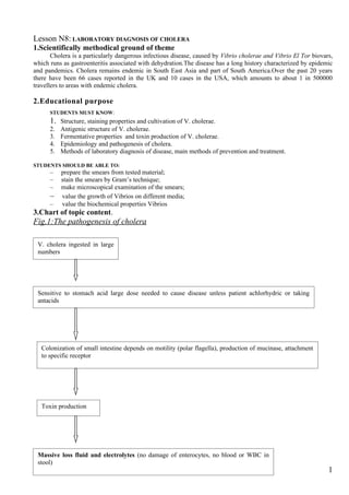
- 1. Lesson N8: LABORATORY DIAGNOSIS OF CHOLERA 1.Scientifically methodical ground of theme Cholera is a particularly dangerous infectious disease, caused by Vibrio cholerae and Vibrio El Tor biovars, which runs as gastroenteritis associated with dehydration.The disease has a long history characterized by epidemic and pandemics. Cholera remains endemic in South East Asia and part of South America.Over the past 20 years there have been 66 cases reported in the UK and 10 cases in the USA, which amounts to about 1 in 500000 travellers to areas with endemic cholera. 2.Educational purpose STUDENTS MUST KNOW: 1. Structure, staining properties and cultivation of V. cholerae. 2. Antigenic structure of V. cholerae. 3. Fermentative properties and toxin production of V. cholerae. 4. Epidemiology and pathogenesis of cholera. 5. Methods of laboratory diagnosis of disease, main methods of prevention and treatment. STUDENTS SHOULD BE ABLE TO: – prepare the smears from tested material; – stain the smears by Gram’s technique; – make microscopical examination of the smears; – value the growth of Vibrios on different media; – value the biochemical properties Vibrios 3.Chart of topic content. Fig.1:The pathogenesis of cholera V. cholera ingested in large numbers Sensitive to stomach acid large dose needed to cause disease unless patient achlorhydric or taking antacids Colonization of small intestine depends on motility (polar flagella), production of mucinase, attachment to specific receptor Toxin production Massive loss fluid and electrolytes (no damage of enterocytes, no blood or WBC in stool) 1
- 2. Fig.2: Mode action of Vibrio cholera exotoxin Diarrhoea V. cholera Cholera Cholera toxin B B Loss of cell nutrients A Na+ H2O Cl - K+ HCO3 - B B B B Ganglioside receptor Cell membrane A A2 A1 Increased adenylate cyclase activity cAMP Fig.N3: Comparison between biovar V.Cholera and biovar El Tor Biovars of V. cholerae Signs cholerae El Tor Lysis by phages: cholerae phage “C” + – “Ël Tor” – + Hemagglutination of erythrocytes – + Sensitivity to 50 U of polymixin + – M Haemolysis of sheep erythrocytes – + Bacteriological examination. Stage I. Using the material collected, prepare smears, dry them in the air, fix with alcohol or Nikiforov's mixture, stain by the Gram technique, and examine under the microscope. Later on, if laboratory findings confirm the diagnosis of cholera in at least one case, stain the smears with Pfeiffer's fuchsine only. Cholera vibrios appear as thin curved Gram-negative rods. Because of great polymorphism the smear may, along with typical cells, contain coccal, rod-shaped, and spiral forms, which diminishes the value of this method. 2
- 3. The first preliminary answer is given after the microscopic examination of the smear. It refers to the presence of vibrios and the nature of their Gram-staining. At this stage of bacteriological examination, one can also perform the immunoftuorescence test, using specific labelled 0-cholera sera. Simultaneously with bacterioscopy, the material tested is inoculated onto liquid and solid nutrient media. Enrichment liquid media that are usually recommended for use include alkaline 1 per cent peptone water, 1 per cent peptone water with potassium tellurite in a ratio of 1 to 100 000, and alkaline taurocholate-tellurite-peptone medium (Monsur's liquid medium), etc. Solid nutrient media usually employed are alkaline meat-peptone agar and one of the selective nutrient media: Aronson's medium, Monsur's alkaline taurocholate-lellurite-gelatine-agar medium, TCBS, etc. To isolate the vibrio from carriers or patients with subclinical forms of cholera, use media which improve the growth of vibrios and suppress the attendant flora (predominantly E. coli). All inoculated cultures are placed in an incubator at 37 °C. Aronson's medium consists of 2-3 per cent of meat-peptone agar to which sucrose and destained fuchsine are added. Monsur's alkaline taurocholate-tellurite-gelatine-agar medium contains 10 g of trypticase, l0 g of sodium chloride, 50 g of sodium taurocholate, 30g of sodium carbonate, 1 g of gelatin, 15 g of agar-agar, and 1 L of distilled water. TCBS (thiosulphate-citrate-bromthymol sucrose) is manufactured in the form ready for use; 69 g of the dry medium is taken per 1 L of distilled water. Stage II. Some 5-6 hours after inoculation examine the film on the peptone water. To do it, tilt the test tube or the vial so that a delicate bluish film is attached to the wall. Prepare smears from the film or the surface of the medium, stain them by the Gram method, evaluate motility, and conduct presumptive slide agglutination test with 0-cholera (0-1) serum diluted 1:100 or the reaction of cholera vibrio immobilization with 0-cholera serum. The results of the latter are estimated by phase-contrast microscopy. Inhibition of vibrios motility and the formation of agglutinate occur within 1-2 min. On the basis of the results obtained give a second preliminary result referring to the motility of the vibrio and its relation to the agglutinating serum. Subculture the material from the film onto plates with alkaline agar or selective medium and simultaneously onto the second peptone water and look for changes in 5-6 hrs. Stage III. Some 10-16 hrs after inoculation, examine the growth in the second enrichment medium (peptone water) and on the plates with the culture of the native material. The film formed on the peptone water is examined as described above. On an alkaline agar the cholera vibrio grows with the formation of round, smooth, flat, bluish, homogeneous colonies which are 1-2 mm in diameter, transparent in the transmitted light and have smooth edges. They are oily in consistence, are readily removed and emulsified. Examination of the material from convalescents, bacteria carriers, and individuals treated with antibiotics may reveal atypical colonies. On Aronson's medium colonies of cholera vibrios are scarlet in the centre and pale-pink or colourless at the periphery. On Monsur's medium colonies are transparent or semitransparent, or they may be of a greyish-black colour with turbid edges. On the TCBS medium they appear as flat and yellow against a bluish-grey background. The selected colonies are introduced into test tubes with Oikenitsky's medium or onto an agar slant for enrichment of pure culture and placed in an incubator. Preliminary identification of cholera vibrios grown on plates with solid media is based on the study of cultural and morphological characteristics and on a presumptive slide agglutination test with 0-cholera serum diluted 1:100 and with Ogawa's and Inaba's sera in a 1 to 60 dilution, which is carried out to determine the serovar. If the examination demonstrates signs typical of the cholera vibrio, a third preliminary answer about the positive result of the investigation is issued. Some material from the typical colonies may be transferred to a broth; then, using a 3-4-hour old culture, perform a standard agglutination test, check fermentation of carbohydrates, and determine whether the isolated culture belongs to Group I according to Heiberg and whether it is liable to phagolysis by cholera phages C and El Tor 2. If the results are positive, an answer concerning the isolation of the causative agent is given within 18-24 hrs from the beginning of the study. Stage IV. After the results of the standard agglutination test and the reaction of phagolysis and fermentation of carbohydrates by 3-4-hour broth culture have been analysed, a preliminary conclusion about the isolation of the cholera vibrio is made. Plates with the inoculated culture on the second peptone water are examined, using the scheme which is employed in examining the plates with the culture of the native material. On Oikenitsky's medium the vibrio breaks down sucrose without gas formation and does not ferment lactose (reddening of the medium in the column without gas formation). 3
- 4. To distinguish vibrios from homogeneous species of microorganisms (Aeromonas, Pseudomonas, Plesiomonas), a number of tests may be employed: the oxidase test, glucose oxidation-fermentation reaction, the "strand" test Fig.N4:Differential-Diagnostic Signs of Vibrios and Related Types of Bacteria Glucose reduction- "Strand" Microorganis Osidase fermentation test ms test reduction fermentation Rapid method of Vibrio ++++ + + (gas is + wide-scale absent) screening for Aeromonas ++++ + + (gas ±) + carriers. During an Pseudomonas ++ + – – outbreak of cholera Plesiomonas + – – – wide-scale screening for carriers of the cholera vibrio is performed. When a large number of analyses is to be made in the laboratory, faeces from ten subjects are examined simultaneously. Faeces are collected with wire loops and placed into one flask containing 200 ml of peptone water and 0-cholera agglutinating serum which is diluted to half the titre. The flask is placed into a 37 "C incubator. In 3-4 hrs the multiplied cholera vibrios begin to agglutinate and fall to the bottom in the form of flakes. If this is the case, faecal material is taken from each of the ten individuals, and the examination is repeated with each sample. Serological diagnosis of cholera is supportive and relies on detecting agglutinins and vibriocidal antibodies in the patient's serum. It is recommended that paired sera obtained from the patients at a 6-8 day interval be used for these reactions. Titres of agglutinins and vibriocidal antibodies usually tend to increase simultaneously. The most sensitive test is demonstration of vibriocidal antibodies. The presence of agglutinating antibodies in the titre of 1:80-1:320 and vibriocidal ones in the titre of 1:1000 is considered diagnostically positive. Fig.N5: Heiberg grouping of vibrios Group Mannose fermantation Sucrose fermantation Arabinose fermantation I A A - II - A - III A A A IV - A A V A - - VI - - - VII A - A VIII - - A Fig.N6 O serotyps of cholera vibrions Serotype O antigen Ogawa AB Inaba AC Hikojima ABC 4. Student’s independent study program 1. Structure and staining properties of causative agents of cholera. 2. Cultivation of Vibrio cholerae. Main nutrient media. 3. Biochemical properties of Vibrio cholerae. 4. Antigenic structure and classification of Vibrio cholerae. 5. Toxin formation of Vibrio cholerae. 6. Resistance to light, heat, low temperature, desiccation, hydrochloric acid (1: 10.000), etc. 7. Epidemiology of cholera: a – source of infectious agents; b – mechanism of transmission; 4
- 5. c – factors of transmission. 8. Pathogenesis and clinical findings of cholera. 9. Laboratory diagnosis of cholera: a – features of collection of tested materials, their transportation to the bacteriological laboratory, organization of work of laboratory; b – stages of bacteriological examination; c – rapid methods of diagnosis of cholera (immunofluorescent test, reaction of immobilization of vibrios under the influence of O1 cholera serum; RIHA test with antibody diagnosticum); d – main characteristic features allowed to differentiate biovars of cholera vibrios (sensitivity to diagnostic phages, hemagglutination of chicken erythrocytes, sensitivity to polymyxin, hemolysis of sheep erythrocytes); e – inagglutinated cholera-like vibrios; f – fast method of wide-scale screening for carriers. 10. Treatment and prophylaxis of a cholera: a – complex of antiepidemic measures in the focus of cholera; b – specific prophylaxis of cholera; c – urgent prophylaxis of cholera; d – treatment (rehydration, remineralization, etiotropic therapy). 5. Students’ practical activities: 1. Stain by Gram’s technique ready smears of vibrios, examine them under the microscope. Draw and record you observation. 2. To familiarize with biological properties of Vibrio cholerae biovars. (See figureN3) Draw and record you observation 3.To familiarize with biological preparation used for laboratory diagnosis and specific prophylaxis of cholera 4. To carry out slide agglutination testwith Osubgroup cholera antisera (OI) and then with type specific cholera antisera(Ogava, Inaba) Make up conclusion . Drow and record you observation. (See figureN6) 6. Control questions and tests Select the correct answers: I. The V. cholerae has such properties: 1. A – peritrichous; b – monotrichous; c – motile; d– grows on alkaline nutrient media; e – grows on acid nutrient media. 2. A – belongs to 1– st Heiberg’s group; b – ferments mannose and saccharose; c – does not ferment arabinose; d – on ТСВS medium forms colourless colonies; e – on ТСВS medium forms yellow colonies. 3. A – has O- and Н-antigens; b – Н-antigen is various; c – there are more than 60 types of O- antigens; d – V. cholerae vibrio has O1-antigen; e – according to Н-antigen V. cholerae is divided into groups. 4. A – both biovars have O-1 antigen; b – every biovar has own O-antigen; c – every biovar has three serovars; d – serovars have combinations of different types of O-1 antigens: A, B, and C; e –serovars differ in Н-antigen. 5. A – V. cholerae produces cholerogen; b – clinical findings of an illness are depend on an endotoxin; c – toxin stimulates the activity of enzyme adenyl cyclase; d – V. cholerae produces mucinase e – V. cholerae does not produces enterotoxin. 6. Differentiation of V. cholerae biovara: а – Vibrio El Tor is marked by high resistance; b –V. El Tor causes erased and mild forms of cholera; c – classical V. cholerae causes vibrio carriage; d – classical cholera vibrio is more resistant then V. El Tor; e – V. cholerae biovar El Tor and V. cholerae cause identical forms of disease. 7. Prophylaxis and treatment: a – for specific prophylaxis – choleragen-anatoxin; b – for specific prophylaxis – live vaccine; c – for treatment – infusion of saline solutions; d – for treatment – tetracycline; e – for treatment – immunoglobulin. 11. Gram negative, comma-shaped rods are revealed in faeces of a patient with diarrhea. What properties should be studied with microscope first of all to receive the additional information on the revealed microbes? A. Presence of spores B. Presence of capsules. C. Mobility. D. Presence of volutin inclusions E. Presence of glycogen inclusions. 5
- 6. 12. The patient is hospitalized to infectious department with suspicion for cholera. What basic method of research is necessary to use for confirmation of the diagnosis? A. Immunological. B. Bacteriological. C. Biological. D. Serological. E. Allergic. 13. Excrements of the patient with cholera were delivered to the laboratory of extremely dangerous infections. What method of microbiological diagnostics needs to be used to confirm or deny the diagnosis? A. Virological. B. Allergic. C. Bacterioscopic. D. Biological. E. Bacteriological. 14. From vomit mass of the patient there were isolated very motile, curved, “comma-shaped” gram-negative rods, which react with diagnostic serum Inaba. The culture gives positive reaction with diagnostic serum Inaba. What symptoms, most probably, will appear at the patient at the absence of treatment? A. Endotoxic shock syndrome. B. Bacteremia. C. Dehydration of organism. D. Spotting on skin. E. Ulcer damages of intestines. 15. The patient with complaints of repeated diarrhea and vomiting, pain in muscles of legs, weakness, dizziness is hospitalized to infectious department. After survey the doctor has made provisional diagnosis – cholera. Which one of the following methods of investigation of the material from the patient should be used for express diagnostics? A. Immunofluorescence test. B. Agglutination test. C. A bacteriological method. D. An allergic method. E. A biological method. 16. Cholera toxin is known to: A. Bind to cell-associated GM ganglioside by it subunit B. Activate adenylate cyclase C. Cause luminal accumulation of electrolyte-rich fluid and subsequent water diarrhea D. Produce necrotizing ulcers in gastrointestinal tract mucosa 17. Activity of cholera exotoxin is associated with A. Inhibited cation (Na+ for H+) and anion (Cl- for HCO3- ) axchange B. Suppressed adenylate cyclase activity C. Enhanced chloride secretion Real-life situation to be solved: 1. From the patient’s feces with diagnosis of acute gastroenteritis on the TCBS medium yellow colonies were grown. Pure culture of bacteria fermented saccharose and mannose and was agglutinated with O-1 cholera serum. A. Can we make diagnosis according these findings? What diagnosis can we make? B. What findings can confirm your diagnosis? C. What tests can we use to determine of serovar of bacteria? 2. From the feces of the patient with acute gastroenteritis the culture of bacteria was isolated. Six-hour-old culture on 1 % alkaline peptone water produced delicate bluish pellicle. There were gram-negative, motile, curved rods. A. What are these microbes? B. What tests help to identify the species of disease agents? 6
- 7. 3. In the seaport some cases of acute enteric disease were registered. The patients had diarrhoea (to 25 times a day), rice-water stool, vomiting, marked dehydratation, sometimes clonicotonic cramps. A. What disease may be in these patients? B. What rapid methods can help to reveal the disease agents? C. How can we perform wide-scale screening for carriers? D. How can we perform urgent prophylaxis? 7. List of literature: 1.I. S. Gaidash, V.V. Flegontova, Microbiology, virology and immunology, Lugansk, 2004, chapter27, p. 208-224. 7