SlideShare uma empresa Scribd logo
1 de 12
CELLS

All living things are made of cells, and cells are the smallest units that can be alive. Life on Earth is
classified into five kingdoms, and they each have their own characteristic kind of cell. However the
biggest division is between the cells of the prokaryote kingdom (the bacteria) and those of the other four
kingdoms (animals, plants, fungi and protoctista), which are all eukaryotic cells. Prokaryotic cells are
smaller and simpler than eukaryotic cells, and do not have a nucleus.

        Prokaryote = without a nucleus
        Eukaryote = with a nucleus


  Eukaryotic Cells


        Cytoplasm (or Cytosol). This is the solution within the cell membrane. It contains enzymes for
        metabolic reactions together with sugars, salts, amino acids, nucleotides and everything else
        needed for the cell to function.
        Nucleus. This is the largest organelle. Surrounded by a nuclear envelope, which is a double
        membrane with nuclear pores - large holes containing proteins that control the exit of substances
such as RNA from the nucleus. The interior is called the nucleoplasm, which is full of chromatin- a
DNA/protein complex containing the genes. During cell division the chromatin becomes
condensed into discrete observable chromosomes. The nucleolus is a dark region of chromatin,
involved in making ribosomes.
Mitochondrion (pl. Mitochondria). This is a sausage-shaped organelle (8µm long), and is
where aerobic respiration takes place in all eukaryotic cells. Mitochondria are surrounded by a
double membrane: the outer membrane is simple, while the inner membrane is highly folded
into cristae, which give it a large surface area. The space enclosed by the inner membrane is
called the matrix, and contains small circular strands of DNA. The inner membrane is studded
with stalked particles, which are the site of ATP synthesis.
Chloroplast. Bigger and fatter than mitochondria, chloroplasts are where photosynthesis takes
place, so are only found in photosynthetic organisms (plants and algae). Like mitochondria they
are enclosed by a double membrane, but chloroplasts also have a third membrane called
the thylakoid membrane. The thylakoid membrane is folded into thylakoid disks, which are then
stacked into piles called grana. The space between the inner membrane and the thylakoid is
called the stroma. The thylakoid membrane contains chlorophyll and stalked particles, and is the
site of photosynthesis and ATP synthesis. Chloroplasts also contain starch grains, ribosomes and
circular DNA.
Ribosomes. These are the smallest and most numerous of the cell organelles, and are the sites
of protein synthesis. They are composed of protein and RNA, and are manufactured in the
nucleolus of the nucleus. Ribosomes are either found free in the cytoplasm, where they make
proteins for the cell's own use, or they are found attached to the rough endoplasmic reticulum,
where they make proteins for export from the cell. They are often found in groups
called polysomes. All eukaryotic ribosomes are of the larger, "80S", type.
Smooth Endoplasmic Reticulum (SER). Series of membrane channels involved in synthesising
and transporting materials, mainly lipids, needed by the cell.
Rough Endoplasmic Reticulum (RER). Similar to the SER, but studded with numerous
ribosomes, which give it its rough appearance. The ribosomes synthesise proteins, which are
processed in the RER (e.g. by enzymatically modifying the polypeptide chain, or adding
carbohydrates), before being exported from the cell via the Golgi Body.
Golgi Body (or Golgi Apparatus). Another series of flattened membrane vesicles, formed from
the endoplasmic reticulum. Its job is to transport proteins from the RER to the cell membrane for
export. Parts of the RER containing proteins fuse with one side of the Golgi body membranes,
while at the other side small vesicles bud off and move towards the cell membrane, where they
fuse, releasing their contents by exocytosis.
Vacuoles. These are membrane-bound sacs containing water or dilute solutions of salts and
other solutes. Most cells can have small vacuoles that are formed as required, but plant cells
usually have one very large permanent vacuole that fills most of the cell, so that the cytoplasm
(and everything else) forms a thin layer round the outside. Plant cell vacuoles are filled with cell
sap, and are very important in keeping the cell rigid, or turgid. Some unicellular protoctists
have feeding vacuoles for digesting food, or contractile vacuoles for expelling water.
Lysosomes. These are small membrane-bound vesicles formed from the RER containing a
cocktail of digestive enzymes. They are used to break down unwanted chemicals, toxins,
organelles or even whole cells, so that the materials may be recycled. They can also fuse with a
feeding vacuole to digest its contents.
Cytoskeleton. This is a network of protein fibres extending throughout all eukaryotic cells, used
for support, transport and motility. The cytoskeleton is attached to the cell membrane and gives
the cell its shape, as well as holding all the organelles in position. There are three types of protein
fibres     (microfilaments, intermediate     filaments and microtubules),     and    each     has     a
corresponding motor protein that can move along the fibre carrying a cargo such as organelles,
chromosomes or other cytoskeleton fibres. These motor proteins are responsible for such actions
as: chromosome movement in mitosis, cytoplasm cleavage in cell division, cytoplasmic streaming
in plant cells, cilia and flagella movements, cell crawling and even muscle contraction in animals.
Centriole. This is a pair of short microtubules involved in cell division.
Cilium and Flagellum. These are flexible tails present in some cells and used for motility. They
are an extension of the cytoplasm, surrounded by the cell membrane, and are full of microtubules
and motor proteins so are capable of complex swimming movements. There are two
kinds: flagella (pl.) (no relation of the bacterial flagellum) are longer than the cell, and there are
usually only one or two of them, while cilia (pl.) are identical in structure, but are much smaller
and there are usually very many of them.
Microvilli. These are small finger-like extensions of the cell membrane found in certain cells such
as in the epithelial cells of the intestine and kidney, where they increase the surface area for
absorption of materials. They are just visible under the light microscope as a brush border.
Cell Membrane (or Plasma Membrane). This is a thin, flexible layer round the outside of all cells
made of phospholipids and proteins. It separates the contents of the cell from the outside
environment, and controls the entry and exit of materials. The membrane is examined in detail
later.
Cell Wall. This is a thick layer outside the cell membrane used to give a cell strength and rigidity.
Cell walls consist of a network of fibres, which give strength but are freely permeable to solutes
(unlike membranes). Plant cell walls are made mainly of cellulose, but can also contain
hemicellulose, pectin, lignin and other polysaccharides. There are often channels through plant
cell walls called plasmodesmata, which link the cytoplasms of adjacent cells. Fungal cell walls are
made of chitin. Animal cells do not have a cell wall.
Prokaryotic Cells


     Cytoplasm. Contains all the enzymes needed for all metabolic reactions, since there are no
     organelles
     Ribosomes. The smaller (70 S) type.
     Nuclear Zone. The region of the cytoplasm that contains DNA. It is not surrounded by a nuclear
     membrane.
     DNA. Always circular, and not associated with any proteins to form chromatin.
     Plasmid. Small circles of DNA, used to exchange DNA between bacterial cells, and very useful
     for genetic engineering.
     Cell membrane. made of phospholipids and proteins, like eukaryotic membranes.
     Mesosome. A tightly-folded region of the cell membrane containing all the membrane-bound
     proteins required for respiration and photosynthesis.
     Cell Wall. Made of murein, which is a glycoprotein (i.e. a protein/carbohydrate complex). There
     are two kinds of cell wall, which can be distinguished by a Gram stain: Gram positive bacteria
     have a thick cell wall and stain purple, while Gram negative bacteria have a thin cell wall with an
     outer lipid layer and stain pink.
     Capsule (or Slime Layer). A thick polysaccharide layer outside of the cell wall. Used for sticking
     cells together, as a food reserve, as protection against desiccation and chemicals, and as
     protection against phagocytosis.
     Flagellum. A rigid rotating helical-shaped tail used for propulsion. The motor is embedded in the
                                          +
     cell membrane and is driven by a H gradient across the membrane. Clockwise rotation drives the
     cell forwards, while anticlockwise rotation causes a chaotic spin. This is an example of a rotating
     motor in nature.


Summary of the Differences Between Prokaryotic and Eukaryotic Cells
PROKARYOTIC CELLS                                 EUKARYOTIC CELLS
  small cells (< 5 m)                               larger cells (> 10 m)
  always unicellular                                often multicellular
  no nucleus        or   any    membrane-bound always have nucleus and other membrane-
  organelles                                   bound organelles
  DNA is circular, without proteins                 DNA is linear and associated with proteins to
                                                    form chromatin
  ribosomes are small (70S)                         ribosomes are large (80S)
  no cytoskeleton                                   always has a cytoskeleton
  cell division is by binary fission                cell division is by mitosis or meiosis
  reproduction is always asexual                    reproduction is asexual or sexual


Endosymbiosis
Prokaryotic cells are far older and more diverse than eukaryotic cells. Prokaryotic cells have probably
been around for 3.5 billion years - 2.5 billion years longer than eukaryotic cells. It is thought that
eukaryotic cell organelles like mitochondria and chloroplasts are derived from prokaryotic cells that
became incorporated inside larger prokaryotic cells. This idea is called endosymbiosis, and is supported
by these observations:

       organelles contain circular DNA, like bacteria cells.
       organelles contain 70S ribosomes, like bacteria cells.
       organelles have double membranes, as though a single-membrane cell had been engulfed and
       surrounded by a larger cell.
The Cell Membrane
The cell membrane (or plasma membrane) surrounds all living cells. It controls how substances can move
in and out of the cell and is responsible for many other properties of the cell as well. The membranes that
surround the nucleus and other organelles are almost identical to the cell membrane. Membranes are
composed of phospholipids, proteins and carbohydrates arranged in a fluid mosaic structure, as shown in
this diagram.


The phospholipids form a thin, flexible sheet, while the proteins "float" in the phospholipid sheet like
icebergs, and the carbohydrates extend out from the proteins.

The phospholipids are arranged in a bilayer, with their polar, hydrophilic phosphate heads facing
outwards, and their non-polar, hydrophobic fatty acid tails facing each other in the middle of the bilayer.
This hydrophobic layer acts as a barrier to all but the smallest molecules, effectively isolating the two
sides of the membrane. Different kinds of membranes can contain phospholipids with different fatty acids,
affecting the strength and flexibility of the membrane, and animal cell membranes also contain cholesterol
linking the fatty acids together and so stabilising and strengthening the membrane.

The proteins usually span from one side of the phospholipid bilayer to the other (intrinsic proteins), but
can also sit on one of the surfaces (extrinsic proteins). They can slide around the membrane very quickly
and collide with each other, but can never flip from one side to the other. The proteins have hydrophilic
amino acids in contact with the water on the outside of membranes, and hydrophobic amino acids in
contact with the fatty chains inside the membrane. Proteins comprise about 50% of the mass of
membranes, and are responsible for most of the membrane's properties.

        Proteins that span the membrane are usually involved in transporting substances across the
        membrane (more details below).
        Proteins on the inside surface of cell membranes are often attached to the cytoskeleton and are
        involved in maintaining the cell's shape, or in cell motility. They may also be enzymes, catalysing
        reactions in the cytoplasm.
        Proteins on the outside surface of cell membranes can act as receptors by having a specific
        binding site where hormones or other chemicals can bind. This binding then triggers other events
        in the cell. They may also be involved in cell signalling and cell recognition, or they may be
        enzymes, such as maltase in the small intestine (more in digestion).
The carbohydrates are found on the outer surface of all eukaryotic cell membranes, and are usually
attached to the membrane proteins. Proteins with carbohydrates attached are called glycoproteins. The
carbohydrates are short polysaccharides composed of a variety of different monosaccharides, and form
a cell coat or glycocalyx outside the cell membrane. The glycocalyx is involved in protection and cell
recognition, and antigens such as the ABO antigens on blood cells are usually cell-surface glycoproteins.

Remember that a membrane is not just a lipid bilayer, but comprises the lipid, protein and carbohydrate
parts.


  Transport Across The Membrane
Cell membranes are a barrier to most substances, and this property allows materials to be concentrated
inside cells, excluded from cells, or simply separated from the outside environment. This
is compartmentalization is essential for life, as it enables reactions to take place that would otherwise be
impossible. Eukaryotic cells can also compartmentalize materials inside organelles. Obviously materials
need to be able to enter and leave cells, and there are five main methods by which substances can move
across a cell membrane:

        1. Simple Diffusion
        2. Osmosis
        3. Facilitated Diffusion
        4. Active Transport
        5. Vesicles

1. Simple Diffusion


A few substances can diffuse directly through the lipid bilayer part of the membrane. The only substances
that can do this are lipid-soluble molecules such as steroids, or very small molecules, such as H 2O,
O2 and CO2. For these molecules the membrane is no barrier at all. Since lipid diffusion is (obviously) a
passive diffusion process, no energy is involved and substances can only move down their concentration
gradient. Lipid diffusion cannot be controlled by the cell, in the sense of being switched on or off.

2. Osmosis
Osmosis is the diffusion of water across a membrane. It is in fact just normal lipid diffusion, but since
water is so important and so abundant in cells (its concentration is about 50 M), the diffusion of water has
its own name - osmosis. The contents of cells are essentially solutions of numerous different solutes, and
the more concentrated the solution, the more solute molecules there are in a given volume, so the fewer
water molecules there are. Water molecules can diffuse freely across a membrane, but always down their
concentration gradient, so water therefore diffuses from a dilute to a concentrated solution.


Water Potential. Osmosis can be quantified using water potential, so we can calculate which way water
will move, and how fast. Water potential ( , the Greek letter psi, pronounced "sy") is a measure of the
water molecule potential for movement in a solution. It is measured in units of pressure (Pa, or usually
kPa), and the rule is that water always moves by osmosis from less negative to more negative water
potential (in other words it's a bit like gravity potential or electrical potential). 100% pure water has = 0,
which is the highest possible water potential, so all solutions have < 0 (i.e. a negative number), and you
cannot get > 0.


Cells and Osmosis. The concentration (or OP) of the solution that surrounds a cell will affect the state of
the cell, due to osmosis. There are three possible concentrations of solution to consider:

        Isotonic solution a solution of equal OP (or concentration) to a cell
        Hypertonic solution a solution of higher OP (or concentration) than a cell
        Hypotonic solution a solution of lower OP (or concentration) than a cell
        The effects of these solutions on cells are shown in this diagram:
The diagram below shows what happens when 2 fresh raw eggs with their shells removed with acid are
placed into sucrose solution (hypertonic) and distilled water (hypotonic). Water enters the egg in water
(endosmosis) causing it to swell and water leaves the egg in sucrose causing it to shrink (exosmosis).


These are problems that living cells face all the time. For example:

        Simple animal cells (protozoans) in fresh water habitats are surrounded by a hypotonic solution
        and constantly need to expel water using contractile vacuoles to prevent swelling and lysis.
        Cells in marine environments are surrounded by a hypertonic solution, and must actively pump
        ions into their cells to reduce their water potential and so reduce water loss by osmosis.
        Young non-woody plants rely on cell turgor for their support, and without enough water they wilt.
        Plants take up water through their root hair cells by osmosis, and must actively pump ions into
        their cells to keep them hypertonic compared to the soil. This is particularly difficult for plants
        rooted in salt water.
3. Facilitated Diffusion.


Facilitated diffusion is the transport of substances across a membrane by a trans-membrane protein
molecule. The transport proteins tend to be specific for one molecule (a bit like enzymes), so substances
can only cross a membrane if it contains the appropriate protein. As the name suggests, this is a passive
diffusion process, so no energy is involved and substances can only move down their concentration
gradient. There are two kinds of transport protein:

        Channel Proteins form a water-filled pore or channel in the membrane. This allows charged
        substances (usually ions) to diffuse across membranes. Most channels can be gated (opened or
        closed), allowing the cell to control the entry and exit of ions.
        Carrier Proteins have a binding site for a specific solute and constantly flip between two states so
        that the site is alternately open to opposite sides of the membrane. The substance will bind on the
        side where it at a high concentration and be released where it is at a low concentration.

The rate of diffusion of a substance across a membrane increases as its concentration gradient
increases, but whereas lipid diffusion shows a linear relationship, facilitated diffusion has a curved
relationship with a maximum rate. This is due to the rate being limited by the number of transport
proteins.

4. Active Transport (or Pumping).


Active transport is the pumping of substances across a membrane by a trans-membrane protein
pump molecule. The protein binds a molecule of the substance to be transported on one side of the
membrane, changes shape, and releases it on the other side. The proteins are highly specific, so there is
a different protein pump for each molecule to be transported. The protein pumps are also ATPase
enzymes, since they catalyse the splitting of ATP into ADP + phosphate (Pi), and use the energy released
to change shape and pump the molecule. Pumping is therefore an active process, and is the only
transport mechanism that can transport substances up their concentration gradient.
+   +
The Na K Pump. This transport protein is present in the cell membranes of all animal cells and is the
most abundant and important of all membrane pumps. We look at it in more detail in module 4 (A2
course)


5. Vesicles
The processes described so far only apply to small molecules. Large molecules (such as proteins,
polysaccharides and nucleotides) and even whole cells are moved in and out of cells by using membrane
vesicles.

Endocytosis is the transport of materials into a cell. Materials are enclosed by a fold of the cell
membrane, which then pinches shut to form a closed vesicle. Strictly speaking the material has not yet
crossed the membrane, so it is usually digested and the small product molecules are absorbed by the
methods above. When the materials and the vesicles are small (such as a protein molecule) the process
is known as pinocytosis (cell drinking), and if the materials are large (such as a white blood cell ingesting
a bacterial cell) the process is known as phagocytosis (cell eating).


Exocytosis is the transport of materials out of a cell. It is the exact reverse of endocytosis. Materials to
be exported must first be enclosed in a membrane vesicle, usually from the RER and Golgi Body.
Hormones and digestive enzymes are secreted by exocytosis from the secretory cells of the intestine and
endocrine glands.

Sometimes materials can pass straight through cells without ever making contact with the cytoplasm by
being taken in by endocytosis at one end of a cell and passing out by exocytosis at the other end.
Summary of Membrane Transport


                         USES
      METHOD                     USES PROTEINS   SPECIFIC   CONTROLLABLE
                        ENERGY

Simple Diffusion          N           N             N            N

Osmosis                   N           N             Y            N

Facilitated Diffusion     N           Y             Y            Y

Active Transport          Y           Y             Y            Y

Vesicles                  Y           N             Y            Y

Mais conteúdo relacionado

Mais procurados (20)

Cell membrane
Cell membraneCell membrane
Cell membrane
 
Structure of cell membrane and Transport
Structure of cell membrane and TransportStructure of cell membrane and Transport
Structure of cell membrane and Transport
 
Cytoskeleton
CytoskeletonCytoskeleton
Cytoskeleton
 
Plasma membrane
Plasma membranePlasma membrane
Plasma membrane
 
Transportation through cell membrane.
Transportation through cell membrane.Transportation through cell membrane.
Transportation through cell membrane.
 
Transport mechanism
Transport mechanismTransport mechanism
Transport mechanism
 
Mitosis
MitosisMitosis
Mitosis
 
The cell membrane
The cell membraneThe cell membrane
The cell membrane
 
Animal Tissues
Animal TissuesAnimal Tissues
Animal Tissues
 
Cell junctions ppt
Cell junctions pptCell junctions ppt
Cell junctions ppt
 
Transport across cell membrane
Transport across cell membraneTransport across cell membrane
Transport across cell membrane
 
Gen. histology (cytoplasm) 2
Gen. histology (cytoplasm) 2Gen. histology (cytoplasm) 2
Gen. histology (cytoplasm) 2
 
Cytoskeleton
CytoskeletonCytoskeleton
Cytoskeleton
 
Cell membrane bch405 mic_3
Cell membrane bch405 mic_3Cell membrane bch405 mic_3
Cell membrane bch405 mic_3
 
Nucleus
NucleusNucleus
Nucleus
 
Cell junction
Cell junctionCell junction
Cell junction
 
Chapter 7: Cell Membrane
Chapter 7: Cell MembraneChapter 7: Cell Membrane
Chapter 7: Cell Membrane
 
Cell membrane and transport
Cell membrane and transportCell membrane and transport
Cell membrane and transport
 
Biology lecture 2
Biology lecture 2Biology lecture 2
Biology lecture 2
 
Intermediate filaments
Intermediate filamentsIntermediate filaments
Intermediate filaments
 

Semelhante a cells structure and transport mechanisms

Lecture 1 -_cells_structure_and_transport_mechanisms
Lecture 1 -_cells_structure_and_transport_mechanismsLecture 1 -_cells_structure_and_transport_mechanisms
Lecture 1 -_cells_structure_and_transport_mechanismsKingJC899_GUNplay
 
DIFFERENCE BETWEEN PROKARYOTIC AND EUKARYOTIC
DIFFERENCE BETWEEN PROKARYOTIC AND EUKARYOTICDIFFERENCE BETWEEN PROKARYOTIC AND EUKARYOTIC
DIFFERENCE BETWEEN PROKARYOTIC AND EUKARYOTICMicrobiology Jam
 
2. cell structure and functions
2. cell structure and functions2. cell structure and functions
2. cell structure and functionsjoy blanco
 
Introduction Biochemistry for BPT Students
Introduction Biochemistry for BPT StudentsIntroduction Biochemistry for BPT Students
Introduction Biochemistry for BPT StudentsKalaivanisathishr
 
Animal Cell Structure
Animal Cell StructureAnimal Cell Structure
Animal Cell StructureAchuth B S
 
Cell physiology
Cell physiologyCell physiology
Cell physiologyvmailj
 
Cell structure and function.pdfx
Cell structure and function.pdfxCell structure and function.pdfx
Cell structure and function.pdfxMukul Kumar
 
S C I E N C E P R O J E C T W O R K ( I T E R M )
S C I E N C E  P R O J E C T  W O R K (  I  T E R M )S C I E N C E  P R O J E C T  W O R K (  I  T E R M )
S C I E N C E P R O J E C T W O R K ( I T E R M )Nandeesh Laxetty
 
Science project work ( i term )
Science project work ( i term )Science project work ( i term )
Science project work ( i term )Nandeesh Laxetty
 
Cell and tissue
Cell and tissueCell and tissue
Cell and tissueVivek Dev
 
Structure of plant cell
Structure of plant cellStructure of plant cell
Structure of plant cellAnhad Naad
 

Semelhante a cells structure and transport mechanisms (20)

Lecture 1 -_cells_structure_and_transport_mechanisms
Lecture 1 -_cells_structure_and_transport_mechanismsLecture 1 -_cells_structure_and_transport_mechanisms
Lecture 1 -_cells_structure_and_transport_mechanisms
 
Unit1-Cell_Str.pptx
Unit1-Cell_Str.pptxUnit1-Cell_Str.pptx
Unit1-Cell_Str.pptx
 
DIFFERENCE BETWEEN PROKARYOTIC AND EUKARYOTIC
DIFFERENCE BETWEEN PROKARYOTIC AND EUKARYOTICDIFFERENCE BETWEEN PROKARYOTIC AND EUKARYOTIC
DIFFERENCE BETWEEN PROKARYOTIC AND EUKARYOTIC
 
Cells
CellsCells
Cells
 
Cells
CellsCells
Cells
 
Cells
CellsCells
Cells
 
Prokaryotic Cells Essay
Prokaryotic Cells EssayProkaryotic Cells Essay
Prokaryotic Cells Essay
 
Cell
CellCell
Cell
 
2. cell structure and functions
2. cell structure and functions2. cell structure and functions
2. cell structure and functions
 
Introduction Biochemistry for BPT Students
Introduction Biochemistry for BPT StudentsIntroduction Biochemistry for BPT Students
Introduction Biochemistry for BPT Students
 
Cellppt
CellpptCellppt
Cellppt
 
Animal Cell Structure
Animal Cell StructureAnimal Cell Structure
Animal Cell Structure
 
Cell physiology
Cell physiologyCell physiology
Cell physiology
 
Cell structure and function.pdfx
Cell structure and function.pdfxCell structure and function.pdfx
Cell structure and function.pdfx
 
Animal Cells.pdf
Animal Cells.pdfAnimal Cells.pdf
Animal Cells.pdf
 
S C I E N C E P R O J E C T W O R K ( I T E R M )
S C I E N C E  P R O J E C T  W O R K (  I  T E R M )S C I E N C E  P R O J E C T  W O R K (  I  T E R M )
S C I E N C E P R O J E C T W O R K ( I T E R M )
 
Science project work ( i term )
Science project work ( i term )Science project work ( i term )
Science project work ( i term )
 
Cell project
Cell projectCell project
Cell project
 
Cell and tissue
Cell and tissueCell and tissue
Cell and tissue
 
Structure of plant cell
Structure of plant cellStructure of plant cell
Structure of plant cell
 

Último

Choosing the Right CBSE School A Comprehensive Guide for Parents
Choosing the Right CBSE School A Comprehensive Guide for ParentsChoosing the Right CBSE School A Comprehensive Guide for Parents
Choosing the Right CBSE School A Comprehensive Guide for Parentsnavabharathschool99
 
How to do quick user assign in kanban in Odoo 17 ERP
How to do quick user assign in kanban in Odoo 17 ERPHow to do quick user assign in kanban in Odoo 17 ERP
How to do quick user assign in kanban in Odoo 17 ERPCeline George
 
EMBODO Lesson Plan Grade 9 Law of Sines.docx
EMBODO Lesson Plan Grade 9 Law of Sines.docxEMBODO Lesson Plan Grade 9 Law of Sines.docx
EMBODO Lesson Plan Grade 9 Law of Sines.docxElton John Embodo
 
Presentation Activity 2. Unit 3 transv.pptx
Presentation Activity 2. Unit 3 transv.pptxPresentation Activity 2. Unit 3 transv.pptx
Presentation Activity 2. Unit 3 transv.pptxRosabel UA
 
Visit to a blind student's school🧑‍🦯🧑‍🦯(community medicine)
Visit to a blind student's school🧑‍🦯🧑‍🦯(community medicine)Visit to a blind student's school🧑‍🦯🧑‍🦯(community medicine)
Visit to a blind student's school🧑‍🦯🧑‍🦯(community medicine)lakshayb543
 
Activity 2-unit 2-update 2024. English translation
Activity 2-unit 2-update 2024. English translationActivity 2-unit 2-update 2024. English translation
Activity 2-unit 2-update 2024. English translationRosabel UA
 
Q4-PPT-Music9_Lesson-1-Romantic-Opera.pptx
Q4-PPT-Music9_Lesson-1-Romantic-Opera.pptxQ4-PPT-Music9_Lesson-1-Romantic-Opera.pptx
Q4-PPT-Music9_Lesson-1-Romantic-Opera.pptxlancelewisportillo
 
ICS2208 Lecture6 Notes for SL spaces.pdf
ICS2208 Lecture6 Notes for SL spaces.pdfICS2208 Lecture6 Notes for SL spaces.pdf
ICS2208 Lecture6 Notes for SL spaces.pdfVanessa Camilleri
 
How to Add Barcode on PDF Report in Odoo 17
How to Add Barcode on PDF Report in Odoo 17How to Add Barcode on PDF Report in Odoo 17
How to Add Barcode on PDF Report in Odoo 17Celine George
 
Millenials and Fillennials (Ethical Challenge and Responses).pptx
Millenials and Fillennials (Ethical Challenge and Responses).pptxMillenials and Fillennials (Ethical Challenge and Responses).pptx
Millenials and Fillennials (Ethical Challenge and Responses).pptxJanEmmanBrigoli
 
INTRODUCTION TO CATHOLIC CHRISTOLOGY.pptx
INTRODUCTION TO CATHOLIC CHRISTOLOGY.pptxINTRODUCTION TO CATHOLIC CHRISTOLOGY.pptx
INTRODUCTION TO CATHOLIC CHRISTOLOGY.pptxHumphrey A Beña
 
4.16.24 Poverty and Precarity--Desmond.pptx
4.16.24 Poverty and Precarity--Desmond.pptx4.16.24 Poverty and Precarity--Desmond.pptx
4.16.24 Poverty and Precarity--Desmond.pptxmary850239
 
Transaction Management in Database Management System
Transaction Management in Database Management SystemTransaction Management in Database Management System
Transaction Management in Database Management SystemChristalin Nelson
 
Student Profile Sample - We help schools to connect the data they have, with ...
Student Profile Sample - We help schools to connect the data they have, with ...Student Profile Sample - We help schools to connect the data they have, with ...
Student Profile Sample - We help schools to connect the data they have, with ...Seán Kennedy
 
Measures of Position DECILES for ungrouped data
Measures of Position DECILES for ungrouped dataMeasures of Position DECILES for ungrouped data
Measures of Position DECILES for ungrouped dataBabyAnnMotar
 
ROLES IN A STAGE PRODUCTION in arts.pptx
ROLES IN A STAGE PRODUCTION in arts.pptxROLES IN A STAGE PRODUCTION in arts.pptx
ROLES IN A STAGE PRODUCTION in arts.pptxVanesaIglesias10
 

Último (20)

Choosing the Right CBSE School A Comprehensive Guide for Parents
Choosing the Right CBSE School A Comprehensive Guide for ParentsChoosing the Right CBSE School A Comprehensive Guide for Parents
Choosing the Right CBSE School A Comprehensive Guide for Parents
 
How to do quick user assign in kanban in Odoo 17 ERP
How to do quick user assign in kanban in Odoo 17 ERPHow to do quick user assign in kanban in Odoo 17 ERP
How to do quick user assign in kanban in Odoo 17 ERP
 
EMBODO Lesson Plan Grade 9 Law of Sines.docx
EMBODO Lesson Plan Grade 9 Law of Sines.docxEMBODO Lesson Plan Grade 9 Law of Sines.docx
EMBODO Lesson Plan Grade 9 Law of Sines.docx
 
Presentation Activity 2. Unit 3 transv.pptx
Presentation Activity 2. Unit 3 transv.pptxPresentation Activity 2. Unit 3 transv.pptx
Presentation Activity 2. Unit 3 transv.pptx
 
Visit to a blind student's school🧑‍🦯🧑‍🦯(community medicine)
Visit to a blind student's school🧑‍🦯🧑‍🦯(community medicine)Visit to a blind student's school🧑‍🦯🧑‍🦯(community medicine)
Visit to a blind student's school🧑‍🦯🧑‍🦯(community medicine)
 
YOUVE_GOT_EMAIL_PRELIMS_EL_DORADO_2024.pptx
YOUVE_GOT_EMAIL_PRELIMS_EL_DORADO_2024.pptxYOUVE_GOT_EMAIL_PRELIMS_EL_DORADO_2024.pptx
YOUVE_GOT_EMAIL_PRELIMS_EL_DORADO_2024.pptx
 
INCLUSIVE EDUCATION PRACTICES FOR TEACHERS AND TRAINERS.pptx
INCLUSIVE EDUCATION PRACTICES FOR TEACHERS AND TRAINERS.pptxINCLUSIVE EDUCATION PRACTICES FOR TEACHERS AND TRAINERS.pptx
INCLUSIVE EDUCATION PRACTICES FOR TEACHERS AND TRAINERS.pptx
 
Activity 2-unit 2-update 2024. English translation
Activity 2-unit 2-update 2024. English translationActivity 2-unit 2-update 2024. English translation
Activity 2-unit 2-update 2024. English translation
 
Q4-PPT-Music9_Lesson-1-Romantic-Opera.pptx
Q4-PPT-Music9_Lesson-1-Romantic-Opera.pptxQ4-PPT-Music9_Lesson-1-Romantic-Opera.pptx
Q4-PPT-Music9_Lesson-1-Romantic-Opera.pptx
 
ICS2208 Lecture6 Notes for SL spaces.pdf
ICS2208 Lecture6 Notes for SL spaces.pdfICS2208 Lecture6 Notes for SL spaces.pdf
ICS2208 Lecture6 Notes for SL spaces.pdf
 
How to Add Barcode on PDF Report in Odoo 17
How to Add Barcode on PDF Report in Odoo 17How to Add Barcode on PDF Report in Odoo 17
How to Add Barcode on PDF Report in Odoo 17
 
Millenials and Fillennials (Ethical Challenge and Responses).pptx
Millenials and Fillennials (Ethical Challenge and Responses).pptxMillenials and Fillennials (Ethical Challenge and Responses).pptx
Millenials and Fillennials (Ethical Challenge and Responses).pptx
 
INTRODUCTION TO CATHOLIC CHRISTOLOGY.pptx
INTRODUCTION TO CATHOLIC CHRISTOLOGY.pptxINTRODUCTION TO CATHOLIC CHRISTOLOGY.pptx
INTRODUCTION TO CATHOLIC CHRISTOLOGY.pptx
 
4.16.24 Poverty and Precarity--Desmond.pptx
4.16.24 Poverty and Precarity--Desmond.pptx4.16.24 Poverty and Precarity--Desmond.pptx
4.16.24 Poverty and Precarity--Desmond.pptx
 
Transaction Management in Database Management System
Transaction Management in Database Management SystemTransaction Management in Database Management System
Transaction Management in Database Management System
 
Student Profile Sample - We help schools to connect the data they have, with ...
Student Profile Sample - We help schools to connect the data they have, with ...Student Profile Sample - We help schools to connect the data they have, with ...
Student Profile Sample - We help schools to connect the data they have, with ...
 
Measures of Position DECILES for ungrouped data
Measures of Position DECILES for ungrouped dataMeasures of Position DECILES for ungrouped data
Measures of Position DECILES for ungrouped data
 
LEFT_ON_C'N_ PRELIMS_EL_DORADO_2024.pptx
LEFT_ON_C'N_ PRELIMS_EL_DORADO_2024.pptxLEFT_ON_C'N_ PRELIMS_EL_DORADO_2024.pptx
LEFT_ON_C'N_ PRELIMS_EL_DORADO_2024.pptx
 
ROLES IN A STAGE PRODUCTION in arts.pptx
ROLES IN A STAGE PRODUCTION in arts.pptxROLES IN A STAGE PRODUCTION in arts.pptx
ROLES IN A STAGE PRODUCTION in arts.pptx
 
Paradigm shift in nursing research by RS MEHTA
Paradigm shift in nursing research by RS MEHTAParadigm shift in nursing research by RS MEHTA
Paradigm shift in nursing research by RS MEHTA
 

cells structure and transport mechanisms

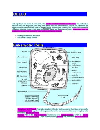
  • 1. CELLS All living things are made of cells, and cells are the smallest units that can be alive. Life on Earth is classified into five kingdoms, and they each have their own characteristic kind of cell. However the biggest division is between the cells of the prokaryote kingdom (the bacteria) and those of the other four kingdoms (animals, plants, fungi and protoctista), which are all eukaryotic cells. Prokaryotic cells are smaller and simpler than eukaryotic cells, and do not have a nucleus. Prokaryote = without a nucleus Eukaryote = with a nucleus Eukaryotic Cells Cytoplasm (or Cytosol). This is the solution within the cell membrane. It contains enzymes for metabolic reactions together with sugars, salts, amino acids, nucleotides and everything else needed for the cell to function. Nucleus. This is the largest organelle. Surrounded by a nuclear envelope, which is a double membrane with nuclear pores - large holes containing proteins that control the exit of substances
  • 2. such as RNA from the nucleus. The interior is called the nucleoplasm, which is full of chromatin- a DNA/protein complex containing the genes. During cell division the chromatin becomes condensed into discrete observable chromosomes. The nucleolus is a dark region of chromatin, involved in making ribosomes. Mitochondrion (pl. Mitochondria). This is a sausage-shaped organelle (8µm long), and is where aerobic respiration takes place in all eukaryotic cells. Mitochondria are surrounded by a double membrane: the outer membrane is simple, while the inner membrane is highly folded into cristae, which give it a large surface area. The space enclosed by the inner membrane is called the matrix, and contains small circular strands of DNA. The inner membrane is studded with stalked particles, which are the site of ATP synthesis. Chloroplast. Bigger and fatter than mitochondria, chloroplasts are where photosynthesis takes place, so are only found in photosynthetic organisms (plants and algae). Like mitochondria they are enclosed by a double membrane, but chloroplasts also have a third membrane called the thylakoid membrane. The thylakoid membrane is folded into thylakoid disks, which are then stacked into piles called grana. The space between the inner membrane and the thylakoid is called the stroma. The thylakoid membrane contains chlorophyll and stalked particles, and is the site of photosynthesis and ATP synthesis. Chloroplasts also contain starch grains, ribosomes and circular DNA. Ribosomes. These are the smallest and most numerous of the cell organelles, and are the sites of protein synthesis. They are composed of protein and RNA, and are manufactured in the nucleolus of the nucleus. Ribosomes are either found free in the cytoplasm, where they make proteins for the cell's own use, or they are found attached to the rough endoplasmic reticulum, where they make proteins for export from the cell. They are often found in groups called polysomes. All eukaryotic ribosomes are of the larger, "80S", type. Smooth Endoplasmic Reticulum (SER). Series of membrane channels involved in synthesising and transporting materials, mainly lipids, needed by the cell. Rough Endoplasmic Reticulum (RER). Similar to the SER, but studded with numerous ribosomes, which give it its rough appearance. The ribosomes synthesise proteins, which are processed in the RER (e.g. by enzymatically modifying the polypeptide chain, or adding carbohydrates), before being exported from the cell via the Golgi Body. Golgi Body (or Golgi Apparatus). Another series of flattened membrane vesicles, formed from the endoplasmic reticulum. Its job is to transport proteins from the RER to the cell membrane for export. Parts of the RER containing proteins fuse with one side of the Golgi body membranes, while at the other side small vesicles bud off and move towards the cell membrane, where they fuse, releasing their contents by exocytosis. Vacuoles. These are membrane-bound sacs containing water or dilute solutions of salts and other solutes. Most cells can have small vacuoles that are formed as required, but plant cells usually have one very large permanent vacuole that fills most of the cell, so that the cytoplasm (and everything else) forms a thin layer round the outside. Plant cell vacuoles are filled with cell sap, and are very important in keeping the cell rigid, or turgid. Some unicellular protoctists have feeding vacuoles for digesting food, or contractile vacuoles for expelling water. Lysosomes. These are small membrane-bound vesicles formed from the RER containing a cocktail of digestive enzymes. They are used to break down unwanted chemicals, toxins, organelles or even whole cells, so that the materials may be recycled. They can also fuse with a feeding vacuole to digest its contents. Cytoskeleton. This is a network of protein fibres extending throughout all eukaryotic cells, used for support, transport and motility. The cytoskeleton is attached to the cell membrane and gives the cell its shape, as well as holding all the organelles in position. There are three types of protein fibres (microfilaments, intermediate filaments and microtubules), and each has a corresponding motor protein that can move along the fibre carrying a cargo such as organelles, chromosomes or other cytoskeleton fibres. These motor proteins are responsible for such actions as: chromosome movement in mitosis, cytoplasm cleavage in cell division, cytoplasmic streaming in plant cells, cilia and flagella movements, cell crawling and even muscle contraction in animals. Centriole. This is a pair of short microtubules involved in cell division.
  • 3. Cilium and Flagellum. These are flexible tails present in some cells and used for motility. They are an extension of the cytoplasm, surrounded by the cell membrane, and are full of microtubules and motor proteins so are capable of complex swimming movements. There are two kinds: flagella (pl.) (no relation of the bacterial flagellum) are longer than the cell, and there are usually only one or two of them, while cilia (pl.) are identical in structure, but are much smaller and there are usually very many of them. Microvilli. These are small finger-like extensions of the cell membrane found in certain cells such as in the epithelial cells of the intestine and kidney, where they increase the surface area for absorption of materials. They are just visible under the light microscope as a brush border. Cell Membrane (or Plasma Membrane). This is a thin, flexible layer round the outside of all cells made of phospholipids and proteins. It separates the contents of the cell from the outside environment, and controls the entry and exit of materials. The membrane is examined in detail later. Cell Wall. This is a thick layer outside the cell membrane used to give a cell strength and rigidity. Cell walls consist of a network of fibres, which give strength but are freely permeable to solutes (unlike membranes). Plant cell walls are made mainly of cellulose, but can also contain hemicellulose, pectin, lignin and other polysaccharides. There are often channels through plant cell walls called plasmodesmata, which link the cytoplasms of adjacent cells. Fungal cell walls are made of chitin. Animal cells do not have a cell wall.
  • 4. Prokaryotic Cells Cytoplasm. Contains all the enzymes needed for all metabolic reactions, since there are no organelles Ribosomes. The smaller (70 S) type. Nuclear Zone. The region of the cytoplasm that contains DNA. It is not surrounded by a nuclear membrane. DNA. Always circular, and not associated with any proteins to form chromatin. Plasmid. Small circles of DNA, used to exchange DNA between bacterial cells, and very useful for genetic engineering. Cell membrane. made of phospholipids and proteins, like eukaryotic membranes. Mesosome. A tightly-folded region of the cell membrane containing all the membrane-bound proteins required for respiration and photosynthesis. Cell Wall. Made of murein, which is a glycoprotein (i.e. a protein/carbohydrate complex). There are two kinds of cell wall, which can be distinguished by a Gram stain: Gram positive bacteria have a thick cell wall and stain purple, while Gram negative bacteria have a thin cell wall with an outer lipid layer and stain pink. Capsule (or Slime Layer). A thick polysaccharide layer outside of the cell wall. Used for sticking cells together, as a food reserve, as protection against desiccation and chemicals, and as protection against phagocytosis. Flagellum. A rigid rotating helical-shaped tail used for propulsion. The motor is embedded in the + cell membrane and is driven by a H gradient across the membrane. Clockwise rotation drives the cell forwards, while anticlockwise rotation causes a chaotic spin. This is an example of a rotating motor in nature. Summary of the Differences Between Prokaryotic and Eukaryotic Cells
  • 5. PROKARYOTIC CELLS EUKARYOTIC CELLS small cells (< 5 m) larger cells (> 10 m) always unicellular often multicellular no nucleus or any membrane-bound always have nucleus and other membrane- organelles bound organelles DNA is circular, without proteins DNA is linear and associated with proteins to form chromatin ribosomes are small (70S) ribosomes are large (80S) no cytoskeleton always has a cytoskeleton cell division is by binary fission cell division is by mitosis or meiosis reproduction is always asexual reproduction is asexual or sexual Endosymbiosis Prokaryotic cells are far older and more diverse than eukaryotic cells. Prokaryotic cells have probably been around for 3.5 billion years - 2.5 billion years longer than eukaryotic cells. It is thought that eukaryotic cell organelles like mitochondria and chloroplasts are derived from prokaryotic cells that became incorporated inside larger prokaryotic cells. This idea is called endosymbiosis, and is supported by these observations: organelles contain circular DNA, like bacteria cells. organelles contain 70S ribosomes, like bacteria cells. organelles have double membranes, as though a single-membrane cell had been engulfed and surrounded by a larger cell.
  • 6. The Cell Membrane The cell membrane (or plasma membrane) surrounds all living cells. It controls how substances can move in and out of the cell and is responsible for many other properties of the cell as well. The membranes that surround the nucleus and other organelles are almost identical to the cell membrane. Membranes are composed of phospholipids, proteins and carbohydrates arranged in a fluid mosaic structure, as shown in this diagram. The phospholipids form a thin, flexible sheet, while the proteins "float" in the phospholipid sheet like icebergs, and the carbohydrates extend out from the proteins. The phospholipids are arranged in a bilayer, with their polar, hydrophilic phosphate heads facing outwards, and their non-polar, hydrophobic fatty acid tails facing each other in the middle of the bilayer. This hydrophobic layer acts as a barrier to all but the smallest molecules, effectively isolating the two sides of the membrane. Different kinds of membranes can contain phospholipids with different fatty acids, affecting the strength and flexibility of the membrane, and animal cell membranes also contain cholesterol linking the fatty acids together and so stabilising and strengthening the membrane. The proteins usually span from one side of the phospholipid bilayer to the other (intrinsic proteins), but can also sit on one of the surfaces (extrinsic proteins). They can slide around the membrane very quickly and collide with each other, but can never flip from one side to the other. The proteins have hydrophilic amino acids in contact with the water on the outside of membranes, and hydrophobic amino acids in contact with the fatty chains inside the membrane. Proteins comprise about 50% of the mass of membranes, and are responsible for most of the membrane's properties. Proteins that span the membrane are usually involved in transporting substances across the membrane (more details below). Proteins on the inside surface of cell membranes are often attached to the cytoskeleton and are involved in maintaining the cell's shape, or in cell motility. They may also be enzymes, catalysing reactions in the cytoplasm. Proteins on the outside surface of cell membranes can act as receptors by having a specific binding site where hormones or other chemicals can bind. This binding then triggers other events in the cell. They may also be involved in cell signalling and cell recognition, or they may be enzymes, such as maltase in the small intestine (more in digestion).
  • 7. The carbohydrates are found on the outer surface of all eukaryotic cell membranes, and are usually attached to the membrane proteins. Proteins with carbohydrates attached are called glycoproteins. The carbohydrates are short polysaccharides composed of a variety of different monosaccharides, and form a cell coat or glycocalyx outside the cell membrane. The glycocalyx is involved in protection and cell recognition, and antigens such as the ABO antigens on blood cells are usually cell-surface glycoproteins. Remember that a membrane is not just a lipid bilayer, but comprises the lipid, protein and carbohydrate parts. Transport Across The Membrane Cell membranes are a barrier to most substances, and this property allows materials to be concentrated inside cells, excluded from cells, or simply separated from the outside environment. This is compartmentalization is essential for life, as it enables reactions to take place that would otherwise be impossible. Eukaryotic cells can also compartmentalize materials inside organelles. Obviously materials need to be able to enter and leave cells, and there are five main methods by which substances can move across a cell membrane: 1. Simple Diffusion 2. Osmosis 3. Facilitated Diffusion 4. Active Transport 5. Vesicles 1. Simple Diffusion A few substances can diffuse directly through the lipid bilayer part of the membrane. The only substances that can do this are lipid-soluble molecules such as steroids, or very small molecules, such as H 2O, O2 and CO2. For these molecules the membrane is no barrier at all. Since lipid diffusion is (obviously) a passive diffusion process, no energy is involved and substances can only move down their concentration gradient. Lipid diffusion cannot be controlled by the cell, in the sense of being switched on or off. 2. Osmosis Osmosis is the diffusion of water across a membrane. It is in fact just normal lipid diffusion, but since water is so important and so abundant in cells (its concentration is about 50 M), the diffusion of water has its own name - osmosis. The contents of cells are essentially solutions of numerous different solutes, and the more concentrated the solution, the more solute molecules there are in a given volume, so the fewer
  • 8. water molecules there are. Water molecules can diffuse freely across a membrane, but always down their concentration gradient, so water therefore diffuses from a dilute to a concentrated solution. Water Potential. Osmosis can be quantified using water potential, so we can calculate which way water will move, and how fast. Water potential ( , the Greek letter psi, pronounced "sy") is a measure of the water molecule potential for movement in a solution. It is measured in units of pressure (Pa, or usually kPa), and the rule is that water always moves by osmosis from less negative to more negative water potential (in other words it's a bit like gravity potential or electrical potential). 100% pure water has = 0, which is the highest possible water potential, so all solutions have < 0 (i.e. a negative number), and you cannot get > 0. Cells and Osmosis. The concentration (or OP) of the solution that surrounds a cell will affect the state of the cell, due to osmosis. There are three possible concentrations of solution to consider: Isotonic solution a solution of equal OP (or concentration) to a cell Hypertonic solution a solution of higher OP (or concentration) than a cell Hypotonic solution a solution of lower OP (or concentration) than a cell The effects of these solutions on cells are shown in this diagram:
  • 9. The diagram below shows what happens when 2 fresh raw eggs with their shells removed with acid are placed into sucrose solution (hypertonic) and distilled water (hypotonic). Water enters the egg in water (endosmosis) causing it to swell and water leaves the egg in sucrose causing it to shrink (exosmosis). These are problems that living cells face all the time. For example: Simple animal cells (protozoans) in fresh water habitats are surrounded by a hypotonic solution and constantly need to expel water using contractile vacuoles to prevent swelling and lysis. Cells in marine environments are surrounded by a hypertonic solution, and must actively pump ions into their cells to reduce their water potential and so reduce water loss by osmosis. Young non-woody plants rely on cell turgor for their support, and without enough water they wilt. Plants take up water through their root hair cells by osmosis, and must actively pump ions into their cells to keep them hypertonic compared to the soil. This is particularly difficult for plants rooted in salt water.
  • 10. 3. Facilitated Diffusion. Facilitated diffusion is the transport of substances across a membrane by a trans-membrane protein molecule. The transport proteins tend to be specific for one molecule (a bit like enzymes), so substances can only cross a membrane if it contains the appropriate protein. As the name suggests, this is a passive diffusion process, so no energy is involved and substances can only move down their concentration gradient. There are two kinds of transport protein: Channel Proteins form a water-filled pore or channel in the membrane. This allows charged substances (usually ions) to diffuse across membranes. Most channels can be gated (opened or closed), allowing the cell to control the entry and exit of ions. Carrier Proteins have a binding site for a specific solute and constantly flip between two states so that the site is alternately open to opposite sides of the membrane. The substance will bind on the side where it at a high concentration and be released where it is at a low concentration. The rate of diffusion of a substance across a membrane increases as its concentration gradient increases, but whereas lipid diffusion shows a linear relationship, facilitated diffusion has a curved relationship with a maximum rate. This is due to the rate being limited by the number of transport proteins. 4. Active Transport (or Pumping). Active transport is the pumping of substances across a membrane by a trans-membrane protein pump molecule. The protein binds a molecule of the substance to be transported on one side of the membrane, changes shape, and releases it on the other side. The proteins are highly specific, so there is a different protein pump for each molecule to be transported. The protein pumps are also ATPase enzymes, since they catalyse the splitting of ATP into ADP + phosphate (Pi), and use the energy released to change shape and pump the molecule. Pumping is therefore an active process, and is the only transport mechanism that can transport substances up their concentration gradient.
  • 11. + + The Na K Pump. This transport protein is present in the cell membranes of all animal cells and is the most abundant and important of all membrane pumps. We look at it in more detail in module 4 (A2 course) 5. Vesicles The processes described so far only apply to small molecules. Large molecules (such as proteins, polysaccharides and nucleotides) and even whole cells are moved in and out of cells by using membrane vesicles. Endocytosis is the transport of materials into a cell. Materials are enclosed by a fold of the cell membrane, which then pinches shut to form a closed vesicle. Strictly speaking the material has not yet crossed the membrane, so it is usually digested and the small product molecules are absorbed by the methods above. When the materials and the vesicles are small (such as a protein molecule) the process is known as pinocytosis (cell drinking), and if the materials are large (such as a white blood cell ingesting a bacterial cell) the process is known as phagocytosis (cell eating). Exocytosis is the transport of materials out of a cell. It is the exact reverse of endocytosis. Materials to be exported must first be enclosed in a membrane vesicle, usually from the RER and Golgi Body. Hormones and digestive enzymes are secreted by exocytosis from the secretory cells of the intestine and endocrine glands. Sometimes materials can pass straight through cells without ever making contact with the cytoplasm by being taken in by endocytosis at one end of a cell and passing out by exocytosis at the other end.
  • 12. Summary of Membrane Transport USES METHOD USES PROTEINS SPECIFIC CONTROLLABLE ENERGY Simple Diffusion N N N N Osmosis N N Y N Facilitated Diffusion N Y Y Y Active Transport Y Y Y Y Vesicles Y N Y Y