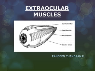
2. ORBITAL MUSCLES
Extrinsic muscles of eyeball.
Involved in movement of eyeball.
Intrinsic muscles
Controls shape of lens and size of pupil.
3. Extraocular muscles are special
The motor units are small, with only from 5 to 18 muscle
fibers contact by each motor nerve
4. Motion of an Eye
To describe eye motions we need a set of defined axes (Fick’s
Axes - draw on board)
X axis : nasal -> temporal
Y axis: anterior -> posterior
Z axis: superior -> inferior
These axes intersect at the center of rotation - a fixed point,
defined as 13.5 mm behind cornea.
6. Ductions (single eye movements)
Rotation about the Z axis (Z axis runs vertically superior to
inferior)
Medial Rotation - adduction toward midline
Lateral Rotation - abduction away from midline
Rotation about the X axis (X axis runs horizontally, from
nasal to temporal)
Upward, elevation (supraduction)
Downward, depression (infraduction)
7. Torsion - cyclorotations
Rotation about the Y axis (Y axis runs horizontally, from anterior
to posterior)
These are described with respect to a point at 12 o‘clock on the
superior limbus
Intorsion (incyclorotation) rotation nasally
Extorsion (excyclorotation) rotation of the 12 o’clock position
temporally.
Counteracting head tilt (up to 7-9°)
8. Version & Vergences
Some eye movements are paired, that is both eyes do
the same thing. . . . Versions
Sometimes eyes move in the opposite directions
simultaneously. . . Vergences
10. Versions (conjugate eye
movement)
Dextroversion - rightward gaze (demo)
Levoversion - leftward gaze
Supraversion - elevation
Infraversion - depression
Also up and right, up and left
Down and right, down and left
ALL BEHAVIOR IS THAT OF YOLKED
EYES
16. Origin-Inferior surface of lesser wing of sphenoid.
Insertion-
1. Upper lamina-Anterior surface of
superior tarsus and skin of upper eyelid.
2. Middle lamina-superior margin of
superior tarsus.
3. Lower lamina-Superior conjunctival
fornix
NERVE SUPPLY-
Upper division of occulomotor nerve.
18. Ptosis
Drooping of upper eyelid.
Complete ptosis-injury to occulomotor nerve.
Partial ptosis-disruption of postganglionic sympathetic
fibres from superior cervical sympathetic ganglion.
19. SUPERIOR RECTUS MUSCLE
Origin-Superior part of common tendon of zinn.
Insertion-inserted into sclera by flat tendinous
insertion(10mm broad)about 7.7 mm behind sclero-
corneal junction.
Nerve supply-superior division of occulomotor nerve.
21. Frontal nerve runs above the superior rectus & levator.
The nasociliary nerve and ophthalmic artery run
below.
The tendon for insertion of the superior oblique
muscle runs below the anterior part of the superior
rectus.
22. Action of Superior Rectus
Primary action is elevation . . But since the insertion on
the globe is lateral as well as superior, contraction will
produce rotation about the vertical axis toward midline
Thus secondary action is adduction
Finally, because the insertion is oblique, contraction
produces torsion nasally Intorsion.
23. INFERIOR RECTUS
Origin-inferior part of common tendon of zinn
Insertion-in the sclera 6.5 mm behind sclero corneal
junction.
Nerve supply-inferior division occulomotor nerve.
24. Fascial attachments below attached to inferior lid
coordinate depression and lid opening.
Fascia below Inf. Rectus and Inf. Oblique contribute to
the suspensory ligament of lockwood.
ACTIONS-
Primary depressor.
Subsidiary actions are adduction and extorsion.
25. MEDIAL RECTUS
Origin-annulus of zinn and from optic nerve sheath.
Insertion-in sclera 5.5mm behind sclero-corneal
junction.
Nerve supply-lower division of occulomotor nerve.
Fascial expansion from muscle sheath forms the medial
check ligament and attach to medial wall of orbit.
26. Innervation is via cranial nerve III, the oculomotor
nerve, and the specific branch runs along the inside of
the muscle cone, on the lateral surface.
The superior oblique, ophthalmic artery and nasociliary
nerve all lie above the medial rectus.
ACTION-
Primary adductor of the eye.
27. LATERAL RECTUS
Origin-annulus of zinn.
Insertion-in the sclera 6.9mm behind sclerocorneal
junction.
Nerve supply-abducens nerve which enters the muscle
on the medial surface.
28. The lacrimal artery and nerve run along the superior
border.
The abducens nerve, ophthalmic artery and ciliary
ganglion lie medial to the lateral rectus and between it
and the optic nerve.
ACTION-
Primary abductor of eye.
30. SUPERIOR OBLIQUE
Longest and thinnest intraorbital muscle, the muscle
ends before the trochlea, tendon is 2.5 cm, smooth
movement through trochlea.
Origin-body of sphenoid above and medial to optic
canal.Passes along superomedial part of orbit and ends
in a tendon.
Insertion-Posterosuperior quadrant of sclera behind
equator of eyeball.
Nerve supply-trochlear nerve entering it approximately
one third of the distance from the origin to the trochlea.
32. INFERIOR OBLIQUE
Origin-Anteromedial part of orbital floor lateral to
nasolacrimal groove.
Insertion-posteroinferior surface of globe near the
macula.
Nerve supply-inferior division of occulomotor nerve
enters the muscle laterally at the junction of the inferior
oblique and inferior rectus muscles.