SlideShare uma empresa Scribd logo
1 de 13
Baixar para ler offline
UK Journal of Pharmaceutical and Biosciences Vol. 3(2), 29-41, 2015 RESEARCH ARTICLE
Ameliorative Effect of Salicin Against Gamma Irradiation Induced Electrophoretic
Changes in Brain Tissue in Male Rats
Mohga Shafik Abdalla, Hayat Mohamed Sharada, Ibrahim Abulyazid, Monira Abdel-Latif Abdel-Karim,
Wael Mahmoud Kamel*
Genetic Engineering and Biotechnology Division, National Research Centre, Dokki, Giza, Egypt-12622
Article Information
Received 10 Feburary 2015
Received in revised form 27 April 2015
Accepted 28 April 2015
Abstract
The aim of this study was to investigate the radioprotective effect of salicin against irradiation
effect on brain tissue of male rats. Lipid peroxidation level was measured as thiobarbituric acid
reactive substance in brain tissue. The polyacrylamide gel electrophoresis for native protein,
lipoprotein and zymogram were carried out in brain homogenate. As expected, salicin resisted
the irradiation effect and declined the MDA level in brain homogenate of all treated groups
(especially in the irradiated salicin post-treated group). Salicin minimized the qualitative
mutagenic effect of irradiation on the electrophoretic protein pattern in all irradiated salicin
treated groups and it showed the highest antagonistic effect against irradiation in irradiated
salicin post-treated group (SI = 0.57). It could not prevent the abnormalities occurred qualitatively
and quantitatively as a result of irradiation in lipoprotein pattern in all irradiated salicin treated
groups. In the electrophoretic esterase pattern, salicin prevented the qualitative effect of
irradiation in irradiated salicin post-treated group (SI = 1.00). Salicin minimized the qualitative
irradiation effect on the catalase pattern in the irradiated salicin pre-treated group (SI = 0.73).
While in the peroxidase pattern, salicin adminstration resisted the irradiation effect in the
irrradiated post-treated groups (SI = 0.67). The results suggested the radioprotective ability of
salicin against gamma irradiation effect on various electrophoretic patterns in brain tissue of male
rats.
Keywords:
Gamma irradiation,
Salicin,
Brain,
Protein electrophoresis,
Isozymes
*
Corresponding Author:
E-mail: wmkamel83@hotmail.com
Tel.: 00201126682686
1 Introduction
The use of this radiation as a source to enhance the mutation
frequency was recognized as early as 19301
.These rays act either
directly or by secondary reactions to produce biochemical lesions
that initiate series of physiological symptoms. They are known to
induce oxidative stress through the generation of reactive oxygen
species (ROS) resulting in imbalance of the prooxidant and
antioxidant activities, ultimately resulting in cell death2
.Studies
showed that radiation can affect a wide variety of tissues, particularly
those with greater levels of cellular turnover and divisions3
.
Irradiation causes similar damage at a cellular level but gamma rays
are more penetrating, causing diffuse damage throughout the body4
.
This type of rays also used for diagnostic purposes in nuclear
medicine in imaging techniques5
.
Irradiation causes damage to living tissue through a series of
molecular events. The formation of ROS as a result of interaction of
irradiation with cellular macromolecules is the cause of dysfunction
and death, in both normal as well as tumor cells exposed to
radiation6,7
.The energy exchange between the rays and the targeted
molecules leads to changes produced in deoxyribonucleic acid
(DNA), lipids, and proteins and then cell inactivation8,9
.
Gamma irradiation was found to interrupt energy supplies and
blocking all key enzymes, which may stop normal metabolism of the
exposed tissue10
. The radiation exposure whether occupational or
during radiotherapy leads to serious systemic damage to various
cellular and subcellular structures11
.
Irradiation causes induction of lipid peroxidation as evidenced by
increased malondialdhyde (MDA)12
. It causes damage of cells
UK Journal of Pharmaceutical and Biosciences
Available at www.ukjpb.com
ISSN: 2347-9442
Kamel et al. Ameliorative Effect of Salicin Against Gamma Irradiation
UK J Pharm & Biosci, 2015: 3(2); 30
directly by ionizing DNA and other cellular targets and indirectly by
effect through ROS13
. It produces oxygen-derived free radicals in
tissue environment: these include hydroxyl radicals (the most
damaging), superoxide anion radicals and other oxidants such as
hydrogen peroxide14
.
Major radiation damage is due to the aqueous free radicals
generated by the water radiolysis. These free radicals act as
molecular marauders and in turn damage DNA which is considered
to be the primary target15
.This gives rise to genomic instability
leading to mutagenesis, carcinogenesis and cell death16
.
The most important consequences of OS are lipid peroxidation,
protein oxidation and depletion of antioxidants17,18
. The latter authors
showed thatthe increase of MDA level is probably due to the
interaction of •OH resulting as a bi-product of water radiolysis with
the polyunsaturated fatty acids presents in the phospholipids portion
of cellular membranes. The excessive free radicals can damage
crucial macromolecules, including DNA, cell membranes and
enzymes, and can cause cell death. DNA damage includes
genotoxicity, chromosomal abnormalities, gene mutations and cell
death if the damage is beyond repair19,20
.
Catalase (CAT) and glutathione peroxidase (GPx) play an important
role in detoxification of hydrogen peroxide and ROS21
(Kula et al.,
2000). The irradiation caused significant increase in markers of the
oxidative stress as malondialdehyde (MDA) and alterations in activity
of antioxidant enzymes such as CAT and GPx in a dose-dependent
manner22,23
.
Salicin was discovered in 1831. It was isolated from the willow bark
and leaves. It is a prodrug and thus a precursor of salicylic
acid24
.Salicin is a phenolic glycoside. It exhibits analgesic effects,
and it is used for the treatment of rheumatic pain. Its occurrence in
willow (Salix) species is the major reason willow bark and its extracts
are popular products. High-Performance Liquid Chromatography
(HPLC) has been the standard method of analysis of quantifying
salicin in the different Salix species25
.
The present experiment was concerned with studying the ability of
salicin which represents the most abundant fraction of aqueous
willow extract to impress radiation-induced electrophoretic alterations
in brain tissue of male rats.
2 Materials and Methods
2.1 Salicin isolation
Fresh young leaves of the willow trees (Salix subserrata, Salix
safsaf) were collected from Orman garden, Giza, Egypt. This species
was well authenticated by qualified specialists in plant taxonomy.
Salicin was extracted and isolated from fresh leaves according to
method described by Mabry et al26
and purified according to method
suggested by Kur'yanov et al27
then identified qualitatively by
traditional and advanced chromatographic techniques.
2.2 Acute toxicity test
The lethal dose 50 (LD50) was evaluated on 6 groups of female
albino mice (8 animals / group) receiving progressively increasing
oral dose levels (500, 1000, 2000, 3000, 4000 and 5000 mg/kg body
weight) of aqueous solution of salicin solution. Mortality was
recorded 24 hrs post treatment. The LD50 was calculated according
to the equation suggested by Paget and Barnes28
.
2.3 Animals
Seven groups of male rats weighing between 150-200 gm per one
obtained from the animal house laboratory of the national research
centre. Ten rats in each group. All the animals were kept under
normal environmental and nutritional conditions. The animal groups
were divided into Control group: Rats were non-irradiated and non-
treated with salicin, Salicin treated group: Rats were non-irradiated
but treated with the safe dose of salicin which was about 150 mg /
Kg taking in the consideration weight of each rat, Irradiated group:
Rats were irradiated at the dose 7 Gy and non-treated with salicin,
Irradiated salicin pre-treated group: Rats were treated with salicin for
15 days followed by irradiation at the 15th
day, Irradiated salicin
prepost-treated group: Rats were treated with salicin for 15 days
followed by irradiation at the 15th
day then the treatment was
continued daily for another 15 days, Irradiated salicin simultaneous
treated group: Rats were irradiated and treated with salicin at the
same time of irradiation and continue daily for 15 days and Irradiated
salicin post-treated group: Rats were irradiated at the same gamma
dose then left without treatment for 15 days. At the 15th
day, the rats
were treated with salicin for another 15 days.
2.4 Irradiation
Whole body was gamma irradiated at Middle Eastern Regional
Radioisotope Centre for the Arab Countries, Dokki, Egypt. Using
Cobalt 60 (Co 60
) as a suitable gamma source. Rats were irradiated
at a single dose of 7 Gy delivered at the dose rate of 1.167 Rad /
Sec.
2.5 Biochemical Assay
Lipid peroxidation level was measured as thiobarbituric acid reactive
substance in brain homogenate according to method of Ohkawa et
al29
.
2.6 Electrophoretic protein pattern
Total protein was determined in brain homogenate according to
Bradford30
.The sample was mixed with the sample buffer. The
protein concentration in each well must be about 70 μg protein.
Proteins were separated through polyacrylamide gel electrophoresis
Kamel et al. Ameliorative Effect of Salicin Against Gamma Irradiation
UK J Pharm & Biosci, 2015: 3(2); 31
(PAGE) with different concentrations. Electrode and gel buffer and
polyacrylamide stock were prepared according to Laemmli31
. After
electrophoretic separation, the gel was gently removed from the
apparatus and put into a staining solution of coomasie brilliant blue
for native protein pattern32
and staining solution of sudan black B
(SBB) for lipoprotein pattern33
.
2.7 Electrophoretic isozyme
Native protein gel was stained for peroxidase pattern using certain
stain prepared according to the method suggested by Rescigno et
al34
. It was stained for catalase pattern according to the method
described by Siciliano and Shaw35
.For esterase pattern, the native
gel was stained according to the method suggested by Baker and
Manwell36
.
2.8 Data analysis
The polyacrylamide gel plate was photographed, scanned and then
analyzed using Phoretix 1D pro software (Version 12.3). The
similarity index (S.I.) compares patterns within, as well as, between
irradiated and non-irradtated samples. The similarity values were
converted into genetic distance (GD) according the method
suggested by Nei and Li37
.
2.9 Statistical analysis
All the grouped data were statistically evaluated with SPSS/16.00
software. The results were expressed as mean ± SE of studied
groups using the analysis of variance test (one-way ANOVA)
followed by student’s t-test34
. P values of less than 0.05 were
considered to indicate statistical significance. The means of
irradiated groups and the salicin treated groups were individually
compared with those of control group. The irradiated group was
compared with irradiated salicin treated groups
3 Results and Discussions
3.1 Lipid peroxidation
As compared to control, irradiation caused the severe increase in the
lipid peroxidation product (MDA) level in the brain tissue. Salicin
administration showed the ameliorative effect against irradiation by
reducing MDA level in all irradiated salicin treated rats. From the
data compiled in table 1, it was found that salicin showed the most
suitable antagonistic effect against irradiation on brain of irradiated
salicin post-treated group.
3.2 Electrophoretic protein pattern
Protein pattern in the control sample produced 9 bands with Rfs
ranged between 0.09 – 0.98 (Mwts 5.21 – 212.46 KDa and B %
values 0.20 – 25.09). There were no common bands but there were
2 characteristic bands appeared in irradiated salicin pre-treated
group with Rf 0.56 (Mwt 20.09 KDa and B % 21.13) and in irradiated
salicin simultaneous treated group with Rf 0.75 (Mwt 14.58 KDa and
B % 10.85).
As shown in table 2 and illustrated in fig. 1, irradiation caused
disappearance of 6 normal bands without appearance of abnormal
bands. The 3rd
and 5th
bands might be deviated to be appeared with
Rf 0.22 and 0.43 (Mwts 113.48 and 30.53 KDa and B % 17.28 and
80.91). It caused quantitative mutation represented by increasing B
% of the 9th
band (Rf 0.98, Mwt 5.02 KDa and B % 1.81). Salicin
administration resisted the qualitative mutagenic effect of irradiation
in the irradiated salicin simultaneous treated and post-treated
groups. It retained 4 normal bands with Rfs ranged between Rf 0.23 -
0.98 (Mwts 5.08 - 112.21 KDa and B % 0.20 - 15.81) in irradiated
salicin simultaneous treated group and with Rf 0.45 - 0.99 (Mwts 4.75
- 27.06 KDa and B % 0.59 - 56.41) in irradiated salicin post-treated
group. It was probable that the 5th
band was deviated to be appeared
with Rf 0.45 (Mwt 28.20 KDa and B % 37.70) in irradiated salicin
simultaneous treated group. It could not prevent the quantitative
mutation which was represented by increasing B % of the 3rd
and 7th
bands (Rfs 0.23 and 0.85, Mwts 112.21 and 10.65 KDa and B %
15.81 and 14.00) and by decreasing B % of the 6th
band (Rf 0.63,
Mwt 18.00 KDa and B% 11.99) in irradiated salicin simultaneous
treated group. Also, it could not prevent the quantitative mutation
which was represented by increasing B % of the 5th
, 6th
, 7th
and 9th
bands (Rfs 0.45, 0.65, 0.86 and 0.99 (Mwts 27.06, 17.68, 10.38 and
4.75 KDa and B % values 56.41, 0.59, 34.49 and 1.54) respectively.
It was found that the lowest SI value (SI = 0.17) was recorded with
irradiated group and the highest SI value (SI = 0.70) with salicin
treated group. Salicin improved the SI values in all irradiated salicin
treated groups, and it showed the highest antagonistic effect against
irradiation in irradiated salicin post-treated group (SI = 0.57). The
overall results showed that salicin prevented the irradiation effect on
number and arrangement of the bands in all irradiated salicin treated
groups.
3.3 Electrophoretic lipoprotein pattern
As revealed in table 3 and illustrated in fig. 2, lipoprotein pattern in
the control sample produced 3 bands with Rfs 0.22, 0.47 and 0.69 (B
% 67.28, 18.91 and 13.82). There were no common bands appeared
in all groups. Salicin alone caused severe qualitative alterations
represented by disappearance of the normal bands with appearance
of 3 abnormal bands with Rf 0.25, 0.33 and 0.72 (B % 48.27, 31.12
and 20.61).
Irradiation caused severe abnormalities represented disappearance
of all the normal bands without appearance of abnormal bands in the
irradiated, irradiated salicin simultaneous treated and post-treated
groups. Salicin adminstration could not prevent the irradiation effect
Kamel et al. Ameliorative Effect of Salicin Against Gamma Irradiation
UK J Pharm & Biosci, 2015: 3(2); 32
which was represented qualitatively by disappearance of 2 normal
bands and quantitatively by increasing B % of the normal band
appeared with Rf 0.22 and B % 100.00 in the irradiated salicin pre-
treated group and by disappearance of 2 normal bands with
appearance of one abnormal band with Rf 0.35 (B % 29.73) in the
irradiated salicin prepost-treated group. It was observed that all the
bands were not matched with all bands of the other groups in the
salicin treated, irrradiated, irradiated salicin simultaneous treated and
post-treated groups.
Table 1: Effect of irradiation, salicin and their combination in various treatment modes on the level of lipid peroxidation in spleen
tissue of male rats
Organ Control Sal. Irr.
Irradiated salicin treated groups
Pre-treated Simultaneous Prepost-treated Post-treated
Brain
(nmol/g)
162.02
± 2.28
137.84a
± 2.17
239.15a
± 6.55
232.90a
± 6.24
227.21a
± 4.78
228.10a
± 7.95
165.24a,b
± 2.60…
a : Different from control at P < 0.05, b : Different from the irradiated group at P < 0.05.
Note : Sal. ; salicin, Irr. : Irradiated
3.4 Electrophoretic esterase pattern
The electrophoretic esterase pattern in control of brain tissue
produced 3 types with Rfs 0.22, 0.48 and 0.64 (B % 36.85, 45.38 and
17.77). As shown in table 4 and illustrated in fig. 3, there were no
common bands in all groups. Salicin alone caused qualitative
mutation represented by appearance of one abnormal characteristic
band with Rf 0.11 (B % 21.93) and decreasing B % of the 2nd
normal
type (Rf 0.49 and B % 19.00). Irradiation caused disturbances
represented qualitatively by disappearance of 2 normal types of the
enzyme and quantitatively by increasing B % of the 1st
normal band
(Rf 0.21 and B % 100.00).
Salicin administration prevented the qualitative or quantitative
alterations occurred as a result of irradiation in the irradiated salicin
post-treated group. It could not prevent the irradiation effect which
was represented qualitatively by deviation of the 3rd
type to be
appeared with Rf 0.60 (B % 23.33) and quantitatively by increasing B
% of the 1st
band (Rf 0.21 and B % 51.38) and decreasing B % of the
2nd
type (Rf 0.47 and B % 25.29) in the irradiated salicin pre-treated
group, represented by disappearance of the 3rd
type with deviation of
the 1st
and 2nd
normal types of the enzyme to be appeared with Rfs
0.23 and 0.48 (B % 55.23 and 44.77) in the irradiated salicin
simultaneous treated group and represented by deviation of the 2nd
and 3rd
normal types of the enzyme to be appeared with Rfs 0.45 and
Kamel et al. Ameliorative Effect of Salicin Against Gamma Irradiation
UK J Pharm & Biosci, 2015: 3(2); 33
0.62 (B % 38.34 and 21.20) in the irradiated salicin prepost- treated
group.
Fig.1:Electrophoretic pattern showing effect of salicin against
the irradiation effect on protein pattern in brain tissue of male
rats
Fig. 2:Electrophoretic pattern showing effect of salicin against
the irradiation effect on lipoprotein pattern in brain tissue of
male rats
Table 3: Data of the electrophoretic lipoprotein pattern in brain tissue of control, irradiated and irradiated salicin treated groups in
male rats
Control Salicin Irradiated
Irradiated salicin treated
Pre-treated Simultaneous Prepost-treated Post-treated
Rf. B. % Rf. B. % Rf. B. % Rf. B. % Rf. B. % Rf. B. % Rf. B. %
0.22 67.28 0.25 48.27 — — 0.22 100.00 — — 0.22 70.27 — —
0.47 18.91 0.33 31.12 — — — — — — 0.35 29.73 — —
0.69 13.82 0.72 20.61 — — — — — — — — — —
Rf.: Rate of Flow, B.%: Band Percent
Table 4: Data of the electrophoretic esterase pattern in brain tissue of control, irradiated and irradiated salicin treated groups in male
rats
Control Salicin Irradiated
Irradiated salicin treated
Pre-treated Simultaneous Prepost-treated Post-treated
Rf. B. % Rf. B. % Rf. B. % Rf. B. % Rf. B. % Rf. B. % Rf. B. %
0.22 36.85 0.11 21.93 0.21 100.00 0.21 51.38 0.23 55.23 0.21 40.46 0.22 40.27
0.48 45.38 0.22 40.13 — — 0.47 25.29 0.48 44.77 0.45 38.34 0.47 36.92
0.64 17.77 0.49 19.00 — — 0.60 23.33 — — 0.62 21.20 0.62 22.81
— — 0.64 18.94 — — — — — — — — — —
Rf.: Rate of Flow, B.%: Band Percent
Kamel et al. Ameliorative Effect of Salicin Against Gamma Irradiation
UK J Pharm & Biosci, 2015: 3(2); 34
It was found that the highest SI value (SI = 1.00) was noticed in
irradiated salicin post-treated group. In the irradiated salicin
simultaneous treated group, it was observed that all the bands were
not matched with all bands of the other groups.
Fig. 3:Electrophoretic pattern showing effect of salicin against
the irradiation effect on esterase pattern in brain tissue of male
rats
3.5 Electrophoretic catalase pattern
As shown in table 5 and illustrated in fig. 4, 5 types of catalase
enzyme were produced in the control sample with Rfs ranged
between 0.21 - 0.97 (B % 8.11 – 47.12). There were 2 common
bands appeared in all the groups with Rfs 0.45 and 0.97 (B % 8.47
and 15.17). Salicin alone caused alterations represented by
appearance of one abnormal band with Rf 0.18 (B % 23.47) with
decreasing the B % of the 1st
type (Rf 0.22 and B % 6.12) and
increasing the B % of the 2nd
type (Rf 0.44 and B % 25.89).
Irradiation caused qualitative alterations represented by
disappearance of the 1st
type of the enzyme with deviation of the 3rd
band to be appeared with Rf 0.78 (B % 15.01) and quantitative
mutation occurred by increasing B % of the 2nd
band (Rf 0.45 and B
% 64.42).
Salicin administration could not prevent the irradiation effect which
was represented by alteration occurred qualitatively by appearance
of one abnormal band with Rf 0.15 (B % 29.71) with deviation of the
3rd
type of the enzyme to be appeared with Rf 0.82 (B % 28.32) and
quantitatively by decreasing B % of the band appeared with Rf 0.22
(B % 9.44) in the irradiated salicin pre-treated group, by
disappearance of the 1st
and 4th
types of the enzyme with appearance
of one abnormal band with Rf 0.17 (B % 31.52) and deviation of the
3rd
type to be appeared with Rf 0.82 (B % 38.60) in the irradiated
salicin prepost-treated group and represented by disappearance of
the 1st
type with appearance of one abnormal band with Rf 0.12 (B %
20.99) deviation of the 3rd
type to be appeared with Rf 0.83 (B %
19.39) in the irradiated salicin simultaneous treated group. While in
the irradiated salicin post-treated group, salicin could not overcome
the mutation represented qualitatively by disappearance of 3 normal
types with appearance of one abnormal band with Rf 0.66 (B %
57.05) and quantitatively by increasing B % of the bnds appeared
with Rfs 0.45 and 0.96 (B % 20.65 and 22.30).
From the SI values, it was found that the lowest SI value (SI = 0.44)
was observed with irradiated salicin prepost-treated group and the
highest value (SI = 0.91) observed with salicin treated group. Salicin
treatment minimized the qualitative irradiation effect in the irradiated
salicin pre-treated group (SI = 0.73) more than the other irradiated
salicin treated groups.
3.6 Electrophoretic peroxidase pattern
As shown in table 6 and illustrated in fig. 5, it was found that 5 types
of peroxidase enzyme were produced in the control sample with Rfs
ranged between 0.28 - 0.72 (B % 0.46 - 41.66). There were no
common band appeared in all groups. Salicin alone caused no
obvious qualitative or quantitative alterations. Irradiation caused
severe disturbances represented qualitatively by disappearance of
the 2nd
, 3rd
and 5th
normal types of the enzyme and quantitatively by
increasing B % of the 1st
type (Rf 0.28 and B % 58.28). Salicin could
not remove the abnormalities which were represented qualitatively
by disappearance of the 1st
, 3rd
and 5th
types with increasing B % of
the 2nd
and 4th
normal types of the enzyme (Rfs 0.35 and 0.58 and B
% 34.34 and 65.66) in irradiated salicin pre-treated group and
represented qualitatively by disappearance of the 3rd
type of the
enzyme with appearance of one abnormal characteristic band with Rf
0.69 (B % 44.83) and quantitatively by decreasing the B % of the 1st
type (Rf 0.27 and B % 14.08) and increasing B % of the 2nd
and 4th
types of the enzyme (Rfs 0.38 and 0.69 and B % 25.82 and 44.83) in
irradiated salicin prepost-treated group. In the irradiated salicin post-
treated group, salicin could not prevent the mutagenic effect which
was represented qualitatively by disappearance of the 5th
type and
deviation of the 4th
type to be appeared with Rf 0.56 (B % 61.82) and
quantitatively by decreasing B % of the 1st
type appeared with Rf
0.27 and 14.47.
From the SI values, it was found that salicin minimized the irradiation
effect on the band number and arrangement in the irradiated salicin
prepost-treated groups (SI = 0.44) and post-treated groups (SI =
0.67). In the irrradiated pre-treated group, it was observed that all the
bands were not matched with all bands of the other groups. It
showed the highest antagonistic effect in the irradiated salicin post-
treated group.
4 Discussions
Kamel et al. Ameliorative Effect of Salicin Against Gamma Irradiation
UK J Pharm & Biosci, 2015: 3(2); 35
During results of the present study, the MDA level elevated
significantly as a result of radiation exposure in the brain tissue. This
was in accordance with the results obtained by Saada and Azab39
that showed that the MDA level increased due to production of ROS
associatedwith the increase in lipid peroxidation. ROS are known to
attack the highly unsaturated fatty acids of the cell membrane to
induce peroxidation reactions, which considereda key process in
many pathological events and is one ofthe reactions induced by
oxidative stress40
. The increasein intracellular ROS concentration
leads subsequently tooxidative stress41
and decrease in activity of
antioxidantenzymes with possible damage of cellular membranes42
.
The present study showed that irradiation increased the MDA level.
This was in agreement with Dixit et al 43
who showed that the doses
2, 6 and 10 Gy of irradiation enhanced the MDA level. This may be
due to reducing the antioxidant enzymes as superoxide dismutase,
catalase and glutathione-S-transferase.
It was demonstrated that brain belonged to the most biosensitive
organs to low doses of irradiation in rats44
. The oxidative stress
affects some brain regions more than others45
.
Irradiation induced lesions tend to occur more frequently in the
cerebral brain white matter. It caused necrosis in the cortex and
subcortical area in the brain46
. This because white matter tissue is
more affected than gray matter tissue47
. This means that the brain
represents one of the most important targets of irradiation. So, it was
selected to be under study during the current experiment.
The elevation of the levels of antioxidant enzymes in the irradiated
animals when compared with the control suggests that these
enzymes were up regulated to respond to the toxicity induced by the
radiation48
.
Proteins are the most complex compounds and at the same time the
most characteristic of living matter. They are present in all viable
cells; they are the compounds which, as nucleoproteins, are
essential for cell division and, as enzymes and hormones, control
many chemical reactions in the metabolism of cells. Thus, the
separation and characterization of the individual proteins facilitate
the study of the chemical nature and physiological function of each
protein49
.
Changes in the protein patterns of the tissues may reflect
specialization and adaptation in the organisms. It is worthy to note
that each protein is considered as reflect to the activity of specific
gene through the production of enzyme, which act as a catalyst to
produce the demanded protein; this type of produced protein is
responsible for a specific biological character50
. Proteins are major
targets for oxidative damage due to their abundance and rapid rates
of reaction with a wide range of radicals and excited state species51
.
It is worthy to note that each protein type has a biological role, due to
this role, the DNA secrets enzymes which act as catalysts to produce
the specific type of protein. Oxidative protein damage could also
affect the activity of DNA repair enzymes. Another possible
mutagenic effect of ROS involves their attack on lipids, to initiate lipid
peroxidation. The peroxides can decompose to a range of mutagenic
carbonyl products52
.
The proteins in brains of all rats were separated electrophoretically
and abundances were measured by using image analysis technique.
Data in the present study indicated that specific protein bands in
tissues of the irradiated rats differed (through disappearence in some
protein bands or appearance of new ones). Disappearance of some
protein bands in treated rats may be attributed to the effects of
irradiation, which inhibits the synthesis and expression process of
these deleted proteins (qualitative effect). In addition, even the band
remained after irradiation, it usually differs in the amount of protein,
and this may be explained by that irradiation could not inhibit the
synthesis of this protein type, but it may be affected only on the
quantitative level.
The representative electrophoretic profile of the proteins was carried
out in serum and different tissue homogenates. This profile showed
different types of mutations occurred as a result of radiation
exposure. As compared to control, some normal proteins were
represented by the fastest migrating band and had the highest
staining intensity. There were some proteins showed the
intermediate bands and the slowest migrating bands. On the other
hand, there was type of proteins represented as the biggest protein
band close to the well comb (origin). All these proteins differ from
each other in the mobility, molecular weight, optical density and band
intensity of each protein band.
In the present study, the similarity index between the control and all
the irradiated samples and between the irradiated samples
themselves recorded low values, indicating to apparent effect of the
irradiation and the differences in the protein pattern. It was stated by
many previous studies that the irradiation created a great genetic
distance between the control and the irradiated samples that may be
due to the activation of some genes. These genes produce different
types of proteins not produced in the control. These protein types
may lead to variation of the different biological processes.
The current study showed that there were different mutations was
detected by the appearance of new proteins or by the quantitative
decrease in abundance of normally occurring proteins. This was in
agreement with the results reported by Giometti et al 53
who reported
that the electrophoresis can be used to detect the mutations
reflected as quantitative changes in the protein expression.
Kamel et al. Ameliorative Effect of Salicin Against Gamma Irradiation
UK J Pharm & Biosci, 2015: 3(2); 36
Table 5: Data of the electrophoretic Catalase pattern in brain tissue of control, irradiated and irradiated salicin treated groups in male
rats
Control Salicin Irradiated
Irradiated salicin treated
Pre-treated Simultaneous Prepost-treated Post-treated
Rf. B. % Rf. B. % Rf. B. % Rf. B. % Rf. B. % Rf. B. % Rf. B. %
0.21 47.12 0.18 23.47 0.45 64.42 0.15 29.71 0.12 20.99 0.17 31.52 0.45 20.65
0.45 8.47 0.22 6.12 0.78 15.01 0.22 9.44 0.45 41.66 0.44 13.82 0.66 57.05
0.85 21.13 0.44 25.89 0.92 8.84 0.45 9.96 0.83 19.39 0.82 38.60 0.96 22.30
0.92 8.11 0.85 26.36 0.97 11.74 0.82 28.32 0.92 6.94 0.97 16.07 — —
0.97 15.17 0.93 7.04 — — 0.91 9.16 0.95 11.02 — — — —
— — 0.97 11.12 — — 0.97 13.41 — — — — — —
Rf.: Rate of Flow, B.%: Band Percent
Table 6: Data of the electrophoretic peroxidase pattern in brain tissue of control, irradiated and irradiated salicin treated groups in
male rats
Control Salicin Irradiated
Irradiated salicin treated
Pre-treated Simultaneous Prepost-treated Post-treated
Rf. B. % Rf. B. % Rf. B. % Rf. B. % Rf. B. % Rf. B. % Rf. B. %
0.28 35.62 0.27 35.91 0.28 58.28 0.35 34.34 0.27 11.68 0.27 14.08 0.27 14.47
0.36 12.62 0.37 11.01 0.59 41.72 0.58 65.66 0.36 28.20 0.38 25.82 0.37 14.82
0.49 9.63 0.47 12.02 — — — — 0.57 60.12 0.58 15.28 0.48 8.89
0.59 0.46 0.60 0.63 — — — — — — 0.69 44.83 0.56 61.82
0.72 41.66 0.72 40.44 — — — — — — — — — —
Rf.: Rate of Flow, B.%: Band Percent
Fig. 4:Electrophoretic pattern showing effect of salicin against
the irradiation effect on catalase pattern in brain tissue of male
rats
Fig. 5:Electrophoretic pattern showing effect of salicin against
the irradiation effect on peroxidase in brain tissue of male rats
Kamel et al. Ameliorative Effect of Salicin Against Gamma Irradiation
UK J Pharm & Biosci, 2015: 3(2); 37
During the current experiment, irradiation-dependent accumulation of
oxidatively modified proteins was studied in the brain tissues.
Alterations in the protein increased as a result of irradiation. This
may be related to an extensive modification of lysine and arginine
residues in histone molecules54
. The authors observed activation of
histone-specific proteases in the nuclei of γ-irradiated rats. The lack
of carbonyl accumulation in the nuclear proteins isolated from tissues
of γ-irradiated animals may be explained by the degradation of
oxidized histones by these proteases.
All lipoproteins carry all types of lipid, but in different proportions, so
that the density is directly proportional to the protein content and
inversely proportional to the lipid content55
. The lipoproteins were
more susceptible to oxidative modifications resulting in small
lipoproteins56
. The ROS can initiate one-electron oxidation or one-
electron reduction reactions on numerous biological systems. The
oxidative hypothesis classically admits the involvement of the
lipoproteins oxidation radiolytically57
. There was natural binding
between protein and lipoproteins in the rat tissues. These two tissues
known to be involved in the processing of the lipoproteins. The
lipoproteins-binding protein has previously been identified in adrenal
cortical plasma membranesandconcentration of the binding protein
was strongest in kidneys58
. So the alterations in the protein pattern
were associated with altering the lipoprotein pattern in these tissues.
The alterations in the lipoprotein pattern may refer to the
disturbances in the cholesteryl esterase required or cholesterol
hydrolysis. It was suggested that non-parenchymal liver cells
possess the enzymic equipment (cholesteryl esterase) to hydrolyze
very efficiently internalized cholesterol esters and this supported that
these cell types are an important site for lipoprotein catabolism59,60
.
Salicin administration showed the protective effect against the
irradiation. This may be due to its antioxidative effect against attack
of the free radicals. It prevented the alterations in the proteins and
hence the lipoproteins fractions in the brain tissues.
In the current experiment, polyacrylamide gel electrophoresis was
used for separation of different enzymes, which help in explanation
of different biological processes that occur inside the living
organisms due to the deleterious effect of irradiation and the
ameliorated effect of salicin.
According to the present data, the activities of CAT and GPx in
brains of irradiated rats were altered. These results strongly
suggested that irradiation has the capability to induce free radicals
and oxidative damage as evidenced by perturbations in various
antioxidant enzymes. Depletion of these enzymes activity could be
due to the direct effect on the enzymes by irradiation-induced ROS
generation, direct inhibition of the enzymes by irradiation61
.
The present work evaluated the enzymatic activity of the AO
enzymes enzymes as CAT and GPx and electrophoretic detection of
oxidative damage in proteins (carbonyl groups) in brains of rats. This
was in agreement with Ehrenbrink et al62
. The authors suggested
that the patterns of activity and accumulation of damages can be
sex-specific and related to the cycle of reproductive life of the rats.
Esterases with active thiol groups detected electrophoretically in
brain of different animal species. It exhibits wide distribution among
these species63
.The brain was selected to show the esterase activity
because it is rich in esterase enzyme due to its role in the
neurotransmission and communication of messages64
.
Electrophoresis plays a major role in identifying esterases. So, this
technique was selected to identify the esterase types in male and
female reproductive organs. The present study showed that
irradiation caused alterations in the electrophoretic esterase pattern
representing great differences in a number of zones of esterase
activity and in substrate specificity between all treated groups
compared to control.
During the current the experiment, irradiation caused alterations in
the electrophoretic esterase pattern. This may refer to effect of
irradiation on the protein pattern. As regards changes in
electrophoretic mobility demonstrated in the present study, it seemed
that free radicals affect the integrity of the polypeptide chain in the
protein molecule causing fragmentation of the polypeptide chain due
to sulfhydral-mediated cross linking of the labile amino acids as
claimed by Bedwell et al65
. The changes in the fractional activity of
different isoenzymes seemed to be correlated with changes in the
rate of protein expression secondary to DNA damage initiated by
free radicals66
.
During the current experiment, salicin administration showed the
highest ameliorative effect against irradiation in the irradiated salicin
treated rats. This may be due to trapping of these free radicals by
salicin, thus preventing DNA damage. Salicin and salicylic acid
belonged to the phenolic compounds which showed AO activity due
to their ability to scavenge free radicals67
.So, they are able to
overcome the disturbances in the esterase pattern in the tissues
selected to be under study.
Salicin belonged to the phenolic glycosides which are characterized
by their antioxidant activity in biological systems. The antioxidant
activity of the phenolic compounds refers to their ability to scavenge
free radicals67,68
. The authors suggested that the phenolic molecules
undergo redox reactions because phenolic hydroxyl groups readily
donate hydrogen to reducing agents. Ronca et al68
showed that
antioxidants suppressed hydroxyl radical production in the fenton
reaction, probably by chelating the iron required in the generation of
hydroxyl radicals. Results of the present study were in agreement
Kamel et al. Ameliorative Effect of Salicin Against Gamma Irradiation
UK J Pharm & Biosci, 2015: 3(2); 38
with that reported by Das et al69
who confirmed that elevation of MDA
concentration might be a consequence of decreased production of
antioxidants in the irradiated rats’ tissues. The antioxidant activities
of salicin fraction varied markedly. This may be due to the
differences in structures of phenolic compounds and primarily related
to their hydroxylation and methylation patterns70
.The total antioxidant
activity of the total aqueous extract of willow leaves was much more
than salicin alone. This may be due to the aqueous extract of willow
leaves contain other antioxidants such as tannins, flavenoids and
proanthocyanidins in addition to salicin which could play some role in
the increase of the total antioxidant activity71
. Salicin may induce
elevation in activities of the antioxidants as glutathione peroxidase in
the tissues homogenates which were selected to be under study.
The antioxidants have a major role in the antioxidants defense
mechanisms against irradiation injury72
.
So, the maintenance of normal electrophoretic protein and
zymogram levels after the treatment with salicin may be due to
trapping of these free radicals by this fraction, thus preventing DNA
damage. Salicin was able to overcome the disturbances in the
protein pattern in the tissues selected to be under study.
5 Conclusions
The study concluded that salicin showed radioprotective effect
against the gamma irradiation effect on various electrophoretic
patterns in brain tissue of male rats.
6 Competing interest
The study aimed to suggest new compounds obtained from the
nature and act as radioprotector to minimize or resist the irradiation
effect.
7 Author’s contributions
MSA and MALAK carried out literature review and draft the
manuscript. HMS participated in collection of data and arranged in
tabular form. IA and WMK carried out the experimental work. All
authors read and approved the final manuscript.
8 References
1. Casarett AP. Radiation Biology. Prentice-Hall, New Jersey.
1968; 368.
2. Srinivasan M, Sudheer AR, Pillail KR, Kumar PR,
Sudhakaran PR, Menon VP. Influence of ferulic acid on
gamma-radiation induced DNA damage, lipid peroxidation
and antioxidant status in primary culture of isolated rat
hepatocytes. Toxicology. 2006; 228(2-3): 249-258.
3. DeAngelis LM, Gutin PH, Leibel SA. Murtin Dunitz Ltd..
Interacranial tumors. Dignosis and Treatment. 2002.
4. Bock RK. Very high energy gamma rays fromdistant quasar:
How transparent is the universe?. Science. 2008; 320(5884):
1752-1754.
5. Dwyer J, David MS. Dealy rays from clouds. Scientific
American. 2012; 307(2): 55-59.
6. Mobbs SF, Muirhead CR, Harrison JD. Risks from ionising
radiation: an HPA viewpoint paper for safegrounds, J. Radiol.
Prot., 2011; 31 : 289–307.
7. Moores BM, Regulla D. A review of the scientific basis for
radiation protection of the patient, Radiat. Prot. Dosimetry.
2011; 147: 22–29.
8. Burlakova EB, Mikhaĭlov VF, Azurik VK. The redox
homeostasis system in radiation-induced genomic instability.
Radiats Biol. Radioecol. 2001; 41(5) :489 - 99.
9. Di Pietro C, Piro S, Tabbì G, Ragusa M, Di Pietro V, Zimmitti
V, Cuda F, Anello M, Consoli U, Salinaro ET, Caruso
M, Vancheri C, Crimi N, Sabini MG, Cirrone GA, Raffaele
L, Privitera G, Pulvirenti A, Giugno R, Ferro A, Cuttone G, Lo
Nigro S, Purrello R, Purrello F, Purrello M. Cellular and
molecular effects of protons: apoptosis induction and
potential implications for cancer therapy. Apoptosis.
2006;11(1): 57-66.
10. Thornburn CC. Isotopes and radiation in biology, New York,
Halstead Press Division, Wiley. 1972; 287.
11. Ezz MK. The Ameliorative Effect of Echinacea Purpurea
Against Gamma Radiation Induced Oxidative Stress and
Immune Responses in Male Rats. Australian Journal of Basic
and Applied Sciences. 2011; 5(10): 506-512.
12. Nwozo SO, Okameme PE, Oyinloye BE. Potential of Piper
guineense and Aframomum longiscapum to reduce radiation
induced hepatic damage in male Wistar rats. Radiats Biol.
Radioecol. 2012; 52(4): 363-369.
13. Borek C. Antioxidants and radiation therapy. J. Nutr. 2004;
134 : 3207S-3209S.
14. Konopacka M, Rogolinski J. Thiamine prevents X-ray
induction of genetic changes in human lymphocytes in vitro.
Acta. Biochem. Pol. 2004; 51 : 839 – 843.
15. Arora R, Gupta D, Chawla R, Sagar R, Sharma A, Kumar R,
Prasad J, Singh S, Samanta N, Sharma RK. Radioprotection
by plant products: present status and future prospects,
Phytother. Res. 2005; 19 : 1–22.
Kamel et al. Ameliorative Effect of Salicin Against Gamma Irradiation
UK J Pharm & Biosci, 2015: 3(2); 39
16. Snyder AR, Morgan WF. Persistent oxidative stress and
gene expression changes in radiation-induced genomic
instability. Int. Congr. Ser. 2003; 1258: 199–206.
17. Spitz DR, Azzam EI, Li JJ, Gius D. Metabolic
oxidation/reduction reactions and cellular responses to
ionizing radiation: a unifying concept in stress response
biology. Cancer Metastasis Rev. 2004; 23 (3-4): 311-22.
18. Fedorova M, Kuleva N, Hoffmann R. Identification,
quantification, and functional aspects of skeletal muscle
protein-carbonylation in vivo during acute oxidative stress. J.
Proteome Res. 2010; 9 (5):2516 - 2526.
19. Seyed H. Flavonoids and genomic instability induced by
ionizing radiation. Drug Discov. 2010; 15: 907–918.
20. Zhang H, Wang ZY, Zhang Z, Wang X. Purified Auricularia
auricular-judae polysaccharide (AAP I-a) prevents oxidative
stress in an ageing mouse model. Carbohyd. Polym. 2011;
84 :638–648.
21. Kula B, Sobczak A, Kuska R. Effect of static and ELF
magnetic fields on free radical processes in rat liver and
kidney. Electro-Magnetobiol. 2000; 19: 99 - 105.
22. Agrawal A, Kale RK. Radiation induced peroxidative damage:
mechanism and significance. Indian J. Exp. Biol. 2001; 39:
209–291.
23. Bhatia A.L, Manda K. Study of pre-treatment of melatonin
against radiation-induced oxidative stress in mice. Environ.
Toxicol. Pharmacol. 2004; 18: 13 – 20.
24. Mayer RA, Mayer, M. Biologische Salicyltherapie mit Cortex
Salicis. Pharmazie. 1949; 4: 77–81.
25. Zaugg SE, Cefalo D, Walker EB. Capillary electrophoretic
analysis of salicin in Salix spp. J. of Chromatography A.
1997; 781: 487 – 490.
26. Mabry TJ, Markham KR, Thomaas, M. B. The Systematic
Identification of flavonoids, Springer-Verlag, Berlin. 1970
27. Kur'yanov AA, Bondarenko LT, Kurkin VA, Zapesochnaya
GG, Dubichev AA, Vorontsov ED. Determination of the
biologically active components of the rhizomes of Rhodiola
rosea. Translated from Khimiya Prirodnykh Soedinenii. 1991
; 3:320-323.
28. Paget and Barnes. Evaluation of drug activities
pharmacometrics. Edited by Laurence, D.R. and Bacharach,
A.L. Academic Press, London and New York, 1974; 135.
29. Ohkawa H, Ohishi N, Yagi K Assay for lipid peroxides in
animal tissues by thiobarbituric acid reaction. Anal. Biochem.
1979; 95: 351 – 358.
30. Bradford MM. A rapid and sensitive method for the
quantitation of microgram quantities of protein utilizing the
principle of protein-dye binding. Anal. Biochem. 1976; 72:
248-254.
31. Laemmli UK. Cleavage of structural proteins during the
assembly of the head of Bacteriophage T4. Nature. 1970;
227: 680-685.
32. Hames BD. One-dimensional polyacrylamide gel
electrophoresis. In: Gel electrophoresis of proteins: B.D.
Hames B.D. and Rickwood D., 2nd ed.. Oxford university
press, NY. 1990; 1-147.
33. Chippendale GM, Beak SD. Haemolymph proteins of
Osirinla nubilalis (Hubner): during diapauses prepupa
differentiation . J. Insect Physiolo. 1966; 12 : 1629-1638.
34. Rescigno A, Sanjust E, Montanari L, Sollai F, Soddu G,
Rinaldi AC, Oliva S, Rinaldi A. Detection of laccase,
peroxidase, and polyphenol oxidase on a single
polyacrylamide gel electrophoresis, Anal. Lett. 1997; 30(12):
2211.
35. Siciliano MJ, Shaw CR. Separation and visualization of
enzymes on gels, in Chromatographic and Electrophoretic
Techniques, Vol. 2, Zone Electrophoresis, Smith, I., Ed.,
Heinemann, London. 1976; p.185.
36. Baker CMA, Manwell, C. Heterozygosity of the sheep:
Polymorphism of 'malic enzyme', isocitrate dehydrogenase
(NADP+
), catalase and esterase. Aust. J. Biol. Sci. 1977;
30(1-2): 127-40.
37. Nei M, Li WS. Mathematical model for studing genetic
variation in terms of restriction endonuclease. Proc. Natl.
Acad. Sci., USA. 1979; 76: 5269 – 5273.
38. Snedecor CW, Cochran WG. Statistical Methods.7th ed.
Iowa, Univ. Press Ames USA. 1982.
39. Saada HN, Azab KS. Role of lycopene in recovery of
radiation induced injury to mammalian cellular organelles.
Pharmazie. 2001; 56 (3): 239-241.
40. Schinella GR, Tounier HA, Prieto JM, Mordujovich de
Buschiazzo P, Rios JL. Antioxidant activity of anti-
inflammatory plant extracts. Life Sci. 2002; 70: 1023 – 1033.
41. Maurel A, Hernandez C, Kunduzova O. Age-dependent
increase in hydrogen peroxide production by cardiac
Kamel et al. Ameliorative Effect of Salicin Against Gamma Irradiation
UK J Pharm & Biosci, 2015: 3(2); 40
monoamine oxidase A in rats. Am. J. Physiol. Heart Circ.
Physiol. 2003; 284: H1460 – H1467.
42. El Habit OHM, Saada HN, Azab KS, Abdel Rahman M, El
Malah DF. The modifying effect of B-carotene on gamma
radiation-induced elevation of oxidative reactions and
genotoxicity in male rats. Mutation Research. 2000; 466:
179-186.
43. Dixit AK, Bhatnagar D, Kumar V, Chawla D, Fakhruddin K,
Bhatnagar D. Antioxidant potential and radioprotective effect
of soy isoflavone against gamma irradiation-induced
oxidative stress. J. Funct. Foods. 2012; 4: 196–206.
44. Hawas AM. The biosensitivity of certain organs in rats
exposed to low doses of γ-radiation. Journal
of Radiation Research and Applied Sciences. 2013; 6(2): 56
– 62.
45. Pajovic SB, Saicici ZS, Spasic MB, Petrovic VM. The Effect
of Ovarian Hormones on Antioxidant Enzyme Activities in the
Brain of Male Rats. Physiol. Res. 2003; 52: 189-194.
46. Hopwell JW, Vander Kogel AJ. Pathophysiology mechanisms
leading to the development of late radiation-induced damage
to the central nervous system. Fron Radiat The Oncol. 1999;
33: 265-275.
47. Vong L, Venita J, Shung W. Oligodendrocytes in the in the
adult rat spinal cord undergo radiation –induced apoptosis.
American Association for cancer Research. 1996; 56: 5417-
5422.
48. Cuevas P, Gimenez-Gallego G. Role of fibrolast growth
factors in neural trauma. Neurological Research. 1997; 19:
254-256.
49. Mohamed MI. Sterility and some associated physiological
changes in the adult cowpea weevil, Callosobruchus
maculatus (F.). Ph. D. Thesis, Dept. Entomol., Fac, Sci., Ain
Shams Univ. 1990.
50. Hassan Heba A, AbdEl-Hafez Hanan F. The comparison
effects of two acetylcholine receptor modulator on some
biological aspects, protein pattern and detoxification enzyme
of the cotton leafworm, spodoptera littoralis. Egypt. J. Agric.
Res. 2009; 87(2):103-117.
51. Hawkins CL, Morgan PE, Davies MJ. Quantification of
protein modification by oxidants. Free Radical Biology and
Medicine. 2009; 46: 965 - 988.
52. Cheeseman K In DNA and Free Radicals (Halliwell, B. and
Aruoma, O. I., eds.). 1993; pp. 109-144, Ellis Horwood,
Chichester.
53. Giometti CS, Gemmell MA, Nance SL, Tollaksen SL, Taylor
J. Detection of heritable mutations as quantitative changes in
protein expression. J. Biol. Chem. 1987; 262: 12764 – 12767.
54. Pleshakova OV, Kutsyi MP, Sukharev SA, Sadovnikov VB,
Gaziev AI. Study of protein carbonyls in subcellular fractions
isolated from liver and spleen of old and γ-irradiated rats.
Mechanisms of Ageing and Development. 1998; 103(1): 45-
55.
55. Bass KM, Newschaffer CJ, Klag MJ, Bush TL. Plasma
lipoprotein levels as predictors of cardiovascular death in
women. Arch. Intern. Med. 1993; 153(19): 2209-16.
56. Tsumura M, Kinouchi T, Ono S, Nakajima T, Komoda T.
Serum lipid metabolism abnormalities and change in
lipoprotein contents in patients with advanced-stage renal
disease. Clinica. Chimica. Acta. 2001; 314: 27 – 37.
57. Bonnefont-Rousselot D. Gamma radiolysis as a tool to study
lipoprotein oxidation mechanisms. Biochimie. 2004; 86: 903-
911.
58. Fidge NH. Partial purification of a high density lipoprotein-
binding protein from rat liver and kidney membranes.
Federation of European Biochemical Societies. 1986 ; 199:
265 - 268.
59. Theo JC, Berkel V, Vaandrager H, Kruijt JK, Koster JF.
Characteristics of acid lipase and acid cholesteryl esterase
activity in parenchymal and non-parenchymal rat liver cells.
Biochimica et Biophysica Acta (BBA) - Lipids and Lipid
Metabolism. 1980; 617: 446 - 457.
60. Satoh T. Toxicological implications of esterases—From
molecular structures to functions. Toxicology and Applied
Pharmacology. 2005; 207: 11 – 18.
61. Schreinemachers DD. Birth malformations and other adverse
perinatal outcomes in four US wheat-producing states.
Environ. Health. Perspect. 2003; 111(9): 1259–64.
62. Ehrenbrink G, Hakenhaar FS, Salomon TB, Petrucci AP,
Sandri MR, Benfato MS. Antioxidant enzymes activities and
protein damage in rat brain of both sexes. Experimental
Gerontology. 2006: 41(4): 368-371.
63. Lakshmipathi V, Reddy T M. Comparative study
of esterases in brains of the vertebrates. Brain Research.
1990; 521 (1–2): 321-324.
Kamel et al. Ameliorative Effect of Salicin Against Gamma Irradiation
UK J Pharm & Biosci, 2015: 3(2); 41
64. Srividhya R, Gayathri R, Kalaiselvi P. Impact of epigallo
catechin-3-gallate on acetylcholine-
acetylcholineesterase cycle in aged rat brain.
Neurochemistry International. 2012; 60(5): 517-522.
65. Bedwell S, Dean RT, Jessup W. The action of defined
oxygen centered free radicals on human low-density
lipoprotein. Biochem. J. 1989; 262: 707-712.
66. El-Zayat EM. Isoenzyme Pattern and Activity in Oxidative
Stress-Induced Hepatocarcinogenesis: The Protective Role
of Selenium and Vitamin E. Research Journal of Medicine
and Medical Sciences. 2007; 2(2): 62-71.
67. Madrigal-Carballo S, Rodriguez G, Krueger CG, Dreher M,
Reed JD. Pomegranate (Punica granatum L.) supplements:
authenticity, antioxidant and polyphenol composition. J.
Funct. Food. 2009; 1: 324 - 329.
68. Ronca G, Ronca F, Yu G, Zucchi R, Bertelli A. Protection of
isolated perfused working rat heart from oxidative stress by
exogenous L-propionyl carnitine. Drugs Exp. Clin. Res. 1992;
18: 475-480.
69. Das S, Chakraborty SP, Roy S, Roy S. Nicotine induced pro-
oxidant and antioxidant imbalance in rat lymphocytes: In vivo
dose and time dependent approaches. Toxicol. Mech.
Methods. 2012; 22: 711-20.
70. Meyer AS, Donovan JL, Pearson DA, Waterhouse AL,
Frankel EN Fruit hydroxycinnamic acids inhibit human low
density lipoprotein oxidation. J. Agric. Food Chem. 1998;
46(5): 1783 - 1787.
71. Arab L, Steck S. Lycopene and cardiovascular disease. Am.
J. Clin. Nutr. 2000; 71: 1691 - 1697.
72. Ibrahim K, Seyithan T, Mustafa E, Ihsan K, Akcahan G,
Orhan S, Korkmaz S. The effect of L- carnitine in the
prevention of ionizing radiation induced cataracts; a rat
model.Graefe _s. Archive Clinic. and Exp. Ophthalm. 2007;
245(4): 588- 594.

Mais conteúdo relacionado

Mais procurados

Mechanisms involved in Reactive oxygen species induced sperm damages and thei...
Mechanisms involved in Reactive oxygen species induced sperm damages and thei...Mechanisms involved in Reactive oxygen species induced sperm damages and thei...
Mechanisms involved in Reactive oxygen species induced sperm damages and thei...rahulvet27
 
Comp Of Chem&amp;Cell Based Antiox Jafc Sep 2008
Comp Of Chem&amp;Cell Based Antiox Jafc Sep 2008Comp Of Chem&amp;Cell Based Antiox Jafc Sep 2008
Comp Of Chem&amp;Cell Based Antiox Jafc Sep 2008joecali26
 
Acute and chronic hyperammonemia modulate antioxidant
Acute and chronic hyperammonemia modulate antioxidantAcute and chronic hyperammonemia modulate antioxidant
Acute and chronic hyperammonemia modulate antioxidantrkkoiri
 
PBL 490 Publication
PBL 490 PublicationPBL 490 Publication
PBL 490 PublicationJen Klabis
 
RecA foscused antibacterial screening
RecA foscused antibacterial screeningRecA foscused antibacterial screening
RecA foscused antibacterial screeningtimwigle
 
Aitken 2016 causes and consequences of oxidative stress in spermatozoa
Aitken 2016 causes and consequences of oxidative stress in spermatozoaAitken 2016 causes and consequences of oxidative stress in spermatozoa
Aitken 2016 causes and consequences of oxidative stress in spermatozoaGloria María Lévano Sánchez
 
Ultrasound triggered release of anticancer agents from alginate chitosan hydr...
Ultrasound triggered release of anticancer agents from alginate chitosan hydr...Ultrasound triggered release of anticancer agents from alginate chitosan hydr...
Ultrasound triggered release of anticancer agents from alginate chitosan hydr...Tomsk Polytechnic University
 
Resveratrol - Improve Cellular & Heart Health
Resveratrol - Improve Cellular & Heart HealthResveratrol - Improve Cellular & Heart Health
Resveratrol - Improve Cellular & Heart HealthMichael J. Daras
 
Atividade antioxidante2
Atividade antioxidante2Atividade antioxidante2
Atividade antioxidante2Sarah Silveira
 
SELF-ASSEMBLY AMPHIPHILIC CHITOSAN NANOCARRIERS FOR ORAL CO-DELIVERY OF HYDRO...
SELF-ASSEMBLY AMPHIPHILIC CHITOSAN NANOCARRIERS FOR ORAL CO-DELIVERY OF HYDRO...SELF-ASSEMBLY AMPHIPHILIC CHITOSAN NANOCARRIERS FOR ORAL CO-DELIVERY OF HYDRO...
SELF-ASSEMBLY AMPHIPHILIC CHITOSAN NANOCARRIERS FOR ORAL CO-DELIVERY OF HYDRO...Tomsk Polytechnic University
 
Ting Endogenous Glutathione And Catalase Protect Cultured Rat Astrocytes From...
Ting Endogenous Glutathione And Catalase Protect Cultured Rat Astrocytes From...Ting Endogenous Glutathione And Catalase Protect Cultured Rat Astrocytes From...
Ting Endogenous Glutathione And Catalase Protect Cultured Rat Astrocytes From...Laboratório
 
Examining Neurobehavioral Toxicity of Patulin in Adult Zebrafish
Examining Neurobehavioral Toxicity of Patulin in Adult ZebrafishExamining Neurobehavioral Toxicity of Patulin in Adult Zebrafish
Examining Neurobehavioral Toxicity of Patulin in Adult ZebrafishQuang Nguyen
 
Pancetti et al 2004 methilene
Pancetti et al 2004 methilenePancetti et al 2004 methilene
Pancetti et al 2004 methileneJorge Parodi
 
Olivia4915UURAF_FINALPoster4
Olivia4915UURAF_FINALPoster4Olivia4915UURAF_FINALPoster4
Olivia4915UURAF_FINALPoster4Olivia Goethe
 
DIETARY ANTI-OXIDANTS IN CHEMOPREVENTION
DIETARY ANTI-OXIDANTS IN CHEMOPREVENTIONDIETARY ANTI-OXIDANTS IN CHEMOPREVENTION
DIETARY ANTI-OXIDANTS IN CHEMOPREVENTIONEDITOR IJCRCPS
 
IRJET- Subcellular Localization of Transmembrane E-cadherin-GFP Fusion Pr...
IRJET-  	  Subcellular Localization of Transmembrane E-cadherin-GFP Fusion Pr...IRJET-  	  Subcellular Localization of Transmembrane E-cadherin-GFP Fusion Pr...
IRJET- Subcellular Localization of Transmembrane E-cadherin-GFP Fusion Pr...IRJET Journal
 
Clarkson et al. Biochem J 1991
Clarkson et al. Biochem J 1991Clarkson et al. Biochem J 1991
Clarkson et al. Biochem J 1991George Clarkson
 

Mais procurados (20)

Mechanisms involved in Reactive oxygen species induced sperm damages and thei...
Mechanisms involved in Reactive oxygen species induced sperm damages and thei...Mechanisms involved in Reactive oxygen species induced sperm damages and thei...
Mechanisms involved in Reactive oxygen species induced sperm damages and thei...
 
Comp Of Chem&amp;Cell Based Antiox Jafc Sep 2008
Comp Of Chem&amp;Cell Based Antiox Jafc Sep 2008Comp Of Chem&amp;Cell Based Antiox Jafc Sep 2008
Comp Of Chem&amp;Cell Based Antiox Jafc Sep 2008
 
Acute and chronic hyperammonemia modulate antioxidant
Acute and chronic hyperammonemia modulate antioxidantAcute and chronic hyperammonemia modulate antioxidant
Acute and chronic hyperammonemia modulate antioxidant
 
PBL 490 Publication
PBL 490 PublicationPBL 490 Publication
PBL 490 Publication
 
RecA foscused antibacterial screening
RecA foscused antibacterial screeningRecA foscused antibacterial screening
RecA foscused antibacterial screening
 
Aitken 2016 causes and consequences of oxidative stress in spermatozoa
Aitken 2016 causes and consequences of oxidative stress in spermatozoaAitken 2016 causes and consequences of oxidative stress in spermatozoa
Aitken 2016 causes and consequences of oxidative stress in spermatozoa
 
Ultrasound triggered release of anticancer agents from alginate chitosan hydr...
Ultrasound triggered release of anticancer agents from alginate chitosan hydr...Ultrasound triggered release of anticancer agents from alginate chitosan hydr...
Ultrasound triggered release of anticancer agents from alginate chitosan hydr...
 
Resveratrol - Improve Cellular & Heart Health
Resveratrol - Improve Cellular & Heart HealthResveratrol - Improve Cellular & Heart Health
Resveratrol - Improve Cellular & Heart Health
 
Atividade antioxidante2
Atividade antioxidante2Atividade antioxidante2
Atividade antioxidante2
 
life sci paper
life sci paperlife sci paper
life sci paper
 
SELF-ASSEMBLY AMPHIPHILIC CHITOSAN NANOCARRIERS FOR ORAL CO-DELIVERY OF HYDRO...
SELF-ASSEMBLY AMPHIPHILIC CHITOSAN NANOCARRIERS FOR ORAL CO-DELIVERY OF HYDRO...SELF-ASSEMBLY AMPHIPHILIC CHITOSAN NANOCARRIERS FOR ORAL CO-DELIVERY OF HYDRO...
SELF-ASSEMBLY AMPHIPHILIC CHITOSAN NANOCARRIERS FOR ORAL CO-DELIVERY OF HYDRO...
 
Ting Endogenous Glutathione And Catalase Protect Cultured Rat Astrocytes From...
Ting Endogenous Glutathione And Catalase Protect Cultured Rat Astrocytes From...Ting Endogenous Glutathione And Catalase Protect Cultured Rat Astrocytes From...
Ting Endogenous Glutathione And Catalase Protect Cultured Rat Astrocytes From...
 
Examining Neurobehavioral Toxicity of Patulin in Adult Zebrafish
Examining Neurobehavioral Toxicity of Patulin in Adult ZebrafishExamining Neurobehavioral Toxicity of Patulin in Adult Zebrafish
Examining Neurobehavioral Toxicity of Patulin in Adult Zebrafish
 
Pancetti et al 2004 methilene
Pancetti et al 2004 methilenePancetti et al 2004 methilene
Pancetti et al 2004 methilene
 
Olivia4915UURAF_FINALPoster4
Olivia4915UURAF_FINALPoster4Olivia4915UURAF_FINALPoster4
Olivia4915UURAF_FINALPoster4
 
DIETARY ANTI-OXIDANTS IN CHEMOPREVENTION
DIETARY ANTI-OXIDANTS IN CHEMOPREVENTIONDIETARY ANTI-OXIDANTS IN CHEMOPREVENTION
DIETARY ANTI-OXIDANTS IN CHEMOPREVENTION
 
Antioxidant
AntioxidantAntioxidant
Antioxidant
 
IRJET- Subcellular Localization of Transmembrane E-cadherin-GFP Fusion Pr...
IRJET-  	  Subcellular Localization of Transmembrane E-cadherin-GFP Fusion Pr...IRJET-  	  Subcellular Localization of Transmembrane E-cadherin-GFP Fusion Pr...
IRJET- Subcellular Localization of Transmembrane E-cadherin-GFP Fusion Pr...
 
Clarkson et al. Biochem J 1991
Clarkson et al. Biochem J 1991Clarkson et al. Biochem J 1991
Clarkson et al. Biochem J 1991
 
F1804013742
F1804013742F1804013742
F1804013742
 

Semelhante a Ameliorative effect of salicin against gamma irradiation induced

International Journal of Pharmaceutical Science Invention (IJPSI)
International Journal of Pharmaceutical Science Invention (IJPSI)International Journal of Pharmaceutical Science Invention (IJPSI)
International Journal of Pharmaceutical Science Invention (IJPSI)inventionjournals
 
Radiation Protection : Phospholipase A
Radiation Protection : Phospholipase ARadiation Protection : Phospholipase A
Radiation Protection : Phospholipase ADmitri Popov
 
Does allicin combined with vitamin B-complex have superior potentials than al...
Does allicin combined with vitamin B-complex have superior potentials than al...Does allicin combined with vitamin B-complex have superior potentials than al...
Does allicin combined with vitamin B-complex have superior potentials than al...Prof. Hesham N. Mustafa
 
Medical Management of Acute Radiation Syndromes : Comparison of Anti-Radiatio...
Medical Management of Acute Radiation Syndromes : Comparison of Anti-Radiatio...Medical Management of Acute Radiation Syndromes : Comparison of Anti-Radiatio...
Medical Management of Acute Radiation Syndromes : Comparison of Anti-Radiatio...Dmitri Popov
 
Plant responses to oxidative stress
Plant responses to oxidative stressPlant responses to oxidative stress
Plant responses to oxidative stressShahzad Sial
 
Evaluation of In-vitro neuroprotective effect of Ethanolic extract of Canariu...
Evaluation of In-vitro neuroprotective effect of Ethanolic extract of Canariu...Evaluation of In-vitro neuroprotective effect of Ethanolic extract of Canariu...
Evaluation of In-vitro neuroprotective effect of Ethanolic extract of Canariu...AI Publications
 
Bryophyllum Pinnatum: A Potential Attenuator of Cadmium-Induced Oxidative Str...
Bryophyllum Pinnatum: A Potential Attenuator of Cadmium-Induced Oxidative Str...Bryophyllum Pinnatum: A Potential Attenuator of Cadmium-Induced Oxidative Str...
Bryophyllum Pinnatum: A Potential Attenuator of Cadmium-Induced Oxidative Str...IOSR Journals
 
Does allicin combined with vitamin B-complex have superior potentials than α-...
Does allicin combined with vitamin B-complex have superior potentials than α-...Does allicin combined with vitamin B-complex have superior potentials than α-...
Does allicin combined with vitamin B-complex have superior potentials than α-...Prof. Hesham N. Mustafa
 
Sodium Thiosulfate (Hydrogen Sulfide Donor): Ameliorates the Pituitary-testic...
Sodium Thiosulfate (Hydrogen Sulfide Donor): Ameliorates the Pituitary-testic...Sodium Thiosulfate (Hydrogen Sulfide Donor): Ameliorates the Pituitary-testic...
Sodium Thiosulfate (Hydrogen Sulfide Donor): Ameliorates the Pituitary-testic...BRNSSPublicationHubI
 
Nico wanandy unsw mechanism of antioxidant for the skin
Nico wanandy   unsw mechanism of antioxidant for the skinNico wanandy   unsw mechanism of antioxidant for the skin
Nico wanandy unsw mechanism of antioxidant for the skinNico Wanandy
 
Radiation protection: Sphingolipids
Radiation protection: SphingolipidsRadiation protection: Sphingolipids
Radiation protection: SphingolipidsDmitri Popov
 
Neuroprotective Herbal Plants - A Review
Neuroprotective Herbal Plants - A ReviewNeuroprotective Herbal Plants - A Review
Neuroprotective Herbal Plants - A ReviewAI Publications
 
Early Diagnosis and Treatment for Alzheimer's
Early Diagnosis and Treatment for Alzheimer'sEarly Diagnosis and Treatment for Alzheimer's
Early Diagnosis and Treatment for Alzheimer'sLisa Michaels
 
Morphohistometric analysis of the effects of Coriandrum sativum on cortical a...
Morphohistometric analysis of the effects of Coriandrum sativum on cortical a...Morphohistometric analysis of the effects of Coriandrum sativum on cortical a...
Morphohistometric analysis of the effects of Coriandrum sativum on cortical a...Prof. Hesham N. Mustafa
 
Morphohistometric analysis of the effects of Coriandrum sativum on cortical a...
Morphohistometric analysis of the effects of Coriandrum sativum on cortical a...Morphohistometric analysis of the effects of Coriandrum sativum on cortical a...
Morphohistometric analysis of the effects of Coriandrum sativum on cortical a...Hesham N Mustafa
 

Semelhante a Ameliorative effect of salicin against gamma irradiation induced (20)

International Journal of Pharmaceutical Science Invention (IJPSI)
International Journal of Pharmaceutical Science Invention (IJPSI)International Journal of Pharmaceutical Science Invention (IJPSI)
International Journal of Pharmaceutical Science Invention (IJPSI)
 
Radiation Protection : Phospholipase A
Radiation Protection : Phospholipase ARadiation Protection : Phospholipase A
Radiation Protection : Phospholipase A
 
Does allicin combined with vitamin B-complex have superior potentials than al...
Does allicin combined with vitamin B-complex have superior potentials than al...Does allicin combined with vitamin B-complex have superior potentials than al...
Does allicin combined with vitamin B-complex have superior potentials than al...
 
Medical Management of Acute Radiation Syndromes : Comparison of Anti-Radiatio...
Medical Management of Acute Radiation Syndromes : Comparison of Anti-Radiatio...Medical Management of Acute Radiation Syndromes : Comparison of Anti-Radiatio...
Medical Management of Acute Radiation Syndromes : Comparison of Anti-Radiatio...
 
Plant responses to oxidative stress
Plant responses to oxidative stressPlant responses to oxidative stress
Plant responses to oxidative stress
 
Evaluation of In-vitro neuroprotective effect of Ethanolic extract of Canariu...
Evaluation of In-vitro neuroprotective effect of Ethanolic extract of Canariu...Evaluation of In-vitro neuroprotective effect of Ethanolic extract of Canariu...
Evaluation of In-vitro neuroprotective effect of Ethanolic extract of Canariu...
 
s41598-020-75873-1.pdf
s41598-020-75873-1.pdfs41598-020-75873-1.pdf
s41598-020-75873-1.pdf
 
Effect of Deltamethrin Toxicity on Rat Retina and Examination of FAS and NOS ...
Effect of Deltamethrin Toxicity on Rat Retina and Examination of FAS and NOS ...Effect of Deltamethrin Toxicity on Rat Retina and Examination of FAS and NOS ...
Effect of Deltamethrin Toxicity on Rat Retina and Examination of FAS and NOS ...
 
Bryophyllum Pinnatum: A Potential Attenuator of Cadmium-Induced Oxidative Str...
Bryophyllum Pinnatum: A Potential Attenuator of Cadmium-Induced Oxidative Str...Bryophyllum Pinnatum: A Potential Attenuator of Cadmium-Induced Oxidative Str...
Bryophyllum Pinnatum: A Potential Attenuator of Cadmium-Induced Oxidative Str...
 
Does allicin combined with vitamin B-complex have superior potentials than α-...
Does allicin combined with vitamin B-complex have superior potentials than α-...Does allicin combined with vitamin B-complex have superior potentials than α-...
Does allicin combined with vitamin B-complex have superior potentials than α-...
 
Autophagy Is Involved in the Apoptosis of Human Retinal Pigment Epithelial Ce...
Autophagy Is Involved in the Apoptosis of Human Retinal Pigment Epithelial Ce...Autophagy Is Involved in the Apoptosis of Human Retinal Pigment Epithelial Ce...
Autophagy Is Involved in the Apoptosis of Human Retinal Pigment Epithelial Ce...
 
Sodium Thiosulfate (Hydrogen Sulfide Donor): Ameliorates the Pituitary-testic...
Sodium Thiosulfate (Hydrogen Sulfide Donor): Ameliorates the Pituitary-testic...Sodium Thiosulfate (Hydrogen Sulfide Donor): Ameliorates the Pituitary-testic...
Sodium Thiosulfate (Hydrogen Sulfide Donor): Ameliorates the Pituitary-testic...
 
Caspase Dependent Apoptosis is Only Inhibited on Γ Irradiation of Cells Condi...
Caspase Dependent Apoptosis is Only Inhibited on Γ Irradiation of Cells Condi...Caspase Dependent Apoptosis is Only Inhibited on Γ Irradiation of Cells Condi...
Caspase Dependent Apoptosis is Only Inhibited on Γ Irradiation of Cells Condi...
 
Nico wanandy unsw mechanism of antioxidant for the skin
Nico wanandy   unsw mechanism of antioxidant for the skinNico wanandy   unsw mechanism of antioxidant for the skin
Nico wanandy unsw mechanism of antioxidant for the skin
 
Radiation protection: Sphingolipids
Radiation protection: SphingolipidsRadiation protection: Sphingolipids
Radiation protection: Sphingolipids
 
Neuroprotective Herbal Plants - A Review
Neuroprotective Herbal Plants - A ReviewNeuroprotective Herbal Plants - A Review
Neuroprotective Herbal Plants - A Review
 
I0554754
I0554754I0554754
I0554754
 
Early Diagnosis and Treatment for Alzheimer's
Early Diagnosis and Treatment for Alzheimer'sEarly Diagnosis and Treatment for Alzheimer's
Early Diagnosis and Treatment for Alzheimer's
 
Morphohistometric analysis of the effects of Coriandrum sativum on cortical a...
Morphohistometric analysis of the effects of Coriandrum sativum on cortical a...Morphohistometric analysis of the effects of Coriandrum sativum on cortical a...
Morphohistometric analysis of the effects of Coriandrum sativum on cortical a...
 
Morphohistometric analysis of the effects of Coriandrum sativum on cortical a...
Morphohistometric analysis of the effects of Coriandrum sativum on cortical a...Morphohistometric analysis of the effects of Coriandrum sativum on cortical a...
Morphohistometric analysis of the effects of Coriandrum sativum on cortical a...
 

Mais de Ram Sahu

Therapeutic Properties and Nutritive Values of Some Fruit Bearing Medicinal P...
Therapeutic Properties and Nutritive Values of Some Fruit Bearing Medicinal P...Therapeutic Properties and Nutritive Values of Some Fruit Bearing Medicinal P...
Therapeutic Properties and Nutritive Values of Some Fruit Bearing Medicinal P...Ram Sahu
 
Catharanthus roseus Combination Therapy with Orthodox Oral Hypoglycemic Drugs...
Catharanthus roseus Combination Therapy with Orthodox Oral Hypoglycemic Drugs...Catharanthus roseus Combination Therapy with Orthodox Oral Hypoglycemic Drugs...
Catharanthus roseus Combination Therapy with Orthodox Oral Hypoglycemic Drugs...Ram Sahu
 
Effects of atorvastatin and streptozocin on immunohistochemical markers in hi...
Effects of atorvastatin and streptozocin on immunohistochemical markers in hi...Effects of atorvastatin and streptozocin on immunohistochemical markers in hi...
Effects of atorvastatin and streptozocin on immunohistochemical markers in hi...Ram Sahu
 
Comprehensive report on phytochemistry and pharmacological prominence
Comprehensive report on phytochemistry and pharmacological prominenceComprehensive report on phytochemistry and pharmacological prominence
Comprehensive report on phytochemistry and pharmacological prominenceRam Sahu
 
Mycoflora associated with different varieties of cotton
Mycoflora associated with different varieties of cottonMycoflora associated with different varieties of cotton
Mycoflora associated with different varieties of cottonRam Sahu
 
Evaluation of lipid profile, calcium and alkaline phosphatase in pregnancy in...
Evaluation of lipid profile, calcium and alkaline phosphatase in pregnancy in...Evaluation of lipid profile, calcium and alkaline phosphatase in pregnancy in...
Evaluation of lipid profile, calcium and alkaline phosphatase in pregnancy in...Ram Sahu
 
Evaluation of anti atherosclerotic activity of virgin coconut oil
Evaluation of anti atherosclerotic activity of virgin coconut oilEvaluation of anti atherosclerotic activity of virgin coconut oil
Evaluation of anti atherosclerotic activity of virgin coconut oilRam Sahu
 

Mais de Ram Sahu (7)

Therapeutic Properties and Nutritive Values of Some Fruit Bearing Medicinal P...
Therapeutic Properties and Nutritive Values of Some Fruit Bearing Medicinal P...Therapeutic Properties and Nutritive Values of Some Fruit Bearing Medicinal P...
Therapeutic Properties and Nutritive Values of Some Fruit Bearing Medicinal P...
 
Catharanthus roseus Combination Therapy with Orthodox Oral Hypoglycemic Drugs...
Catharanthus roseus Combination Therapy with Orthodox Oral Hypoglycemic Drugs...Catharanthus roseus Combination Therapy with Orthodox Oral Hypoglycemic Drugs...
Catharanthus roseus Combination Therapy with Orthodox Oral Hypoglycemic Drugs...
 
Effects of atorvastatin and streptozocin on immunohistochemical markers in hi...
Effects of atorvastatin and streptozocin on immunohistochemical markers in hi...Effects of atorvastatin and streptozocin on immunohistochemical markers in hi...
Effects of atorvastatin and streptozocin on immunohistochemical markers in hi...
 
Comprehensive report on phytochemistry and pharmacological prominence
Comprehensive report on phytochemistry and pharmacological prominenceComprehensive report on phytochemistry and pharmacological prominence
Comprehensive report on phytochemistry and pharmacological prominence
 
Mycoflora associated with different varieties of cotton
Mycoflora associated with different varieties of cottonMycoflora associated with different varieties of cotton
Mycoflora associated with different varieties of cotton
 
Evaluation of lipid profile, calcium and alkaline phosphatase in pregnancy in...
Evaluation of lipid profile, calcium and alkaline phosphatase in pregnancy in...Evaluation of lipid profile, calcium and alkaline phosphatase in pregnancy in...
Evaluation of lipid profile, calcium and alkaline phosphatase in pregnancy in...
 
Evaluation of anti atherosclerotic activity of virgin coconut oil
Evaluation of anti atherosclerotic activity of virgin coconut oilEvaluation of anti atherosclerotic activity of virgin coconut oil
Evaluation of anti atherosclerotic activity of virgin coconut oil
 

Último

Scientific Writing :Research Discourse
Scientific  Writing :Research  DiscourseScientific  Writing :Research  Discourse
Scientific Writing :Research DiscourseAnita GoswamiGiri
 
Transaction Management in Database Management System
Transaction Management in Database Management SystemTransaction Management in Database Management System
Transaction Management in Database Management SystemChristalin Nelson
 
Oppenheimer Film Discussion for Philosophy and Film
Oppenheimer Film Discussion for Philosophy and FilmOppenheimer Film Discussion for Philosophy and Film
Oppenheimer Film Discussion for Philosophy and FilmStan Meyer
 
Daily Lesson Plan in Mathematics Quarter 4
Daily Lesson Plan in Mathematics Quarter 4Daily Lesson Plan in Mathematics Quarter 4
Daily Lesson Plan in Mathematics Quarter 4JOYLYNSAMANIEGO
 
Textual Evidence in Reading and Writing of SHS
Textual Evidence in Reading and Writing of SHSTextual Evidence in Reading and Writing of SHS
Textual Evidence in Reading and Writing of SHSMae Pangan
 
ClimART Action | eTwinning Project
ClimART Action    |    eTwinning ProjectClimART Action    |    eTwinning Project
ClimART Action | eTwinning Projectjordimapav
 
Using Grammatical Signals Suitable to Patterns of Idea Development
Using Grammatical Signals Suitable to Patterns of Idea DevelopmentUsing Grammatical Signals Suitable to Patterns of Idea Development
Using Grammatical Signals Suitable to Patterns of Idea Developmentchesterberbo7
 
Q-Factor HISPOL Quiz-6th April 2024, Quiz Club NITW
Q-Factor HISPOL Quiz-6th April 2024, Quiz Club NITWQ-Factor HISPOL Quiz-6th April 2024, Quiz Club NITW
Q-Factor HISPOL Quiz-6th April 2024, Quiz Club NITWQuiz Club NITW
 
Reading and Writing Skills 11 quarter 4 melc 1
Reading and Writing Skills 11 quarter 4 melc 1Reading and Writing Skills 11 quarter 4 melc 1
Reading and Writing Skills 11 quarter 4 melc 1GloryAnnCastre1
 
ICS2208 Lecture6 Notes for SL spaces.pdf
ICS2208 Lecture6 Notes for SL spaces.pdfICS2208 Lecture6 Notes for SL spaces.pdf
ICS2208 Lecture6 Notes for SL spaces.pdfVanessa Camilleri
 
Team Lead Succeed – Helping you and your team achieve high-performance teamwo...
Team Lead Succeed – Helping you and your team achieve high-performance teamwo...Team Lead Succeed – Helping you and your team achieve high-performance teamwo...
Team Lead Succeed – Helping you and your team achieve high-performance teamwo...Association for Project Management
 
Narcotic and Non Narcotic Analgesic..pdf
Narcotic and Non Narcotic Analgesic..pdfNarcotic and Non Narcotic Analgesic..pdf
Narcotic and Non Narcotic Analgesic..pdfPrerana Jadhav
 
4.16.24 Poverty and Precarity--Desmond.pptx
4.16.24 Poverty and Precarity--Desmond.pptx4.16.24 Poverty and Precarity--Desmond.pptx
4.16.24 Poverty and Precarity--Desmond.pptxmary850239
 
Q-Factor General Quiz-7th April 2024, Quiz Club NITW
Q-Factor General Quiz-7th April 2024, Quiz Club NITWQ-Factor General Quiz-7th April 2024, Quiz Club NITW
Q-Factor General Quiz-7th April 2024, Quiz Club NITWQuiz Club NITW
 
Tree View Decoration Attribute in the Odoo 17
Tree View Decoration Attribute in the Odoo 17Tree View Decoration Attribute in the Odoo 17
Tree View Decoration Attribute in the Odoo 17Celine George
 
Sulphonamides, mechanisms and their uses
Sulphonamides, mechanisms and their usesSulphonamides, mechanisms and their uses
Sulphonamides, mechanisms and their usesVijayaLaxmi84
 
Decoding the Tweet _ Practical Criticism in the Age of Hashtag.pptx
Decoding the Tweet _ Practical Criticism in the Age of Hashtag.pptxDecoding the Tweet _ Practical Criticism in the Age of Hashtag.pptx
Decoding the Tweet _ Practical Criticism in the Age of Hashtag.pptxDhatriParmar
 
How to Fix XML SyntaxError in Odoo the 17
How to Fix XML SyntaxError in Odoo the 17How to Fix XML SyntaxError in Odoo the 17
How to Fix XML SyntaxError in Odoo the 17Celine George
 

Último (20)

Scientific Writing :Research Discourse
Scientific  Writing :Research  DiscourseScientific  Writing :Research  Discourse
Scientific Writing :Research Discourse
 
Transaction Management in Database Management System
Transaction Management in Database Management SystemTransaction Management in Database Management System
Transaction Management in Database Management System
 
Oppenheimer Film Discussion for Philosophy and Film
Oppenheimer Film Discussion for Philosophy and FilmOppenheimer Film Discussion for Philosophy and Film
Oppenheimer Film Discussion for Philosophy and Film
 
Daily Lesson Plan in Mathematics Quarter 4
Daily Lesson Plan in Mathematics Quarter 4Daily Lesson Plan in Mathematics Quarter 4
Daily Lesson Plan in Mathematics Quarter 4
 
Textual Evidence in Reading and Writing of SHS
Textual Evidence in Reading and Writing of SHSTextual Evidence in Reading and Writing of SHS
Textual Evidence in Reading and Writing of SHS
 
ClimART Action | eTwinning Project
ClimART Action    |    eTwinning ProjectClimART Action    |    eTwinning Project
ClimART Action | eTwinning Project
 
Using Grammatical Signals Suitable to Patterns of Idea Development
Using Grammatical Signals Suitable to Patterns of Idea DevelopmentUsing Grammatical Signals Suitable to Patterns of Idea Development
Using Grammatical Signals Suitable to Patterns of Idea Development
 
prashanth updated resume 2024 for Teaching Profession
prashanth updated resume 2024 for Teaching Professionprashanth updated resume 2024 for Teaching Profession
prashanth updated resume 2024 for Teaching Profession
 
Q-Factor HISPOL Quiz-6th April 2024, Quiz Club NITW
Q-Factor HISPOL Quiz-6th April 2024, Quiz Club NITWQ-Factor HISPOL Quiz-6th April 2024, Quiz Club NITW
Q-Factor HISPOL Quiz-6th April 2024, Quiz Club NITW
 
Reading and Writing Skills 11 quarter 4 melc 1
Reading and Writing Skills 11 quarter 4 melc 1Reading and Writing Skills 11 quarter 4 melc 1
Reading and Writing Skills 11 quarter 4 melc 1
 
ICS2208 Lecture6 Notes for SL spaces.pdf
ICS2208 Lecture6 Notes for SL spaces.pdfICS2208 Lecture6 Notes for SL spaces.pdf
ICS2208 Lecture6 Notes for SL spaces.pdf
 
Team Lead Succeed – Helping you and your team achieve high-performance teamwo...
Team Lead Succeed – Helping you and your team achieve high-performance teamwo...Team Lead Succeed – Helping you and your team achieve high-performance teamwo...
Team Lead Succeed – Helping you and your team achieve high-performance teamwo...
 
Narcotic and Non Narcotic Analgesic..pdf
Narcotic and Non Narcotic Analgesic..pdfNarcotic and Non Narcotic Analgesic..pdf
Narcotic and Non Narcotic Analgesic..pdf
 
4.16.24 Poverty and Precarity--Desmond.pptx
4.16.24 Poverty and Precarity--Desmond.pptx4.16.24 Poverty and Precarity--Desmond.pptx
4.16.24 Poverty and Precarity--Desmond.pptx
 
INCLUSIVE EDUCATION PRACTICES FOR TEACHERS AND TRAINERS.pptx
INCLUSIVE EDUCATION PRACTICES FOR TEACHERS AND TRAINERS.pptxINCLUSIVE EDUCATION PRACTICES FOR TEACHERS AND TRAINERS.pptx
INCLUSIVE EDUCATION PRACTICES FOR TEACHERS AND TRAINERS.pptx
 
Q-Factor General Quiz-7th April 2024, Quiz Club NITW
Q-Factor General Quiz-7th April 2024, Quiz Club NITWQ-Factor General Quiz-7th April 2024, Quiz Club NITW
Q-Factor General Quiz-7th April 2024, Quiz Club NITW
 
Tree View Decoration Attribute in the Odoo 17
Tree View Decoration Attribute in the Odoo 17Tree View Decoration Attribute in the Odoo 17
Tree View Decoration Attribute in the Odoo 17
 
Sulphonamides, mechanisms and their uses
Sulphonamides, mechanisms and their usesSulphonamides, mechanisms and their uses
Sulphonamides, mechanisms and their uses
 
Decoding the Tweet _ Practical Criticism in the Age of Hashtag.pptx
Decoding the Tweet _ Practical Criticism in the Age of Hashtag.pptxDecoding the Tweet _ Practical Criticism in the Age of Hashtag.pptx
Decoding the Tweet _ Practical Criticism in the Age of Hashtag.pptx
 
How to Fix XML SyntaxError in Odoo the 17
How to Fix XML SyntaxError in Odoo the 17How to Fix XML SyntaxError in Odoo the 17
How to Fix XML SyntaxError in Odoo the 17
 

Ameliorative effect of salicin against gamma irradiation induced

  • 1. UK Journal of Pharmaceutical and Biosciences Vol. 3(2), 29-41, 2015 RESEARCH ARTICLE Ameliorative Effect of Salicin Against Gamma Irradiation Induced Electrophoretic Changes in Brain Tissue in Male Rats Mohga Shafik Abdalla, Hayat Mohamed Sharada, Ibrahim Abulyazid, Monira Abdel-Latif Abdel-Karim, Wael Mahmoud Kamel* Genetic Engineering and Biotechnology Division, National Research Centre, Dokki, Giza, Egypt-12622 Article Information Received 10 Feburary 2015 Received in revised form 27 April 2015 Accepted 28 April 2015 Abstract The aim of this study was to investigate the radioprotective effect of salicin against irradiation effect on brain tissue of male rats. Lipid peroxidation level was measured as thiobarbituric acid reactive substance in brain tissue. The polyacrylamide gel electrophoresis for native protein, lipoprotein and zymogram were carried out in brain homogenate. As expected, salicin resisted the irradiation effect and declined the MDA level in brain homogenate of all treated groups (especially in the irradiated salicin post-treated group). Salicin minimized the qualitative mutagenic effect of irradiation on the electrophoretic protein pattern in all irradiated salicin treated groups and it showed the highest antagonistic effect against irradiation in irradiated salicin post-treated group (SI = 0.57). It could not prevent the abnormalities occurred qualitatively and quantitatively as a result of irradiation in lipoprotein pattern in all irradiated salicin treated groups. In the electrophoretic esterase pattern, salicin prevented the qualitative effect of irradiation in irradiated salicin post-treated group (SI = 1.00). Salicin minimized the qualitative irradiation effect on the catalase pattern in the irradiated salicin pre-treated group (SI = 0.73). While in the peroxidase pattern, salicin adminstration resisted the irradiation effect in the irrradiated post-treated groups (SI = 0.67). The results suggested the radioprotective ability of salicin against gamma irradiation effect on various electrophoretic patterns in brain tissue of male rats. Keywords: Gamma irradiation, Salicin, Brain, Protein electrophoresis, Isozymes * Corresponding Author: E-mail: wmkamel83@hotmail.com Tel.: 00201126682686 1 Introduction The use of this radiation as a source to enhance the mutation frequency was recognized as early as 19301 .These rays act either directly or by secondary reactions to produce biochemical lesions that initiate series of physiological symptoms. They are known to induce oxidative stress through the generation of reactive oxygen species (ROS) resulting in imbalance of the prooxidant and antioxidant activities, ultimately resulting in cell death2 .Studies showed that radiation can affect a wide variety of tissues, particularly those with greater levels of cellular turnover and divisions3 . Irradiation causes similar damage at a cellular level but gamma rays are more penetrating, causing diffuse damage throughout the body4 . This type of rays also used for diagnostic purposes in nuclear medicine in imaging techniques5 . Irradiation causes damage to living tissue through a series of molecular events. The formation of ROS as a result of interaction of irradiation with cellular macromolecules is the cause of dysfunction and death, in both normal as well as tumor cells exposed to radiation6,7 .The energy exchange between the rays and the targeted molecules leads to changes produced in deoxyribonucleic acid (DNA), lipids, and proteins and then cell inactivation8,9 . Gamma irradiation was found to interrupt energy supplies and blocking all key enzymes, which may stop normal metabolism of the exposed tissue10 . The radiation exposure whether occupational or during radiotherapy leads to serious systemic damage to various cellular and subcellular structures11 . Irradiation causes induction of lipid peroxidation as evidenced by increased malondialdhyde (MDA)12 . It causes damage of cells UK Journal of Pharmaceutical and Biosciences Available at www.ukjpb.com ISSN: 2347-9442
  • 2. Kamel et al. Ameliorative Effect of Salicin Against Gamma Irradiation UK J Pharm & Biosci, 2015: 3(2); 30 directly by ionizing DNA and other cellular targets and indirectly by effect through ROS13 . It produces oxygen-derived free radicals in tissue environment: these include hydroxyl radicals (the most damaging), superoxide anion radicals and other oxidants such as hydrogen peroxide14 . Major radiation damage is due to the aqueous free radicals generated by the water radiolysis. These free radicals act as molecular marauders and in turn damage DNA which is considered to be the primary target15 .This gives rise to genomic instability leading to mutagenesis, carcinogenesis and cell death16 . The most important consequences of OS are lipid peroxidation, protein oxidation and depletion of antioxidants17,18 . The latter authors showed thatthe increase of MDA level is probably due to the interaction of •OH resulting as a bi-product of water radiolysis with the polyunsaturated fatty acids presents in the phospholipids portion of cellular membranes. The excessive free radicals can damage crucial macromolecules, including DNA, cell membranes and enzymes, and can cause cell death. DNA damage includes genotoxicity, chromosomal abnormalities, gene mutations and cell death if the damage is beyond repair19,20 . Catalase (CAT) and glutathione peroxidase (GPx) play an important role in detoxification of hydrogen peroxide and ROS21 (Kula et al., 2000). The irradiation caused significant increase in markers of the oxidative stress as malondialdehyde (MDA) and alterations in activity of antioxidant enzymes such as CAT and GPx in a dose-dependent manner22,23 . Salicin was discovered in 1831. It was isolated from the willow bark and leaves. It is a prodrug and thus a precursor of salicylic acid24 .Salicin is a phenolic glycoside. It exhibits analgesic effects, and it is used for the treatment of rheumatic pain. Its occurrence in willow (Salix) species is the major reason willow bark and its extracts are popular products. High-Performance Liquid Chromatography (HPLC) has been the standard method of analysis of quantifying salicin in the different Salix species25 . The present experiment was concerned with studying the ability of salicin which represents the most abundant fraction of aqueous willow extract to impress radiation-induced electrophoretic alterations in brain tissue of male rats. 2 Materials and Methods 2.1 Salicin isolation Fresh young leaves of the willow trees (Salix subserrata, Salix safsaf) were collected from Orman garden, Giza, Egypt. This species was well authenticated by qualified specialists in plant taxonomy. Salicin was extracted and isolated from fresh leaves according to method described by Mabry et al26 and purified according to method suggested by Kur'yanov et al27 then identified qualitatively by traditional and advanced chromatographic techniques. 2.2 Acute toxicity test The lethal dose 50 (LD50) was evaluated on 6 groups of female albino mice (8 animals / group) receiving progressively increasing oral dose levels (500, 1000, 2000, 3000, 4000 and 5000 mg/kg body weight) of aqueous solution of salicin solution. Mortality was recorded 24 hrs post treatment. The LD50 was calculated according to the equation suggested by Paget and Barnes28 . 2.3 Animals Seven groups of male rats weighing between 150-200 gm per one obtained from the animal house laboratory of the national research centre. Ten rats in each group. All the animals were kept under normal environmental and nutritional conditions. The animal groups were divided into Control group: Rats were non-irradiated and non- treated with salicin, Salicin treated group: Rats were non-irradiated but treated with the safe dose of salicin which was about 150 mg / Kg taking in the consideration weight of each rat, Irradiated group: Rats were irradiated at the dose 7 Gy and non-treated with salicin, Irradiated salicin pre-treated group: Rats were treated with salicin for 15 days followed by irradiation at the 15th day, Irradiated salicin prepost-treated group: Rats were treated with salicin for 15 days followed by irradiation at the 15th day then the treatment was continued daily for another 15 days, Irradiated salicin simultaneous treated group: Rats were irradiated and treated with salicin at the same time of irradiation and continue daily for 15 days and Irradiated salicin post-treated group: Rats were irradiated at the same gamma dose then left without treatment for 15 days. At the 15th day, the rats were treated with salicin for another 15 days. 2.4 Irradiation Whole body was gamma irradiated at Middle Eastern Regional Radioisotope Centre for the Arab Countries, Dokki, Egypt. Using Cobalt 60 (Co 60 ) as a suitable gamma source. Rats were irradiated at a single dose of 7 Gy delivered at the dose rate of 1.167 Rad / Sec. 2.5 Biochemical Assay Lipid peroxidation level was measured as thiobarbituric acid reactive substance in brain homogenate according to method of Ohkawa et al29 . 2.6 Electrophoretic protein pattern Total protein was determined in brain homogenate according to Bradford30 .The sample was mixed with the sample buffer. The protein concentration in each well must be about 70 μg protein. Proteins were separated through polyacrylamide gel electrophoresis
  • 3. Kamel et al. Ameliorative Effect of Salicin Against Gamma Irradiation UK J Pharm & Biosci, 2015: 3(2); 31 (PAGE) with different concentrations. Electrode and gel buffer and polyacrylamide stock were prepared according to Laemmli31 . After electrophoretic separation, the gel was gently removed from the apparatus and put into a staining solution of coomasie brilliant blue for native protein pattern32 and staining solution of sudan black B (SBB) for lipoprotein pattern33 . 2.7 Electrophoretic isozyme Native protein gel was stained for peroxidase pattern using certain stain prepared according to the method suggested by Rescigno et al34 . It was stained for catalase pattern according to the method described by Siciliano and Shaw35 .For esterase pattern, the native gel was stained according to the method suggested by Baker and Manwell36 . 2.8 Data analysis The polyacrylamide gel plate was photographed, scanned and then analyzed using Phoretix 1D pro software (Version 12.3). The similarity index (S.I.) compares patterns within, as well as, between irradiated and non-irradtated samples. The similarity values were converted into genetic distance (GD) according the method suggested by Nei and Li37 . 2.9 Statistical analysis All the grouped data were statistically evaluated with SPSS/16.00 software. The results were expressed as mean ± SE of studied groups using the analysis of variance test (one-way ANOVA) followed by student’s t-test34 . P values of less than 0.05 were considered to indicate statistical significance. The means of irradiated groups and the salicin treated groups were individually compared with those of control group. The irradiated group was compared with irradiated salicin treated groups 3 Results and Discussions 3.1 Lipid peroxidation As compared to control, irradiation caused the severe increase in the lipid peroxidation product (MDA) level in the brain tissue. Salicin administration showed the ameliorative effect against irradiation by reducing MDA level in all irradiated salicin treated rats. From the data compiled in table 1, it was found that salicin showed the most suitable antagonistic effect against irradiation on brain of irradiated salicin post-treated group. 3.2 Electrophoretic protein pattern Protein pattern in the control sample produced 9 bands with Rfs ranged between 0.09 – 0.98 (Mwts 5.21 – 212.46 KDa and B % values 0.20 – 25.09). There were no common bands but there were 2 characteristic bands appeared in irradiated salicin pre-treated group with Rf 0.56 (Mwt 20.09 KDa and B % 21.13) and in irradiated salicin simultaneous treated group with Rf 0.75 (Mwt 14.58 KDa and B % 10.85). As shown in table 2 and illustrated in fig. 1, irradiation caused disappearance of 6 normal bands without appearance of abnormal bands. The 3rd and 5th bands might be deviated to be appeared with Rf 0.22 and 0.43 (Mwts 113.48 and 30.53 KDa and B % 17.28 and 80.91). It caused quantitative mutation represented by increasing B % of the 9th band (Rf 0.98, Mwt 5.02 KDa and B % 1.81). Salicin administration resisted the qualitative mutagenic effect of irradiation in the irradiated salicin simultaneous treated and post-treated groups. It retained 4 normal bands with Rfs ranged between Rf 0.23 - 0.98 (Mwts 5.08 - 112.21 KDa and B % 0.20 - 15.81) in irradiated salicin simultaneous treated group and with Rf 0.45 - 0.99 (Mwts 4.75 - 27.06 KDa and B % 0.59 - 56.41) in irradiated salicin post-treated group. It was probable that the 5th band was deviated to be appeared with Rf 0.45 (Mwt 28.20 KDa and B % 37.70) in irradiated salicin simultaneous treated group. It could not prevent the quantitative mutation which was represented by increasing B % of the 3rd and 7th bands (Rfs 0.23 and 0.85, Mwts 112.21 and 10.65 KDa and B % 15.81 and 14.00) and by decreasing B % of the 6th band (Rf 0.63, Mwt 18.00 KDa and B% 11.99) in irradiated salicin simultaneous treated group. Also, it could not prevent the quantitative mutation which was represented by increasing B % of the 5th , 6th , 7th and 9th bands (Rfs 0.45, 0.65, 0.86 and 0.99 (Mwts 27.06, 17.68, 10.38 and 4.75 KDa and B % values 56.41, 0.59, 34.49 and 1.54) respectively. It was found that the lowest SI value (SI = 0.17) was recorded with irradiated group and the highest SI value (SI = 0.70) with salicin treated group. Salicin improved the SI values in all irradiated salicin treated groups, and it showed the highest antagonistic effect against irradiation in irradiated salicin post-treated group (SI = 0.57). The overall results showed that salicin prevented the irradiation effect on number and arrangement of the bands in all irradiated salicin treated groups. 3.3 Electrophoretic lipoprotein pattern As revealed in table 3 and illustrated in fig. 2, lipoprotein pattern in the control sample produced 3 bands with Rfs 0.22, 0.47 and 0.69 (B % 67.28, 18.91 and 13.82). There were no common bands appeared in all groups. Salicin alone caused severe qualitative alterations represented by disappearance of the normal bands with appearance of 3 abnormal bands with Rf 0.25, 0.33 and 0.72 (B % 48.27, 31.12 and 20.61). Irradiation caused severe abnormalities represented disappearance of all the normal bands without appearance of abnormal bands in the irradiated, irradiated salicin simultaneous treated and post-treated groups. Salicin adminstration could not prevent the irradiation effect
  • 4. Kamel et al. Ameliorative Effect of Salicin Against Gamma Irradiation UK J Pharm & Biosci, 2015: 3(2); 32 which was represented qualitatively by disappearance of 2 normal bands and quantitatively by increasing B % of the normal band appeared with Rf 0.22 and B % 100.00 in the irradiated salicin pre- treated group and by disappearance of 2 normal bands with appearance of one abnormal band with Rf 0.35 (B % 29.73) in the irradiated salicin prepost-treated group. It was observed that all the bands were not matched with all bands of the other groups in the salicin treated, irrradiated, irradiated salicin simultaneous treated and post-treated groups. Table 1: Effect of irradiation, salicin and their combination in various treatment modes on the level of lipid peroxidation in spleen tissue of male rats Organ Control Sal. Irr. Irradiated salicin treated groups Pre-treated Simultaneous Prepost-treated Post-treated Brain (nmol/g) 162.02 ± 2.28 137.84a ± 2.17 239.15a ± 6.55 232.90a ± 6.24 227.21a ± 4.78 228.10a ± 7.95 165.24a,b ± 2.60… a : Different from control at P < 0.05, b : Different from the irradiated group at P < 0.05. Note : Sal. ; salicin, Irr. : Irradiated 3.4 Electrophoretic esterase pattern The electrophoretic esterase pattern in control of brain tissue produced 3 types with Rfs 0.22, 0.48 and 0.64 (B % 36.85, 45.38 and 17.77). As shown in table 4 and illustrated in fig. 3, there were no common bands in all groups. Salicin alone caused qualitative mutation represented by appearance of one abnormal characteristic band with Rf 0.11 (B % 21.93) and decreasing B % of the 2nd normal type (Rf 0.49 and B % 19.00). Irradiation caused disturbances represented qualitatively by disappearance of 2 normal types of the enzyme and quantitatively by increasing B % of the 1st normal band (Rf 0.21 and B % 100.00). Salicin administration prevented the qualitative or quantitative alterations occurred as a result of irradiation in the irradiated salicin post-treated group. It could not prevent the irradiation effect which was represented qualitatively by deviation of the 3rd type to be appeared with Rf 0.60 (B % 23.33) and quantitatively by increasing B % of the 1st band (Rf 0.21 and B % 51.38) and decreasing B % of the 2nd type (Rf 0.47 and B % 25.29) in the irradiated salicin pre-treated group, represented by disappearance of the 3rd type with deviation of the 1st and 2nd normal types of the enzyme to be appeared with Rfs 0.23 and 0.48 (B % 55.23 and 44.77) in the irradiated salicin simultaneous treated group and represented by deviation of the 2nd and 3rd normal types of the enzyme to be appeared with Rfs 0.45 and
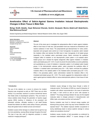
  • 5. Kamel et al. Ameliorative Effect of Salicin Against Gamma Irradiation UK J Pharm & Biosci, 2015: 3(2); 33 0.62 (B % 38.34 and 21.20) in the irradiated salicin prepost- treated group. Fig.1:Electrophoretic pattern showing effect of salicin against the irradiation effect on protein pattern in brain tissue of male rats Fig. 2:Electrophoretic pattern showing effect of salicin against the irradiation effect on lipoprotein pattern in brain tissue of male rats Table 3: Data of the electrophoretic lipoprotein pattern in brain tissue of control, irradiated and irradiated salicin treated groups in male rats Control Salicin Irradiated Irradiated salicin treated Pre-treated Simultaneous Prepost-treated Post-treated Rf. B. % Rf. B. % Rf. B. % Rf. B. % Rf. B. % Rf. B. % Rf. B. % 0.22 67.28 0.25 48.27 — — 0.22 100.00 — — 0.22 70.27 — — 0.47 18.91 0.33 31.12 — — — — — — 0.35 29.73 — — 0.69 13.82 0.72 20.61 — — — — — — — — — — Rf.: Rate of Flow, B.%: Band Percent Table 4: Data of the electrophoretic esterase pattern in brain tissue of control, irradiated and irradiated salicin treated groups in male rats Control Salicin Irradiated Irradiated salicin treated Pre-treated Simultaneous Prepost-treated Post-treated Rf. B. % Rf. B. % Rf. B. % Rf. B. % Rf. B. % Rf. B. % Rf. B. % 0.22 36.85 0.11 21.93 0.21 100.00 0.21 51.38 0.23 55.23 0.21 40.46 0.22 40.27 0.48 45.38 0.22 40.13 — — 0.47 25.29 0.48 44.77 0.45 38.34 0.47 36.92 0.64 17.77 0.49 19.00 — — 0.60 23.33 — — 0.62 21.20 0.62 22.81 — — 0.64 18.94 — — — — — — — — — — Rf.: Rate of Flow, B.%: Band Percent
  • 6. Kamel et al. Ameliorative Effect of Salicin Against Gamma Irradiation UK J Pharm & Biosci, 2015: 3(2); 34 It was found that the highest SI value (SI = 1.00) was noticed in irradiated salicin post-treated group. In the irradiated salicin simultaneous treated group, it was observed that all the bands were not matched with all bands of the other groups. Fig. 3:Electrophoretic pattern showing effect of salicin against the irradiation effect on esterase pattern in brain tissue of male rats 3.5 Electrophoretic catalase pattern As shown in table 5 and illustrated in fig. 4, 5 types of catalase enzyme were produced in the control sample with Rfs ranged between 0.21 - 0.97 (B % 8.11 – 47.12). There were 2 common bands appeared in all the groups with Rfs 0.45 and 0.97 (B % 8.47 and 15.17). Salicin alone caused alterations represented by appearance of one abnormal band with Rf 0.18 (B % 23.47) with decreasing the B % of the 1st type (Rf 0.22 and B % 6.12) and increasing the B % of the 2nd type (Rf 0.44 and B % 25.89). Irradiation caused qualitative alterations represented by disappearance of the 1st type of the enzyme with deviation of the 3rd band to be appeared with Rf 0.78 (B % 15.01) and quantitative mutation occurred by increasing B % of the 2nd band (Rf 0.45 and B % 64.42). Salicin administration could not prevent the irradiation effect which was represented by alteration occurred qualitatively by appearance of one abnormal band with Rf 0.15 (B % 29.71) with deviation of the 3rd type of the enzyme to be appeared with Rf 0.82 (B % 28.32) and quantitatively by decreasing B % of the band appeared with Rf 0.22 (B % 9.44) in the irradiated salicin pre-treated group, by disappearance of the 1st and 4th types of the enzyme with appearance of one abnormal band with Rf 0.17 (B % 31.52) and deviation of the 3rd type to be appeared with Rf 0.82 (B % 38.60) in the irradiated salicin prepost-treated group and represented by disappearance of the 1st type with appearance of one abnormal band with Rf 0.12 (B % 20.99) deviation of the 3rd type to be appeared with Rf 0.83 (B % 19.39) in the irradiated salicin simultaneous treated group. While in the irradiated salicin post-treated group, salicin could not overcome the mutation represented qualitatively by disappearance of 3 normal types with appearance of one abnormal band with Rf 0.66 (B % 57.05) and quantitatively by increasing B % of the bnds appeared with Rfs 0.45 and 0.96 (B % 20.65 and 22.30). From the SI values, it was found that the lowest SI value (SI = 0.44) was observed with irradiated salicin prepost-treated group and the highest value (SI = 0.91) observed with salicin treated group. Salicin treatment minimized the qualitative irradiation effect in the irradiated salicin pre-treated group (SI = 0.73) more than the other irradiated salicin treated groups. 3.6 Electrophoretic peroxidase pattern As shown in table 6 and illustrated in fig. 5, it was found that 5 types of peroxidase enzyme were produced in the control sample with Rfs ranged between 0.28 - 0.72 (B % 0.46 - 41.66). There were no common band appeared in all groups. Salicin alone caused no obvious qualitative or quantitative alterations. Irradiation caused severe disturbances represented qualitatively by disappearance of the 2nd , 3rd and 5th normal types of the enzyme and quantitatively by increasing B % of the 1st type (Rf 0.28 and B % 58.28). Salicin could not remove the abnormalities which were represented qualitatively by disappearance of the 1st , 3rd and 5th types with increasing B % of the 2nd and 4th normal types of the enzyme (Rfs 0.35 and 0.58 and B % 34.34 and 65.66) in irradiated salicin pre-treated group and represented qualitatively by disappearance of the 3rd type of the enzyme with appearance of one abnormal characteristic band with Rf 0.69 (B % 44.83) and quantitatively by decreasing the B % of the 1st type (Rf 0.27 and B % 14.08) and increasing B % of the 2nd and 4th types of the enzyme (Rfs 0.38 and 0.69 and B % 25.82 and 44.83) in irradiated salicin prepost-treated group. In the irradiated salicin post- treated group, salicin could not prevent the mutagenic effect which was represented qualitatively by disappearance of the 5th type and deviation of the 4th type to be appeared with Rf 0.56 (B % 61.82) and quantitatively by decreasing B % of the 1st type appeared with Rf 0.27 and 14.47. From the SI values, it was found that salicin minimized the irradiation effect on the band number and arrangement in the irradiated salicin prepost-treated groups (SI = 0.44) and post-treated groups (SI = 0.67). In the irrradiated pre-treated group, it was observed that all the bands were not matched with all bands of the other groups. It showed the highest antagonistic effect in the irradiated salicin post- treated group. 4 Discussions
  • 7. Kamel et al. Ameliorative Effect of Salicin Against Gamma Irradiation UK J Pharm & Biosci, 2015: 3(2); 35 During results of the present study, the MDA level elevated significantly as a result of radiation exposure in the brain tissue. This was in accordance with the results obtained by Saada and Azab39 that showed that the MDA level increased due to production of ROS associatedwith the increase in lipid peroxidation. ROS are known to attack the highly unsaturated fatty acids of the cell membrane to induce peroxidation reactions, which considereda key process in many pathological events and is one ofthe reactions induced by oxidative stress40 . The increasein intracellular ROS concentration leads subsequently tooxidative stress41 and decrease in activity of antioxidantenzymes with possible damage of cellular membranes42 . The present study showed that irradiation increased the MDA level. This was in agreement with Dixit et al 43 who showed that the doses 2, 6 and 10 Gy of irradiation enhanced the MDA level. This may be due to reducing the antioxidant enzymes as superoxide dismutase, catalase and glutathione-S-transferase. It was demonstrated that brain belonged to the most biosensitive organs to low doses of irradiation in rats44 . The oxidative stress affects some brain regions more than others45 . Irradiation induced lesions tend to occur more frequently in the cerebral brain white matter. It caused necrosis in the cortex and subcortical area in the brain46 . This because white matter tissue is more affected than gray matter tissue47 . This means that the brain represents one of the most important targets of irradiation. So, it was selected to be under study during the current experiment. The elevation of the levels of antioxidant enzymes in the irradiated animals when compared with the control suggests that these enzymes were up regulated to respond to the toxicity induced by the radiation48 . Proteins are the most complex compounds and at the same time the most characteristic of living matter. They are present in all viable cells; they are the compounds which, as nucleoproteins, are essential for cell division and, as enzymes and hormones, control many chemical reactions in the metabolism of cells. Thus, the separation and characterization of the individual proteins facilitate the study of the chemical nature and physiological function of each protein49 . Changes in the protein patterns of the tissues may reflect specialization and adaptation in the organisms. It is worthy to note that each protein is considered as reflect to the activity of specific gene through the production of enzyme, which act as a catalyst to produce the demanded protein; this type of produced protein is responsible for a specific biological character50 . Proteins are major targets for oxidative damage due to their abundance and rapid rates of reaction with a wide range of radicals and excited state species51 . It is worthy to note that each protein type has a biological role, due to this role, the DNA secrets enzymes which act as catalysts to produce the specific type of protein. Oxidative protein damage could also affect the activity of DNA repair enzymes. Another possible mutagenic effect of ROS involves their attack on lipids, to initiate lipid peroxidation. The peroxides can decompose to a range of mutagenic carbonyl products52 . The proteins in brains of all rats were separated electrophoretically and abundances were measured by using image analysis technique. Data in the present study indicated that specific protein bands in tissues of the irradiated rats differed (through disappearence in some protein bands or appearance of new ones). Disappearance of some protein bands in treated rats may be attributed to the effects of irradiation, which inhibits the synthesis and expression process of these deleted proteins (qualitative effect). In addition, even the band remained after irradiation, it usually differs in the amount of protein, and this may be explained by that irradiation could not inhibit the synthesis of this protein type, but it may be affected only on the quantitative level. The representative electrophoretic profile of the proteins was carried out in serum and different tissue homogenates. This profile showed different types of mutations occurred as a result of radiation exposure. As compared to control, some normal proteins were represented by the fastest migrating band and had the highest staining intensity. There were some proteins showed the intermediate bands and the slowest migrating bands. On the other hand, there was type of proteins represented as the biggest protein band close to the well comb (origin). All these proteins differ from each other in the mobility, molecular weight, optical density and band intensity of each protein band. In the present study, the similarity index between the control and all the irradiated samples and between the irradiated samples themselves recorded low values, indicating to apparent effect of the irradiation and the differences in the protein pattern. It was stated by many previous studies that the irradiation created a great genetic distance between the control and the irradiated samples that may be due to the activation of some genes. These genes produce different types of proteins not produced in the control. These protein types may lead to variation of the different biological processes. The current study showed that there were different mutations was detected by the appearance of new proteins or by the quantitative decrease in abundance of normally occurring proteins. This was in agreement with the results reported by Giometti et al 53 who reported that the electrophoresis can be used to detect the mutations reflected as quantitative changes in the protein expression.
  • 8. Kamel et al. Ameliorative Effect of Salicin Against Gamma Irradiation UK J Pharm & Biosci, 2015: 3(2); 36 Table 5: Data of the electrophoretic Catalase pattern in brain tissue of control, irradiated and irradiated salicin treated groups in male rats Control Salicin Irradiated Irradiated salicin treated Pre-treated Simultaneous Prepost-treated Post-treated Rf. B. % Rf. B. % Rf. B. % Rf. B. % Rf. B. % Rf. B. % Rf. B. % 0.21 47.12 0.18 23.47 0.45 64.42 0.15 29.71 0.12 20.99 0.17 31.52 0.45 20.65 0.45 8.47 0.22 6.12 0.78 15.01 0.22 9.44 0.45 41.66 0.44 13.82 0.66 57.05 0.85 21.13 0.44 25.89 0.92 8.84 0.45 9.96 0.83 19.39 0.82 38.60 0.96 22.30 0.92 8.11 0.85 26.36 0.97 11.74 0.82 28.32 0.92 6.94 0.97 16.07 — — 0.97 15.17 0.93 7.04 — — 0.91 9.16 0.95 11.02 — — — — — — 0.97 11.12 — — 0.97 13.41 — — — — — — Rf.: Rate of Flow, B.%: Band Percent Table 6: Data of the electrophoretic peroxidase pattern in brain tissue of control, irradiated and irradiated salicin treated groups in male rats Control Salicin Irradiated Irradiated salicin treated Pre-treated Simultaneous Prepost-treated Post-treated Rf. B. % Rf. B. % Rf. B. % Rf. B. % Rf. B. % Rf. B. % Rf. B. % 0.28 35.62 0.27 35.91 0.28 58.28 0.35 34.34 0.27 11.68 0.27 14.08 0.27 14.47 0.36 12.62 0.37 11.01 0.59 41.72 0.58 65.66 0.36 28.20 0.38 25.82 0.37 14.82 0.49 9.63 0.47 12.02 — — — — 0.57 60.12 0.58 15.28 0.48 8.89 0.59 0.46 0.60 0.63 — — — — — — 0.69 44.83 0.56 61.82 0.72 41.66 0.72 40.44 — — — — — — — — — — Rf.: Rate of Flow, B.%: Band Percent Fig. 4:Electrophoretic pattern showing effect of salicin against the irradiation effect on catalase pattern in brain tissue of male rats Fig. 5:Electrophoretic pattern showing effect of salicin against the irradiation effect on peroxidase in brain tissue of male rats
  • 9. Kamel et al. Ameliorative Effect of Salicin Against Gamma Irradiation UK J Pharm & Biosci, 2015: 3(2); 37 During the current experiment, irradiation-dependent accumulation of oxidatively modified proteins was studied in the brain tissues. Alterations in the protein increased as a result of irradiation. This may be related to an extensive modification of lysine and arginine residues in histone molecules54 . The authors observed activation of histone-specific proteases in the nuclei of γ-irradiated rats. The lack of carbonyl accumulation in the nuclear proteins isolated from tissues of γ-irradiated animals may be explained by the degradation of oxidized histones by these proteases. All lipoproteins carry all types of lipid, but in different proportions, so that the density is directly proportional to the protein content and inversely proportional to the lipid content55 . The lipoproteins were more susceptible to oxidative modifications resulting in small lipoproteins56 . The ROS can initiate one-electron oxidation or one- electron reduction reactions on numerous biological systems. The oxidative hypothesis classically admits the involvement of the lipoproteins oxidation radiolytically57 . There was natural binding between protein and lipoproteins in the rat tissues. These two tissues known to be involved in the processing of the lipoproteins. The lipoproteins-binding protein has previously been identified in adrenal cortical plasma membranesandconcentration of the binding protein was strongest in kidneys58 . So the alterations in the protein pattern were associated with altering the lipoprotein pattern in these tissues. The alterations in the lipoprotein pattern may refer to the disturbances in the cholesteryl esterase required or cholesterol hydrolysis. It was suggested that non-parenchymal liver cells possess the enzymic equipment (cholesteryl esterase) to hydrolyze very efficiently internalized cholesterol esters and this supported that these cell types are an important site for lipoprotein catabolism59,60 . Salicin administration showed the protective effect against the irradiation. This may be due to its antioxidative effect against attack of the free radicals. It prevented the alterations in the proteins and hence the lipoproteins fractions in the brain tissues. In the current experiment, polyacrylamide gel electrophoresis was used for separation of different enzymes, which help in explanation of different biological processes that occur inside the living organisms due to the deleterious effect of irradiation and the ameliorated effect of salicin. According to the present data, the activities of CAT and GPx in brains of irradiated rats were altered. These results strongly suggested that irradiation has the capability to induce free radicals and oxidative damage as evidenced by perturbations in various antioxidant enzymes. Depletion of these enzymes activity could be due to the direct effect on the enzymes by irradiation-induced ROS generation, direct inhibition of the enzymes by irradiation61 . The present work evaluated the enzymatic activity of the AO enzymes enzymes as CAT and GPx and electrophoretic detection of oxidative damage in proteins (carbonyl groups) in brains of rats. This was in agreement with Ehrenbrink et al62 . The authors suggested that the patterns of activity and accumulation of damages can be sex-specific and related to the cycle of reproductive life of the rats. Esterases with active thiol groups detected electrophoretically in brain of different animal species. It exhibits wide distribution among these species63 .The brain was selected to show the esterase activity because it is rich in esterase enzyme due to its role in the neurotransmission and communication of messages64 . Electrophoresis plays a major role in identifying esterases. So, this technique was selected to identify the esterase types in male and female reproductive organs. The present study showed that irradiation caused alterations in the electrophoretic esterase pattern representing great differences in a number of zones of esterase activity and in substrate specificity between all treated groups compared to control. During the current the experiment, irradiation caused alterations in the electrophoretic esterase pattern. This may refer to effect of irradiation on the protein pattern. As regards changes in electrophoretic mobility demonstrated in the present study, it seemed that free radicals affect the integrity of the polypeptide chain in the protein molecule causing fragmentation of the polypeptide chain due to sulfhydral-mediated cross linking of the labile amino acids as claimed by Bedwell et al65 . The changes in the fractional activity of different isoenzymes seemed to be correlated with changes in the rate of protein expression secondary to DNA damage initiated by free radicals66 . During the current experiment, salicin administration showed the highest ameliorative effect against irradiation in the irradiated salicin treated rats. This may be due to trapping of these free radicals by salicin, thus preventing DNA damage. Salicin and salicylic acid belonged to the phenolic compounds which showed AO activity due to their ability to scavenge free radicals67 .So, they are able to overcome the disturbances in the esterase pattern in the tissues selected to be under study. Salicin belonged to the phenolic glycosides which are characterized by their antioxidant activity in biological systems. The antioxidant activity of the phenolic compounds refers to their ability to scavenge free radicals67,68 . The authors suggested that the phenolic molecules undergo redox reactions because phenolic hydroxyl groups readily donate hydrogen to reducing agents. Ronca et al68 showed that antioxidants suppressed hydroxyl radical production in the fenton reaction, probably by chelating the iron required in the generation of hydroxyl radicals. Results of the present study were in agreement
  • 10. Kamel et al. Ameliorative Effect of Salicin Against Gamma Irradiation UK J Pharm & Biosci, 2015: 3(2); 38 with that reported by Das et al69 who confirmed that elevation of MDA concentration might be a consequence of decreased production of antioxidants in the irradiated rats’ tissues. The antioxidant activities of salicin fraction varied markedly. This may be due to the differences in structures of phenolic compounds and primarily related to their hydroxylation and methylation patterns70 .The total antioxidant activity of the total aqueous extract of willow leaves was much more than salicin alone. This may be due to the aqueous extract of willow leaves contain other antioxidants such as tannins, flavenoids and proanthocyanidins in addition to salicin which could play some role in the increase of the total antioxidant activity71 . Salicin may induce elevation in activities of the antioxidants as glutathione peroxidase in the tissues homogenates which were selected to be under study. The antioxidants have a major role in the antioxidants defense mechanisms against irradiation injury72 . So, the maintenance of normal electrophoretic protein and zymogram levels after the treatment with salicin may be due to trapping of these free radicals by this fraction, thus preventing DNA damage. Salicin was able to overcome the disturbances in the protein pattern in the tissues selected to be under study. 5 Conclusions The study concluded that salicin showed radioprotective effect against the gamma irradiation effect on various electrophoretic patterns in brain tissue of male rats. 6 Competing interest The study aimed to suggest new compounds obtained from the nature and act as radioprotector to minimize or resist the irradiation effect. 7 Author’s contributions MSA and MALAK carried out literature review and draft the manuscript. HMS participated in collection of data and arranged in tabular form. IA and WMK carried out the experimental work. All authors read and approved the final manuscript. 8 References 1. Casarett AP. Radiation Biology. Prentice-Hall, New Jersey. 1968; 368. 2. Srinivasan M, Sudheer AR, Pillail KR, Kumar PR, Sudhakaran PR, Menon VP. Influence of ferulic acid on gamma-radiation induced DNA damage, lipid peroxidation and antioxidant status in primary culture of isolated rat hepatocytes. Toxicology. 2006; 228(2-3): 249-258. 3. DeAngelis LM, Gutin PH, Leibel SA. Murtin Dunitz Ltd.. Interacranial tumors. Dignosis and Treatment. 2002. 4. Bock RK. Very high energy gamma rays fromdistant quasar: How transparent is the universe?. Science. 2008; 320(5884): 1752-1754. 5. Dwyer J, David MS. Dealy rays from clouds. Scientific American. 2012; 307(2): 55-59. 6. Mobbs SF, Muirhead CR, Harrison JD. Risks from ionising radiation: an HPA viewpoint paper for safegrounds, J. Radiol. Prot., 2011; 31 : 289–307. 7. Moores BM, Regulla D. A review of the scientific basis for radiation protection of the patient, Radiat. Prot. Dosimetry. 2011; 147: 22–29. 8. Burlakova EB, Mikhaĭlov VF, Azurik VK. The redox homeostasis system in radiation-induced genomic instability. Radiats Biol. Radioecol. 2001; 41(5) :489 - 99. 9. Di Pietro C, Piro S, Tabbì G, Ragusa M, Di Pietro V, Zimmitti V, Cuda F, Anello M, Consoli U, Salinaro ET, Caruso M, Vancheri C, Crimi N, Sabini MG, Cirrone GA, Raffaele L, Privitera G, Pulvirenti A, Giugno R, Ferro A, Cuttone G, Lo Nigro S, Purrello R, Purrello F, Purrello M. Cellular and molecular effects of protons: apoptosis induction and potential implications for cancer therapy. Apoptosis. 2006;11(1): 57-66. 10. Thornburn CC. Isotopes and radiation in biology, New York, Halstead Press Division, Wiley. 1972; 287. 11. Ezz MK. The Ameliorative Effect of Echinacea Purpurea Against Gamma Radiation Induced Oxidative Stress and Immune Responses in Male Rats. Australian Journal of Basic and Applied Sciences. 2011; 5(10): 506-512. 12. Nwozo SO, Okameme PE, Oyinloye BE. Potential of Piper guineense and Aframomum longiscapum to reduce radiation induced hepatic damage in male Wistar rats. Radiats Biol. Radioecol. 2012; 52(4): 363-369. 13. Borek C. Antioxidants and radiation therapy. J. Nutr. 2004; 134 : 3207S-3209S. 14. Konopacka M, Rogolinski J. Thiamine prevents X-ray induction of genetic changes in human lymphocytes in vitro. Acta. Biochem. Pol. 2004; 51 : 839 – 843. 15. Arora R, Gupta D, Chawla R, Sagar R, Sharma A, Kumar R, Prasad J, Singh S, Samanta N, Sharma RK. Radioprotection by plant products: present status and future prospects, Phytother. Res. 2005; 19 : 1–22.
  • 11. Kamel et al. Ameliorative Effect of Salicin Against Gamma Irradiation UK J Pharm & Biosci, 2015: 3(2); 39 16. Snyder AR, Morgan WF. Persistent oxidative stress and gene expression changes in radiation-induced genomic instability. Int. Congr. Ser. 2003; 1258: 199–206. 17. Spitz DR, Azzam EI, Li JJ, Gius D. Metabolic oxidation/reduction reactions and cellular responses to ionizing radiation: a unifying concept in stress response biology. Cancer Metastasis Rev. 2004; 23 (3-4): 311-22. 18. Fedorova M, Kuleva N, Hoffmann R. Identification, quantification, and functional aspects of skeletal muscle protein-carbonylation in vivo during acute oxidative stress. J. Proteome Res. 2010; 9 (5):2516 - 2526. 19. Seyed H. Flavonoids and genomic instability induced by ionizing radiation. Drug Discov. 2010; 15: 907–918. 20. Zhang H, Wang ZY, Zhang Z, Wang X. Purified Auricularia auricular-judae polysaccharide (AAP I-a) prevents oxidative stress in an ageing mouse model. Carbohyd. Polym. 2011; 84 :638–648. 21. Kula B, Sobczak A, Kuska R. Effect of static and ELF magnetic fields on free radical processes in rat liver and kidney. Electro-Magnetobiol. 2000; 19: 99 - 105. 22. Agrawal A, Kale RK. Radiation induced peroxidative damage: mechanism and significance. Indian J. Exp. Biol. 2001; 39: 209–291. 23. Bhatia A.L, Manda K. Study of pre-treatment of melatonin against radiation-induced oxidative stress in mice. Environ. Toxicol. Pharmacol. 2004; 18: 13 – 20. 24. Mayer RA, Mayer, M. Biologische Salicyltherapie mit Cortex Salicis. Pharmazie. 1949; 4: 77–81. 25. Zaugg SE, Cefalo D, Walker EB. Capillary electrophoretic analysis of salicin in Salix spp. J. of Chromatography A. 1997; 781: 487 – 490. 26. Mabry TJ, Markham KR, Thomaas, M. B. The Systematic Identification of flavonoids, Springer-Verlag, Berlin. 1970 27. Kur'yanov AA, Bondarenko LT, Kurkin VA, Zapesochnaya GG, Dubichev AA, Vorontsov ED. Determination of the biologically active components of the rhizomes of Rhodiola rosea. Translated from Khimiya Prirodnykh Soedinenii. 1991 ; 3:320-323. 28. Paget and Barnes. Evaluation of drug activities pharmacometrics. Edited by Laurence, D.R. and Bacharach, A.L. Academic Press, London and New York, 1974; 135. 29. Ohkawa H, Ohishi N, Yagi K Assay for lipid peroxides in animal tissues by thiobarbituric acid reaction. Anal. Biochem. 1979; 95: 351 – 358. 30. Bradford MM. A rapid and sensitive method for the quantitation of microgram quantities of protein utilizing the principle of protein-dye binding. Anal. Biochem. 1976; 72: 248-254. 31. Laemmli UK. Cleavage of structural proteins during the assembly of the head of Bacteriophage T4. Nature. 1970; 227: 680-685. 32. Hames BD. One-dimensional polyacrylamide gel electrophoresis. In: Gel electrophoresis of proteins: B.D. Hames B.D. and Rickwood D., 2nd ed.. Oxford university press, NY. 1990; 1-147. 33. Chippendale GM, Beak SD. Haemolymph proteins of Osirinla nubilalis (Hubner): during diapauses prepupa differentiation . J. Insect Physiolo. 1966; 12 : 1629-1638. 34. Rescigno A, Sanjust E, Montanari L, Sollai F, Soddu G, Rinaldi AC, Oliva S, Rinaldi A. Detection of laccase, peroxidase, and polyphenol oxidase on a single polyacrylamide gel electrophoresis, Anal. Lett. 1997; 30(12): 2211. 35. Siciliano MJ, Shaw CR. Separation and visualization of enzymes on gels, in Chromatographic and Electrophoretic Techniques, Vol. 2, Zone Electrophoresis, Smith, I., Ed., Heinemann, London. 1976; p.185. 36. Baker CMA, Manwell, C. Heterozygosity of the sheep: Polymorphism of 'malic enzyme', isocitrate dehydrogenase (NADP+ ), catalase and esterase. Aust. J. Biol. Sci. 1977; 30(1-2): 127-40. 37. Nei M, Li WS. Mathematical model for studing genetic variation in terms of restriction endonuclease. Proc. Natl. Acad. Sci., USA. 1979; 76: 5269 – 5273. 38. Snedecor CW, Cochran WG. Statistical Methods.7th ed. Iowa, Univ. Press Ames USA. 1982. 39. Saada HN, Azab KS. Role of lycopene in recovery of radiation induced injury to mammalian cellular organelles. Pharmazie. 2001; 56 (3): 239-241. 40. Schinella GR, Tounier HA, Prieto JM, Mordujovich de Buschiazzo P, Rios JL. Antioxidant activity of anti- inflammatory plant extracts. Life Sci. 2002; 70: 1023 – 1033. 41. Maurel A, Hernandez C, Kunduzova O. Age-dependent increase in hydrogen peroxide production by cardiac
  • 12. Kamel et al. Ameliorative Effect of Salicin Against Gamma Irradiation UK J Pharm & Biosci, 2015: 3(2); 40 monoamine oxidase A in rats. Am. J. Physiol. Heart Circ. Physiol. 2003; 284: H1460 – H1467. 42. El Habit OHM, Saada HN, Azab KS, Abdel Rahman M, El Malah DF. The modifying effect of B-carotene on gamma radiation-induced elevation of oxidative reactions and genotoxicity in male rats. Mutation Research. 2000; 466: 179-186. 43. Dixit AK, Bhatnagar D, Kumar V, Chawla D, Fakhruddin K, Bhatnagar D. Antioxidant potential and radioprotective effect of soy isoflavone against gamma irradiation-induced oxidative stress. J. Funct. Foods. 2012; 4: 196–206. 44. Hawas AM. The biosensitivity of certain organs in rats exposed to low doses of γ-radiation. Journal of Radiation Research and Applied Sciences. 2013; 6(2): 56 – 62. 45. Pajovic SB, Saicici ZS, Spasic MB, Petrovic VM. The Effect of Ovarian Hormones on Antioxidant Enzyme Activities in the Brain of Male Rats. Physiol. Res. 2003; 52: 189-194. 46. Hopwell JW, Vander Kogel AJ. Pathophysiology mechanisms leading to the development of late radiation-induced damage to the central nervous system. Fron Radiat The Oncol. 1999; 33: 265-275. 47. Vong L, Venita J, Shung W. Oligodendrocytes in the in the adult rat spinal cord undergo radiation –induced apoptosis. American Association for cancer Research. 1996; 56: 5417- 5422. 48. Cuevas P, Gimenez-Gallego G. Role of fibrolast growth factors in neural trauma. Neurological Research. 1997; 19: 254-256. 49. Mohamed MI. Sterility and some associated physiological changes in the adult cowpea weevil, Callosobruchus maculatus (F.). Ph. D. Thesis, Dept. Entomol., Fac, Sci., Ain Shams Univ. 1990. 50. Hassan Heba A, AbdEl-Hafez Hanan F. The comparison effects of two acetylcholine receptor modulator on some biological aspects, protein pattern and detoxification enzyme of the cotton leafworm, spodoptera littoralis. Egypt. J. Agric. Res. 2009; 87(2):103-117. 51. Hawkins CL, Morgan PE, Davies MJ. Quantification of protein modification by oxidants. Free Radical Biology and Medicine. 2009; 46: 965 - 988. 52. Cheeseman K In DNA and Free Radicals (Halliwell, B. and Aruoma, O. I., eds.). 1993; pp. 109-144, Ellis Horwood, Chichester. 53. Giometti CS, Gemmell MA, Nance SL, Tollaksen SL, Taylor J. Detection of heritable mutations as quantitative changes in protein expression. J. Biol. Chem. 1987; 262: 12764 – 12767. 54. Pleshakova OV, Kutsyi MP, Sukharev SA, Sadovnikov VB, Gaziev AI. Study of protein carbonyls in subcellular fractions isolated from liver and spleen of old and γ-irradiated rats. Mechanisms of Ageing and Development. 1998; 103(1): 45- 55. 55. Bass KM, Newschaffer CJ, Klag MJ, Bush TL. Plasma lipoprotein levels as predictors of cardiovascular death in women. Arch. Intern. Med. 1993; 153(19): 2209-16. 56. Tsumura M, Kinouchi T, Ono S, Nakajima T, Komoda T. Serum lipid metabolism abnormalities and change in lipoprotein contents in patients with advanced-stage renal disease. Clinica. Chimica. Acta. 2001; 314: 27 – 37. 57. Bonnefont-Rousselot D. Gamma radiolysis as a tool to study lipoprotein oxidation mechanisms. Biochimie. 2004; 86: 903- 911. 58. Fidge NH. Partial purification of a high density lipoprotein- binding protein from rat liver and kidney membranes. Federation of European Biochemical Societies. 1986 ; 199: 265 - 268. 59. Theo JC, Berkel V, Vaandrager H, Kruijt JK, Koster JF. Characteristics of acid lipase and acid cholesteryl esterase activity in parenchymal and non-parenchymal rat liver cells. Biochimica et Biophysica Acta (BBA) - Lipids and Lipid Metabolism. 1980; 617: 446 - 457. 60. Satoh T. Toxicological implications of esterases—From molecular structures to functions. Toxicology and Applied Pharmacology. 2005; 207: 11 – 18. 61. Schreinemachers DD. Birth malformations and other adverse perinatal outcomes in four US wheat-producing states. Environ. Health. Perspect. 2003; 111(9): 1259–64. 62. Ehrenbrink G, Hakenhaar FS, Salomon TB, Petrucci AP, Sandri MR, Benfato MS. Antioxidant enzymes activities and protein damage in rat brain of both sexes. Experimental Gerontology. 2006: 41(4): 368-371. 63. Lakshmipathi V, Reddy T M. Comparative study of esterases in brains of the vertebrates. Brain Research. 1990; 521 (1–2): 321-324.
  • 13. Kamel et al. Ameliorative Effect of Salicin Against Gamma Irradiation UK J Pharm & Biosci, 2015: 3(2); 41 64. Srividhya R, Gayathri R, Kalaiselvi P. Impact of epigallo catechin-3-gallate on acetylcholine- acetylcholineesterase cycle in aged rat brain. Neurochemistry International. 2012; 60(5): 517-522. 65. Bedwell S, Dean RT, Jessup W. The action of defined oxygen centered free radicals on human low-density lipoprotein. Biochem. J. 1989; 262: 707-712. 66. El-Zayat EM. Isoenzyme Pattern and Activity in Oxidative Stress-Induced Hepatocarcinogenesis: The Protective Role of Selenium and Vitamin E. Research Journal of Medicine and Medical Sciences. 2007; 2(2): 62-71. 67. Madrigal-Carballo S, Rodriguez G, Krueger CG, Dreher M, Reed JD. Pomegranate (Punica granatum L.) supplements: authenticity, antioxidant and polyphenol composition. J. Funct. Food. 2009; 1: 324 - 329. 68. Ronca G, Ronca F, Yu G, Zucchi R, Bertelli A. Protection of isolated perfused working rat heart from oxidative stress by exogenous L-propionyl carnitine. Drugs Exp. Clin. Res. 1992; 18: 475-480. 69. Das S, Chakraborty SP, Roy S, Roy S. Nicotine induced pro- oxidant and antioxidant imbalance in rat lymphocytes: In vivo dose and time dependent approaches. Toxicol. Mech. Methods. 2012; 22: 711-20. 70. Meyer AS, Donovan JL, Pearson DA, Waterhouse AL, Frankel EN Fruit hydroxycinnamic acids inhibit human low density lipoprotein oxidation. J. Agric. Food Chem. 1998; 46(5): 1783 - 1787. 71. Arab L, Steck S. Lycopene and cardiovascular disease. Am. J. Clin. Nutr. 2000; 71: 1691 - 1697. 72. Ibrahim K, Seyithan T, Mustafa E, Ihsan K, Akcahan G, Orhan S, Korkmaz S. The effect of L- carnitine in the prevention of ionizing radiation induced cataracts; a rat model.Graefe _s. Archive Clinic. and Exp. Ophthalm. 2007; 245(4): 588- 594.