Gloved finger sign and cervicothoracic sign
•Download as PPTX, PDF•
20 likes•8,055 views

Gloved finger sign and Cervicothoracic sign

Recommended
More Related Content
What's hot
What's hot (20)
Viewers also liked
Viewers also liked (11)
Similar to Gloved finger sign and cervicothoracic sign
Similar to Gloved finger sign and cervicothoracic sign (20)
More from Minstry of health ,Ibn alnafis hoapital, Damascus
More from Minstry of health ,Ibn alnafis hoapital, Damascus (10)
Recently uploaded
Recently uploaded (20)
Gloved finger sign and cervicothoracic sign
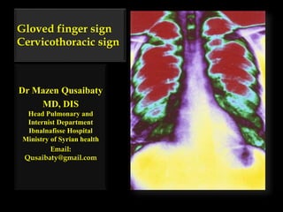
- 1. Gloved finger sign Cervicothoracic sign Dr Mazen Qusaibaty MD, DIS Head Pulmonary and Internist Department Ibnalnafisse Hospital Ministry of Syrian health Email: Qusaibaty@gmail.com
- 2. Topic Outline 1. Gloved finger sign 2. Cervicothoracic sign 2
- 3. Gloved finger sign القفاز إصبع عالمة
- 4. Gloved finger sign • Refers to the branching finger like opacities. 4
- 5. Gloved finger sign • Gloved finger shadows" due to intrabronchial exudates with bronchial wall thickening 5
- 6. Gloved finger sign • These appear as branched tubular radiodensities: 2 to 3 cm long 5 to 8 mm wide that extend from the hilus 6
- 7. Gloved finger sign • Representing dilated bronchi filled with mucus (mucoid impaction) radiating from the hila towards the periphery 7
- 8. 8 Schematic diagram depicts four grades of bronchial wall thickening scores
- 9. Central bronchiectasis • Central bronchiectasis in a patient with allergic bronchopulmonary aspergillosis 9
- 10. Central bronchiectasis • Multiple dilated third and fourth generation bronchi are seen. 10
- 11. Central bronchiectasis • Smaller peripheral bronchi filled with mucus account for the branching linear opacities in the distal lung parenchyma. 11 Courtesy of Paul Stark, MD
- 12. Gloved finger sign Mucoid Impaction 12
- 13. Gloved finger sign Mucoid impaction of underlying bronchiectatic airway in a patient with Allergic BronchoPulmonary Aspergillosis (ABPA). 13
- 14. Gloved finger sign Mucoid impactions are: A characteristic finding in ABPA And typically occur distal to the diseased central airways. 14
- 16. Gloved finger sign • In Allergic Bronchopulmonary Aspergillosis 16
- 17. • Close-up frontal radiograph of the right upper lobe obtained in a patient with asthma and allergic bronchopulmonary aspergillosis (ABPA) 17
- 18. • Note the branching tubular opacities (arrows) emanating from the right hilum, which compose the gloved finger sign. 18
- 20. • Two contiguous 5-mm thick transverse images obtained at contrast material-enhanced (CT) of the chest just above the left hemidiaphragm 20
- 21. • A tubular and a branching structure in the posterior basal segment of the LLL 21
- 22. • A congenital atresia of this bronchus. 22
- 23. • The vessels in the lung surrounding the mucoid impaction are decreased in size due to hypoxic vasoconstriction 23
- 24. Transverse CT image in 1-year-old boy • A round opacity (arrow) • An area of hypoattenuation (arrowheads) and decreased vascularity 24
- 25. Transverse CT image in 1-year-old boy • A congenital atresia of this bronchus 25
- 26. Bronchial atresia • Bronchial atresia is a developmental anomaly 26
- 27. Bronchial atresia • Characterised by focal obliteration of the proximal segment of a bronchus 27
- 28. Bronchial atresia • It is typically at the: o Segmental o Or subsegmental level o And most commonly occurs in the upper lobes. 28
- 29. Bronchial atresia • The bronchi distal to the atresia become filled with mucus and may form a mucocoele 29
- 30. Bronchial atresia • The lung distal to the atretic bronchus o Develops normally 30
- 31. Bronchial atresia The lung distal is overinflated due to collateral air drift with air trapping. 31
- 32. Bronchial atresia • It may cause Shortness of breath Cough Or rarely infection. 32
- 33. Conclusion • Gloved finger sign - indicates bronchial impaction, which can be seen in allergic bronchopulmonary aspergillosis 33
- 34. Cervicothoracic sign الصدرية الرقبية العالمة 34
- 35. Cervicothoracic sign 35 A mediastinal opacity that projects above the clavicles is retrotracheal and posteriorly
- 36. Cervicothoracic sign 36 while an opacity effaced along its superior aspect and projecting at or below the clavicles is situated anteriorly
- 37. Cervicothoracic sign • This 74 year-old female presented with mild dyspnoea 37
- 38. Cervicothoracic sign A superior mediastinal mass Displaces the trachea to the right38
- 39. This mediastinal mass is seen in A. Anterior mediastinal B. Posterior mediastinal 39
- 40. This mediastinal mass is seen in A. Anterior mediastinal B. Posterior mediastinal 40
- 41. The margins of the mass fade out at the level of the clavicles, the cervicothoracic sign, indicating an anterior location. 41
- 42. Positive Cervicothoracic sign (Ant) 42
- 43. What is your diagnosis?
- 44. The most common anterior superior mediastinal mass is a retrosternal goitre, as in this case. 44
- 45. Cervicothoracic sign This mediastinal mass is seen in A. Anterior mediastinal B. Posterior mediastinal45
- 46. Negative Cervicothoracic sign • This mediastinal mass is seen in A. Anterior mediastinal B. Posterior mediastinal46
- 47. What is your diagnosis?
- 49. This mediastinal mass is seen in A. Anterior mediastinal B. Posterior mediastinal 49
- 50. This mediastinal mass is seen in A. Anterior mediastinal B. Posterior mediastinal 50
- 51. This mediastinal mass is seen in A. Anterior mediastinal B. Posterior mediastinal 51
- 52. This mediastinal mass is seen in A. Anterior mediastinal B. Posterior mediastinal 52
- 53. This mediastinal mass is seen in A. Anterior mediastinal B. Posterior mediastinal 53
- 54. This mediastinal mass is seen in A. Anterior mediastinal B. Posterior mediastinal 54
- 55. • This mediastinal mass is seen in A. Anterior mediastinal B. Posterior mediastinal 55
- 56. • This mediastinal mass is seen in A. Anterior mediastinal B. Posterior mediastinal 56
- 57. What is your diagnosis?
- 58. Schwannoma 58
- 59. REFERENCES • 1. Marshall GB, Farnquist BA, MacGregor JH, Burrowes PW. Signs in thoracic imaging. J.Thorac.Imaging 2006;21:76-90 • 2. Webb WR. Thin-section CT of the secondary pulmonary lobule: anatomy and the image—the 2004 Fleischner lecture.Radiology. 2006 May;239(2):322-38 • 3. Austin JH, Muller NL, Friedman PJ, Hansell DM, Naidich DP, Remy-Jardin M, Webb WR, Zerhouni EA. Glossary of terms for CT of the lungs: recommendations of the Nomenclature Committee of the Fleischner Society. Radiology 1996;200(2):327-31 59
Editor's Notes
- A Pictorial Review of “Signs in Thoracic Imaging” Karuppasamy, K.1, Abhyankar-Gupta, M.1, Fewins, H.1, Curtis, J.2 1The Cardiothoracic Centre - Liverpool NHS Trust, 2Aintree University Hospitals NHS Foundation Trust, Liverpool, United Kingdom
- prominence : بروزRefers to the branching finger like opacities representing dilated bronchi filled with mucus (mucoid impaction) radiating from the hila towards the periphery; e.g. ABPA
- prominence : بروزRefers to the branching finger like opacities representing dilated bronchi filled with mucus (mucoid impaction) radiating from the hila towards the periphery; e.g. ABPA
- prominence : بروزRefers to the branching finger like opacities representing dilated bronchi filled with mucus (mucoid impaction) radiating from the hila towards the periphery; e.g. ABPA
- prominence : بروزRefers to the branching finger like opacities representing dilated bronchi filled with mucus (mucoid impaction) radiating from the hila towards the periphery; e.g. ABPA
- Central bronchiectasis in a patient with allergic bronchopulmonary aspergillosis. Multiple dilated third and fourth generation bronchi are seen. Smaller peripheral bronchi filled with mucus account for the branching linear opacities in the distal lung parenchyma. Courtesy of Paul Stark, MD
- Central bronchiectasis in a patient with allergic bronchopulmonary aspergillosis. Multiple dilated third and fourth generation bronchi are seen. Smaller peripheral bronchi filled with mucus account for the branching linear opacities in the distal lung parenchyma. Courtesy of Paul Stark, MD
- Central bronchiectasis in a patient with allergic bronchopulmonary aspergillosis. Multiple dilated third and fourth generation bronchi are seen. Smaller peripheral bronchi filled with mucus account for the branching linear opacities in the distal lung parenchyma. Courtesy of Paul Stark, MD
- Mucoid Impaction. Mucoid impaction of underlying bronchiectatic airway in a patient with ABPA. Mucoid impactions are a characteristic finding in ABPA and typically occur distal to the diseased central airways. Tubular branching opacities extend from the hilum and form a "gloved-finger" appearance
- Mucoid Impaction. Mucoid impaction of underlying bronchiectatic airway in a patient with ABPA. Mucoid impactions are a characteristic finding in ABPA and typically occur distal to the diseased central airways. Tubular branching opacities extend from the hilum and form a "gloved-finger" appearance
- Mucoid Impaction. Mucoid impaction of underlying bronchiectatic airway in a patient with ABPA. Mucoid impactions are a characteristic finding in ABPA and typically occur distal to the diseased central airways. Tubular branching opacities extend from the hilum and form a "gloved-finger" appearance
- The finger in glove sign seen on CXR and CT chest and refers to the characteristic sign of a bronchocoele, as seen in allergic bronchopulmonary aspergillosis (ABPA). Rarely a similar appearance can occur with bronchial atresia.
- In allergic bronchopulmonary aspergillosis. The impacted bronchi appear radiographically as opacities with distinctive shapes.
- Close-up frontal radiograph of the right upper lobe obtained in a patient with asthma and allergic bronchopulmonary aspergillosis (ABPA). Note the branching tubular opacities (arrows) emanating from the right hilum, which compose the gloved finger sign.
- Close-up frontal radiograph of the right upper lobe obtained in a patient with asthma and allergic bronchopulmonary aspergillosis (ABPA). Note the branching tubular opacities (arrows) emanating from the right hilum, which compose the gloved finger sign.
- Two contiguous 5-mm thick transverse images obtained at contrast material-enhanced computed tomography (CT) of the chest just above the left hemidiaphragm show (a) a tubular and (b) a branching structure in the posterior basal segment of the left lower lobe due to a congenital atresia of this bronchus. The vessels in the lung surrounding the mucoid impaction are decreased in size due to hypoxic vasoconstriction.
- Two contiguous 5-mm thick transverse images obtained at contrast material-enhanced computed tomography (CT) of the chest just above the left hemidiaphragm show (a) a tubular and (b) a branching structure in the posterior basal segment of the left lower lobe due to a congenital atresia of this bronchus. The vessels in the lung surrounding the mucoid impaction are decreased in size due to hypoxic vasoconstriction.
- Two contiguous 5-mm thick transverse images obtained at contrast material-enhanced computed tomography (CT) of the chest just above the left hemidiaphragm show (a) a tubular and (b) a branching structure in the posterior basal segment of the left lower lobe due to a congenital atresia of this bronchus. The vessels in the lung surrounding the mucoid impaction are decreased in size due to hypoxic vasoconstriction. رتق
- Two contiguous 5-mm thick transverse images obtained at contrast material-enhanced computed tomography (CT) of the chest just above the left hemidiaphragm show (a) a tubular and (b) a branching structure in the posterior basal segment of the left lower lobe due to a congenital atresia of this bronchus. The vessels in the lung surrounding the mucoid impaction are decreased in size due to hypoxic vasoconstriction.
- Transverse CT image in 1-year-old boy with known right lower lobe nodular lesion, representing mucus accumulation within the patent bronchus distal to the atretic segment, shows characteristic CT appearance of a congenital bronchial atresia manifested as a round opacity (arrow) associated with an area of hypoattenuation (arrowheads) and decreased vascularity. Note its somewhat atypical location, since congenital bronchial atresia is typically located in the apical or apicoposterior segment of the upper lobes.
- Transverse CT image in 1-year-old boy with known right lower lobe nodular lesion, representing mucus accumulation within the patent bronchus distal to the atretic segment, shows characteristic CT appearance of a congenital bronchial atresia manifested as a round opacity (arrow) associated with an area of hypoattenuation (arrowheads) and decreased vascularity. Note its somewhat atypical location, since congenital bronchial atresia is typically located in the apical or apicoposterior segment of the upper lobes.
- Bronchial atresia is a developmental anomaly characterised by focal obliteration of the proximal segment of a bronchus. It is typically at the segmental or subsegmental level and most commonly occurs in the upper lobes. The bronchi distal to the atresia become filled with mucus and may form a mucocoele. The lung distal to the atretic bronchus develops normally but is overinflated due to collateral air drift with air trapping. Bronchial atresia is usually asymptomatic, as with this case found incidentally on the CT chest of a trauma patient. If symptomatic, it may cause shortness of breath, cough or rarely infection. Reference: Berrocal T et al, Congenital Anomalies of the Tracheobronchial Tree, Lung, and Mediastinum: Embryology, Radiology, and Pathology, Radiographics 2003;24:e1
- Bronchial atresia is a developmental anomaly characterised by focal obliteration of the proximal segment of a bronchus. It is typically at the segmental or subsegmental level and most commonly occurs in the upper lobes. The bronchi distal to the atresia become filled with mucus and may form a mucocoele. The lung distal to the atretic bronchus develops normally but is overinflated due to collateral air drift with air trapping. Bronchial atresia is usually asymptomatic, as with this case found incidentally on the CT chest of a trauma patient. If symptomatic, it may cause shortness of breath, cough or rarely infection. Reference: Berrocal T et al, Congenital Anomalies of the Tracheobronchial Tree, Lung, and Mediastinum: Embryology, Radiology, and Pathology, Radiographics 2003;24:e1
- Bronchial atresia is a developmental anomaly characterised by focal obliteration of the proximal segment of a bronchus. It is typically at the segmental or subsegmental level and most commonly occurs in the upper lobes. The bronchi distal to the atresia become filled with mucus and may form a mucocoele. The lung distal to the atretic bronchus develops normally but is overinflated due to collateral air drift with air trapping. Bronchial atresia is usually asymptomatic, as with this case found incidentally on the CT chest of a trauma patient. If symptomatic, it may cause shortness of breath, cough or rarely infection. Reference: Berrocal T et al, Congenital Anomalies of the Tracheobronchial Tree, Lung, and Mediastinum: Embryology, Radiology, and Pathology, Radiographics 2003;24:e1
- Bronchial atresia is a developmental anomaly characterised by focal obliteration of the proximal segment of a bronchus. It is typically at the segmental or subsegmental level and most commonly occurs in the upper lobes. The bronchi distal to the atresia become filled with mucus and may form a mucocoele. The lung distal to the atretic bronchus develops normally but is overinflated due to collateral air drift with air trapping. Bronchial atresia is usually asymptomatic, as with this case found incidentally on the CT chest of a trauma patient. If symptomatic, it may cause shortness of breath, cough or rarely infection. Reference: Berrocal T et al, Congenital Anomalies of the Tracheobronchial Tree, Lung, and Mediastinum: Embryology, Radiology, and Pathology, Radiographics 2003;24:e1
- Bronchial atresia is a developmental anomaly characterised by focal obliteration of the proximal segment of a bronchus. It is typically at the segmental or subsegmental level and most commonly occurs in the upper lobes. The bronchi distal to the atresia become filled with mucus and may form a mucocoele. The lung distal to the atretic bronchus develops normally but is overinflated due to collateral air drift with air trapping. Bronchial atresia is usually asymptomatic, as with this case found incidentally on the CT chest of a trauma patient. If symptomatic, it may cause shortness of breath, cough or rarely infection. Reference: Berrocal T et al, Congenital Anomalies of the Tracheobronchial Tree, Lung, and Mediastinum: Embryology, Radiology, and Pathology, Radiographics 2003;24:e1
- Bronchial atresia is a developmental anomaly characterised by focal obliteration of the proximal segment of a bronchus. It is typically at the segmental or subsegmental level and most commonly occurs in the upper lobes. The bronchi distal to the atresia become filled with mucus and may form a mucocoele. The lung distal to the atretic bronchus develops normally but is overinflated due to collateral air drift with air trapping. Bronchial atresia is usually asymptomatic, as with this case found incidentally on the CT chest of a trauma patient. If symptomatic, it may cause shortness of breath, cough or rarely infection. Reference: Berrocal T et al, Congenital Anomalies of the Tracheobronchial Tree, Lung, and Mediastinum: Embryology, Radiology, and Pathology, Radiographics 2003;24:e1
- Bronchial atresia is a developmental anomaly characterised by focal obliteration of the proximal segment of a bronchus. It is typically at the segmental or subsegmental level and most commonly occurs in the upper lobes. The bronchi distal to the atresia become filled with mucus and may form a mucocoele. The lung distal to the atretic bronchus develops normally but is overinflated due to collateral air drift with air trapping. Bronchial atresia is usually asymptomatic, as with this case found incidentally on the CT chest of a trauma patient. If symptomatic, it may cause shortness of breath, cough or rarely infection. Reference: Berrocal T et al, Congenital Anomalies of the Tracheobronchial Tree, Lung, and Mediastinum: Embryology, Radiology, and Pathology, Radiographics 2003;24:e1
- Two contiguous 5-mm thick transverse images obtained at contrast material-enhanced computed tomography (CT) of the chest just above the left hemidiaphragm show (a) a tubular and (b) a branching structure in the posterior basal segment of the left lower lobe due to a congenital atresia of this bronchus. The vessels in the lung surrounding the mucoid impaction are decreased in size due to hypoxic vasoconstriction.
- cervicothoracic sign - a mediastinal opacity that projects above the clavicles is retrotracheal and posteriorly situated while an opacity effaced along its superior aspect and projecting at or below the clavicles is situated anteriorly Cervicothoracic sign The anterior mediastinum stops at the level of the superior clavicle. Therefore, when a mass extends above the superior clavicle, it is located either in the neck or in the posterior mediastinum. When lung tissue comes between the mass and the neck, the mass is probably in the posterior mediastinum. This is known as the Cervicothoracic Sign. If we study the image on the frontal view on the left, we see a mass extending above the level of the clavicle and there is lung tissue in front of it, so this must be a mass in the posterior mediastinum.
- cervicothoracic sign - a mediastinal opacity that projects above the clavicles is retrotracheal and posteriorly situated while an opacity effaced along its superior aspect and projecting at or below the clavicles is situated anteriorly
- This 74 year-old female presented with mild dyspnoea. The chest x-ray above shows a superior mediastinal mass which displaces the trachea to the right. The margins of the mass fade out at the level of the clavicles, the cervicothoracic sign, indicating an anterior location. The most common anterior superior mediastinal mass is a retrosternal goitre, as in this case. Not all goitres are anterior - some may insinuate between trachea and oesophagus, in which case the margins are visible above the clavicles. Other anterior mediastinal masses (thymic tumours, germ cell tumours) tend to be more caudad. Lymphadenopathy in lymphoma may be at the same level. Reference: Gurney JW, Winer-Muram HT. PocketRadiologist Chest: Top 100 Diagnoses. Amirsys 2003
- This 74 year-old female presented with mild dyspnoea. The chest x-ray above shows a superior mediastinal mass which displaces the trachea to the right. The margins of the mass fade out at the level of the clavicles, the cervicothoracic sign, indicating an anterior location. The most common anterior superior mediastinal mass is a retrosternal goitre, as in this case. Not all goitres are anterior - some may insinuate between trachea and oesophagus, in which case the margins are visible above the clavicles. Other anterior mediastinal masses (thymic tumours, germ cell tumours) tend to be more caudad. Lymphadenopathy in lymphoma may be at the same level. Reference: Gurney JW, Winer-Muram HT. PocketRadiologist Chest: Top 100 Diagnoses. Amirsys 2003
- This 74 year-old female presented with mild dyspnoea. The chest x-ray above shows a superior mediastinal mass which displaces the trachea to the right. The margins of the mass fade out at the level of the clavicles, the cervicothoracic sign, indicating an anterior location. The most common anterior superior mediastinal mass is a retrosternal goitre, as in this case. Not all goitres are anterior - some may insinuate between trachea and oesophagus, in which case the margins are visible above the clavicles. Other anterior mediastinal masses (thymic tumours, germ cell tumours) tend to be more caudad. Lymphadenopathy in lymphoma may be at the same level. Reference: Gurney JW, Winer-Muram HT. PocketRadiologist Chest: Top 100 Diagnoses. Amirsys 2003
- This 74 year-old female presented with mild dyspnoea. The chest x-ray above shows a superior mediastinal mass which displaces the trachea to the right. The margins of the mass fade out at the level of the clavicles, the cervicothoracic sign, indicating an anterior location. The most common anterior superior mediastinal mass is a retrosternal goitre, as in this case. Not all goitres are anterior - some may insinuate between trachea and oesophagus, in which case the margins are visible above the clavicles. Other anterior mediastinal masses (thymic tumours, germ cell tumours) tend to be more caudad. Lymphadenopathy in lymphoma may be at the same level. Reference: Gurney JW, Winer-Muram HT. PocketRadiologist Chest: Top 100 Diagnoses. Amirsys 2003
- This 74 year-old female presented with mild dyspnoea. The chest x-ray above shows a superior mediastinal mass which displaces the trachea to the right. The margins of the mass fade out at the level of the clavicles, the cervicothoracic sign, indicating an anterior location. The most common anterior superior mediastinal mass is a retrosternal goitre, as in this case. Not all goitres are anterior - some may insinuate between trachea and oesophagus, in which case the margins are visible above the clavicles. Other anterior mediastinal masses (thymic tumours, germ cell tumours) tend to be more caudad. Lymphadenopathy in lymphoma may be at the same level. Reference: Gurney JW, Winer-Muram HT. PocketRadiologist Chest: Top 100 Diagnoses. Amirsys 2003
- This 74 year-old female presented with mild dyspnoea. The chest x-ray above shows a superior mediastinal mass which displaces the trachea to the right. The margins of the mass fade out at the level of the clavicles, the cervicothoracic sign, indicating an anterior location. The most common anterior superior mediastinal mass is a retrosternal goitre, as in this case. Not all goitres are anterior - some may insinuate between trachea and oesophagus, in which case the margins are visible above the clavicles. Other anterior mediastinal masses (thymic tumours, germ cell tumours) tend to be more caudad. Lymphadenopathy in lymphoma may be at the same level. Reference: Gurney JW, Winer-Muram HT. PocketRadiologist Chest: Top 100 Diagnoses. Amirsys 2003
- This 74 year-old female presented with mild dyspnoea. The chest x-ray above shows a superior mediastinal mass which displaces the trachea to the right. The margins of the mass fade out at the level of the clavicles, the cervicothoracic sign, indicating an anterior location. The most common anterior superior mediastinal mass is a retrosternal goitre, as in this case. Not all goitres are anterior - some may insinuate between trachea and oesophagus, in which case the margins are visible above the clavicles. Other anterior mediastinal masses (thymic tumours, germ cell tumours) tend to be more caudad. Lymphadenopathy in lymphoma may be at the same level. Reference: Gurney JW, Winer-Muram HT. PocketRadiologist Chest: Top 100 Diagnoses. Amirsys 2003
- Cervicothoracic sign The anterior mediastinum stops at the level of the superior clavicle. Therefore, when a mass extends above the superior clavicle, it is located either in the neck or in the posterior mediastinum. When lung tissue comes between the mass and the neck, the mass is probably in the posterior mediastinum. This is known as the Cervicothoracic Sign. If we study the image on the frontal view on the left, we see a mass extending above the level of the clavicle and there is lung tissue in front of it, so this must be a mass in the posterior mediastinum.
- Cervicothoracic sign The anterior mediastinum stops at the level of the superior clavicle. Therefore, when a mass extends above the superior clavicle, it is located either in the neck or in the posterior mediastinum. When lung tissue comes between the mass and the neck, the mass is probably in the posterior mediastinum. This is known as the Cervicothoracic Sign. If we study the image on the frontal view on the left, we see a mass extending above the level of the clavicle and there is lung tissue in front of it, so this must be a mass in the posterior mediastinum.
- Cervicothoracic sign The anterior mediastinum stops at the level of the superior clavicle. Therefore, when a mass extends above the superior clavicle, it is located either in the neck or in the posterior mediastinum. When lung tissue comes between the mass and the neck, the mass is probably in the posterior mediastinum. This is known as the Cervicothoracic Sign. If we study the image on the frontal view on the left, we see a mass extending above the level of the clavicle and there is lung tissue in front of it, so this must be a mass in the posterior mediastinum.
- Cervicothoracic sign The anterior mediastinum stops at the level of the superior clavicle. Therefore, when a mass extends above the superior clavicle, it is located either in the neck or in the posterior mediastinum. When lung tissue comes between the mass and the neck, the mass is probably in the posterior mediastinum. This is known as the Cervicothoracic Sign. If we study the image on the frontal view on the left, we see a mass extending above the level of the clavicle and there is lung tissue in front of it, so this must be a mass in the posterior mediastinum.
- Cervicothoracic sign The anterior mediastinum stops at the level of the superior clavicle. Therefore, when a mass extends above the superior clavicle, it is located either in the neck or in the posterior mediastinum. When lung tissue comes between the mass and the neck, the mass is probably in the posterior mediastinum. This is known as the Cervicothoracic Sign. If we study the image on the frontal view on the left, we see a mass extending above the level of the clavicle and there is lung tissue in front of it, so this must be a mass in the posterior mediastinum.