Kolkata Call Girls Naktala 💯Call Us 🔝 8005736733 🔝 💃 Top Class Call Girl Se...
Coronary Artery Disease Sec 3
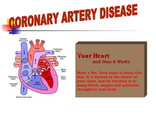
1. CORONARY ARTERY DISEASE Your Heart and How it Works Make a fist. Your heart is about this size. It is located in the center of your chest, and its function is to pump blood, oxygen and nutrients throughout your body.
2. Look at the illustration. As you can see, the heart is divided into four chambers: the two top chambers ( the right atrium and the left atrium) are receiving chambers. The bottom chambers ( the right ventricle and the left ventricle ) are pumping chambers. Cross Section of the Heart Your Heart
3.
4. In your heart, specialized cells carry impulses along a specific pathway in much the same way as electrical wiring carries current. When the impulse completes its course along the pathway, it causes the ventricles to contract, pumping blood through the aorta to the rest the body. Each contraction of the heart begins with a single electrical impulse that is started in the sinoatrial or SA node , located in the upper right atrium of the heart. The SA node is called the heart’s “natural pacemaker” because it begins each impulse and sets the timing between the impulses that cause the heart to contract. When the SA node begins an impulse, the impulse travels along the electrical pathways in the heart’s atria to the atrioventricular or AV node , located between the heart’s atria and ventricles. Here the impulse pauses briefly before continuing down the electrical pathways throughout the ventricles. How the heart knows when to contract Your Heart
5. The pathways in the ventricles are called bundle branches : the right branch stimulates the right ventricle, and the left branch stimulates the left ventricle. When the impulse reaches the end of the these branches in the ventricles, it causes the heart muscle to contract. Each contraction pumps blood through the aorta to the rest of the body. The contraction of the heart produces a pulse (heart beat) that you can feel at specific points on your body. How the heart knows when to (continued) contract Your Heart
6. The SA node normally initiates an electrical impulse 60 to 100 times a minute, resulting in 60 to 100 heart beats per minute. During exercise or when you are excited or stressed, the SA node will initiate more impulses per minute in order to speed up the flow of blood throughout the body. After you relax again, impulse frequency or heart rate should return to a rate of 60 to 100 per minute. An abnormal heart rate can occur if something happens to disturb the regular function of the SA node, AV node or the impulse flow along the pathways. There are a variety of causes for abnormal heart rhythm, and most are easily treatable. If your doctor discovers that you experience abnormal heart rhythm, you will receive more specific information about its cause and your treatment. How the heart knows when to contract (continued) Your Heart
15. Heart valves can be damaged at birth or may become weak and damaged with illness or age. Defective valves may not open or close properly, causing the heart to work harder and less efficiently. If the valve opening is narrower than normal, the condition is know as stenosis . If a valve does not close completely, it is described as insufficient, a condition know as regurgitation. Many factors can damage heart valves, including a heart attack. Valve Disease Your Heart
16. When the heart muscle becomes weak, it cannot pump blood forcefully to all parts of the body. This condition, called congestive heart failure (CHF), can be caused by a heart attack, high blood pressure, valve problems, coronary artery disease or a number of other conditions. Congestive heart failure means that the heart is weakened and cannot pump enough blood to meet the needs of the lungs and body tissues. It does not mean death will soon follow. If appropriate measures are taken CHF can be controlled and you can lead a relatively normal life. Congestive Heart Failure Your Heart
17. Congenital Heart Disease Congenital heart disease is a term used to describe an abnormal heart condition that your were born with. Symptoms related to these abnormalities can appear at any point in a lifetime. Cardiac Rehabilitation Your doctor may order cardiac rehabilitation for you. Inpatient cardiac rehabilitation is an education and monitored exercise program that will prepare you for leaving the hospital. The cardiac rehabilitation nurse will give you and our family instructions, activity guidelines and other important information. She will also work with you to gradually increase your activity level while you are in the hospital by walking the hallways with you and monitoring your heart during these exercise periods. Your Heart
18. Heart Problems If your doctor has warned you about coronary artery disease or atherosclerosis, you’ll want to become familiar with the following information .
19.
20.
21.
22.
23.
24.
25. A heart attack is the result of total blockage in one of the coronary arteries. The blockage has stopped the flow of blood to a section of the heart long enough to cause permanent damage. The damaged heart muscle is weaker and pumps less effectively. Your doctor may also refer to your heart attack as a “coronary”, a “myocardial infarction” or an “MI”. The coronary artery can become blocked, stopping the flow of blood, in several ways: Atherosclerosis Fatty cholesterol deposits build up in the artery to the point where little blood can flow through to nourish the heart . Thrombosis A blood clot forms in an artery already narrowed by fatty buildup, blocking the flow of blood through the vessel. Spasm The coronary artery can spasm or constrict, limiting blood flow. Heart Attack Heart Problems
26. Recognizing a Heart Attack Heart attack symptoms are usually more severe than angina. They last longer, and rest offers no relief from pain. Nitroglycerin tablets will not relieve the pain and symptoms of a heart attack as they do for angina. If your doctor has prescribed nitroglycerin tablets, you should always take them if you have chest pain. If 3 tablets do not relieve your pain, call 011 or have someone drive you to the nearest emergency department. You may be having a heart attack. Heart Problems
27.
28.
29.
30.
31.
32. Heart Attack Treatment . There are many different treatments for heart attack. You and your doctor will decide which of these treatments is best for you depending on your condition Thrombolytic Therapy Heart attacks often occur because a blood clot (thrombosis) has blocked one of your coronary arteries. Doctors can administer clot-dissolving medication to restore blood flow and reduce the amount of damage to the heart. This medication, called a thrombolytic, is given intravenously. Thrombolytic therapy is most effective when given as soon as possible after the onset of symptoms, so it is extremely important to seek immediate medical attention . Medical treatment and risk factor modification Your doctor may prescribe medical therapy and lifestyle changes (risk factor modification) to improve the function of your heart. Many medications are available, and your doctor will work closely with you when deciding on medications. Heart Problems
33. Heart Attack Treatment (cont.) . Angioplasty or atherectomy Your doctor may advise balloon and angioplasty (PTCA) or coronary atherectomy to reduce the blockage in you coronary artery and increase blood flow to that part of your heart. You will receive more information about this procedure if your doctor recommends it for you. Coronary artery bypass surgery In this procedure, your doctor will use a healthy vein or artery from another part of your body – usually your leg – to create a new passage for blood around (or bypassing) the affected area of the coronary artery. You will receive more information about this procedure if your doctor recommends it for you. Before proceeding with any treatment, your doctor will discuss your condition and options with you. Be sure to ask questions and express your concerns at that time. Heart Problems
36. ATRIAL FIBRILLATION (cont.) You can see from a normal EKG that the pattern is regular and even. Following an impulse from the SA node, the upper chambers (atria) of the heart gently contract and pump blood into the more powerful lower chambers (ventricles). Then a stronger contraction in the ventricles pumps the blood into the aorta and throughout the body. When artrial fibrillation occurs, the atria do not contract in an even pattern. Instead, the muscles quiver. Irregular contractions mean the the amount of blood pumped from the heart to the body varies from beat to beat. Artrial fibrillation is a common condition following heart surgery and is usually temporary. If you have rheumatic heart disease, you may have long-term (chronic) atrial fibrillation. You may experience one or several of the following symptoms: a feeling of weakness chest discomfort palpitations light-headedness shortness of breath You may not experience any symptoms at all. Heart Problems
37. ATRIAL FIBRILLATION (cont.) Keep your doctor or nurse fully informed about your symptoms and concerns. Your health care providers may use medication or electrical cardioversion to return your heart to its normal rhythm. Your doctor may also prescribe blood-thinning medications to reduce your risk of stroke, a possible complication of atrial fibrillation. Though millions of Americans experience atrial fibrillation, treatment differs according to each individual’s needs. Heart Problems
38. Premature Ventricular Contractions (PVCs) PVCs are early contractions that start in the heart’s ventricles. They occur both in the healthy people and people with heart disease. They may be harmless or the beginning of more serious arrhythmias. PVCs are common in all age groups. Anxiety, excessive caffeine and certain drugs can cause occasional PVCs even in young adult. However, frequent PVCs indicate the need to search for an underlying cause. PVCs may also occur with any type of heart disease: valvular disease, hypertensive disease, or ischemic heart disease with or without a heart attack. PVCs are the most common arrhythmia to occur with heart attack (acute myocardial infarction) and may lead to ventricular tachycardia and ventricular fibrillation. You may experience one or several of the following symptoms: weakness palpitations chest discomfort light-headedness shortness of breath You may not experience any symptoms at all. Keep your doctor or nurse fully informed about your symptoms and concerns. Treatment of PVCs varies according to each individual’s needs. Your doctor will discuss possible treatment options with you. There are many medications that your doctor may prescribe to prevent frequent PVCs. Heart Problems
39. Serious Arrhythmias Heart rhythm abnormalities that prevent the ventricles from contracting and pumping blood effectively are life-threatening. If you have suffered a life-threatening abnormal heart rhythm, your doctor will order a series of diagnostic tests and will recommend further treatment. If the SA node is damaged for some reason or if there is a blockage in the pathway along which the impulse travels, the doctor may recommend surgery to implant a battery-operated pacemaker. This small machine take over the role of the SA node, the heart’s natural pacemaker, and initiates impulses to the heart at a regular, pre-set rate. If you suffer any type of abnormal heart rhythm, your doctor or nurse will discuss the diagnostic tests and treatment option available to you. Heart Problems
40. Congestive Heart Failure When the heart muscle becomes weak, it cannot pump blood forcefully to all parts of the body. This condition, called congestive heart failure (CHF), can be caused by a heart attack, high blood pressure, valve problems, coronary artery disease or a number of other conditions. Congestive heart failure means that the heart is weakened and cannot pump enough blood the meet the needs of the lungs and body tissues. It does not mean death will soon follow. If appropriate measures are taken CHF can be controlled and you can lead a relatively normal life . Heart Problems
41.
42.
43. Managing Congestive Heart Failure Heart Problems Activity Some activity every day can help you feel better, so don’t let congestive heart failure stop you from being active. On days that you feel good, plan an extra activity in addition to your normal routine. Stop and rest if you feel tired or short of breath. Fatigue and shortness of breath with activity are expected symptoms of heart failure. You should not experience worsening of symptoms such as increasing shortness of breath with activity, shortness of breath while lying down or having to sit up to catch your breath. If you do experience any of these symptoms call your doctor or nurse immediately.
44. Managing Congestive Heart Failure Heart Problems Rest Resting allows your heart muscle to regain its strength. Plan frequent rest periods alternating with your activities throughout the day. Put your feet up for a few minutes every couple of hours to help reduce angle swelling Please refer to the Leaving the Hospital section of this book for further guidelines. Daily weight A sudden weight gain is a sign that the body is holding sodium and water. Weigh yourself daily and record your weight. Get on the scale first thing in the morning after you urinate and before your eat or drink. Call your doctor if you gain 2 to 5 pounds in 1 to 4 days. Rapid weight gains may be a sign that you are retaining fluids and need a change in your treatment plan. To be consistent, weigh yourself with the same amount of clothing each day.
45. Managing Congestive Heart Failure Heart Problems Medications Four general categories of medicine are generally prescribed for congestive heart failure: diuretics, digitalis, ace inhibitors and potassium. Your doctor may prescribe a single medication or a combination for you. Here is a general explanation of the medications and their possible side effects. Ask your doctor or nurse if you have questions about how to take your medications.
46.
47.
48.
49.
50. Nutrition for Congestive Heart Failure Patients Heart Problems Sodium is a mineral needed by the body in small amounts to function properly. However, most Americans consume significant more sodium than the body needs. Excess sodium can build up in the body causing increasing blood pressure, fluid retention (edema), weight gain and shortness of breath. Most of the sodium, which is preferred as “salt” comes from salt added to food during or after cooking and in processing. Food preservatives, condiments and other seasonings are other high sources of sodium. One teaspoon of salt contains 2300 milligrams (mg) of sodium. It is recommended that your diet be limited to 2000 milligrams of sodium daily. Do not trust your sense of taste in choosing foods to eat as there may be more sodium in the food than you realize. It will be very important to read and check food labels. The dietitian will be available to help you and your family make positive food choices. Some people use salt substitute which does not contain sodium. The sodium replaced with potassium and may not be safe for everyone. Before using a salt substitute, contact your doctor or dietitian.
51.
52. Diagnostic Tests History And Physical Exam Understanding your symptoms and knowing your health history will help your doctor diagnose and treat your disease. It’s important that you answer questions as completely and clearly as you can. You can expect the doctor to ask specific questions about your daily habits, previous diseases and family health history. This information determine what other diagnostic test your doctor will order. Chest X-ray Your doctor may order a chest X-ray to provide information about the size of your heart, its position and the condition of your lungs.
53. Diagnostic Tests Electrocardiogram (EKG or ECG The EKG records the heart’s electrical activity and detects abnormal heartbeats (rhythms). It may even show a heart attack in progress An EKG technician will position small sensors, called electrodes, on your chest, arms and legs. You will not experience any discomfort. The test takes about 5 minutes and can be performed in your room. Your doctor may ask to have EKG’s repeat at intervals to show the changes that may occur as you recover.
54. Diagnostic Tests Laboratory Test Your doctor will probably order a series of blood tests to help diagnose your heart disease: A cardiac enzyme count will help determine if the heart muscle has been damaged. This test may be repeated several times during your hospital stay. Cholesterol levels, triglyceride levels and glucose (blood sugar) levels can help identify your risk factor for heart disease. Blood sample will be taken several times during your stay in the hospital.
55.
56.
57. Diagnostic Tests Nuclear Cardiology Your doctor may order a cardiac stress test using Thallium 201 or Cardiolite. These tests are similar to the treadmill test with the addition of an IV injection of a radioactive isotope that will allow us to take clear images of your heart after exercise and at rest. Your doctor will determine which of the following tests you should have.
58. Diagnostic Tests Nuclear Cardiology Thallium Cardiac Stress Test You will not be allowed to eat before the test. Certain medications may be held until after you’ve completed the test. If you don’t already have an IV, one will be started in your arm prior to the test. You will exercise on the treadmill. Just as you reach a predetermined heart rate, the isotope will be injected through the IV. Thallium is a weak radioactive isotope used in very limited quantities to “trace” cardiac abnormalities. After the treadmill portion of the test is completed, you will be taken to the nuclear medicine department for a scan. Scanning takes only about 30 minutes. You will return in 3 to 4 hours for another series of images of your heart at rest. You will not be allowed to eat between these tests. Your doctor may order another series of scans after 24 hours. (You may eat during this 24-hour period). Your doctor will share the results of the test with you later.
59. Diagnostic Tests Cardiolite Cardiac Stress Test You will not be allowed to eat before the test. Certain medications may be held until after you’ve completed the test. If you don’t already have an IV, one will be started in your arm prior to the test. This test has two parts: a resting portion and an exercise portion. For each portion you will receive Cardiolite through an IV and have and image scan. For the resting portion, you will receive an IV injection, and an hour later you will have your first scan. For the exercise portion, you will walk on a treadmill. Just as you reach a predetermined heart rate, the isotope will be injected. After completion of exercise, you will be instructed to eat a fatty meal and return in an hour for another scan. Cardiolite is a weak radioactive isotope given in very limited quantities to “trace” cardiac abnormalities. Your doctor will share the results of the test with you later.
60.
61. Diagnostic Tests Cardiac Catheterization Cardiac catheterization (also called coronary angiography or heart catheterization) provides more detailed information about the function of the heart and its arteries than can other diagnostic tests. No other test can provide this information. By combining the information from other tests, catheterization with information from other tests, your doctor can most accurately diagnose your condition and choose the most effective treatment. A cardiologist in the cardiac catheterization lab (cath lab) will perform the test. It involves inserting a thin plastic tube (catheter into a large blood vessel in your groin or arm, the guiding it to the heart to monitor pressures and take pictures . The cardiologist will also inject a contrast material into the coronary arteries and take special X-rays to determine if blockages or any other problems exist.
62. Diagnostic Tests Cardiac Catheterization Before the test A nurse or your doctor will explain the test and ask you to sign consent forms. Do not eat or drink for 6 to 8 hours before the test. You may have blood drawn and a chest X-ray taken. The nurse will insert an IV in your arm to allow medications and/or fluids to be given before and/or during the test. Medications to help you relax may be given by mouth or through the IV before or during the test.
63. Diagnostic Tests Cardiac Catheterization During the test The entire test, including transport to the cath lab, takes about 2 hours. Your family may accompany you to the cath lab waiting area. You will be taken to a special room containing X-ray equipment and heart monitoring devices. You will be asked to lie flat on the X-ray table and your body will be covered with a sterile sheet. Please keep your hands under this sheet. Your heart rate and blood pressure will be monitored continuously during the test. The area chosen for the catheter insertion (your groin our arm) will be shaved and washed with and antiseptic solution. continued
64. Diagnostic Tests Cardiac Catheterization During the test (cont.) The cardiologist will give you a local anesthetic to numb the insertion area. The area will sting for a few moments, then become numb. The doctor will place plastic “introducer” sheats through your skin at the insertion sites to allow access to the artery. If you have any discomfort in the area of insertion or any chest pain, tell the doctor . Additional medication can be given through your IV to ease any discomfort. Catheters will be used to inject a contrast agent into the coronary arteries and left ventricle. During the test, you will be asked to hold your breath momentarily to ensure clear pictures. You may be asked to cough. continued
65. Diagnostic Tests Cardiac Catheterization During the test (cont.) You may feel a warm sensation (flush), nausea or have the urge to urinate when the contrast material is injected to the heart chamber. These sensations pass quickly. After pictures have been taken, the doctor will remove the catheter. The sheaths may also be removed and pressure will be applied over the insertion sites for 15 to 20 minutes to prevent bleeding. The nurse will then apply a pressure bandage. Or the sheaths may remain in place until you and your doctor make a decision about your treatment.
66. Diagnostic Tests Cardiac Catheterization After the test You will return to your room to a special monitoring area, and your family may visit. The nurse will frequently check your blood pressure, pulse and the site where the catheter was inserted for bleeding. You will be asked to lie flat and not bend your arm or leg for several hours following the test. To relieve stiffness, you may move your ankle and wiggle your toes. The head of your bed may be raised slightly. You will be able to bend your other arm or leg. You will be able to eat shortly after returning to your room. Drink plenty of fluids to flush to contrast material from your body.
67. Diagnostic Tests Cardiac Catherization After the test (cont.) If you notice any bleeding or a warm, wet feeling at an insertion site, your should apply pressure to the area and call the nurse immediately. If you have any chest, arm or neck discomfort, nausea, numbness or tingling in your leg or foot, call the nurse immediately. After the period of bed rest, you will be permitted to get out of bed and walk with nurse’s help. Do not bend over, strain or lift heavy objects (10 pounds or more) for the next 24 hours. Avoid climbing stairs. If you have to sneeze or cough, place your fingers over the bandage and hold it firmly. Your doctor will discuss the findings with you soon after the test.
68. Diagnostic Tests Cardiac Catherization Outpatient Instructions If you have the test as an outpatient, you may be able to go home in 6 to 8 hours, depending on your doctor’s instruction. You will be discharged from the hospital in a wheelchair. You will need to have a friend or relative drive you home and stay with you for the next 8 to 12 hours. During the first 8 to 12 hours, limit you activity. Wear loose clothing and stay in bed, on the sofa or in a recliner with your feet up. If the catheter was inserted in your arm for the test, you should stretch and bend the arm, but avoid strenuous movement. Do not bend over, strain or lift heavy objects (10 pounds or more) for the next 24 hours. Avoid climbing stairs. If you have to sneeze or cough, place your fingers over the bandage and hold it firmly.
69. Diagnostic Tests Outpatient Instructions (cont.) If you notice any bleeding (bright red blood from the dressing, or severe swelling, place your fingers over the site and press firmly to apply pressure. Have your helper call EMS (911 in the Phoenix area) for immediate help. Write your Emergency Response number here: Phone #: ___________________________________________ Cardiac Catheterization
70. Diagnostic Tests Cardiac Catherization Outpatient Instructions (cont.) If you experience severe pain, call your doctor immediately . Do not go for long walks or lift heavy objects for 3 days following the test. Leave the pressure bandage on your groin or arm until the next day. As your doctor when it will be OK to take a shower . Removing the bandage can be painful. It is best to remove it in the shower. Soak the area thoroughly using soap. Gently peel the bandage away from the skin a little at a time . A black and blue bruise or a bump under the skin may form at the catheter insertion site. This is normal, and the bruise or bump should disappear within 2 to 3 weeks. Ask your doctor when you can return to your normal activities and whether there are any specific restrictions. Usually people are able resume normal activities a few days after the test. Make an appointment to see your doctor for a follow-up visit.
71. Diagnostic Tests Cardiac Catheterization Outpatient Instructions (cont.) If you experience severe pain, call your doctor immediately . Do not go for long walks or lift heavy objects for 3 days following the test. Leave the pressure bandage on your groin or arm until the next day. As your doctor when it will be OK to take a shower . Removing the bandage can be painful. It is best to remove it in the shower. Soak the area thoroughly using soap. Gently peel the bandage away from the skin a little at a time . A black and blue bruise or a bump under the skin may form at the catheter insertion site. This is normal, and the bruise or bump should disappear within 2 to 3 weeks. Ask your doctor when you can return to your normal activities and whether there are any specific restrictions. Usually people are able resume normal activities a few days after the test. Make an appointment to see your doctor for a follow-up visit.
72.
73.
74. Medical Treatment of Valve Disease (cont.) Open Heart Surgery Open heart surgery refers to any surgery during which a heart-lung bypass machine temporarily replaces the normal function of the heart and lungs. During the surgery, the blood is diverted from the heart to the machine which performs the same function as the heart and lungs. The most common open heart procedures are coronary artery bypass and valve replacement or repair surgery.
75. Medical Treatment of Valve Disease (cont.) Coronary Artery Bypass Graft Surgery (CABG
76. Medical Treatment of Valve Disease (cont.) Coronary Artery Bypass Graft Surgery (CABG Coronary artery bypass graft surgery is the most frequently performed heart operation today. There are many reasons why it is recommended, but the goal in each case is the same – to route the blood around the blockage in the artery or arteries.
77. Medical Treatment of Valve Disease (cont.) Coronary Artery Bypass Graft Surgery (CABG
78. Medical Treatment of Valve Disease (cont.) Coronary Artery Bypass Graft Surgery (CABG
79. Medical Treatment of Valve Disease (cont.) Coronary Artery Bypass Graft Surgery (CABG Reasons for surgery Life-threatening coronary artery blockage Complications following a heart attack Angina that can no longer be controlled by medication. Goal Coronary artery bypass surgery provides a new route for blood to flow around, or bypass, the blockage in the coronary artery. The surgeon will use a healthy vessel from your leg or chest wall to supply blood to your heart muscle. One end of this blood vessel is attached to the aorta and the other end to the coronary artery beyond the point of blockage. The number of bypasses needed and the amount of heart muscle affected. The vessel most often used for replacement is the saphenous vein which is located in the inner part of the leg. After the vein is removed, your body will develop new vessels in the area.
80. Medical Treatment of Valve Disease (cont.) Coronary Artery Bypass Graft Surgery (CABG During the surgery When you arrive in the operating room, the surgical staff will move you to the operating table, and the anesthesiologist will administer a sedative (anesthesia) to put you to sleep. A number of tubes and drains need to be inserted during surgery: A breathing tube and respirator (breathing machine) will ensure that you breathe regularly during surgery and until you are fully awake. A nasogastric (NG) tube from your nose to your stomach will empty stomach secretions. A catheter will be inserted into the natural opening through which you urinate. This catheter will keep the bladder empty and allows us to measure your urine output exactly. An IV will be inserted into a major vein in your neck. During and after surgery, monitoring equipment attached to this line will help the doctors and nurses monitor the pressures within your heart. The line will also provide IV fluid and medications.
81. Medical Treatment of Valve Disease (cont.) Coronary Artery Bypass Graft Surgery (CABG During the surgery (cont.) A chest tube (or tubes) will drain the chest cavity of normal post-operative bleeding. A temporary pacing wire may be placed in your heart to increase your heart rate if necessary. When the anesthesiologist is sure that you are asleep, the surgeon will make an incision down the center of your chest. Then he will cut down the center of the breast bone (sternum) and spread it apart, allowing access the your heart. The surgeon may take the internal mammary artery from your chest or a long vein from your leg ( the saphenous vein) to use as the bypass for your coronary artery. If the surgeon uses the saphenous vein, you will have an incision on the inside of your leg. One end of this vessel will be grafted (sewn) to the major artery leaving your heart (the aorta) and the other end to the coronary artery below the blockage. When the surgeon is sure that the bypass is secure, the surgical team will check that blood flows through it correctly, the secure your breast bone with surgical wires and suture the chest incision.
82. Medical Treatment of Valve Disease (cont.) Coronary Artery Bypass Graft Surgery (CABG The length of time you will be in surgery depends on the number of bypasses you need. During the surgery
83. Medical Treatment of Valve Disease (cont.) Coronary Artery Bypass Graft Surgery (CABG
84. Medical Treatment of Valve Disease (cont.) Coronary Artery Bypass Graft Surgery (CABG