

Recomendados
Mais conteúdo relacionado
Mais procurados
Mais procurados (20)
Destaque
Destaque (20)
Semelhante a Leucocytozoon & Hepatozoon
Semelhante a Leucocytozoon & Hepatozoon (20)
Mais de Osama Zahid
Mais de Osama Zahid (20)
Último
Último (20)
Leucocytozoon & Hepatozoon
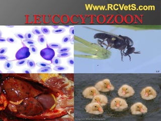
- 2. Introduction A protozoan parasite of avian erythrocytes transmitted by the 'blackfly' Simulium spp. Infection causes the disease leucocytozoonosis. The parasites were first seen by Danilewsky in 1884 in blood from an owl. There are over 100 species in this genus.
- 4. Major Species There are over 100 species in this genus, but the 3 most important species that’ve infected the most common domestic birds are; Leucocytozoon caulleryi(chickens) Leucocytozoon smithi(turkeys) Leucocytozoon simondi(ducks and geese)
- 5. Geographical Distribution In Thailand, India, Taiwan, Japan, Burma, Sri Lanka, the Philippines, Singapore, Malaysia, Indonesia, China, and Korea, USA, Canada and Africa most cases have been reported. Acute outbreaks of leucocytozoonosis have been reported in chickens, turkeys, waterfowl and wild birds worldwide.
- 6. Hosts & Vectors Definitive host Ducks Turkey Geese Swans Similar waterfowl and Chickens Vector Black Fly(Simulium spp)
- 7. Leucocytozoon use Black flies (Simulium species) as their intermediate host and Birds as their definitive host. Over 100 species of birds have been recorded as hosts to these parasites. Leucocytozoon does not threaten human populations in terms of potential infection, infected poultry is not pathogenic in humans. Still, the parasite's economic impact could hurt poultry farmers' revenue as mortality rates are extremely high, especially among young birds
- 8. Transmission in Host Leucocytozoon's involves an intermediate host (the blackfly) which carries the parasite from one avian host to another. When the blackfly vector bites a bird, perhaps around the unfeathered eye area, sporozoites are released in the vector's saliva and into the bird's circulatory system.
- 10. Life Cycle It completes it’s life cycle in Two different hosts i;e Intermediate host(black fly) and Definitive host(birds) In Black fly, it enters in it’s body when the fly feeds on the bird already been infected by the parasite. After penetrating in the fly it’s gametophytes get mature and reproduce in Midgut.
- 11. In the insect vector these elongated gametocytes become either a female (macro)gametocyte with a red-staining nucleus Male (micro)gametocyte with a palestaining diffuse nucleus, which come together to form an ookinete. This ookinete invades an intestinal cell of the fly vector, where it matures into an oocyst. This oocyst produces sporozoites which migrate to the salivary glands of the blackfly, thus reinitiating the cycle.
- 12. In Birds, sporozoites invade the liver cells and develop into small schizonts, which in turn produce merozoites. Schizonts release merozoites or secondary schizonts in the endothelial cells of the blood vessels and in essentially any of the viscera. The merozoites either enter red blood cells or macrophages. In the red blood cells, merozoites develop into round gametocytes. In the macrophage, merozoites develop into megaloschizonts, which then divide into primary cytomeres.
- 14. Pathogenesis Five days post infection: Many schizonts develop in hepatocytes Cells rupture Seven days post infection: Megaloschizonts begin to appear in spleen Also appear in Lymph and other tissues Gametocytes accumulate in liver 12+ days post infection Hemorrhagic scars from rupturing megaloschizonts
- 15. Area of Infection Leucocytozoon’s infect the Heart Liver Lungs Spleen Brain
- 16. Diagnosis A diagnosis can be made by The demonstration of gametocytes in blood smears. Histopathological examination of the liver, spleen and brain can show developing megaloschizonts. Necropsy may reveal an enlarged spleen and liver. Since the majority of birds are sub clinically infected with Leucocytozoon, other causes of death must be ruled out even with the presence of Leucocytozoon gametocytes in peripheral blood smears.
- 18. Clinical signs/symptoms The majority of birds affected with leucocytozoonosis exhibit no clinical signs. Visibly affected show mild to severe signs of anorexia, luekocytosis, weakness, anemia, emaciation, and have difficult breathing. Young birds manifest in appetence, weakness, dyspnea, and sometimes death within 24hrs.
- 19. Signs in adults appear less abruptly and consist of listlessness and a low mortality rate. The mortality in adult birds occurs as a result of debilitation and increased susceptibility to secondary infection. Granulomatous and lymphocytic lesions are seen in the lungs, heart, brain and peripheral nerves. Large gametocytes can block capillaries of the lungs.
- 21. Treatment and Control Treatment usually is not effective, control of the Leucocytozoon in domestic avian species has, until recently, been limited to control of the blackfly vector. Preventive medication using Pyrimethamine(1 ppm) and Sulfadimethoxine(10 ppm) combined in the feed controls it.
- 22. Clopidol (0.0125%–0.025%) controls invertebrate vectors are helpful. Quinacrine hydrochloride or Trimethoprim/Sulfamethoxazole solution have been used i;e parasitemia is reduced, but the infection is not cleared. Oral Anti-LEUCOCYTOZOONOSIS Vaccines based on TRANSGENIC plants.
- 23. Hepatozoon
- 24. Introduction Hepatozoon is a protozoan parasite of peripheral blood neutrophils. It’s genus Apicomplexan protozoa which incorporates over 300 species obligate intra erythrocytic parasites. Infection caused by hepatozoon is called Hepatozoonosis. A Hepatozoon parasite was initially reported from a cat in India in 1908.
- 26. Geographical Distribution It’s widely distributed in the world mainly found in; Africa Southwestern Asia Southwestern Eastern Europe North and South America. Brazil United States
- 27. Hosts and Vectors Dogs Cats Rabbits Rats Snakes And other domestic animals Vectors Ticks
- 28. Major Species Hepatozoon has more than 300 species but the most imp. Of them which cause infection is; Hepatozoon canis.(dogs, jackals and all canines) Hepatozoon atticorae(birds) And very recently discovered Hepatozoon sipedon.(infect Snakes and mosquitoes)
- 29. Transmission in Host Transmission of the infection occurs via ingestion of infected ticks. Dogs are infected by ingesting infected ticks during grooming behavior or eating prey that has attached ticks. It’s not transmitted by Tick biting. In Snakes, it is transmitted through infected mosquitoes that are ingested by some frogs that later on, eaten by the snake transmitting it. Mosquitoes get infected by feeding on infected snakes.
- 31. Life Cycle Members of the genus Hepatozoon possess particularly complex life cycles which vary considerably among species. Sexual reproduction and sporogenic development occur in haemocoel of the invertebrate host. It also has two hosts; Intermediate(ticks)&Definitive(Dogs/snakes)
- 32. Vertebrate host, consumes the infected invertebrate. The sporozoites then migrate to the liver of the vertebrate, where they undergo multiple fission (asexual reproduction) to produce merozoites. Merozoites are released into the bloodstream where they form gametocytes. Gamonts are large, conspicuous organisms which occupy significant portion of the erythrocyte, and are easily visible on simple blood films.
- 33. The invertebrate vector feeds on the blood of the infected vertebrate The gamonts are taken up into the gut once more, where they undergo gametogenesis and the cycle begins once more. In case of snakes, they do not typically feed on mosquitoes, a third, intermediate host is required, in this case a frog. The frog ingests the infected mosquito, and the snake acquires the infection by feeding on the now infected frog. Another mosquito can then feed on the snake, thus continuing the life cycle.
- 35. Diagnosis Is based on, Blood tests Clinical signs Biopsy of skeletal muscles Serological tests Molecular diagnosis
- 36. Clinical Signs & Symptoms Periodic or persistent fever Weakness muscle atrophy Generalized pain Stiff gait and hind limb paralysis. Pus-filled eye discharge.
- 37. Treatment & Control The current therapy for hepatozoonosis includes; Trimethoprim Sulfadiazine Clindamycin Pyrimethamine (TCP) drugs for two weeks to destroy the parasites. Www.RCVetS.com