epistaxis
•Transferir como DOCX, PDF•
9 gostaram•2,705 visualizações

The document summarizes the causes, evaluation, and management of epistaxis (nosebleeds). Key points include: - The most common causes are local trauma, dry air, and hypertension. Weather has a proven association with incidence. - Evaluation involves examination of the nasal cavity to identify the bleeding site. Imaging and labs may be needed to investigate underlying causes. - Management begins with first aid and may involve cautery, nasal packing, medications, or surgery to control active bleeding and address its cause. Continued or severe bleeding may require embolization or ligation of arteries supplying the nasal cavity.

Recomendados
Mais conteúdo relacionado
Mais procurados
Mais procurados (20)
Destaque
Destaque (20)
Semelhante a epistaxis
Semelhante a epistaxis (20)
Último
Último (20)
epistaxis
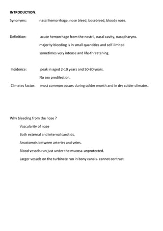
- 1. INTRODUCTION: Synonyms: nasal hemorrhage, nose bleed, bosebleed, bloody nose. Definition: acute hemorrhage from the nostril, nasal cavity, nasopharynx. majority bleeding is in small quantities and self-limited sometimes very intense and life-threatening. Incidence: peak in aged 2-10 years and 50-80 years. No sex predilection. Climates factor: most common occurs during colder month and in dry colder climates. Why bleeding from the nose ? Vascularity of nose Both external and internal carotids. Anastomsis between arteries and veins. Blood vessels run just under the mucosa-unprotected. Larger vessels on the turbinate run in bony canals- cannot contract
- 2. Historical aspect: Carl Michel (1871), James Little (1879) , and Wilhelm Kiesselbach First to identify nasal septum anterior plexus Pilz : (1869) first to surgically treat epistaxis Ligation of common carotid artery. Seiffert: (1928) via maxillary sinus ligated internal maxillary artery Henry Goodyear: First anterior ethmoid artery ligation. Hippocratic technique : pinching the ala nasi Vascular anatomy of nasal cavity: Respiratory mucosa with its underlying vascular supply serve to regulate heat exchange and humidification during respiration.
- 3. Blood supply: 1.External Carotid Artery -Sphenopalatine artery -Greater palatine artery -Ascending pharyngeal artery -Posterior nasal artery -Superior Labial artery 2.Internal Carotid Artery -Anterior Ethmoid artery -Posterior Ethmoid artery Arteries intercommunicate in rich plexuses Kisselbach,s plexus(anterior bleed) Woodruff,s plexus(posterior bleed)
- 4. Kesselbach’s Plexus/Little’s Area: 1/2 inch from the caudal border of the septum antero-inferiorly. Vessels anastomosing are -Anterior Ethmoid (Opth) -Superior Labial A (Facial) -Sphenopalatine A (IMAX) -Greater Palatine (IMAX) Bleeding may be arterial or venous. Commonest site of bleeding Exposed to drying of inspiratory current & finger nail trauma
- 5. Woodruff’s Plexus: Lying just inferior to posterior end of inferior turbinate Pharyngeal & Post. Nasal AA of Sphenopalatine A (IMAX) ? venous plexu
- 6. Retrocolluellar vein : Run vertically downwards just behind the collumella Crosses floor of nose and joins venous plexus on lateral nasal wall. Common site of venous bleeding in young people Classification of Epistaxis According to Age: Childhood <16 years Adult >16 years Common in childhood, less common in early adult life,peaks in 6th decade According to causal factor: Primary no proven causal factor. Secondary proven causal factor According to area: Anterior bleeding point anterior to piriform aperture Posterior bleeding point posterior to piriform aperture Anterior epistaxis Posterior epistaxis Incidence More common Less common Site Mostly from little,s area Posteriosuperior part of nasal cavity Age In children and young adults After 40 yrs of age Cause Mostly trauma Spontaneous(HTN,arteriosclerosis) Bleeding Mild,ctrl by local pressure or anterior Severe,hospitalization,post nasal pack pack often required
- 7. Etiology of epistaxis Local General Idiopathic Local causes Congenital Hereditary telengectesia (osler- weber – rendu syndrome) Trauma Microtrauma by nose picking Facial and skull bone fractures Foreign body in nose Iatrogenic trauma Barotraumas Inflammatory Infective rhinitis Specific Acute infection life diphtheria Chronic granulomatous conditions Tuberculosis leprosy syphilis rhinosporidiosis Rhinoscleroma Wegener,s granulomatous
- 8. Non specific Viral –common cold ,influenza Bacterial-Secondoary bacterial rhinitis sinusitis Fungal rhinosinusitis Atrophic rhinitis Neoplastic Benign Juvenile angiofibroma,angioma of septum,capillary and cavernous hemangioma,inverted papilloma Malignant Squamous cell carcinoma,olfactory neuroblastoma, nasopharyngeal carcinoma Miscellaneous causes Deviated nasal septum and spur Rhinitis sicca Spontaneous rupture of tortuous arteriosclerotic vessels Rhinolith
- 9. Physiological causes High altitude Extreme cold or hot climate Systemic causes Hypertension Cardiac – CCF,mitral stenosis Pulmonary – COPD Cirrhosis – vit K deficiency Renal - nephritis Hormonal – vicarious menstruation,endometiosis,granuloma gravidarum Coagulopathies Clotting disorders like Christmas diseases Von willebrand diseases ,hemophilia Bleeding disorders like thrombocytopenic purpura Agranulocytosis Leukemia Vit K deficiency Exanthematous fevers like measles,mumps,typhoid Idiopathic No obvious cause detected clinically and after investigations Summary of etiology evidence : Factor Weather proven association NSAID proven association Alcohol proven association Hypertension no association Septal deviation no association
- 10. Evaluation of epistaxis: Priority to ctrl bleed before invest FIRST AID ABC of emergency management is followed (Airway ,Breathing and Circulation). Make pt. sit up , pinch the nose for 5-10 min,open mouth and breath Ice pack on nose Sedatory /sublingual antihtn in case of hypertensive epistaxis In profuse bleed, aspiration is prevented by (#facial bones) lateral position/intubation with inflated cuff. Injection or topical use of hemocoagulase Vital sign regularly monitored and concerntration is given on the following: Volume status BP Adequacy of airway Oral and nasal examination Detailed medical and treatment history simultaneously with bleeding ctrl
- 11. Patient history Physical examination equipments • Protective equipment - gloves, safety goggles • Headlight if available • Nasal Speculum • Suction with Frazier tip • Bayonet forceps • Tongue depressor • Vasoconstricting agent (such as oxymetazoline) • Topical anesthetic Physical examination: Anterior rhinoscopy Posterior rhinoscopy Nasal endoscopy Radiological evaluation Xray PNS r/o infective, traumatic and neoplastic condition CT scan Digital subtraction angiography –identification of bleeding vessel
- 12. Hematological investigations CBC with platelet count Clotting and bleeding profiles Blood grp and cross matching
- 13. Management of epistaxiS EPISTAXIS ASSESSEMENT & FIRST AID /RESUSCITATION NASAL PREPARATION IDENTIFY SITE OF BLEEDING ANTERIOR NOT IDENTIFIED POSTERIOR CAUTERY (chemical,electocautery, endoscopiccwutery Antibiotic anterior nasal pack ANP+PNP ointment FOLEYS WITH TAMPONADE WITH ANP Ctl unctrl CTRL UNCTRL UNCTRL CTRL Consider bld transfusion VESSEL LIGATION LOW BLEEDING UIDENTIFIED HIGH BLEEDING SITE SITE IMA LIGATION/ ANT & POST ETHMOIDAL TESPAL LIGATION CTRL UNCTRL CTRL ARTERIOGRAPHY & EMBOLISATION
- 14. Preventive measures: • Keep allergic rhinitis under control. Use saline nasal spray frequently to cleanse and moisturize the nose. • Avoid forceful nose blowing • Avoid digital manipulation of the nose with fingers or other objects • Use saline-based gel intranasally for mucosal dryness • Consider using a humidifier in the bedroom • Keep vasoconstricting spray at home to use only prn epistaxis Direct therapy Silver nitrate cautery - avoid cautery of bilateral nasal septum as this may lead to necrosis and perforation of the septum Collagen Absorbable Hemostat or other topical coagulant Endoscopic control – enables targetted hemostasis using insulated hot wire cautery or modern single fibre bipolar electrodes. Monopolar diathermy should not be used in nasal cavity –blindness Indirect therapies Nasal packing Anterior nasal packing for refractory epistaxis - may use expandable sponge packing or gauze packing Posterior nasal packs
- 15. Usually, 1/2 inch Iodiform or NuGauze is used. Coat the gauze with a topical antibiotic ointment prior to placement Formed expandable sponges are very effective Available in many shapes, sizes and some are impregnated with antibacterial properties
- 16. Correct direction for placement of nasal packing Actual duration will vary according to the patient’s particular needs. Typically, anterior pack at least 24-48 hours, sometimes longer. Best to place patient on a p.o. antibiotic to decrease risk of sinusitis and Toxic Shock Syndrome Advise pt to avoid straining, bending forward or removing packing early If other nostril is unpacked, advise topical saline spray and saline gel to moisturize nasal mucosa Admission may also be prudent for those with CAD, severe HTN or significant anemia. Give supplemental oxygen via humidified face tent. Hot water irrigation - reflex vasodilation and reduction in nasal lumen dimension Cold water irrigation Systemic medical therapy
- 17. Surgical management Continued epistaxis consists of: Posterior packing Ligation techniques Septal surgery techniques Embolisation techniques Posterior nasal packs
- 18. • Always test before placing in patient • Fill “balloons” with water, not air • Orient in direction shown • Fill posterior balloon first, then anterior • Document volumes used to fill balloons Posterior pack- rolling 4 *4 inch gauze sponge into A 1 inch dameter pack secured with 3 heavy silk suture 10 french Foleys used
- 19. Potential complications of PNP Hypovolemic shock Naso-vagal reflux Hypoxia Hypoventilation Respiratory obstruction Local infection Bacteremia TSS Obstuctve sleep apnoea Cardiac arrhythmia Indications for surgery/embolization Continued bleeding despite nasal packing Pt requires transfusion/admit hct of <38% (barlow) Nasal anomaly precluding packing Patient refusal/intolerance of packing Posterior bleed vs. failed medical mgmt after >72hrs Selective Angiography/embolization Helps identify location of bleeding Embolization most effective in patients who Still bleeding after surgical arterial ligation Bleeding site difficult to reach surgically Comorbidities prohibit general anesthetic Effective only when bleeding is >.5 ml/min 90+% success rate, complication rate of 0.1% Only able to embolize external carotid & branches Complications: minor (18-45%)/major (0-2%) Contraindicated in bad atherosclerosis, Ethmoid bleed
- 20. Ligation techniques Transantral IMA ligation Intraoral IMA ligation Anterior/Posterior Ethmoidal ligation Transnasal Sphenopalatine ligation External carotid artery ligation Septodermoplasty/Laser ablation Transatral IMA ligation(SEIFFERT,s operation) Under LA/GA After Caldwell-Luc maxillary antrotomy,posterior maxillary sinus wall identified & removed Periosteum opened via cruciate incision IMA & its 3 major branches explored Vascular clips applied to IMA , Recurrence rate (failure rate) of 10-15% Complication rate of 25-30% -sinusitis , damage to infraorbital nerve,oroantral fistula, dental damage , anaesthesia , rare opthalmoplegia, blindness
- 23. Intraoral IMA ligation Posterior gingivobuccal incision beginning at second molar Temporalis mm split and partially dissected IMAX visualized, clipped and divided Advantages: children/facial fractures Disadvantages: more proximal ligation Complications: trismus, damage to infraorbital n Ant./Post. Ethmoidal ligation Patients s/p IMAX ligation still bleeding, superior nasal cavity epistaxis, or in conjunction when source unclear Lynch incision Fronto-ethmoid suture line
- 24. Transnasal Endoscopic Sphenopalatine Artery ligation Follow Middle Turbinate to posteriormost aspect Vertical mucoperiosteal incision 7-8mm anterior to post middle turb (between mid. and inf. turbs) Elevation of flap—ID neurovascular bundle at foramen Ligation with titanium clip Reapproximate flap Complications –few, Failures—0-13% ECA ligation Effectiveness Anterior border of SCM ID ECA/ICA Ligation after clear that surrounding structures are safe. Septodermoplasty/Laser Remove mucosa from anterior ½ septum, floor of nose, lateral wall STSG vs. cutaneous, myocutaneous, microvascular free flaps vs. Autografts Neodymium-yttrium-garnet (Nd-YAG) laser or Argon laser + topical steroid best nonsurg rx for mild/mod disease Still bleed, but not as bad Definitive treatment (severe disease)—closure of nose
- 25. Special cases: Hereditary hemorrhagic telangiectasia Also called Rendu-Osler-Weber diseaseargon Autosomal dominant Affecting blood vessels in skin , mucous membranes and viscera. Genetic abnormality located to chromosome 9q(HHT1) & 12q(HHT2). Features telengiactias,av malformationsand aneurysms.
- 26. Recurrent epistaxis in HHT No bld transfusions bld transfusion Mild moderate severe Septodermoplasty hormones antifibrinolytics agents arterial ligation selective embolisation Coagulating laser Nasal closure eg.argon
- 27. Haemophilia: – Replace factor VIII, or fresh blood. Other clotting deficiency: – FFP. Purpura: -Platelets Anticoagulants: – Stop drug, or titrate. – Heparin is reversed with protamine sulphate, – warfarin with vitamin K Unconscious head injury; – Dangerous to pack in suspected skull #.