Life
•Transferir como PPSX, PDF•
1 gostou•611 visualizações

This slide collection is intended as illustrations to the (work in progress) book "Life? The Universe! Almost Everything..." Some animations require PowerPoint 7.

Recomendados
Recomendados
Mais conteúdo relacionado
Semelhante a Life
Semelhante a Life (20)
Último
Último (20)
Life
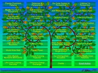
- 1. Popular Prominent Distances In Our Solar System In “Lifelines” In Patterns Perspectyive Perspective Perspective Really Tiny Stuff in Cell Membranes: ATP Production Sites Cell Power Plant Perspective Living Walls The Ultimate Life’s Leading Capturing Energy Liberating Energy Building Blocks Ladder No Waste: Electron Transport Electron & Proton The World’s Smallest Recycling “Staircase” Translocation Motor ATP Generator Cytoskeleton: Cytoskeleton: Cytoskeleton: Myosin (Principles) Collagen Microtubules Filaments Cytoskeleton: Skeletal Muscle Mighty Muscle Marvelous Molecular Actin Filaments Structure Movement Motors Light and Dark Huffing, Puffing… From Nuclear Energy Mind Over Matter Reactions to New Clear Energy World Wide Web Flower Power Gaian Symbiosis or Exercise? Single Entity? More Recycling! DNA: Digitally Forever Fanning Our Distingushed Some Really Close Numbered Aminoacids Features Ancestors Relatives Life Loves Looping- Electronic Energy Credits Email Author the-Loop Exchange Copyright © 2011Henry Norman
- 2. Prominent Patterns… Brain Cell Tree at Sunset Lena River Delta Courtesy Alan Opsahl, Pfizer Courtesy Kevin Schofelt (goo.gl/P0aIb) Courtesy NASA (Landsat 7) Colorado River Delta Fan Coral Purkinje Saguaro Courtesy Google Earth (Digital Globe) Courtesy Denis in Minneapolis (lucky@usfamily.net) Courtesy Anita Gould (Flickr)
- 3. Distances in Perspective… Scanning Electron Microscope Range Optical (Light) Microscope Range 10-11 10-10 10-9 10-8 10-7 10-6 10-5 10-4 10-3 10-2 10-1 100 10 pm 1Å 1 nm 10 nm 100 nm 1 µm 10 µm 100 µm 1 mm 1 cm 1 dm 1m Human Eye Range Atom Lipid Virus Bacterium Cell Human Nucleon Molecule Protein Mitochondrion 10-14 m Scale An atom is to one meter what one meter is to the Earth-Sun distance! Human Eye Range 100 101 102 103 104 105 106 107 108 109 1010 1011 1m 10 m 100 m 1 km 10 km 100 km 1 Mm 10 Mm 100 Mm 1 Gm 10 Gm 100 Gm 300 Mm = 300 000 km = 1 “light second” 150 Gm = 1 Astronomical Unit (AU) = Mean distance Earth-Sun = 150 000 000 km = 8.3 “light minutes”
- 4. Solar System Bodies (Except Pluto): Relative Sizes/Distances Distance Sol — Tellus = 150 Gm (Gigameter) = 150 million kilometer = One Astronomical Unit: With a solar diameter of 32 cm (as in this slide), Earth diameter = 2.9 mm, 34.5 meter removed Tellus Sol (Sun) Mercury Venus Mars Jupiter Saturn Uranus Neptune (Earth) Mass 333 000 0.05 0.8 1 0.1 317.5 95 14.5 17.2 Diameter 109 0.4 0.9 1 0.5 11.2 9.5 4 3.9 Diameter (km×1,000) 1,390 4.9 12.1 12.6 6.8 143 120.5 51.2 49.5 Distance to Sol (AU) – 0.4 0.7 1 1.5 5.2 9.5 19.2 30
- 5. ―Lifelines‖ in Perspective… Multi-Celled Life Forms Relative Organizational Complexity Ten Million Centuries Three Thousand Million Years — Mostly Single-Cell Organisms Evolved Single-Celled Life Forms ? 4 000 3 500 3 000 2 500 2 000 1 500 1 000 500 Now Past Time (Million Years Ago) Multi-Celled Eukarya Single-Celled Archaea LUCA (?) Prokarya Bacteria LUCA = Last Universal Common Ancestor (of all current Earth Life)
- 6. Features Forever Fanning Out… Years Ago Prokaryotes Eukaryotes ( 109) Bacteria Archaea Protists Plants Fungi Animals 0 0.4 One Thousand Hard bodies Million Years 0.8 Soft bodies 1.2 Multi-celled organisms 1.6 Eukaryotes with organelles Chloroplast assimilation (?) 2 Eukaryotes (DNA in nucleus) Mitochondrion assimilation (?) 2.4 Three Thousand Million Years 2.8 3.2 3.6 Prokaryotes (no nucleus, free DNA) Last Universal Common Ancestor (LUCA) of all current Earth Life 4
- 7. Distinguished Ancestors… (3 800 Million Years Ago) Prokaryotes (free DNA) (3 500) Archaea, bacteria Photosynthesis Begins Archaeozoic Era (2 200) Eukaryotes (DNA in nucleus) Three Thousand (1 700) Eukaryotes with organelles Two Hundred Million Years (1 300) Metazoan (multi-celled) organisms (1 200) Plants Proterozoic Era (1 100) Fungi (650) Cnidarians Ediacaran & Vendian fossils (610 MYA) (550) Fishes Cambrian Hard-body Radiation (550 MYA) Ordovician Radiation (500 MYA) Ordovician Extinction (440 MYA) (400) Amphibians Devonian Extinction (370 MYA) Paleozoic Era (300) Reptiles, Birds Permian Extinction (260 MYA) (250) Mammals Six Hundred Triassic Extinction (210 MYA) Million Years (185) Placentals Mesozoic Era Cretaceous/Tertiary Extinction (65 MYA) (60) Primates (7.5) Gorilla gorilla Cenozoic Era (6) Pan troglodyte (Chimpanzee) (2.5) Homo habilis
- 8. Atoms, Molecules, and Energy
- 9. Atomic Distances in Perspective… ―Micro‖ Meter Prefixes: Zepto- Atto- Femto- Pico- 10-21 10-20 10-19 10-18 10-17 10-16 10-15 10-14 10-13 10-12 10-11 10-10 1 zm 10 zm 100 zm 1 am 10 am 100 am 1 fm 10 fm 100 fm 1 pm 10 pm 100 pm Size-wise, an electron is to an atom, what an atom is to a human! Nucleus Nucleons (Proton, Neutron) Atom Electron ≈333 zm ≈1 fm ≈10 fm ≈100 pm < 10-18 m 10-15 m 10-14 m 10-10 m (1 Å)
- 10. About the really tiny stuff… On the “quantum level” a peculiar dualistic nature has been observed: curiously, “quantum size” stuff appears to be both particles and waves, at the same time! The following few slides are misleading: Atoms, and what they’re made of, are not “things” in any ordinary sense. An electron, for example, is not a little sphere orbiting a nucleus, but rather “something, vibrating in nothing”: like a “misty cloud,” it is “smeared out” as a standing wave—and the particle aspect of this wave may be located anywhere within the “cloud.” Note also that so far, this is theory only: Nobody has ever directly “seen” anything as small as an atom.
- 11. Powerful Peculiar Particles… Hydrogen (1H (Protium)) Fundamental part, and motive force, for Life? The Universe! Almost Everything… 1 Electron 1 Proton charge 1 charge +1 (⅔+⅔ ⅓) 1H has no neutrons “Up” Quarks charge +⅔ u u ≈1 fm “Down” Quark d 10-15 m charge ⅓ ≈100 000 fm 2H (Deuterium) has one neutron 10-10 m (1 Å) 3H (Tritium) has two neutrons
- 12. Curiously Commingling Courier ≈100 000 fm ≈10 fm 10-10 m (1 Å) 10-15 m Carbon (12C) Electron ―Cloud‖ Nucleus u d d 6 Neutrons charge 0 ( ⅓ ⅓+⅔) “Up” Quark 6 Electrons charge +⅔ u u 6 Protons charge 6*(⅔+⅔ ⅓) (inner shell: 2, outer shell: 4) charge (1*6) “Down” Quark charge ⅓ d
- 13. Electron: Energy Exchanger Excited State (absorbed photon energy) Photon Emission Type PgUp (or right click/previous) to reset, Energy Quantum (spontaneous or facilitated) then PgDn (or left click) to restart! (massless photon) ―Quantum Leap‖ Life’s Energy (no transitional states!) Source Low EnergyEnergy High (unstable) (stable) Ground State Second Law of Thermodynamics implies: Low probability (close to 0) for spontaneous “leap” to higher state, High probability (close to 1) for spontaneous “leap” to lower state
- 14. DNA: Life’s Linear Ladder 5’ end 3’ end Nitrogenous H C C ―Bricks‖ C O Nucleobases Carbon H C C C C T A C O C H C O H Pyrimidines O O P O (one carbon ring) Hydrogen O P O ~21Å O Oxygen O H C O C C Thymine T H C C G C C C H Phosphorus C C O O H C O P O O C Cytosine Nitrogen O P O O O H C C Purines C O (two carbon rings) H C C C G C O C C H ―Backbone‖ C C H O O O O P O ~3 Å O P O Adenine A O O P O O H C O C C G Guanine O Phosphate H C C C C A T C O C C H O H O Covalent Bonds: 5’ H O P O Oxygen C O 1’ O P O O Nitrogen Hydrogen H C C O H C C C C C O 3’ Deoxyribose H C C C C G C C O C H C H (sugar) 3’ end ~11 Å 5’ end
- 15. DNA Nucleobase Triplets Encoded in Decimal & Quaternal Number Bases (In RNA, T = U (Uracil))4 First quaternal (triplet) symbol position 0 1 2 3 T C A G 0 000 TTT 16 100 CTT 32 200 ATT 48 300 GTT T0 Phe F 1 001 TTC 17 101 CTC 33 201 ATC Ile I 49 301 GTC C1 0T Leu L Val V 2 002 TTA 18 102 CTA 34 202 ATA 50 302 GTA A2 Leu L Second quaternal (triplet) symbol position 3 003 TTG 19 103 CTG 35 203 ATG Met2 M3 51 303 GTG G3 Third quaternal (triplet) symbol position 4 010 TCT 20 110 CCT 36 210 ACT 52 310 GCT T0 5 011 TCC 21 111 CCC 37 211 ACC 53 311 GCC C1 1C Ser S Pro P Thr T Ala A 6 012 TCA 22 112 CCA 38 212 ACA 54 312 GCA A2 7 013 TCG 23 113 CCG 39 213 ACG 55 313 GCG G3 8 020 TAT 24 120 CAT 40 220 AAT 56 320 GAT T0 Tyr Y His H Asn N Asp D 9 021 TAC 25 121 CAC 41 221 AAC 57 321 GAC C1 2A 10 022 TAA 26 122 CAA 42 222 AAA 58 322 GAA A2 Stop1 Gln Q Lys K Glu E 11 023 TAG 27 123 CAG 43 223 AAG 59 323 GAG G3 12 030 TGT 28 130 CGT 44 230 AGT 60 330 GGT T0 Cys C Ser S 13 031 TGC 29 131 CGC 45 231 AGC 61 331 GGC C1 3G Arg R Gly G 14 032 TGA Stop1 30 132 CGA 46 232 AGA 62 332 GGA A2 Arg R 15 033 TGG Trp2 W 31 133 CGG 47 233 AGG 63 333 GGG G3 1) Triplets 10, 11, & 14 serve only as stop codes (no amino acid encodings) 2) Triplets 15 & 35 encode amino acids W and M, respectively 3) Triplet 35 does “double duty” as the universal “start transcription" code 4) Amino acid names shown in their standard 3- and 1-letter codes Note also that the sum of triplet values 10+11+14 (the stop codes) equals 35 (the start code)!
- 16. This slide is an adaptation of an idea originated with Dr. LeeSpetner, from his book “Not By Chance” Room for Evolution: The DNA ―Condominium‖ View DNA Triplet = Binary Sextet = "Room Address" Value (Base 2 (Binary) = Base 4 = DNA Nucleobase) Binary Digit # 0 1 st nd rd 1 = "Floor" 2 = "Condo" 3 = "Room" 1 Lower Upper Floor 0 = 00 = T 0 = 00 = T (North) 2 Even Odd 3 North South 1 = 01 = C 1 = 01 = C (East) "Condo" 4 East West 2 = 10 = A 2 = 10 = A (South) 5 North South Room 3 = 11 = G 3 = 11 = G (West) 6 East West North (DNA) T = 00 G = 11 C = 01 T G C A = 10 3 = G = 11 0 A 00 2 = A = 10 West East (Base 4) 3 1 11 01 (Base 2) 1 = C = 01 2 10 0 = T = 00 South Four floors, four "condos“/floor, four rooms/condo = 4×4×4 = 64. 64 triplets thus uniquely addresses all 64 DNA "condo rooms.” For example: ATG = 2034 = 1000112 = 2nd Floor (102), North Condo (002), West Room (112)
- 17. ADP-ATP Phosphorylation ATP Inorganic Phosphorus (Pi) + Adenosine diphosphate (ADP) + Energy Tiny amount of heat (Increased entropy) H N H N C O O O C N H C H O P O H H O P O P O N C C C O N H C C O H O H O H C C H O O H Water (H2O) + Adenosine triphosphate ( ATP ) (energy “stored” in oxygen-phosphorus phosphoranhydride bond) F0F1 ATP Synthase (ATP “generator”) Type PgUp (or right click/previous) to reset! To animate, type PgDn (or left click)!
- 18. H ATP -ADP Hydrolysis Adenosine triphosphate ( ATP ) + Water (H2O) H N H N C O O O C N H C H H H O PH O P O P O C N C C O N O H C C O H O H O H C C H O O H Inorganic Phosphorus (Pi) + Adenosine diphosphate (ADP) + Energy Energy released: 30.5 kJ (7.3 kcal) / mol To animate, type PgDn (or left click)! Type PgUp (or right click/previous) to reset!
- 19. Energy Conversion and Entropy The Pendulum: Potential Energy Simple Energy Converter Kinetic Energy Max Max Min Min Cell metabolic processes, monitored As heat energy ( ) from friction by the brain, maintain a continuous in the hinge and against the air supply of potential energy, keeping dissipates, the pendulum swings the system alive, far from equilibrium slow down, and eventually stops
- 20. Life’s Primus Motor: Photosynthesis, Origin of most Earthly Life ―fuel‖!
- 21. Assimilated Bacterium: Chloroplast N Imports: Photon Energy + 6 H2O + 6 CO2 Light Reactions: Photosystem II Dark Reactions: Electron Transport Calvin-Benson Cycle Photo System I (Reductive Pentose Electron Transport Phosphate Cycle) Exports: 6 O2 + C6H12O6 (Glucose)
- 22. Why (Most) Plants Are Green 100 Reflected Green ―White‖ Light (all wavelengths) C C C N N C % absorbtion Mg C N N C C C Absorbed Wavelengths (aided by chlorophyll, kinetic light energy is converted to Transmitted Green potential energy, stored in carbohydrate chemical bonds) 0 400 450 500 550 600 650 700 ← Ultraviolet Wavelength (nm) Infrared →
- 23. Flower Power—Photosynthesis Photon hits chlorophyll molecule Chloroplasts: 6 H2O + 6 CO2 + thylakoid = C6H12O6 (Glucose) + 6 O2 (located inside Energy grana) Carbon dioxide (from air Candy Factories via stomata) to chloroplast Chlorophyll captures energy, used to split H2O and to drive cyclic reactions inside stroma, building various carbohydrates Stroma Thylakoid Thylakoid Lumen (PS I/II) membrane H H H O H H H Grana (Thyla- H O C C C C C C H koid ―stacks‖) O Glucose O O H O H H H Inner (C6H12O6) Outer membrane membrane Manufactured carbohydrates are transported to where they are needed by the plant (organism)
- 24. Thylakoid (Light) Reactions Photons Photophosphorylation Photosystem II Photosystem I Excited Energy Levels From/To Fd (Ferredoxin) Stroma pQ (Dark) (Plastoquinone) Fd bound Reactions Cytochrome C C FAD C C pC C N N C (Fd Soluble) C N N C Mg (Plastocyanine) Mg Depleted C N N C C N N C C C C C Oxygen Chlorophyll Molecule (only porphyrin ring shown, and move- (to air) ment added to visualize how electrons get excited to higher energy levels). ATP is synthesized by F0F1 ATP Synthase, driven by hydrogen protons, descending a pH gradient.
- 25. Stroma (Dark) Reactions CO2 From Air Carbohydrate Synthesis Regeneration Glyceraldehyde- Ribulose Reductive Pentose 3-Phosphate Phosphate Cycle Bisphosphate From/To Also known as named after its discoverers, Thylakoid (Light) Calvin-Benson Cycle Reactions Reduction Carboxylation ―Rubisco‖ (Ribulose Bisphosphate Carboxylase-Oxidase) 3-Phospho- Glycerate From Roots
- 27. Cell Membranes—Living ―Walls‖ HOH Pi ADP ATP Polar (hydrophilic, attracts water) “heads” form phospholipid bilayers Non-polar (hydrophobic, repels water) “tails” with embedded transport proteins Na+K+ ATPase Gated (Ion Pump) Uniport Symport Antiport Channel Extracellular matrix + Non-polar interior ~60 mV Cotransports Cytosol _ Passive (“down hill” ion gradients), require no Active (“against” ion gradients), additional energy (“facilitated diffusion”) require additional energy (ATP) Channel gate on/off action may be electrically or chemically controlled
- 28. Mitochondrion—Cell Powerhouse Glycolysis CO2 Inter-membrane Space High H+ concentration (Acidic, low pH) Pyruvate Inorganic Outer Membrane Phosphorus TCA Cycle (PI) Adenosine TriPhosphate (ATP) Adenosine DiPhosphate (ADP) Cristae Matrix Inner Membrane Low H+ concentration (Alkaline (base), high pH) Nucleoids in matrix has unique circular mitochondrial DNA, predominantly (1 000:1) inherited from the female egg cell
- 29. ATP Production Sites Energy Source: Electrochemical Charge (pH) Gradient, created by High Energy Free Electrons ADP and Pi (Phosphorus, Inorganic) TCA Electron Transport, (Kreb’s) Oxidative Phosphorylation, Cycle F0F1 ATP Synthase Glycolysis: Glucose to Pyruvic Acid Mitochondrion ATP ATP ATP Energy stored in chemical bonds (oxygen-phosphorus)
- 30. The Citric Acid (TCA) Cycle NAD+ H H O ATP H OH ATPOHH NAD+ NAD+ O C O O C O O C O H+ H + Cell Cytoplasm H2O Pyruvate Acetate CO2 + Citrate Isocitrate NAD+ Glycolysis H2O Inter-membrane space NADH CO2 NAD+ Oxaloacetate Complex I -ketoglutarate+ NAD NADH NAD+ NADH H+ CO2 NADH Malate FAD Complex II Succinate GDP ADP O C O Lungs GTP ATP H2O Fumarate Cell Energy Mitochondrial Matrix
- 31. H2O Electron Transport ―Staircase‖ ―High energy‖ free electrons (from food) e– Energy level differences used to ―pump‖ e– hydrogen protons (H+) against a charge H+ + gradient, maintaining an ―ion pressure‖ which in turn is used to synthesize ATP from ADP — spontaneous energy flows Inner Membrane Complex I Mitochondrial are coupled to forced energy flows. Complex II Complex III _ At end of transport chain, now ―low Complex IV energy‖ electrons, with one O- and two H+ ions, form H2O (pure water) +
- 32. H+ H+ H+ H+H+ H+ H+ e- e- e- e- e- Electron & Proton Transport Inter-membrane Space (+ side) H+ H+ H+ H+ H+ H+ H+ H+ H+ H+ H+ H+ H+ H+ H+ H+ H+ H+ H+ H+ H+ H+ H+ H+ H+ H+ H+ Complex I Complex II Complex III Complex IV C H+ e- e- H+ Fe S- e- e- H+ H+ - Fe S e- e- Q H+ H+ e- e- H+ H+ QH2 e- e- Q e- e- Q - Fe S Cu+ Cu2+ FADH2 FMNH2 QH2 QH2 e- e- FAD e- e- FMN H+ H+ H+ H+ H+ H+ H+ H O O Succinate Fumarate+2H+ H+ H+ H NAD+ (FAD from Citric Acid Cycle) NADH+ H+ H+ H+ From To H+ Citric Acid Cycle H+ H+ Mitochondrial Matrix (- side)
- 33. H+ H+ H+ F0F1 ATP Synthase (Nano Motor) Inter-membrane Space H+ H+ High H+ concentration (Acidic, low pH) H+ H+ H+ H+ H+ H+ H+ H+ H+ H+ H+ H+ 25 nm H+ H+ H+ H+ H+ H+ H+ H+ H+ H+ ―Rotor‖ H+ H+ H+ H+ H+ H+ H+ F0: Motor, H+ proton ―fuel‖ ―Stator‖ Protein ―shaft‖ F1: Generator, Reaction site Catalytic sites ADP + Pi H+ H+ H+ H+ H+ ATP + H2O H+ H+ H+ Mitochondrial Matrix H+ H+ H+ Low H+ concentration (Alkaline (base), high pH)
- 34. F0F1 ATP Synthase (Principle) Transmembrane Hydrogen protons transporter protein (three for each ATP) ―falling down‖ the charge gradient ―Open‖ ADP/Pi/ATP ―antiport‖ F1 ―generator‖ Torque ―pumping up‖ ―Stator‖ catalytic sites energy to activation phosphorylation conformation level (stored in ATP) reaction site changes ―Tight‖ ―Loose‖ Hydrogen protons are Note: “Turbine shovels” exist only in this ―pumped back up‖ by slide (only to represent the F0 protein “rotor”) complexes I, II, and IV
- 35. Structural Strength—Collagens Endoplasmic Fibre ( < 1 mm) tensile strength ~ 9 000 g Reticulum (stronger than reinforced concrete) (ER) Lumen Collagen Fibre Three precursor -chains… (bundle of fibrils) ~ 70 nm (loose terminal propeptides) Procollagen ~1 mm Self-assembly Collagen Fibril (bundle of ~300 molecules) Collagen also contains: Glycine Hydroxylysine Hydroxyproline Self-assembly ~300 nm ~1.5 nm Procollagen peptidase Secreted enzyme removes Collagen Molecule out of cell propeptides (triple helix -domains)
- 36. Cytoskeleton: Actin Filaments 3 4 Mg++ G-Actin monomer, ATP and Mg++ + ATP ─ sitting in the gap between 2 and 4 1 2 ~5 nm Actin monomer gaps align toward the negative end of the fila- ment, spiraling along the long axis (thirteen monomer units per full helical turn), growing faster at the positive end. ~35 nm + ─ ~7 nm
- 37. Cytoskeleton — Microtubules and tubulin, equal polarity, with a 3-D structure, form heterodimers: + ─ Non-covalently bonded into a linear array, with -tubulin at negative, -tubulin at positive end, with the end growing slower than end, forming sheets, which curl up into tubules. 25 nm 8 nm 15 nm + ─ Again, 13 units per helical turn…
- 38. Cytoskeleton: Myosin Filaments Actin binding sites Myosin bundles up into filaments: Myosin II protein structure Bi-polar thick Myosin filament in muscle cell Sarcomere:
- 39. H Marvelous Molecular Motors HO HOH ADP Pi Pi ADP ATP ATP Kinesin payload examples: Payload vesicle Kinesin Structure (membrane bound) Organelles (Mitochondria) Vesicle Acetylcholin (Nerve Cells) binding β-tubulin binding Chromosomes (at Mitosis) sites sites Much more _ + “Walks” toward negative, 8 nm steps Other Important Motor Protein Families Dynein (also walks on microtubules, from negative to positive) Myosin (binds to Actin filaments, paramount in muscle cells) Myosin Muscle Cell Muscle Cell ATP Generator Microtubules Actin Filaments Filaments Structure Power Cycle
- 40. Skeletal Muscle Structure Attached Muscle to Tendon (bundle of (collagen) Cells) Cell (Fibre), 10-100 μm bundle of Myofibrils More than one nucleus Isolated Myofibril Sarcomere 2.5 μm (contraction unit) 1.5 μm Thick (myosin) filament Sliding Actin and Myosin Thin (actin) filament filaments
- 41. ADP Muscle Cell Power Cycle ADP Pi Pi ATP ATP HOH HOH Relaxed muscle myosin cannot bind to actin, as the binding sites are blocked—by protein tropomysin—and must first be unblocked (accomplished by the regulator protein troponin). Signal receptor molecules open Ca+ ion channels, resulting in troponin release, which exposes the actin binding sites. Myosin now binds to actin, and the myosin head bends (by “spending” ATP ), and the attached actin filament slides along! Relax signals close the Ca+ ion channels, resulting in removal of troponin, allowing tropomysin to reblock the actin binding sites. The myosin heads retract, and the cycle may repeat. Bi-polar thick Myosin filament (in muscle cell Sarcomere) Actin filaments attached to and pulled back by very strong titin protein “springs”
- 42. Life: Mind Over Matter… Physiology Energy ATP Cell Chemistry Brain (conscious Liver converts Actin and myosin mind) “willing” lactate (from muscles) filaments separated muscle to contract to glucose (stored) and relaxed Nerve action (electric) Liver releases Na+/K+ ion flow potential moves from glucose into blood across axon (nerve brain to muscle to muscle cells cell) membranes Nerve signal Muscle cell converts Release of transmission glucose to pyruvate neurotransmitter (via synapses) (through glycolysis) (acetylcholin) Muscle cell Mitochondria uses Muscle receptor membrane pyruvate (TCA cycle), molecules (proteins) depolarization converts ADP to ATP. open Ca+ ion channels Sarcomere ATP hydrolized to ADP, Myosin binds to actin, contraction releasing work energy performs work by commences and lactate (to liver) pulling actin filament
- 43. Cycles, Cycles… Everywhere!
- 44. A World Wide Web… Mammals, Birds, Fishes Insects Carnivorous Insects, Spiders Carnivorous Mites, Spiders Carnivorous Carnivorous Oligochaetae Springtails Roundworms (Earthworms) Collembola Nematoda Protista, Acari (Mites) (Springtails) (Roundworms) Protozoa Fungi (Mildew, Bacteria Mushrooms) Interdependencies, anyone? Plants, Roots, Organic Waste
- 45. NuclearNew Clear Energy Fusion Energy Recycled: H2O, CO2, O2 (low energy: high entropy) Mitochondrion Chloroplast Plant Manufacture: Vegetabilia evolving Cyanobacteria evolving Carbohydrates into animalia… (high energy: low entropy) into vegetabilia… Simultaneous Independent Co-dependency Evolution!
- 46. Symbiosis… or Single Entity? Photosynthesis Respiration CO2 + H2O O2 + Carbohydrates Carbohydrates + O2 H2O + CO2 Input H2O Output Water O2 Output Input Carbohydrates Input CO2 Output Carbon dioxide Oxygen Plants, Carbohydrates, Most algae, other organic other living Energy Output Input some bacteria molecules organisms Survival, Chemical Nuclear fusion energy Propagation, bond energy (See Matter/Energy Cycle) Useful (?) Work Photosynthesis
- 47. Glucose/Lactic Acid Cycle O2 CO2 glucose glucose Lungs glucose-6P glucose-6P glycogen NADH ATP glycogen NADH ATP pyruvate pyruvate Liver, Muscles lactate lactate Liver Blood Muscles
- 48. A-Huffin’ and a-Puffin’… Oxygen O2 Inhaled gases exchanged in alveoli Exhaled From Plants C N C Fe C N C To Plants C N N C C C Carbonic Anhydrase (dehydrates HCO3) Lungs Erythrocytes (Red Blood Cells) Tissues No nucleus (no DNA)—stems from bone marrow Chlorine ion (for electroneutrality) Carbon Carbonic Dioxide Bicarbonate To Lungs Anhydrase (from cells) (hydrates CO2) Water Hydrogen ion To Cells ~7µm
- 49. Life Loves Looping the Loop… Two Haploid Gametes 23 Sperm & Ovum: 46/2 Chromosomes each 23 Meiosis (germ cell division) Fertilization (chromosome addition) 23 46 46 One Diploid Zygote 23+23 Shuffled Chromosomes Consenting Adults 13 ~5 10 (50 trillion!) cells, 2*23 Chromosomes each Mitosis (body cell multiplication) Cell Growth, Differentiation Local Entropy? (subtraction!)
- 50. Some Really Close Relatives… Homo sapiens sapiens 0 1 Pan troglodyte (Chimp) 2 3 Gorilla gorilla 4 † Sivapithecus 5 † Ramapithecus 6 7 Pongo pygmaeus (Orang) Earlier 8 Ancestors Hylobates lar (Gibbon) 9 ―Old World‖ Monkeys 10 30 25 20 15 10 5 0 % DNA Million years ago differences
- 51. Text, Concept, “Artwork,” Graphics, Animations: Henry Norman MicroTech Consulting Tagaytay City Philippine Islands googleplus/henry.ko.norman Copyright © 2011 Henry Norman