SlideShare uma empresa Scribd logo
1 de 25
Baixar para ler offline
CHAPTER
118. _
Breast Augmentation
C. PATRICK MAXWELL, MD, FACS • R. WINFIELD HARTLEY, Jr., MD
EVOLUTIONOF MAMMARYIMPLANTS
Silicone Gel Implants
Saline-Filled Implants
Double-Lumen Implants
Textured-Surface Implants
"Alternative Filler Implants"
Enhanced Cohesive Silicone Gel Implants
Anatomic-Shaped Implants
SAFETYANDREGULATORYISSUESCONCERNING
BREASTIMPLANTS
CURRENTSTATUSOF SILICONEGELIMPLANTS
Female glandular hypomastia is a frequently encoun-
tered entity that occurs either developmentally or by
postpartum involution. Historically, women have long
sought breast enlargement to improve physical pro-
portions, to foster a more feminine appearance, or to
enhance self-image. Following the introduction of the
silicone gel prosthesis in 1962,' breast augmentation
has become the most frequently performed operation
in plastic surgery.' It is estimated that more than 1%
of the adult female population in the United States
(between 1 and 2 million) has undergone breast aug-
mentation.' The women undergoing breast implan-
tation have been scientifically scrutinized since its
inception and found to range from outgoing healthy
individuals with a desire for aesthetic improvement
to women with depression, low self-esteem, negative
body image, and sexual inhibitions.'" The popularity
of the procedure is thought to be based on the satis-
faction of the patients' results.' Women in general
have enhanced self-image, increased self-assurance,
improved sexual functioning, and better interpersonal
relationships after augmentation.'
Czerny8 reported the first augmentation mam-
maplasty, in which he transferred a lipoma to the
breast, in 1895.Longacre9
attempted autogenous"flap"
augmentations in the 1950s, and the use of various
injectable substances such as petroleum jelly,beeswax,
shellac, and epoxy resin soon followed.1O
Uchida 11
reported the use of injectable siJicone in 1961. Solid
materials implanted in the 1950s and early 1960s
included polyurethane, Teflon, and polyvinyl alcohol
formaldehyde (the IvaIon sponge'O). Although none
INFORMEDCONSENT/LITIGATIONPREVENTION
SURGICALGOALS
SURGICALTREATMENT
Assessment
Operative Planning
Technique
Postoperative Care
CLINICALISSUES
Capsular Contracture
Complications
of these methods proved satisfactory, the introduc-
tion of the silicone gel breast implant in 1962 by
Cronin and Gerow' began the modern era of breast
augmentation.
EVOLUTION OF MAMMARY
IMPLANTS
Silicone Gel Implants
The silicone gel implants commercially available in
the United States today are a refined and safer device
than their predecessors. Silicone development evolved
to meet the needs of the aircraft-engineering indus-
try during World War II. Because of its softness and
inert nature, it attracted interest from the medical
sector and was soon evaluated as an implantable
medical device by plastic surgical researchers."·13
The Cronin and Gerow mammary implant of the
1960s, which was manufactured by Dow Corning,'
was composed of a viscous silicone gel contained
within a thick silicone shell in the shape of a teardrop.
Seams were present at the periphery of the device,
and Dacron fixation patches were placed on the
posterior surfaces to help ensure proper position
(Fig. 118-1). These early devices had such a high inci-
dence of capsular contracture that a new generation
of silicone implants was developed by various man-
ufacturers in the mid to late 1970s in an attempt to
produce a more natural result. These implants were
round and characterized by a seamless, thin, smooth
silicone shell. There were no fixation patches, and the
2 VI + TRUNK AND LOWER EXTREMITY
FIGURE 118-1. The original Cronin-Gerow silicone implant introduced in 1962 had an
anatomic (teardrop) shape. smooth surface, and posteriorly placed Dacron patches to help
maintain the implant's position. (From Cronin TD,Gerow FJ: Augmentation mammoplasty:
a new "natural feel" prosthesis. Transactions of the Third International Congress of Plastic
Surgery, October 13-18, 1963. Amsterdam, Excerpta Medica Foundation, 1963:41-49.)
silicone gel was less viscous than in first-generation
implants. Whereas the incidence of capsular con-
tracture may have been improved somewhat, the inci-
dence of silicone gel "bleed" and shell rupture was
enhanced (especially from manufacturers who made
very thin shells"). Gel bleed is a phenomenon whereby
low-molecular-weight particles of silicone gel diffuse
or leak through the silicone elastomer shell, giving a
sticky feel to the surface. It has been theorized that
silicone bleed could promote capsule contracture.IS
,16
Whether it is due to silicone bleed or other factors,
capsule contracture has been the biggest clinical
problem with the use of smooth-surfaced silicone gel
implants.
The third generation of smooth-surfaced silicone
implants, developed in the early to mid 1980s, focused
on improving the strength and integrity of the sili-
cone shell as well as on minimizing the silicone bleed
phenomenon.",18 This generation of implants was
characterized by two layers of "high-performance"
elastomer with a thin fluorosilicone "barrier coat"
in between (produced by McGhan Medical, Heyer-
Schulte, Dow Corning, and Cox-Uphoff). There are
data to suggest that these improvements enhanced shell
life and lessened capsule contracture. Third-genera-
tion silicone gel implants with the application of a
textured surface can be considered fourth-generation
devices, and cohesive silicone gel-filled implants can
be considered fifth-generation devices. These are dis-
cussed in the following paragraphs (Table 118-1).
Saline-Filled Implants
The inflatable saline-filled implant was first reported
by Arion" in France in 1965. The impetus for its
development was to allow smaller incisions through
which a non inflated device could be inserted and then
inflated with its liquid filler material. Saline implants
were subsequently developed by American manufac-
turers and underwent clinical evaluation in the early
1970s.'o,,, The emphasis for the inception of and inter-
est in these devices was focused on their inflatable
118 • BREAST AUGMENTATION 3
TABLE 118-1 + DEVELOPMENT AND
CHARACTERIZATION OF
SILICONE GEL BREAST
IMPLANT
First Generation (1962-1970)
Thickshells
Thickgel
Dacron patches
Teardrop shape
Second Generation (1970-1982)
Thin shells
Thingel
No patches (smooth)
Round shape
Third Generation (1982-present)
Thicker (stronger) shells
"Barrier coat" ("low-bleed") shells
Thicker gel
No patches
Round shape
Fourth Generation (1986-present)
Third-generation technology
Textured silicone surfaces
Round and anatomic shapes
Fifth Generation (1993-present)
Enhanced cohesive silicone gel
Textured silicone surface
Diverse anatomic and round shapes
nature, allowing smaller incisions, not on the charac-
ter or safety of the liquid filler or an attempt to lessen
the rates of capsular contracture.
Although it is generally accepted that the contrac-
ture rate with saline implants is relatively low,two qual-
ities of these devices have plagued their clinical use.
The foremost was their deflation rate. The original
French implant manufactured by Simaplast was found
to have a deflation rate near 75% at 3 years and was
withdrawn from the market. Heyer-Schulte developed
an American saline implant in 1968.Whereas silicone
gel implant shells are high-temperature vulcanized
(HTV) platinum cured, the shells for saline-filled
implants were made thicker and cured by a room-tem-
perature vulcanized (RTV) process. This significantly
decreased the deflation rate, and all American-made
saline implants have since had shells cured by this
process."
A second factor found to increase deflation rates
was valve failure." The original Heyer-Schulte pros-
thesis had a retention (leaflet) valve, which was
subsequently replaced by a diaphragm valve. Saline
implants currently manufactured in the United States
by Mentor (which purchased Heyer-Schulte) and
INAMED (formerly McGhan Medical) have diaphragm
valves and RTV-cured shells.
The other characteristic of saline implants that has
been a problem relates to the saline itself, which may
transmit visible surface wrinkles and a knuckle-like
feel in volumetrically underfilled devices. When the
device is overfilled, it may feel and look like a firm ball
and transmit a peripheral "scalloping" look. For these
reasons, saline implants historically perform better
under thicker tissue, and surgeons generally fill
implants to the recommended volume or just beyond.
Saline implants are also heavier than silicone gel
implants on a volumetric basis and may cause more
tissue thinning with inferior displacement of the
implant over time.
Double-Lumen Implants
The original double-lumen implant was developed by
Hartley" as a means of countering capsular contrac-
ture. It was constructed of an inner silicone gel-filled
lumen surrounded by an outer saline inflatable shell.
The conceptual use of the device employed the initial
inflation of the outer saline shell to make a larger
pocket, with subsequent percutaneous deflation to
leave the smaller silicone gel-filled shell within a larger
pocket. The device became popular without going
through these machinations as a fixed-volume, two-
chamber device or as a drug delivery device, which
allowed the addition of steroids or antibiotics to the
outer saline-filled chamber.
Cox-Uphoff developed a "reverse double-lumen"
implant," which had an outer silicone gel-filled shell
surrounding an inner inflatable shell. Today, the
only double-lumen device on the U.S. market is the
Mentor Becker, an expander-implant used primarily
for reconstruction.'· This device was originally devel-
oped as a saline device but subsequently converted to
a reverse gel and saline double-lumen design to min-
imize deflation rates. INAMED (McGhan) makes
a similar round design and additionally has an
anatomic version in which the postoperative addi-
tion of saline selectively enhances only breast projec-
tion. The McGhan style 153 anatomic silicone gel
implant is also structurally composed of two lumens
with different shapes to enhance its anatomic
form.
Textured-Surface Implants
Early attempts at augmentation with polyurethane
sponge were not successful, but in 1970, Ashley"
reported the favorable use of a silicone gel implant
covered with a thin layer of polyurethane foam.
4 VI • TRUNK AND LOWER EXTREMITY
Although the foam was placed on the implant prima-
rilyto maintain its position, clinical use seemed to show
a lessened incidence of capsular contracture.",'9
Throughout the 1980s, increasing numbers of plastic
surgeons found polyurethane-covered silicone gel
implants to produce aesthetically pleasing results with
low capsular contracture rates.'o,,, The polyurethane
surface adhered to the surrounding tissues, subse-
quently delaminated, and created a relatively non-
contractible capsule." Unlike smooth-surfaced
implants that had to be mobile within their pocket,
polyurethane-covered implants could be immobile yet
soft, These devices had reached a zenith of popularity
by 1990, when questions of the safety of polyurethane
foam breakdown products caused Bristol-Myers
Squibb, which owned Surgitek (the company manu-
facturing the implants), to withdraw from the breast
implant market."
The favorable clinical outcomes and commercial
success of polyurethane-covered implants (Fig. 118-
2) led American implant manufacturers to develop
textured silicone surfaces in the hope of achieving
similar results. In 1986, McGhan Medical introduced
Biocell textured implants and expanders, and Mentor
introduced Siltex textured implants. These remain the
two textured surfaces available in the United States
today. Dow Corning subsequently introduced its MSI
"structured surface" in 1990, but the company with-
drew from the market in 1992.
Biocell is an aggressive open-pore textured silicone
surface composed of irregular pores having an average
density of 3.1 pores/mm' with a mean pore size of
28911m (range, 3711m to 64811m) (see Fig. 118-2).
Created by a lost-salt technique, these interconnected
pores promote adherence to the surrounding, devel-
oping capsule through an "adhesive effect.""·" This
tissue adherence, which is clinically similar to that seen
with the polyurethane foam surface, differs in that
there is no delamination of the texture as occurs with
polyurethane. The adhesive effect and tissue adher-
ence are enhanced in Biocell-covered expanders; these
have the added mechanical advantage of expansion
pressure, which pushes the textured surface into the
developing capsule and imparts its mirror image into
the surrounding tissue." Whereas adherence may not
occur around the entire device or with all Biocell breast
implants, there is a high friction coefficient around
these devices, making them relatively immobile. Thus,
similar to the polyurethane implants, "immobility with
softness" characterizes Biocell-covered implants.
Prospective clinical studies have demonstrated that
Biocelltextured implants have a significantly lower inci-
dence of capsule contracture than do their smooth
counterparts, whether they are filled with silicone gel'9
or saline.'o
Siltex is a less aggressive textured silicone surface
created as a negative contact imprint off texturing foam
(see Fig. 118-2). It is characterized by a raised, dense
pattern of irregular nodules ranging in height'6 from
65 to 150l1m and in width from 60 to 27511m, Siltex
does not adhere to the surrounding tissue and thus is
not characterized by immobility with softness, as are
polyurethane and BioceiL" Whereas Siltex-covered
implants thus move within their surrounding pocket
similar to smooth-walled implants, prospective clin-
ical studies have shown a significantly lower incidence
of capsule contracture compared with their smooth
counterparts, whether they are filled with silicone
gel"'" or saline"
Other textured-surface devices that have been
available in the past or are currently available outside
the United States include the MSI pillar-structured
texture previously manufactured by Dow Corning"
and the polyurethane foam-covered implant manu-
factured by Silimed in BraziL"
"Alternative Filler Implants"
When safety issues with silicone gel implants became
a concern, investigators looked for alternative filler
substances. Three actually came to market.
Polyvinylpyrrolidone is a low-molecular-weight"bio-
oncotic" gel thought to be more radiolucent than sil-
icone. It composed the fill material of the Misti Gold
implant introduced in 1991 by Bioplasty'6 NovaMed
purchased this company, and the polyvinylpyrrolidone
implant is currently still available outside the United
States under the name NovaGold.ln December 2000,
the British Medical Devices Agency issued a device alert
regarding this implant and other alternative filler
devices, citing the opinion that studies demonstrating
the safety of these devices are lacking,"
LipoMatrix manufactured triglyceride-filled im-
plants termed Trilucent implants in 1994. Soybean oil
composed the fill material, which was said to be radi-
olucent. Problems with oil bleed," tissue irritation,
and a rancid or foul smell'9 were reported, and the
implants were withdrawn from the market in 1999.
Hydrogel implants are implants filled with an
organic polymer, which is a mixture of polysaccha-
ride and water. These implants have been manufac-
tured in France by PIP and Arion. There have been
reports of swelling of hydrogel (as well as polyvinyl-
pyrrolidone) implants after implantation due to
osmotic gradient pressure." The British Medical
Devices Agency alert of 2000 also applied to these
devices. None of these alternative filler devices is
available in the United States.
Enhanced Cohesive Silicone
Cellmplants
All silicone gel implants are cross-linked to maintain
a gel consistency, and thus all silicone gel has cohe-
118 • BREAST AUGMENTATION 5
A B
c
FIGURE 118-2. A, Polyurethane foam gains tissue adherence and delaminates from the implant. No
longer available inthe United States, this texture fostered the development of textured silicone surfaces.
B, Biocellis an aggressive silicone textured surface that adheres to surrounding tissue by an adhesive
effect. C, Siltex is a less aggressive silicone textured surface that does not demonstrate any adhesive
effect and does not gain tissue adherence. (From MaxwellGP, Hammond DC: Breast implants: smooth
vs. textured. Adv Plast Reconstr Surg 1993;9:209.)
sive properties. As the cross-linking is increased, the
consistency or firmness of the "liquid-feeling" gel
changes to that of a soft cheese. The enhanced cohe-
sive nature of these implants makes them "form
stable." This refers to the implant's maintaining its
shape in all positions (shape maintenance). These
implants are designed in various anatomic dimensions
in addition to round shapes and are collectively
referred to as cohesive silicone gel implants. These
form-stable implants are currently popular worldwide
and undergoing Food and Drug Administration
(FOA)-approved clinical trials in the United States
(Fig. 118-3).45
Anatomic-Shaped Implants
The original Cronin and Gerow silicone gel implants
had a teardrop shape, as did a number of the early
6 VI • TRUNK AND LOWER EXTREMI1Y
FIGURE 118-3. Style 410 Matrix (INAMED)of enhanced cohesive silicone gel implants offers varying heights and
projections of shaped devices for breast augmentation and reconstruction. (L,low;M, moderate; F,full;X,extra). (Cour-
tesy of INAMEDHealth, Santa Barbara, Calif.)
saline- and gel-filled devices. Problems with capsular
contracture, however, led manufacturers to design
round, smooth-surfaced low-profile implants, which
would move within their surgical pockets. These
round, smooth designs dominated the market for nearly
20 years. Only when the phenomenon of immobility
with softness was appreciated was the creation of
anatomic devices clinically appropriate.38,51The poly-
urethane Optimum and Replicon devices (no longer
available) were early-generation anatomic-shaped
implants popular in the 1980s.28
,29 The adherence
of the polyurethane surface, in fact, lent itself to the
"stacking" of these implants, one on top of another, to
produce an anatomic shape with enhanced projection.51
The tissue adherence observed with tissue expanders
that had the Biocell surface led McGhan to develop
anatomically shaped expanders and subsequently an
internally stacked style 153 gel anatomic-shaped
implant.J5,38,51," Favorable clinical experience and
advanced product design led to a matrix of variable
height-to-width ratio anatomic expanders and
implants, the Style 133 expanders and Style 410
Matrix cohesive implants (see Fig. 118-3). The latter
enjoy widespread international use in aesthetic
surgery"·55 and have completed their initial FDA clin-
ical Investigative Device Exemption study in the
United States, awaiting longer follow-up.
Silimed (Brazil) markets polyurethane-covered
cohesive silicone gel implants in anatomic shapes."
These devices also enjoy international popularity, but
to date, no clinical investigative studies have taken place
in the United States.
Mentor introduced a midheight Siltex anatomic-
shaped tissue expander in 1997 and other height
options in 2003. In the fall of 2002, an Investigative
Device Exemption study on a midheight anatomic
cohesive gel implant was initiated. These "contour"-
shaped devices are covered with the Siltex texture.
Because tissue adherence does not generally occur, the
pocket must be exact and only minimally larger than
the footprint of the reduced height device to mini-
mize the possibility of implant rotation.s6
1 18 • BREAST AUGMENTATION 7
Anatomic-shaped saline inflatable implants are
available in the United States manufactured by both
Mentor and INAMED (McGhan), and there is debate
among plastic surgeons about the merit of each rela-
tive to the resultant breast form.58
'" This debate seems
confined to saline-filled implants alone as virtually all
tissue expanders marketed for breast reconstruction
in the United States are textured and anatomically
shaped. It is predicted that once cohesive gel anatomic
implants and other gel implants are available in the
United States, the issue will be of less concern as
evidenced by surgeons' preferences worldwide.
SAFETY AND REGULATORY
ISSUES CONCERNING
BREAST IMPLANTS
In 1976, the U.S. Congress passed a Medical Device
Amendment to the Food, Drug, and Cosmetic Act that
gave the FDA authority over medical devices. Implants
on the market at the time or those considered "sub-
stantially equivalent" to those marketed before 1976
were "grandfathered" in and allowed to remain in use
until the FDA could formally review their safety and
efficacy. In 1988, the FDA called for the manufactur-
ers of silicone gel-filled implants to submit their Pre-
market Approval Applications containing data
adequate to substantiate the safety and efficacy of the
devices they were marketing. In November 1991, the
FDA convened an advisory panel of experts to hold
public hearings and evaluate the manufacturers' data.
The panel concluded that more research was neces-
sary (to establish safety and efficacy) but recommended
continued availability of implants while that research
was carried out. In January 1992, however, the FDA
Commissioner went against the recommendation of
the advisory panel and called for a voluntary mora-
torium on the use of silicone gel implants. After further
evaluation of the situation by the advisory panel (who
thought there was a public need for the devices), the
FDA Commissioner, in April 1992, ruled that although
silicone breast implants were not necessarily unsafe,
the law required more data to substantiate safety and
efficacy than the manufacturers had supplied.62.63The
use of silicone gel implants was thus restricted to clin-
ical trials until the data were produced. This was inter-
preted by the media and the public at large that silicone
gel implants were "banned" because they were not safe.
This effectivelytook silicone gel implants off the market
for breast augmentation in the United States for the
next 12 years.
The "media frenzy" surrounding this issue was
further heightened by several lay jury court decisions
that found silicone implants to be responsible for
women's pathologic conditions. This led to the filing
of thousands of product liability lawsuits against the
implant manufacturers. This culminated in a class
action lawsuit involving more than 400,000 women."
Unable to withstand the financial pressure to defend
this massive number of cases, Dow Corning filed
Chapter I I and Bristol-Myers Squibb withdrew from
the market. Ultimately, a settlement of approximately
$4 billion was reached, and Mentor and McGhan were
left as the only two American manufacturers of saline
and silicone breast implants.
Concerns relating to the safety of foreign materi-
als implanted in the female breast began in Japan in
1964 when the term human adjuvant disease was
suggested on speculation of an association between
paraffin breast injections and connective tissue disease-
like symptoms in several women." In the 1980s,several
reports questioned a link between silicone gel breast
implants and various collagen vascular diseases"'-6'
Questions were raised as to whether silicone "leaked"
into the body and caused pathologic conditions.
Whereas increased levels of silicone were found within
the surrounding tissue capsule68,6.and axillary lymph
nodes, no correlation with symptoms or any disease
could be established. Likewise, no specific antibodies
to silicone could be found.,o,'1
Amid this background oflawsuits, public concerns,
and implant restrictions, the scientific data began to
prevail, demonstrating the safety of silicone gel and
the lack of its correlation with any disease or patho-
logic condition. By the late 1990s, approximately 20
epidemiologic studies and other important scientific
investigations found no increased risk for development
of connective tissue disorders in women with breast
implants." In addition, respected independent
scientific groups including the Independent Review
Group in England," the Institute of Medicine," and
the National Science Panel" (appointed by the judge
of the class action litigation), after carefully review-
ing all scientific data available, found no relationship
between silicone gel implants and connective tissue
disease.
The other health issue that clouded the breast
implant arena in the early 1990s was the possibility of
a polyurethane foam breakdown product being car-
cinogenic. Specifically cited was a National Cancer Insti-
tute study in which mice fed extremely high doses of
2,4-toluenediamine showed an increased incidence of
breast cancer." Since the foam used to cover the Sur-
gitek implant was produced by a mixture of 2,4- and
2,6-toluenediisocyanate, the FDA questioned whether
the polyurethane itself or one of its biodegradation
products could be carcinogenic in patients after breast
implantation. Scientific scrutiny of patients in whom
these devices had been implanted found minimal expo-
sure to 2,4-toluenediamine,'6 and the FDA ultimately
concluded that it was unlikely any woman with
polyurethane-covered implants was at increased risk
for development of cancer." Before these scientific
findings of safety, however, Bristol-Myers Squibb failed
8 VI • TRUNK AND LOWER EXTREMITY
to make premarket approval for the FDA in April of
1991 and withdrew these devices from the market.
Despite this decade of turbulence, the future of sil-
icone gel implants looks bright. No fill material has
been found to be as safe and as functional as silicone.
Saline clinical inadequacies (in certain situations) are
well appreciated by American plastic surgeons. Man-
ufacturing practices of silicone gel implants have been
improved and brought into compliance to ensure
better-quality products.
CURRENT STATUS OF SILICONE
GEL IMPLANTS
The Premarket Approval Application for silicone gel
breast implants submitted by [NAMED in December
200 I was heard by the FDA expert advising panel in
October 2003. After intense scrutiny of the data sub-
mitted, as well as public testimony, the panel recom-
mended approval of the application "with conditions;'
setting the stage for the return to market of silicone
gel implants in the United States. The panel found no
evidence to support that silicone gel implants cause
disease. They did, however, question the adequacy of
the length of follow-up on the studies.
In January 2004, the Commissioner of the FDAwent
against the panel's recommendation and asked foraddi-
tional data with longer follow-up from all manufac-
turers on silicone gel implants. * In addition, more
information was requested on life expectancy of
implants, causes and effects of shell failure, and clin-
ical evaluation of possible"siIent rupture" of implants.
In light of these requests from the FDA, silicone gel
implants will probably not be back on the American
market until 2005. They remain available for clinical
use (as they have for the last 12 years) under FDA-
approved clinical studies.
INFORMED CONSENT/LITIGATION
PREVENTION
Because the breast is historically viewed as a symbol of
female sexuality and the quality of the surgical result
is primarily in the eye of the beholder (and her com-
panion) alone, emotional outcome can on occasion be
somewhat volatile. The facts are that almost 40% of
aesthetic plastic surgical claims relate to elective breast
operations and half of these to breast augmentation."
It is thus incumbent on the surgeon to evaluate the
patient's emotional state, timing, and appropriateness
of the desired outcome. It is the surgeon's responsibil-
ity to listen, educate, and evaluate; this process and the
communication that takes place between patient and
surgeon are documented in the medical record.
. http://www.fda.gov/cdrhJode/guidance/1239.html
Informed consent isnot simply the signing of a paper
or contract but refers to the entire process between
patient and physician as well as physician extenders.
To be "informed," the patient must be provided with
adequate information about risks, benefits, and treat-
ment alternatives to the proposed procedure. To
"consent;' the patient must be an adult (by age), be
capable of rational communication, and be able to
understand the information. The informed consent
documentation must be thorough and specific to the
operation and preferably the surgeon. A checklist of
specifics (which must be initialed by the patient) is
considered advisable. "Before and after" photographs
may be shown but should be realistic. Photographs of
the patient are a necessary form of documentation,
requiring appropriate permission. Their confidential-
ity is essential unless permission is given for any use
other than medical review documentation. A male
surgeon should be accompanied by a female chaperon
during all breast photography and examinations.
Because of the multiple options in breast aug-
mentation surgery, a second office visit is advisable.
There must be a clear understanding (which is docu-
mented in the medical record) between patient
and surgeon of the specific desired outcome (size,
shape), the alternative ways by which this can be
achieved, and the risk-to-benefit ratio of the chosen
"pathway."
SURGICAL GOALS
The conceptual goal of breast augmentation is to
enhance the form and volume of the female breast in
the most predictable manner with the fewest possible
complications. The resultant form of the augmented
breast will be determined by dynamic interaction over
time between the compliance and character of the soft
tissue envelope; the quality and consistency of the breast
parenchyma; and the dimensions, volume, and charac-
teristics of the breast prosthesis (Fig. 118_4).'9 To
achieve these goals, experience has shown that a
surgical approach based on dimensional concepts
rather than on volume alone ispreferable. This"biodi-
mensional" approach takes into account the patient's
existing breast dimensions and tissue characteristics
of the form of the patient's desired surgical result."o.81
A breast implant is then selected of appropriate
dimensions, character, and volume to accomplish this
goal (Table 118-2).
SURGICAL TREATMENT
Each year, more than 100,000 women in the United
States elect to have surgical enhancement of their
breasts. Women seeking breast augmentation place
considerable emphasis on their physical appearance,
and time should be taken to understand their
A
(I)
(5
Q.
•..(I)
Q.
Q.
::>
.•....c:
Ol
'(j)
..c:
.•..(J)
ctl
(I)
•...
CD
118 • BREAST AUGMENTATION
Soft tissue envelope
Parenchyma
Prosthesis
B
9
FIGURE 118-4. A, The aesthetic breast form is composed of measurable parameters. This form can be attained by
the careful planning and surgical performance of a breast augmentation. B, The resultant breast form desired after
surgical augmentation is determined by the dynamic interaction between the character and compliance of the soft
tissue envelope; the quality, volume, and consistency of the breast parenchyma; and the dimensions, volume, and char-
acteristics of the breast implant.
motivations for having surgery. Most patients are prop-
erly motivated with realistic goals, but the preopera-
tive visit is the time to identify patients who may have
unrealistic expectations or are using surgery as a crutch
for other problems. The patient's desires and expec-
tations must be weighed against the predictability of
achieving those goals. A high level of satisfaction can
be ensured ifthe patient's aesthetic concerns and expec-
tations are within a predictable and attainable result.
TABLE 118-2 • STEPS IN A BIODIMENSIONAL
APPROACH TO BREAST
AUGMENTATION
Evaluate existing chest and breast form.
Characterize the soft tissue envelope.
Plan the resultant breast form desired.
Select implant and site location to accomplish this goal.
Select incision and approach.
Whereas individual preferences will affect opera-
tive planning and procedural specifics, the goal of breast
augmentation is to enhance the form and volume of
the female breast. The form of the female breast isdeter-
mined by the quality, volume, and dimensions of the
breast parenchyma and the character and compliance
of the soft tissue envelope. The form of the augmented
female breast (assuming that capsuJecontracture is not
present) is based on these in dynamic interaction with
the dimensions, volume, and consistency of the breast
prosthesis (see Fig. 118-4).79 There must be mutual
understanding between the patient and surgeon of the
specific resultant breast form that is desired and the
predictability (and tradeoffs) of achieving that form.
Assessment
After the patient's goals are determined and rea-
sonable expectations are established with regard
to outcome, a thorough physical assessment is
10 VI • TRUNK AND LOWER EXTREMITY
I
--IMD--
I.
FIGURE 118-5. Preoperative mea-
sures (taken before breast augmen-
tation) include SSNto N[suprasternal
notch to nipple). N to IMF (nipple
to inframammary fOld). BW (breast
width). BH (breast height). and IMD
[intermammary distance).
undertaken. The bone and muscle structural founda-
tion of each breast must be assessed. Note the shape
of the thorax as well as whether the patient is "long"
or "short" chested.
The majority of women will have some degree of
asymmetry when the breast and chest wall are criti-
cally evaluated." It is imperative to document and
discuss any amount of nipple-areola complex asym-
metry as well as chest wall asymmetry with the patient.
Precise measurements must be taken (Fig. 118-5). Key
measurements include suprasternal notch to nipple
distance, nipple to inframammary fold distance, base
width or diameter, and breast height.
The compliance of the soft tissue envelope is then
assessed. Characterize the elasticity of the skin by
noting evidence of poor compliance, such as stretch
marks or thin nonelastic dermis. The soft tissue pinch
test is a useful measurement; the superior pole of the
breast is gathered between the thumb and index finger
(skin plus parenchyma), and the distance between the
two is measured (Fig. 118-6).A rough estimateforthe
amount of inherent soft tissue necessary to cover a
subglandular implant is 2 cm. A pinch test result of
less than 2cm may lead to subpectoral implant place-
ment. Skin redundancy may also be present." Older
patients or those with a history of weight loss may
exhibit varying degrees of pseudoptosis or true breast
ptosis. These patients may benefit from a concomi-
tant mastopexy.
It is also important to characterize the breast
parenchyma itself. Determination of the amount,
quality, and distribution of the parenchyma may alter
surgical techniques; thus, these should be evaluated
and documented. It may be necessary to redistribute,
adjust, or reshape the parenchyma to achieve the desired
breast mound form.
Operative Planning
IMPLANT SIZE (DIMENSIONS)
The patient's request for a particular breast size and
shape will largely determine the dimensions of the
FIGURE 118-6. Soft tissue pinch test. Assessment of
the thickness and quality of the soft tissue in the upper
pole of the breast preoperatively willhelp the surgeon in
considering pocket locationoptions for implant placement.
118 • BREAST AUGMENTATION II
Implant
width
Original
breast width
FIGURE 118-7. After the width of
the existing breast is measured and
the desired resultant breast form is
formulated, an implant is selected
(generallyjust narrower than the orig-
inal breast, shown on the patient's
right)that incombination withthe pre-
operative breast tissue willachievethe
desired postoperative dimensions and
form (shown on the patient's left).
breast implant used. In addition to a thorough dis-
cussion with the patient as to her desire for the result-
ant form and size, it is often helpful to have the patient
bring photographs showing the sizeand shape of breast
that she finds appealing.
The most important clinical factor in determining
breast prosthesis size is the base width or diameter of
the patient's native breast. After measuring the patient,
one turns to the manufacturer's published data charts
and generally selects an implant slightly less wide than
the existing breast. Rarely does the selected implant
vary significantly more or less than the measured breast
width to avoid an unnatural postoperative appearance
(Fig. lI8-?). In addition to the desired width of the
implant, one also considers height, projection, and
volume before making an implant selection. Anatomic-
shaped implants allow implant height and projection
options to be much more important operative
considerations.
SILICONE VERSUS SALINE
The decision between a saline-filled prosthesis and a
silicone gel implant is one of the patient's preference
after the surgeon's conveyance of information. Expe-
rience has shown the results of silicone gel implants
in primary breast augmentation to be generally soft
and to have a natural feel and appearance, assuming
capsule contracture is not present. Although the
authors prefer silicone gel implants, saline implants
placed in the subpectoral posItIOn can produce
good results with a low incidence of capsule contrac-
ture. The thicker the soft tissue under which a saline
implant is placed, the better it performs. Despite
our preference for silicone gel, some patients will
undoubtedly continue to have concerns about silicone-
filled devices, and subpectoral saline implants have
proved to be a reasonable alternative (Fig. 118-8).
Ultimately, the patient must feel comfortable with
the implant device, so the final decision rests with
the patient.
TEXTURED VERSUS SMOOTH
The decision between textured and smooth-walled
implants is only applicable for round implants.
Anatomic implants are all textured by design to min-
imize malrotation. With round implants, the choice
between textured and smooth-walled implants is
based primarily on minimizing capsular contracture.
For subpectoral augmentation, either implant can
probably be used with comparable results. When the
device is placed in the subglandular pocket, a smooth-
walled implant offers the best protection from visible
rippling and palpability but runs a greater risk for devel-
opment of capsular contracture. A textured implant
can be used in the subglandular position, but it should
be reserved for those patients with adequate soft tissue
coverage such that it will not be visible or easily
palpable.
12 VI • TRUNK AND LOWER EXTREMITY
A
B
FIGURE 118-8. A. A typical patient presenting for
breast augmentation. B. Postoperative result with
subpectorally positioned, smooth saline implants (275 mL
filled to 300 mL) placed through an inframammary
incision.
ANATOMIC VERSUS ROUND
The decision between anatomic implants and round
implants is determined by the shape and form of the
existing breast. If a patient has hypovolemic breasts
with good natural shape, form, and contour, round
implants will provide the desired final result with the
lowest risk of complications. By augmentation of
volume while shape is maintained, a natural result is
attainable (Fig I I8-9A). In a patient who would
benefit from having the form and shape of her breasts
improved in addition to volumetric enhancement, an
anatomic implant is preferable. Breast parenchymal
maldistributions can be corrected, with a more aes-
thetically pleasing result (Fig. II8-9B).
POCKET SELECTION
The decision of subglandular or subpectoral implant
placement depends on implant selection (fill and
texture) and tissue thickness. In theory, the best posi-
tion for a mammary implant is in the subglandular
plane. This is the most anatomically correct position
to maintain natural shape and form. The reasons for
placement of implants in the subpectoral plane are to
minimize the risk of capsular contracture (primarily
for gel implants) and to minimize implant visibility
and palpability. Other considerations relate to possi-
ble effects on mammography and tissue stretch over
time. There are benefits and tradeoffs to each site (Table
118-3). In practice, saline implants are predominantly
placed in the subpectoral pocket because of their ease
of palpability and visibility. Silicone gel implant place-
ment is determined largely by soft tissue adequacy. In
patients with a pinch test result of more than 2 em,
the implant can safely be placed in the subglandular
plane (Fig. 118-10). Previous data suggest that tex-
tured gel implants have a lower rate of capsular con-
tracture when they are placed subglandularly. If one
chooses smooth gel implants for the subglandular
plane, additional measures to prevent capsular con-
tracture must be taken. These include larger pocket
dissections with displacement exercises or possible
dilute steroid pocket irrigation (Fig. !l8-!lA).
Anatomic-shaped textured implants are placed in the
appropriate pocket as determined by soft tissue thick-
ness. Pockets for these implants are made only mini-
mally larger than the footprint of the device to
minimize displacement or malrotation (Figs. II8-IIB,
118-12, and !l8-13).
When subpectoral pockets are selected (Fig. 118-
14), one generally divides the origin of the pectoralis
major muscle just above the inframammary fold to
allow better projection in the lower pole of the aug-
mented breast and to maintain a natural inframam-
mary fold. This places the superior portion of the
implant in a subpectoral position while the inferior
portion is subglandularly located. In constricted
breasts (tuberous breasts) or ptotic breasts, for which
more parenchymal surgical manipulation is necessary,
or when there is a greater need for the implant to fill
out the lower soft tissue envelope, more dissection
between parenchyma and muscle will allow the muscle
to cover less of the implant with a resultant greater
subglandular implant coverage. Alternatively, the pec-
toral muscle can be divided at a higher level to give a
similar result. These pocket manipulations have been
described as dual-plane maneuvers to allow varying
degrees of subpectoral to subglandular implant
coverage (Table 118-4).8'
An additional pocket more recently introduced and
advanced by some is the subpectoral fascial pocket.
This thin layer of tough tissue is said by some to offer
the advantage of subglandular placement with a
thicker soft tissue cover.8'.
Text continued on p, 19
A
Round
implant
B
118 • BREAST AUGMENTATION 13
Anatomic
implant
FIGURE 118-9. A, When the preoperative breast has an acceptable form and inadequate volume, a round implant
may be preferable to achieve the desired result. 8. When the breast form and volume are inadequate, an anatomic-
shaped implant may be preferable.
14 VI • TRUNKAND LOWEREXTREMI1Y
TABLE 118-3 • ALTERNATIVE POCKET LOCATIONS WITH POTENTIAL TRADEOFFS
Pocket
Retromammary
Partial retropectoral
(without dividing
pectoralis origins
along the
infra mammary
fold)
Total submuscular
Tradeoffs
Increased risk of edge visibility or palpability
Possible increased interference with
mammography
Possible increased incidence of capsular
contracture
Lateral implant displacement over time,
widening the space between the breasts
Less control of upper medial fill
More postoperative tenderness and a more
prolonged recovery
Distortion of breast shape with pectoralis
contraction
Less precise control of infra mammary fold
position, depth, and configuration. This
potential tradeoff is minimized or
eliminated by division of pectoralis origins
along the inframammary fold in patients
who have adequate soft tissue coverage.
Increased risk of superior implant malposition
or displacement (when inferior pectoralis
origins across inframammary fold are not
divided)
Longer time required for deepening of the
inframammary fold (when pectoralis origins
along the infra mammary fold are not divided)
All tradeoffs listed above for partial
retropectoral, plus:
Highest risk of superior implant displacement
or malposition
Longer operative time
Longest postoperative recovery and morbidity
Least accurate and predictable inframammary
fold and longest to achieve depth
Greatest risk of inframammary fold
irregularities, lateral flattening, and fold
level inaccuracies
Potential Benefits
Increased control of breast shape
Usually a more rapid postoperative recovery
Minimal or no distortion with pectoralis
contraction
Increased control of inframammary fold
position and shape
Muscle coverage mandatory if pinch
thickness <2 cm above breast parenchyma
Possibly more accurate mammograms
Less risk of palpable or visible implant edges
Possible decreased risk of capsular
contracture (small difference with saline-
filled implants, greater difference with
silicone gel-filled implants)
Possible increased coverage inferolaterally
but clinically no significant additional
cover long term
From Tebbetts 18: Dual plane breast augmentation: optimizing implant-soft tissue relationships in a wide range of breast types. Plast Reconstr Surg 200 1;107: 1255.
118 • BREAST AUGMENTATION 15
Ample soft tissue
Retroglandular Inadequate soft tissue
Retropectoral
A B
FIGURE 118-10. A, When ample soft tissue is present, implants may be placed in the subglandular position.
8, When there is soft tissue inadequacy, the subpectoral position is generally preferable.
A
Mobile implant moving
within larger capsule
B
Exact pockel
FIGURE 118-11. A, Round pocket dissection for smooth-surfaced implants to allow tissue redraping and encour-
age implant mobility to minimize capsule contracture. 8, Precise pockets only slightly larger than the base of an anatomic
textured implant help maintain implant position. Movement is not desirable when the implant allows immobility with
softness.
16 VI + TRUNK AND LOWER EXTREMllY
A B
c o
E F
FIGURE 118-12. A. The biodimensional approach is demonstrated in this physically fit patient wishing no pec-
toral flexion distortion. B, Minor chest-breast asymmetries are noted on examination. C, Result with subglandular
placement of smooth, 240-mL (width of 10.5 em) round, silicone gel implants. D, The subglandular pocket selected
because of her athleticism requires smooth implant displacement exercises within the large pocket to minimize
capsule contracture. E and F. Oblique preoperative and postoperative appearance.
118 • BREAST AUGMENTATION 17
A B
c D
E F
FIGURE 118-13. A biodimensional approach is demonstrated in this postpartum patient desiring a natural pro-
portionate appearance. A, N-SSN and BW measurements are noted. B, The soft tissue pinch test result is
1.5 cm. C and D, Preoperative and postoperative photographs; style 410 MM cohesive gel implants were placed in
precise subpectoral pockets through an inframammary incision. On the patient's right, a 245-mL implant [11.5-cm
width and 1O.6-cm height) was used; on the patient's left, a 215-mL implant [11-cm width and 10. 1-cm height) was
used. E and F. Oblique views of preoperative and postoperative appearance.
18 VI • TRUNK AND LOWER EXTREMITY
ABC
FIGURE 118-14. Subpectoral implant placement generally involves release or division of the pectoralis major muscle,
resulting in varying coverage relationships of muscles and parenchyma to implant. A, Muscle division near the infra-
mammary fold results in muscle coverage of most of the implant. B, Muscle division (or muscle-parenchymal detach-
ment) to the lower areolar level results in muscle coverage of the upper half of the implant. C. Muscle division (or
muscle-parenchymal detachment) to the upper areolar level results in muscle coverage of the upper third of the implant.
118 + BREAST AUGMENTATION 19
TABLE 118-4 • POTENTIAL BENEFITS AND TRADEOFFS OF THE DUAL-PLANE POCKET LOCATION
Pocket
Dual plane (compared
with retromammary)
Dual plane (compared
with partial
retropectoral)
Tradeoffs
Possible increased risk
of palpable or visible
implant edges inferiorly
Potential Benefits
Preserves the potential increased control of lower breast
shape with retromammary
With proper techniques can have similar recovery as with
retromammary
Reduced risk of edge visibilityor palpability of retromammary
by providing more upper pole coverage
Reduced interference with mammography by retromammary
Reduced possibility of capsular contracture of retromammary
by reducing contact with parenchyma compared with
retromammary
Provides same mandatory muscle coverage if pinch thickness
<2 cm above breast parenchyma
Reduced risk of lateral implant dispiacement over time;
dividing inferior origins decreases pectoralis pressure on
implant
Better control of upper medial fillwith division of inferior
origins to decrease pectoralis tension and pressure on
upper pole of implant
Reduced postoperative tenderness and recovery period with
proper technique
Reduced distortion of breast shape with pectoralis
contraction
Decreased risk of superior implant malposition or
displacement by decreasing pressure of pectoralis on lower
pole of implant by dividing lower pectoralis origins
Increased control of inframammary fold position, depth, and
configuration by decreasing pressure of pectoralis on lower
pole of implant along inframammary fold
Retains possibility of more accurate mammograms,
depending on position of muscle
Possible decreased risk of capsular contracture (small)
From Tebbetts J B: Dual plane breast augmentation: optimizing implant-soft tissue relationships in a wide range of breast types. Plast Reconstr Surg 200 I; 107: 1255.
INCISIONS
Four types of incision are commonly employed in
breast augmentation: transaxillary, inframammary,
periareolar, and transumbilical. After implant selec-
tion, the decision as to which type of incision is to be
used should be made by the patient and surgeon after
the options, risks, and benefits of each have been thor-
oughly explained. Surgeons should offer only the tech-
niques that they are comfortable performing. The final
choice should allow the surgeon optimal control
and visualization to deliver the desired outcome
for the specific patient and the specific implant
(Table 118-5).
The inframammary incision permits complete
visualization of either the prepectoral or subglandu-
lar pocket and allows precise placement of virtually
all implants. The technique does leave a visible scar
within the inframammary fold. Smaller incisions
«3 cm) can be used for saline-filled implants, but
silicone gel implants often require incisions up to
5.5 cm in length. The incision should be placed in
the projected inframammary fold rather than in the
existing fold to avoid visibility and widening of the
subsequent scar (Fig. 118-15).
The periareolar incision is placed at the areolar-
cutaneous juncture and generally heals inconspicu-
ously. The dissection allows easy adjustment of the
inframammary fold and direct access to the lower
parenchyma for scoring and release when a constricted
lower pole is present. Disadvantages include limited
exposure of the surgical field, transection of the
parenchymal ducts (which are often colonized with
Staphylococcus epidermidis), potentially increased risk
of nipple sensitivity changes, and visible scarring on
the breast mound. This technique should not routinely
be used on patients with an areola diameter less than
40 mm and may not allow introduction of larger gel
or enhanced cohesive gel implants.
The transaxillary approach can be performed either
bluntly or with the aid of an endoscope. The endo-
scope allows precise dissection and release of the
inferior musculofascial attachments of the pectoralis
major as well as direct visualization for hemostasis.
This approach avoids any scarring on the breast
mound and can be used with both saline and gel
20 VI • TRUNK AND LOWER EXTREMI1Y
TABLE 118-5 + INCISION OPTIONS IN BREASTAUGMENTATION
Factor Axillary Periareolar Inframammary Periumbilical'
Implant plane
Submuscular + + +
Subglandular + + +
Implant type
Saline round + + + +
Saline shaped + +
Silicone round/shaped + +
Preoperative breast volume
High(>200 g) + + + +
Low«200g) + + +
Preoperative breast base position
High + + + +
Low + + +
Breast shape
Tubular +
Glandular ptosis + + + +
Ptosis (grade I-II) +
Areolar characteristics
Small diameter + + +
Light/indistinct + + +
Inframammary crease
None + + +
High + + +
Low + + + +
Secondary procedure + +
+ indicates applicable; - indicates not generally recommended .
• Included for completeness but not generally recommended.
From Hidalgo DA: Breast augmentation: choosing the optimal incision, implant, and pocket plane. Plast Reconstr Surg 2000;105:2202.
implants in either a subpectoral or subglandular
pocket. Disadvantages include difficulty with
parenchymal alterations and the probable need for a
second incision on the breast mound for revisionary
surgery. Precise implant placement can be more
difficult with this incision, and enhanced cohesive gel
and anatomic implants may be precluded.
Transumbilical breast augmentation has the
obvious advantage of a single, well-hidden, remote inci-
sion. It can be used only with saline implants, however,
and precise pocket dissection requires experience. The
pocket is dissected bluntly, and hemostasis can be
difficult from the remote access port. As with the
transaxillary approach, revisions often necessitate a
second incision on the breast mound.
Technique
INFRAMAMMARY INCISION
The patient is marked preoperatively in the standing
or seated position, with shoulders even and arms
resting comfortably at her sides. The miclline of the
chest is marked as a point of reference from the sternal
notch to the xiphoid process. The existing infra-
mammary folds are marked, as are the proposed limits
of the dissection. The incision site is marked along the
expected new inframammary fold after augmentation.
Frequently, augmentation mammaplasty will lower the
existing inframammary fold, and this must be taken
into account when the patient is marked. The result-
ant location of the inframammary fold is determined
by the dimensions of the selected implant postoper-
atively in interaction with the existing tissues. The inci-
sion should begin on a straight line dropped from the
medial areolar border and extend laterally. For saline
implants, a 3-cm incision is often sufficient, whereas
silicone gel implants require incisions 4 cm or more
in length; enhanced cohesive implants may require 5.5-
to 6-cm incisions (Fig. 118-16).
Proper positioning of the patient is important to
the outcome of the procedure. The patient is placed
in the supine position, well centered on the operating
room table. Her arms should be at 90-degree angles
to her torso and well secured to fixed arm boards. The
patient must be able to flex fully at the waist to 90
degrees during the procedure (Fig. 118-17). Finally,
the patient's shoulders must be visible after sterile
draping to ensure symmetry when the intraoperative
appearance is evaluated. The incision sites are injected
with 2 to 3 mL of 1% lidocaine with 1:1000 dilution
of epinephrine to aid in hemostasis.
118 • BREAST AUGMENTATION 21
A B
c o
FIGURE 118-15. A, Asmall-breasted woman with a soft tissue pinch test result of less than 2 cm desirous of full,
round result. B, A 340-mL round, smooth, silicone gel implant is selected, which willbe placed in the subpectoral
position after the inframammary fold is lowered 1.5 cm. The inframammary incision will be 4.5 cm in length and
placed in the new lowered inframammary fold location. C and D, The patient's postoperative appearance.
The incision is made along the proposed markings,
and the dissection is continued with an insulated elec-
trocautery instrument through Scarpa fascia. A
fiberoptic headlight isworn throughout the procedure,
or a variety of lighted fiberoptic retractors are avail-
able to aid illumination and direct visualization within
the pocket. If the implant is to be placed in the sub-
glandular pocket, the dissection proceeds above the
pectoralis major fascia directly beneath the gland.
Meticulous hemostasis can be maintained during the
complete dissection with the use of the electrocautery.
Several medial intercostal perforating vessels may be
encountered. These should be avoided or coagulated
with insulated forceps if need be. For smooth-walled
implants, a larger pocket is dissected to allow mobil-
ity of the implant. For anatomic implants, the pocket
is precisely dissected to snugly accommodate the
implant. Care should be taken to preserve the lateral
intercostal cutaneous nerves, especially the fourth
intercostal, which contains the primary sensory inner-
vation of the nipple-areola complex.
If a subpectoral pocket is chosen, the dissection is
initially carried out laterally to identify the lateral
border of the pectoralis major muscle. The muscle edge
can be lifted by forceps to allow easy entry into the
submusculofascial plane. This plane is readily identified
by the wispy areolar connective tissue and ease of dis-
section. An extended electrocautery instrument is used
to complete the dissection. The inferior origin of the
pectoralis major is released from lateral to medial at
the level of the inframammary fold. Various slips of
origin of the pectoralis major muscle are generally
encountered and divided. Division of the pectoralis
continues medially to the sternal border. Partial deep
division may selectively be carried out 1to 3 cm above
the xiphoid, depending on which implant is to be used.
22 VI • TRUNK AND LOWER EXTREMITY
A B
c
E
D
FIGURE 118-16. A. A small-breasted woman with a sig-
nificant upper pole concavity. She desired strong projec-
tion without a convex (round) upper pole shape. B, Breast
width measured 12 cm and height 12.5 cm; the soft tissue
pinch test result was less than 2 cm. A subpectoral pocket
was selected for a cohesive gel style 410 FF implant of
290 mL (11.S-cm width and 12-cm height). C,The implants
were placed through a S.B-cm incision at the ievel of the
new infra mammary fold to give this result. D and E, Pre-
operative and postoperative appearance demonstrating the
enhanced projection with control of the slope of the upper
pole.
118 • BREAST AUGMENTATION 23
FIGURE 118-17. Theoperatingtableisflexedtoa90-
degree angle intraoperatively to allowthe surgeon to care-
fullyevaluate the patient with the sizer or final implants
in position before closure.
Lateral dissection can be done bluntly with a finger
to avoid injury to the lateral neurovascular bundles.
The nerves can be stretched to accommodate the
implant but should be preserved to minimize post-
operative sensory changes. When the pectoralis major
muscle is elevated, care must be taken to leave the pec-
toralis minor down on the chest wall. This will min-
imize bleeding and allow proper placement of the
implant.
Exact implant "sizers" (gel or saline) are used when
available to evaluate the pockets and resultant breast
form. After the sizers are in place, the patient is placed
in a 90-degree upright position and evaluated from
various perspectives (see Fig. IIS-17). Any asymme-
try or underdissected areas are marked, and the
patient is placed back in the supine position. Once
adequate hemostasis is obtained and pocket dimen-
sions are finalized, the pocket is irrigated with an antibi-
otic-containing solution, and the implants are carefully
placed by a minimal-touch technique. The final results
are assessed, again with the patient in a sitting posi-
tion, and a multilayer closure is performed with
absorbable suture. It is important to close off the
implant pocket with a separate layer of suture before
closing the skin. Once closure is complete, SteriStrips
are applied along the direction of the incisions.
PERIAREOLAR AUGMENTATION
The markings for a periareolar approach are similar
to those for an inframammary augmentation. The
patient is marked in a seated or standing position with
even shoulder position. The sternal midline is marked
as reference. The existing inframammary fold is
marked, as are the limits of the dissection and the
planned resultant inframammary fold (if it is to be
changed). The incision is marked along the junction
of the areola and the breast skin. The limits of the inci-
sion are the 3-0' clock and 9-0' clock positions.
The positioning of the patient is identical to that
for an inframammary approach. It is imperative that
the patient be able to fully flex at the waist for evalu-
ation of the intraoperative appearance of the implants.
With the patient prepared and draped and after
injection of lidocaine with epinephrine, the precise
incision is made. Wound edges are elevated directly
up from the chest wall with an opposing pair of small
sharp retractors. An insulated electrocautery unit is
used to dissect straight down through the breast
parenchyma to the pectoralis major fascia. The retrac-
tors are repositioned as needed to keep the gland ele-
vated into the wound. For subglandular implant
placement, dissection is carried out on top of the pec-
toralis major and serratus anterior fasciae. Although
it is preferable to use a fiberoptic headlight, fiberop-
tic retractor illumination can also facilitate the dis-
section by aiding visualization. The dissection is
carried out to the extent of the preoperative markings
with use of an extended electrocautery instrument,
which allows meticulous hemostasis. If the inferior
pole of the breast is constricted, radial scoring of the
gland in the inferior pole can allow proper redraping
of the soft tissue over the implant to correct the defor-
mity (Fig. lIS-IS).
For subpectoral implant placement, an identical
incision is made, but the dissection is carried down
through the breast tissue in an oblique plane angled
inferiorly, rather than directly through the gland. When
the dissection plane approaches the inframammary
fold, it is carried directly down to the pectoralis major
fascia and continued laterally to identify the lateral
border of the muscle. This is an easy area in which to
begin the subpectoral dissection and ensure that the
dissection planes are correct. The submuscular dis-
section is done under direct vision with use of the elec-
trocautery, in a lateral to medial direction. The origin
of the pectoralis major muscle onto the chest wall can
then be divided. This release is done approximately I
cm above the origin of the muscle fibers and proceeds
from lateral to medial to allow adequate redraping of
the muscle over the implant. This usually requires divi-
sion of several medial slips of muscle that often contain
perforating vessels. The endpoint of the dissection is
adequate release of the pectoralis muscle, usually at
the sternal border. Dual-plane techniques differ slightly
despite which incision is used.
When the pocket is dry, sizers are used to evaluate
the dissection and to determine the final prosthesis to
24
c
VI • TRUNK AND LOWER EXTREMITY
B
D
E
be implanted. Before final implant placement, the
pocket is once again checked for hemostasis and irri-
gated with an antibiotic solution. The closure is par-
ticularly important with this technique. The gland must
be precisely reapproximated and closed with several
FIGURE 118-18. A Patient witha severely constricted
lower breast pole and high, tight inframammary fold. B,
Round,smooth, siliconegel implantswere selected of 11 .4-
em width and 280-mL volume to be positioned in a sub-
pectoral (dual-plane) pocket with a significantly lowered
inframammary fold, after extensive parenchymal release.
e, Thiswas accomplished through a periareolar approach
to give this result with an inconspicuous scar. D and E,
Preoperative and postoperative appearance.
layers of interrupted absorbable sutures to prevent dis-
tortion of the nipple-areola complex. The skin is closed
with deep everting dermal sutures and a running
subcuticular absorbable monofilament. SteriStrips
are applied to the closed incision.
118 • BREAST AUGMENTATION 25
TRANSAXILLARY AUGMENTATION
The markings for transaxillary breast augmentation
are also made with the patient in the sitting or upright
position. The existing and resultant inframammary
folds are marked, as are the boundaries of the pro-
posed dissection. To locate and mark the incision, the
patient's arm is placed in complete adduction and the
most anterior aspect of the axilla is marked. The inci-
sion should not extend beyond this line. The arm is
then abducted approximately 45 degrees, and a promi-
nent axillary crease is identified. Any fold may be used,
but preference is given to one high in the axilla, which
aids in instrumentation during the procedure. For
saline-filled implants, the incision should generally be
2.5 to 3.5 em. Silicone implants require larger incisions.
The patient is placed on the operating table in the
supine position with arms abducted 90 degrees and
secured to arm boards that allow 10- to IS-degree vari-
ations in abduction and adduction from 90 degrees.
She must be able to flex 90 degrees at the waist during
the procedure. After sterile preparation and draping,
the incision is infiltrated with 1 to 2 mL of 1% lido-
caine containing 1:1000 dilution of epinephrine. The
breast parenchyma is then elevated from the chest wall
by manual traction, and the inframammary fold and
medial sternal border are infiltrated with 10 to 15mL
of the same lidocaine solution to aid hemostasis during
the dissection.
The incision is made, and small sharp retractors are
used to elevate the medial aspect of the incision.
Superficial subcutaneous dissection to the lateral
border of the pectoralis major prevents injury to the
intercostobrachial nerve. Scissor dissection isemployed
with use of the electrocautery and insulated forceps to
control any bleeding. The fascia of the pectoralis major
muscle is visualized at the lateral edge of the muscle,
and the dissection is carried deep to this, with care
taken to identify the wispy areolar plane between the
pectoralis major and the pectoralis minor. One must
be certain the correct plane is entered before contin-
uing the dissection further.
The subpectoral space is developed bluntly with
either an Agris-Dingman breast dissector or a 36 French
urethral sound. For a standard transaxillary augmen-
tation, the origin of the pectoralis major muscle must
be avulsed by the dissectors to allow release of the
muscle from the chest wall to a point I to 2 em up the
medial aspect of the sternum. Complete division of all
the muscle fibers is not always necessary.
For an endoscopically assisted augmentation, the
endoscope is passed into the transaxillary tunnel, and
the subpectoral space is seen under direct vision. This
allows a more controlled release of the pectoralis major
origin with a long insulated electrocautery instru-
ment. The pectoralis muscle fibers are released approx-
imately I em above their origin along the inferior and
inferomedial aspects. Meticulous hemostasis can be
confirmed with the electrocautery, and drain tubes are
seldom necessary.
On completion of the dissection, implant sizers are
used to evaluate the pocket and identify any areas that
need final adjustment. This must be done with the
patient in the sitting position. The pockets are then
irrigated with an antibiotic solution, and the final
implants are inserted. Before closure, the patient isonce
again placed in the sitting position for a final check of
the implant position. The pectoralis muscle fascia is
repaired with a single absorbable suture, and the inci-
sion isclosed in one or two layers. SteriStrips are applied
to the incision."'"
TRANSUMBILICAL BREAST
AUGMENTATION
The markings for transumbilical breast augmentation
are similar to those for a standard inframammary
fold approach. The patient is marked in the seated or
standing position. The existing inframammary fold is
marked, as are the limits of the proposed dissection.
The midline is marked for reference.
The patient is placed on the operating table in the
same manner as for an inframammary augmentation.
An additional mark is made with the patient supine:
a line isdrawn from the umbilicus to the medial border
of the areola bilaterally. An incision is made within
the umbilicus, large enough to easily accommodate
an index finger. An endotube with a blunt obturator
is passed just above the rectus fascia along the line
from the umbilicus to the areola. Care is taken to con-
stantly palpate the progress of the obturator with the
surgeon's other hand, always keeping the force up and
away from the abdominal and thoracic cavities. The
endotube is advanced over the costal margin. For sub-
glandular implant placement, the force applied to the
endo tube isdirected upward at the inframammary fold
to prevent the obturator from slipping beneath the
pectoralis major. The tunnel ends just cephalad to the
nipple. Subpectoral positioning is possible by careful
technique with use of special instruments to enter the
fascial plane high laterally.
The obturator is then removed, and an endoscope
may be used to verify correct pocket identification.
Hemostasis is also ensured. Both the endotube and
endoscope are removed from the tunnel, and an
expander is rolled up and placed within the incision.
The expander is "milked" up the tunnel by manual
external pressure. The expander is filled with saline to
150% of the final volume of the implant. Pocket adjust-
ments can be made manually during filling.
When the expansion is complete, the expander is
drained and removed from the pocket by traction on
the fill tube. The implant is placed and filled in exactly
the same manner as the expander. The endotube isthen

Mais conteúdo relacionado

Semelhante a Book breast

Silicone Biomaterial Applications: Past, Present and Future
Silicone Biomaterial Applications: Past, Present and FutureSilicone Biomaterial Applications: Past, Present and Future
Silicone Biomaterial Applications: Past, Present and FutureUBMCanon
 
Breast enlargement through the years
Breast enlargement through the yearsBreast enlargement through the years
Breast enlargement through the yearsHealth First
 
Breast Augmentation Mentor &amp; Eurosilicone
Breast Augmentation Mentor &amp; EurosiliconeBreast Augmentation Mentor &amp; Eurosilicone
Breast Augmentation Mentor &amp; Eurosiliconeweealia
 
Glaucoma drainage devices
Glaucoma drainage devicesGlaucoma drainage devices
Glaucoma drainage devicesmeenank
 
Glaucoma drainage devices
Glaucoma drainage devicesGlaucoma drainage devices
Glaucoma drainage devicesDinesh Madduri
 
2016-June-15-Glass - An unsung hero of scientific revolution
2016-June-15-Glass - An unsung hero of scientific revolution2016-June-15-Glass - An unsung hero of scientific revolution
2016-June-15-Glass - An unsung hero of scientific revolutionAshutosh Goel
 
Superabsorbents - a very personal perspective
Superabsorbents - a very personal perspectiveSuperabsorbents - a very personal perspective
Superabsorbents - a very personal perspectiveEdmund Carus PhD
 
SILICONE IMPLANT IN AUGMENTATON OF SADDLE NOSE
SILICONE IMPLANT IN AUGMENTATON OF SADDLE NOSESILICONE IMPLANT IN AUGMENTATON OF SADDLE NOSE
SILICONE IMPLANT IN AUGMENTATON OF SADDLE NOSEAakanksha Rathor
 
Plastic surgery Past ,Present and Future- Indian Scenario
Plastic surgery  Past ,Present and Future- Indian ScenarioPlastic surgery  Past ,Present and Future- Indian Scenario
Plastic surgery Past ,Present and Future- Indian ScenarioPGIMER Chandigarh
 
Breast Augmentation - DR Charles Cope
Breast Augmentation - DR Charles CopeBreast Augmentation - DR Charles Cope
Breast Augmentation - DR Charles CopeCharles Cope
 
Headgear /certified fixed orthodontic courses by Indian dental academy
Headgear /certified fixed orthodontic courses by Indian dental academy   Headgear /certified fixed orthodontic courses by Indian dental academy
Headgear /certified fixed orthodontic courses by Indian dental academy Indian dental academy
 
Glaucoma drainage devices[1]
Glaucoma drainage devices[1]Glaucoma drainage devices[1]
Glaucoma drainage devices[1]paresh nichlani
 
Glaucoma drainage devices
Glaucoma drainage devicesGlaucoma drainage devices
Glaucoma drainage devicesvaishusmail
 

Semelhante a Book breast (20)

The Evolution of Silicone Molding
The Evolution of Silicone MoldingThe Evolution of Silicone Molding
The Evolution of Silicone Molding
 
Silicone Biomaterial Applications: Past, Present and Future
Silicone Biomaterial Applications: Past, Present and FutureSilicone Biomaterial Applications: Past, Present and Future
Silicone Biomaterial Applications: Past, Present and Future
 
Breast enlargement through the years
Breast enlargement through the yearsBreast enlargement through the years
Breast enlargement through the years
 
Breast Augmentation Mentor &amp; Eurosilicone
Breast Augmentation Mentor &amp; EurosiliconeBreast Augmentation Mentor &amp; Eurosilicone
Breast Augmentation Mentor &amp; Eurosilicone
 
Glaucoma drainage devices
Glaucoma drainage devicesGlaucoma drainage devices
Glaucoma drainage devices
 
Glaucoma drainage devices
Glaucoma drainage devicesGlaucoma drainage devices
Glaucoma drainage devices
 
2016-June-15-Glass - An unsung hero of scientific revolution
2016-June-15-Glass - An unsung hero of scientific revolution2016-June-15-Glass - An unsung hero of scientific revolution
2016-June-15-Glass - An unsung hero of scientific revolution
 
Tissue expansion
Tissue expansionTissue expansion
Tissue expansion
 
Silicones
SiliconesSilicones
Silicones
 
SAPs NRI 8
SAPs NRI 8SAPs NRI 8
SAPs NRI 8
 
Superabsorbents - a very personal perspective
Superabsorbents - a very personal perspectiveSuperabsorbents - a very personal perspective
Superabsorbents - a very personal perspective
 
SILICONE IMPLANT IN AUGMENTATON OF SADDLE NOSE
SILICONE IMPLANT IN AUGMENTATON OF SADDLE NOSESILICONE IMPLANT IN AUGMENTATON OF SADDLE NOSE
SILICONE IMPLANT IN AUGMENTATON OF SADDLE NOSE
 
Silicone implant
Silicone implantSilicone implant
Silicone implant
 
Plastic surgery Past ,Present and Future- Indian Scenario
Plastic surgery  Past ,Present and Future- Indian ScenarioPlastic surgery  Past ,Present and Future- Indian Scenario
Plastic surgery Past ,Present and Future- Indian Scenario
 
Breast Augmentation - DR Charles Cope
Breast Augmentation - DR Charles CopeBreast Augmentation - DR Charles Cope
Breast Augmentation - DR Charles Cope
 
Headgear /certified fixed orthodontic courses by Indian dental academy
Headgear /certified fixed orthodontic courses by Indian dental academy   Headgear /certified fixed orthodontic courses by Indian dental academy
Headgear /certified fixed orthodontic courses by Indian dental academy
 
Cosmetic Clinic Breast Augmentation
Cosmetic Clinic Breast AugmentationCosmetic Clinic Breast Augmentation
Cosmetic Clinic Breast Augmentation
 
Glaucoma drainage devices[1]
Glaucoma drainage devices[1]Glaucoma drainage devices[1]
Glaucoma drainage devices[1]
 
Glaucoma drainage devices
Glaucoma drainage devicesGlaucoma drainage devices
Glaucoma drainage devices
 
6.pre prosthetic surgery(54) Dr. RAHUL TIWARI
6.pre prosthetic surgery(54) Dr. RAHUL TIWARI6.pre prosthetic surgery(54) Dr. RAHUL TIWARI
6.pre prosthetic surgery(54) Dr. RAHUL TIWARI
 

Último

POST NATAL EXERCISES AND ITS IMPACT.pptx
POST NATAL EXERCISES AND ITS IMPACT.pptxPOST NATAL EXERCISES AND ITS IMPACT.pptx
POST NATAL EXERCISES AND ITS IMPACT.pptxvirengeeta
 
Pharmaceutical Marketting: Unit-5, Pricing
Pharmaceutical Marketting: Unit-5, PricingPharmaceutical Marketting: Unit-5, Pricing
Pharmaceutical Marketting: Unit-5, PricingArunagarwal328757
 
PULMONARY EMBOLISM AND ITS MANAGEMENTS.pdf
PULMONARY EMBOLISM AND ITS MANAGEMENTS.pdfPULMONARY EMBOLISM AND ITS MANAGEMENTS.pdf
PULMONARY EMBOLISM AND ITS MANAGEMENTS.pdfDolisha Warbi
 
Primary headache and facial pain. (2024)
Primary headache and facial pain. (2024)Primary headache and facial pain. (2024)
Primary headache and facial pain. (2024)Mohamed Rizk Khodair
 
SWD (Short wave diathermy)- Physiotherapy.ppt
SWD (Short wave diathermy)- Physiotherapy.pptSWD (Short wave diathermy)- Physiotherapy.ppt
SWD (Short wave diathermy)- Physiotherapy.pptMumux Mirani
 
The next social challenge to public health: the information environment.pptx
The next social challenge to public health:  the information environment.pptxThe next social challenge to public health:  the information environment.pptx
The next social challenge to public health: the information environment.pptxTina Purnat
 
PERFECT BUT PAINFUL TKR -ROLE OF SYNOVECTOMY.pptx
PERFECT BUT PAINFUL TKR -ROLE OF SYNOVECTOMY.pptxPERFECT BUT PAINFUL TKR -ROLE OF SYNOVECTOMY.pptx
PERFECT BUT PAINFUL TKR -ROLE OF SYNOVECTOMY.pptxdrashraf369
 
Radiation Dosimetry Parameters and Isodose Curves.pptx
Radiation Dosimetry Parameters and Isodose Curves.pptxRadiation Dosimetry Parameters and Isodose Curves.pptx
Radiation Dosimetry Parameters and Isodose Curves.pptxDr. Dheeraj Kumar
 
Big Data Analysis Suggests COVID Vaccination Increases Excess Mortality Of ...
Big Data Analysis Suggests COVID  Vaccination Increases Excess Mortality Of  ...Big Data Analysis Suggests COVID  Vaccination Increases Excess Mortality Of  ...
Big Data Analysis Suggests COVID Vaccination Increases Excess Mortality Of ...sdateam0
 
PULMONARY EDEMA AND ITS MANAGEMENT.pdf
PULMONARY EDEMA AND  ITS  MANAGEMENT.pdfPULMONARY EDEMA AND  ITS  MANAGEMENT.pdf
PULMONARY EDEMA AND ITS MANAGEMENT.pdfDolisha Warbi
 
Biomechanics- Shoulder Joint!!!!!!!!!!!!
Biomechanics- Shoulder Joint!!!!!!!!!!!!Biomechanics- Shoulder Joint!!!!!!!!!!!!
Biomechanics- Shoulder Joint!!!!!!!!!!!!ibtesaam huma
 
Culture and Health Disorders Social change.pptx
Culture and Health Disorders Social change.pptxCulture and Health Disorders Social change.pptx
Culture and Health Disorders Social change.pptxDr. Dheeraj Kumar
 
Wessex Health Partners Wessex Integrated Care, Population Health, Research & ...
Wessex Health Partners Wessex Integrated Care, Population Health, Research & ...Wessex Health Partners Wessex Integrated Care, Population Health, Research & ...
Wessex Health Partners Wessex Integrated Care, Population Health, Research & ...Wessex Health Partners
 
Presentació "Real-Life VR Integration for Mild Cognitive Impairment Rehabilit...
Presentació "Real-Life VR Integration for Mild Cognitive Impairment Rehabilit...Presentació "Real-Life VR Integration for Mild Cognitive Impairment Rehabilit...
Presentació "Real-Life VR Integration for Mild Cognitive Impairment Rehabilit...Badalona Serveis Assistencials
 
Basic principles involved in the traditional systems of medicine PDF.pdf
Basic principles involved in the traditional systems of medicine PDF.pdfBasic principles involved in the traditional systems of medicine PDF.pdf
Basic principles involved in the traditional systems of medicine PDF.pdfDivya Kanojiya
 
Hematology and Immunology - Leukocytes Functions
Hematology and Immunology - Leukocytes FunctionsHematology and Immunology - Leukocytes Functions
Hematology and Immunology - Leukocytes FunctionsMedicoseAcademics
 
Music Therapy's Impact in Palliative Care| IAPCON2024| Dr. Tara Rajendran
Music Therapy's Impact in Palliative Care| IAPCON2024| Dr. Tara RajendranMusic Therapy's Impact in Palliative Care| IAPCON2024| Dr. Tara Rajendran
Music Therapy's Impact in Palliative Care| IAPCON2024| Dr. Tara RajendranTara Rajendran
 
PNEUMOTHORAX AND ITS MANAGEMENTS.pdf
PNEUMOTHORAX   AND  ITS  MANAGEMENTS.pdfPNEUMOTHORAX   AND  ITS  MANAGEMENTS.pdf
PNEUMOTHORAX AND ITS MANAGEMENTS.pdfDolisha Warbi
 
METHODS OF ACQUIRING KNOWLEDGE IN NURSING.pptx by navdeep kaur
METHODS OF ACQUIRING KNOWLEDGE IN NURSING.pptx by navdeep kaurMETHODS OF ACQUIRING KNOWLEDGE IN NURSING.pptx by navdeep kaur
METHODS OF ACQUIRING KNOWLEDGE IN NURSING.pptx by navdeep kaurNavdeep Kaur
 
Glomerular Filtration rate and its determinants.pptx
Glomerular Filtration rate and its determinants.pptxGlomerular Filtration rate and its determinants.pptx
Glomerular Filtration rate and its determinants.pptxDr.Nusrat Tariq
 

Último (20)

POST NATAL EXERCISES AND ITS IMPACT.pptx
POST NATAL EXERCISES AND ITS IMPACT.pptxPOST NATAL EXERCISES AND ITS IMPACT.pptx
POST NATAL EXERCISES AND ITS IMPACT.pptx
 
Pharmaceutical Marketting: Unit-5, Pricing
Pharmaceutical Marketting: Unit-5, PricingPharmaceutical Marketting: Unit-5, Pricing
Pharmaceutical Marketting: Unit-5, Pricing
 
PULMONARY EMBOLISM AND ITS MANAGEMENTS.pdf
PULMONARY EMBOLISM AND ITS MANAGEMENTS.pdfPULMONARY EMBOLISM AND ITS MANAGEMENTS.pdf
PULMONARY EMBOLISM AND ITS MANAGEMENTS.pdf
 
Primary headache and facial pain. (2024)
Primary headache and facial pain. (2024)Primary headache and facial pain. (2024)
Primary headache and facial pain. (2024)
 
SWD (Short wave diathermy)- Physiotherapy.ppt
SWD (Short wave diathermy)- Physiotherapy.pptSWD (Short wave diathermy)- Physiotherapy.ppt
SWD (Short wave diathermy)- Physiotherapy.ppt
 
The next social challenge to public health: the information environment.pptx
The next social challenge to public health:  the information environment.pptxThe next social challenge to public health:  the information environment.pptx
The next social challenge to public health: the information environment.pptx
 
PERFECT BUT PAINFUL TKR -ROLE OF SYNOVECTOMY.pptx
PERFECT BUT PAINFUL TKR -ROLE OF SYNOVECTOMY.pptxPERFECT BUT PAINFUL TKR -ROLE OF SYNOVECTOMY.pptx
PERFECT BUT PAINFUL TKR -ROLE OF SYNOVECTOMY.pptx
 
Radiation Dosimetry Parameters and Isodose Curves.pptx
Radiation Dosimetry Parameters and Isodose Curves.pptxRadiation Dosimetry Parameters and Isodose Curves.pptx
Radiation Dosimetry Parameters and Isodose Curves.pptx
 
Big Data Analysis Suggests COVID Vaccination Increases Excess Mortality Of ...
Big Data Analysis Suggests COVID  Vaccination Increases Excess Mortality Of  ...Big Data Analysis Suggests COVID  Vaccination Increases Excess Mortality Of  ...
Big Data Analysis Suggests COVID Vaccination Increases Excess Mortality Of ...
 
PULMONARY EDEMA AND ITS MANAGEMENT.pdf
PULMONARY EDEMA AND  ITS  MANAGEMENT.pdfPULMONARY EDEMA AND  ITS  MANAGEMENT.pdf
PULMONARY EDEMA AND ITS MANAGEMENT.pdf
 
Biomechanics- Shoulder Joint!!!!!!!!!!!!
Biomechanics- Shoulder Joint!!!!!!!!!!!!Biomechanics- Shoulder Joint!!!!!!!!!!!!
Biomechanics- Shoulder Joint!!!!!!!!!!!!
 
Culture and Health Disorders Social change.pptx
Culture and Health Disorders Social change.pptxCulture and Health Disorders Social change.pptx
Culture and Health Disorders Social change.pptx
 
Wessex Health Partners Wessex Integrated Care, Population Health, Research & ...
Wessex Health Partners Wessex Integrated Care, Population Health, Research & ...Wessex Health Partners Wessex Integrated Care, Population Health, Research & ...
Wessex Health Partners Wessex Integrated Care, Population Health, Research & ...
 
Presentació "Real-Life VR Integration for Mild Cognitive Impairment Rehabilit...
Presentació "Real-Life VR Integration for Mild Cognitive Impairment Rehabilit...Presentació "Real-Life VR Integration for Mild Cognitive Impairment Rehabilit...
Presentació "Real-Life VR Integration for Mild Cognitive Impairment Rehabilit...
 
Basic principles involved in the traditional systems of medicine PDF.pdf
Basic principles involved in the traditional systems of medicine PDF.pdfBasic principles involved in the traditional systems of medicine PDF.pdf
Basic principles involved in the traditional systems of medicine PDF.pdf
 
Hematology and Immunology - Leukocytes Functions
Hematology and Immunology - Leukocytes FunctionsHematology and Immunology - Leukocytes Functions
Hematology and Immunology - Leukocytes Functions
 
Music Therapy's Impact in Palliative Care| IAPCON2024| Dr. Tara Rajendran
Music Therapy's Impact in Palliative Care| IAPCON2024| Dr. Tara RajendranMusic Therapy's Impact in Palliative Care| IAPCON2024| Dr. Tara Rajendran
Music Therapy's Impact in Palliative Care| IAPCON2024| Dr. Tara Rajendran
 
PNEUMOTHORAX AND ITS MANAGEMENTS.pdf
PNEUMOTHORAX   AND  ITS  MANAGEMENTS.pdfPNEUMOTHORAX   AND  ITS  MANAGEMENTS.pdf
PNEUMOTHORAX AND ITS MANAGEMENTS.pdf
 
METHODS OF ACQUIRING KNOWLEDGE IN NURSING.pptx by navdeep kaur
METHODS OF ACQUIRING KNOWLEDGE IN NURSING.pptx by navdeep kaurMETHODS OF ACQUIRING KNOWLEDGE IN NURSING.pptx by navdeep kaur
METHODS OF ACQUIRING KNOWLEDGE IN NURSING.pptx by navdeep kaur
 
Glomerular Filtration rate and its determinants.pptx
Glomerular Filtration rate and its determinants.pptxGlomerular Filtration rate and its determinants.pptx
Glomerular Filtration rate and its determinants.pptx
 

Book breast

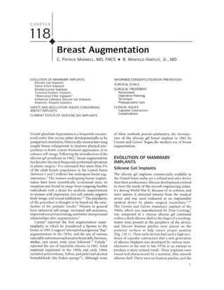
  • 1. CHAPTER 118. _ Breast Augmentation C. PATRICK MAXWELL, MD, FACS • R. WINFIELD HARTLEY, Jr., MD EVOLUTIONOF MAMMARYIMPLANTS Silicone Gel Implants Saline-Filled Implants Double-Lumen Implants Textured-Surface Implants "Alternative Filler Implants" Enhanced Cohesive Silicone Gel Implants Anatomic-Shaped Implants SAFETYANDREGULATORYISSUESCONCERNING BREASTIMPLANTS CURRENTSTATUSOF SILICONEGELIMPLANTS Female glandular hypomastia is a frequently encoun- tered entity that occurs either developmentally or by postpartum involution. Historically, women have long sought breast enlargement to improve physical pro- portions, to foster a more feminine appearance, or to enhance self-image. Following the introduction of the silicone gel prosthesis in 1962,' breast augmentation has become the most frequently performed operation in plastic surgery.' It is estimated that more than 1% of the adult female population in the United States (between 1 and 2 million) has undergone breast aug- mentation.' The women undergoing breast implan- tation have been scientifically scrutinized since its inception and found to range from outgoing healthy individuals with a desire for aesthetic improvement to women with depression, low self-esteem, negative body image, and sexual inhibitions.'" The popularity of the procedure is thought to be based on the satis- faction of the patients' results.' Women in general have enhanced self-image, increased self-assurance, improved sexual functioning, and better interpersonal relationships after augmentation.' Czerny8 reported the first augmentation mam- maplasty, in which he transferred a lipoma to the breast, in 1895.Longacre9 attempted autogenous"flap" augmentations in the 1950s, and the use of various injectable substances such as petroleum jelly,beeswax, shellac, and epoxy resin soon followed.1O Uchida 11 reported the use of injectable siJicone in 1961. Solid materials implanted in the 1950s and early 1960s included polyurethane, Teflon, and polyvinyl alcohol formaldehyde (the IvaIon sponge'O). Although none INFORMEDCONSENT/LITIGATIONPREVENTION SURGICALGOALS SURGICALTREATMENT Assessment Operative Planning Technique Postoperative Care CLINICALISSUES Capsular Contracture Complications of these methods proved satisfactory, the introduc- tion of the silicone gel breast implant in 1962 by Cronin and Gerow' began the modern era of breast augmentation. EVOLUTION OF MAMMARY IMPLANTS Silicone Gel Implants The silicone gel implants commercially available in the United States today are a refined and safer device than their predecessors. Silicone development evolved to meet the needs of the aircraft-engineering indus- try during World War II. Because of its softness and inert nature, it attracted interest from the medical sector and was soon evaluated as an implantable medical device by plastic surgical researchers."·13 The Cronin and Gerow mammary implant of the 1960s, which was manufactured by Dow Corning,' was composed of a viscous silicone gel contained within a thick silicone shell in the shape of a teardrop. Seams were present at the periphery of the device, and Dacron fixation patches were placed on the posterior surfaces to help ensure proper position (Fig. 118-1). These early devices had such a high inci- dence of capsular contracture that a new generation of silicone implants was developed by various man- ufacturers in the mid to late 1970s in an attempt to produce a more natural result. These implants were round and characterized by a seamless, thin, smooth silicone shell. There were no fixation patches, and the
  • 2. 2 VI + TRUNK AND LOWER EXTREMITY FIGURE 118-1. The original Cronin-Gerow silicone implant introduced in 1962 had an anatomic (teardrop) shape. smooth surface, and posteriorly placed Dacron patches to help maintain the implant's position. (From Cronin TD,Gerow FJ: Augmentation mammoplasty: a new "natural feel" prosthesis. Transactions of the Third International Congress of Plastic Surgery, October 13-18, 1963. Amsterdam, Excerpta Medica Foundation, 1963:41-49.) silicone gel was less viscous than in first-generation implants. Whereas the incidence of capsular con- tracture may have been improved somewhat, the inci- dence of silicone gel "bleed" and shell rupture was enhanced (especially from manufacturers who made very thin shells"). Gel bleed is a phenomenon whereby low-molecular-weight particles of silicone gel diffuse or leak through the silicone elastomer shell, giving a sticky feel to the surface. It has been theorized that silicone bleed could promote capsule contracture.IS ,16 Whether it is due to silicone bleed or other factors, capsule contracture has been the biggest clinical problem with the use of smooth-surfaced silicone gel implants. The third generation of smooth-surfaced silicone implants, developed in the early to mid 1980s, focused on improving the strength and integrity of the sili- cone shell as well as on minimizing the silicone bleed phenomenon.",18 This generation of implants was characterized by two layers of "high-performance" elastomer with a thin fluorosilicone "barrier coat" in between (produced by McGhan Medical, Heyer- Schulte, Dow Corning, and Cox-Uphoff). There are data to suggest that these improvements enhanced shell life and lessened capsule contracture. Third-genera- tion silicone gel implants with the application of a textured surface can be considered fourth-generation devices, and cohesive silicone gel-filled implants can be considered fifth-generation devices. These are dis- cussed in the following paragraphs (Table 118-1). Saline-Filled Implants The inflatable saline-filled implant was first reported by Arion" in France in 1965. The impetus for its development was to allow smaller incisions through which a non inflated device could be inserted and then inflated with its liquid filler material. Saline implants were subsequently developed by American manufac- turers and underwent clinical evaluation in the early 1970s.'o,,, The emphasis for the inception of and inter- est in these devices was focused on their inflatable
  • 3. 118 • BREAST AUGMENTATION 3 TABLE 118-1 + DEVELOPMENT AND CHARACTERIZATION OF SILICONE GEL BREAST IMPLANT First Generation (1962-1970) Thickshells Thickgel Dacron patches Teardrop shape Second Generation (1970-1982) Thin shells Thingel No patches (smooth) Round shape Third Generation (1982-present) Thicker (stronger) shells "Barrier coat" ("low-bleed") shells Thicker gel No patches Round shape Fourth Generation (1986-present) Third-generation technology Textured silicone surfaces Round and anatomic shapes Fifth Generation (1993-present) Enhanced cohesive silicone gel Textured silicone surface Diverse anatomic and round shapes nature, allowing smaller incisions, not on the charac- ter or safety of the liquid filler or an attempt to lessen the rates of capsular contracture. Although it is generally accepted that the contrac- ture rate with saline implants is relatively low,two qual- ities of these devices have plagued their clinical use. The foremost was their deflation rate. The original French implant manufactured by Simaplast was found to have a deflation rate near 75% at 3 years and was withdrawn from the market. Heyer-Schulte developed an American saline implant in 1968.Whereas silicone gel implant shells are high-temperature vulcanized (HTV) platinum cured, the shells for saline-filled implants were made thicker and cured by a room-tem- perature vulcanized (RTV) process. This significantly decreased the deflation rate, and all American-made saline implants have since had shells cured by this process." A second factor found to increase deflation rates was valve failure." The original Heyer-Schulte pros- thesis had a retention (leaflet) valve, which was subsequently replaced by a diaphragm valve. Saline implants currently manufactured in the United States by Mentor (which purchased Heyer-Schulte) and INAMED (formerly McGhan Medical) have diaphragm valves and RTV-cured shells. The other characteristic of saline implants that has been a problem relates to the saline itself, which may transmit visible surface wrinkles and a knuckle-like feel in volumetrically underfilled devices. When the device is overfilled, it may feel and look like a firm ball and transmit a peripheral "scalloping" look. For these reasons, saline implants historically perform better under thicker tissue, and surgeons generally fill implants to the recommended volume or just beyond. Saline implants are also heavier than silicone gel implants on a volumetric basis and may cause more tissue thinning with inferior displacement of the implant over time. Double-Lumen Implants The original double-lumen implant was developed by Hartley" as a means of countering capsular contrac- ture. It was constructed of an inner silicone gel-filled lumen surrounded by an outer saline inflatable shell. The conceptual use of the device employed the initial inflation of the outer saline shell to make a larger pocket, with subsequent percutaneous deflation to leave the smaller silicone gel-filled shell within a larger pocket. The device became popular without going through these machinations as a fixed-volume, two- chamber device or as a drug delivery device, which allowed the addition of steroids or antibiotics to the outer saline-filled chamber. Cox-Uphoff developed a "reverse double-lumen" implant," which had an outer silicone gel-filled shell surrounding an inner inflatable shell. Today, the only double-lumen device on the U.S. market is the Mentor Becker, an expander-implant used primarily for reconstruction.'· This device was originally devel- oped as a saline device but subsequently converted to a reverse gel and saline double-lumen design to min- imize deflation rates. INAMED (McGhan) makes a similar round design and additionally has an anatomic version in which the postoperative addi- tion of saline selectively enhances only breast projec- tion. The McGhan style 153 anatomic silicone gel implant is also structurally composed of two lumens with different shapes to enhance its anatomic form. Textured-Surface Implants Early attempts at augmentation with polyurethane sponge were not successful, but in 1970, Ashley" reported the favorable use of a silicone gel implant covered with a thin layer of polyurethane foam.
  • 4. 4 VI • TRUNK AND LOWER EXTREMITY Although the foam was placed on the implant prima- rilyto maintain its position, clinical use seemed to show a lessened incidence of capsular contracture.",'9 Throughout the 1980s, increasing numbers of plastic surgeons found polyurethane-covered silicone gel implants to produce aesthetically pleasing results with low capsular contracture rates.'o,,, The polyurethane surface adhered to the surrounding tissues, subse- quently delaminated, and created a relatively non- contractible capsule." Unlike smooth-surfaced implants that had to be mobile within their pocket, polyurethane-covered implants could be immobile yet soft, These devices had reached a zenith of popularity by 1990, when questions of the safety of polyurethane foam breakdown products caused Bristol-Myers Squibb, which owned Surgitek (the company manu- facturing the implants), to withdraw from the breast implant market." The favorable clinical outcomes and commercial success of polyurethane-covered implants (Fig. 118- 2) led American implant manufacturers to develop textured silicone surfaces in the hope of achieving similar results. In 1986, McGhan Medical introduced Biocell textured implants and expanders, and Mentor introduced Siltex textured implants. These remain the two textured surfaces available in the United States today. Dow Corning subsequently introduced its MSI "structured surface" in 1990, but the company with- drew from the market in 1992. Biocell is an aggressive open-pore textured silicone surface composed of irregular pores having an average density of 3.1 pores/mm' with a mean pore size of 28911m (range, 3711m to 64811m) (see Fig. 118-2). Created by a lost-salt technique, these interconnected pores promote adherence to the surrounding, devel- oping capsule through an "adhesive effect.""·" This tissue adherence, which is clinically similar to that seen with the polyurethane foam surface, differs in that there is no delamination of the texture as occurs with polyurethane. The adhesive effect and tissue adher- ence are enhanced in Biocell-covered expanders; these have the added mechanical advantage of expansion pressure, which pushes the textured surface into the developing capsule and imparts its mirror image into the surrounding tissue." Whereas adherence may not occur around the entire device or with all Biocell breast implants, there is a high friction coefficient around these devices, making them relatively immobile. Thus, similar to the polyurethane implants, "immobility with softness" characterizes Biocell-covered implants. Prospective clinical studies have demonstrated that Biocelltextured implants have a significantly lower inci- dence of capsule contracture than do their smooth counterparts, whether they are filled with silicone gel'9 or saline.'o Siltex is a less aggressive textured silicone surface created as a negative contact imprint off texturing foam (see Fig. 118-2). It is characterized by a raised, dense pattern of irregular nodules ranging in height'6 from 65 to 150l1m and in width from 60 to 27511m, Siltex does not adhere to the surrounding tissue and thus is not characterized by immobility with softness, as are polyurethane and BioceiL" Whereas Siltex-covered implants thus move within their surrounding pocket similar to smooth-walled implants, prospective clin- ical studies have shown a significantly lower incidence of capsule contracture compared with their smooth counterparts, whether they are filled with silicone gel"'" or saline" Other textured-surface devices that have been available in the past or are currently available outside the United States include the MSI pillar-structured texture previously manufactured by Dow Corning" and the polyurethane foam-covered implant manu- factured by Silimed in BraziL" "Alternative Filler Implants" When safety issues with silicone gel implants became a concern, investigators looked for alternative filler substances. Three actually came to market. Polyvinylpyrrolidone is a low-molecular-weight"bio- oncotic" gel thought to be more radiolucent than sil- icone. It composed the fill material of the Misti Gold implant introduced in 1991 by Bioplasty'6 NovaMed purchased this company, and the polyvinylpyrrolidone implant is currently still available outside the United States under the name NovaGold.ln December 2000, the British Medical Devices Agency issued a device alert regarding this implant and other alternative filler devices, citing the opinion that studies demonstrating the safety of these devices are lacking," LipoMatrix manufactured triglyceride-filled im- plants termed Trilucent implants in 1994. Soybean oil composed the fill material, which was said to be radi- olucent. Problems with oil bleed," tissue irritation, and a rancid or foul smell'9 were reported, and the implants were withdrawn from the market in 1999. Hydrogel implants are implants filled with an organic polymer, which is a mixture of polysaccha- ride and water. These implants have been manufac- tured in France by PIP and Arion. There have been reports of swelling of hydrogel (as well as polyvinyl- pyrrolidone) implants after implantation due to osmotic gradient pressure." The British Medical Devices Agency alert of 2000 also applied to these devices. None of these alternative filler devices is available in the United States. Enhanced Cohesive Silicone Cellmplants All silicone gel implants are cross-linked to maintain a gel consistency, and thus all silicone gel has cohe-
  • 5. 118 • BREAST AUGMENTATION 5 A B c FIGURE 118-2. A, Polyurethane foam gains tissue adherence and delaminates from the implant. No longer available inthe United States, this texture fostered the development of textured silicone surfaces. B, Biocellis an aggressive silicone textured surface that adheres to surrounding tissue by an adhesive effect. C, Siltex is a less aggressive silicone textured surface that does not demonstrate any adhesive effect and does not gain tissue adherence. (From MaxwellGP, Hammond DC: Breast implants: smooth vs. textured. Adv Plast Reconstr Surg 1993;9:209.) sive properties. As the cross-linking is increased, the consistency or firmness of the "liquid-feeling" gel changes to that of a soft cheese. The enhanced cohe- sive nature of these implants makes them "form stable." This refers to the implant's maintaining its shape in all positions (shape maintenance). These implants are designed in various anatomic dimensions in addition to round shapes and are collectively referred to as cohesive silicone gel implants. These form-stable implants are currently popular worldwide and undergoing Food and Drug Administration (FOA)-approved clinical trials in the United States (Fig. 118-3).45 Anatomic-Shaped Implants The original Cronin and Gerow silicone gel implants had a teardrop shape, as did a number of the early
  • 6. 6 VI • TRUNK AND LOWER EXTREMI1Y FIGURE 118-3. Style 410 Matrix (INAMED)of enhanced cohesive silicone gel implants offers varying heights and projections of shaped devices for breast augmentation and reconstruction. (L,low;M, moderate; F,full;X,extra). (Cour- tesy of INAMEDHealth, Santa Barbara, Calif.) saline- and gel-filled devices. Problems with capsular contracture, however, led manufacturers to design round, smooth-surfaced low-profile implants, which would move within their surgical pockets. These round, smooth designs dominated the market for nearly 20 years. Only when the phenomenon of immobility with softness was appreciated was the creation of anatomic devices clinically appropriate.38,51The poly- urethane Optimum and Replicon devices (no longer available) were early-generation anatomic-shaped implants popular in the 1980s.28 ,29 The adherence of the polyurethane surface, in fact, lent itself to the "stacking" of these implants, one on top of another, to produce an anatomic shape with enhanced projection.51 The tissue adherence observed with tissue expanders that had the Biocell surface led McGhan to develop anatomically shaped expanders and subsequently an internally stacked style 153 gel anatomic-shaped implant.J5,38,51," Favorable clinical experience and advanced product design led to a matrix of variable height-to-width ratio anatomic expanders and implants, the Style 133 expanders and Style 410 Matrix cohesive implants (see Fig. 118-3). The latter enjoy widespread international use in aesthetic surgery"·55 and have completed their initial FDA clin- ical Investigative Device Exemption study in the United States, awaiting longer follow-up. Silimed (Brazil) markets polyurethane-covered cohesive silicone gel implants in anatomic shapes." These devices also enjoy international popularity, but to date, no clinical investigative studies have taken place in the United States. Mentor introduced a midheight Siltex anatomic- shaped tissue expander in 1997 and other height options in 2003. In the fall of 2002, an Investigative Device Exemption study on a midheight anatomic cohesive gel implant was initiated. These "contour"- shaped devices are covered with the Siltex texture. Because tissue adherence does not generally occur, the pocket must be exact and only minimally larger than the footprint of the reduced height device to mini- mize the possibility of implant rotation.s6
  • 7. 1 18 • BREAST AUGMENTATION 7 Anatomic-shaped saline inflatable implants are available in the United States manufactured by both Mentor and INAMED (McGhan), and there is debate among plastic surgeons about the merit of each rela- tive to the resultant breast form.58 '" This debate seems confined to saline-filled implants alone as virtually all tissue expanders marketed for breast reconstruction in the United States are textured and anatomically shaped. It is predicted that once cohesive gel anatomic implants and other gel implants are available in the United States, the issue will be of less concern as evidenced by surgeons' preferences worldwide. SAFETY AND REGULATORY ISSUES CONCERNING BREAST IMPLANTS In 1976, the U.S. Congress passed a Medical Device Amendment to the Food, Drug, and Cosmetic Act that gave the FDA authority over medical devices. Implants on the market at the time or those considered "sub- stantially equivalent" to those marketed before 1976 were "grandfathered" in and allowed to remain in use until the FDA could formally review their safety and efficacy. In 1988, the FDA called for the manufactur- ers of silicone gel-filled implants to submit their Pre- market Approval Applications containing data adequate to substantiate the safety and efficacy of the devices they were marketing. In November 1991, the FDA convened an advisory panel of experts to hold public hearings and evaluate the manufacturers' data. The panel concluded that more research was neces- sary (to establish safety and efficacy) but recommended continued availability of implants while that research was carried out. In January 1992, however, the FDA Commissioner went against the recommendation of the advisory panel and called for a voluntary mora- torium on the use of silicone gel implants. After further evaluation of the situation by the advisory panel (who thought there was a public need for the devices), the FDA Commissioner, in April 1992, ruled that although silicone breast implants were not necessarily unsafe, the law required more data to substantiate safety and efficacy than the manufacturers had supplied.62.63The use of silicone gel implants was thus restricted to clin- ical trials until the data were produced. This was inter- preted by the media and the public at large that silicone gel implants were "banned" because they were not safe. This effectivelytook silicone gel implants off the market for breast augmentation in the United States for the next 12 years. The "media frenzy" surrounding this issue was further heightened by several lay jury court decisions that found silicone implants to be responsible for women's pathologic conditions. This led to the filing of thousands of product liability lawsuits against the implant manufacturers. This culminated in a class action lawsuit involving more than 400,000 women." Unable to withstand the financial pressure to defend this massive number of cases, Dow Corning filed Chapter I I and Bristol-Myers Squibb withdrew from the market. Ultimately, a settlement of approximately $4 billion was reached, and Mentor and McGhan were left as the only two American manufacturers of saline and silicone breast implants. Concerns relating to the safety of foreign materi- als implanted in the female breast began in Japan in 1964 when the term human adjuvant disease was suggested on speculation of an association between paraffin breast injections and connective tissue disease- like symptoms in several women." In the 1980s,several reports questioned a link between silicone gel breast implants and various collagen vascular diseases"'-6' Questions were raised as to whether silicone "leaked" into the body and caused pathologic conditions. Whereas increased levels of silicone were found within the surrounding tissue capsule68,6.and axillary lymph nodes, no correlation with symptoms or any disease could be established. Likewise, no specific antibodies to silicone could be found.,o,'1 Amid this background oflawsuits, public concerns, and implant restrictions, the scientific data began to prevail, demonstrating the safety of silicone gel and the lack of its correlation with any disease or patho- logic condition. By the late 1990s, approximately 20 epidemiologic studies and other important scientific investigations found no increased risk for development of connective tissue disorders in women with breast implants." In addition, respected independent scientific groups including the Independent Review Group in England," the Institute of Medicine," and the National Science Panel" (appointed by the judge of the class action litigation), after carefully review- ing all scientific data available, found no relationship between silicone gel implants and connective tissue disease. The other health issue that clouded the breast implant arena in the early 1990s was the possibility of a polyurethane foam breakdown product being car- cinogenic. Specifically cited was a National Cancer Insti- tute study in which mice fed extremely high doses of 2,4-toluenediamine showed an increased incidence of breast cancer." Since the foam used to cover the Sur- gitek implant was produced by a mixture of 2,4- and 2,6-toluenediisocyanate, the FDA questioned whether the polyurethane itself or one of its biodegradation products could be carcinogenic in patients after breast implantation. Scientific scrutiny of patients in whom these devices had been implanted found minimal expo- sure to 2,4-toluenediamine,'6 and the FDA ultimately concluded that it was unlikely any woman with polyurethane-covered implants was at increased risk for development of cancer." Before these scientific findings of safety, however, Bristol-Myers Squibb failed
  • 8. 8 VI • TRUNK AND LOWER EXTREMITY to make premarket approval for the FDA in April of 1991 and withdrew these devices from the market. Despite this decade of turbulence, the future of sil- icone gel implants looks bright. No fill material has been found to be as safe and as functional as silicone. Saline clinical inadequacies (in certain situations) are well appreciated by American plastic surgeons. Man- ufacturing practices of silicone gel implants have been improved and brought into compliance to ensure better-quality products. CURRENT STATUS OF SILICONE GEL IMPLANTS The Premarket Approval Application for silicone gel breast implants submitted by [NAMED in December 200 I was heard by the FDA expert advising panel in October 2003. After intense scrutiny of the data sub- mitted, as well as public testimony, the panel recom- mended approval of the application "with conditions;' setting the stage for the return to market of silicone gel implants in the United States. The panel found no evidence to support that silicone gel implants cause disease. They did, however, question the adequacy of the length of follow-up on the studies. In January 2004, the Commissioner of the FDAwent against the panel's recommendation and asked foraddi- tional data with longer follow-up from all manufac- turers on silicone gel implants. * In addition, more information was requested on life expectancy of implants, causes and effects of shell failure, and clin- ical evaluation of possible"siIent rupture" of implants. In light of these requests from the FDA, silicone gel implants will probably not be back on the American market until 2005. They remain available for clinical use (as they have for the last 12 years) under FDA- approved clinical studies. INFORMED CONSENT/LITIGATION PREVENTION Because the breast is historically viewed as a symbol of female sexuality and the quality of the surgical result is primarily in the eye of the beholder (and her com- panion) alone, emotional outcome can on occasion be somewhat volatile. The facts are that almost 40% of aesthetic plastic surgical claims relate to elective breast operations and half of these to breast augmentation." It is thus incumbent on the surgeon to evaluate the patient's emotional state, timing, and appropriateness of the desired outcome. It is the surgeon's responsibil- ity to listen, educate, and evaluate; this process and the communication that takes place between patient and surgeon are documented in the medical record. . http://www.fda.gov/cdrhJode/guidance/1239.html Informed consent isnot simply the signing of a paper or contract but refers to the entire process between patient and physician as well as physician extenders. To be "informed," the patient must be provided with adequate information about risks, benefits, and treat- ment alternatives to the proposed procedure. To "consent;' the patient must be an adult (by age), be capable of rational communication, and be able to understand the information. The informed consent documentation must be thorough and specific to the operation and preferably the surgeon. A checklist of specifics (which must be initialed by the patient) is considered advisable. "Before and after" photographs may be shown but should be realistic. Photographs of the patient are a necessary form of documentation, requiring appropriate permission. Their confidential- ity is essential unless permission is given for any use other than medical review documentation. A male surgeon should be accompanied by a female chaperon during all breast photography and examinations. Because of the multiple options in breast aug- mentation surgery, a second office visit is advisable. There must be a clear understanding (which is docu- mented in the medical record) between patient and surgeon of the specific desired outcome (size, shape), the alternative ways by which this can be achieved, and the risk-to-benefit ratio of the chosen "pathway." SURGICAL GOALS The conceptual goal of breast augmentation is to enhance the form and volume of the female breast in the most predictable manner with the fewest possible complications. The resultant form of the augmented breast will be determined by dynamic interaction over time between the compliance and character of the soft tissue envelope; the quality and consistency of the breast parenchyma; and the dimensions, volume, and charac- teristics of the breast prosthesis (Fig. 118_4).'9 To achieve these goals, experience has shown that a surgical approach based on dimensional concepts rather than on volume alone ispreferable. This"biodi- mensional" approach takes into account the patient's existing breast dimensions and tissue characteristics of the form of the patient's desired surgical result."o.81 A breast implant is then selected of appropriate dimensions, character, and volume to accomplish this goal (Table 118-2). SURGICAL TREATMENT Each year, more than 100,000 women in the United States elect to have surgical enhancement of their breasts. Women seeking breast augmentation place considerable emphasis on their physical appearance, and time should be taken to understand their
  • 9. A (I) (5 Q. •..(I) Q. Q. ::> .•....c: Ol '(j) ..c: .•..(J) ctl (I) •... CD 118 • BREAST AUGMENTATION Soft tissue envelope Parenchyma Prosthesis B 9 FIGURE 118-4. A, The aesthetic breast form is composed of measurable parameters. This form can be attained by the careful planning and surgical performance of a breast augmentation. B, The resultant breast form desired after surgical augmentation is determined by the dynamic interaction between the character and compliance of the soft tissue envelope; the quality, volume, and consistency of the breast parenchyma; and the dimensions, volume, and char- acteristics of the breast implant. motivations for having surgery. Most patients are prop- erly motivated with realistic goals, but the preopera- tive visit is the time to identify patients who may have unrealistic expectations or are using surgery as a crutch for other problems. The patient's desires and expec- tations must be weighed against the predictability of achieving those goals. A high level of satisfaction can be ensured ifthe patient's aesthetic concerns and expec- tations are within a predictable and attainable result. TABLE 118-2 • STEPS IN A BIODIMENSIONAL APPROACH TO BREAST AUGMENTATION Evaluate existing chest and breast form. Characterize the soft tissue envelope. Plan the resultant breast form desired. Select implant and site location to accomplish this goal. Select incision and approach. Whereas individual preferences will affect opera- tive planning and procedural specifics, the goal of breast augmentation is to enhance the form and volume of the female breast. The form of the female breast isdeter- mined by the quality, volume, and dimensions of the breast parenchyma and the character and compliance of the soft tissue envelope. The form of the augmented female breast (assuming that capsuJecontracture is not present) is based on these in dynamic interaction with the dimensions, volume, and consistency of the breast prosthesis (see Fig. 118-4).79 There must be mutual understanding between the patient and surgeon of the specific resultant breast form that is desired and the predictability (and tradeoffs) of achieving that form. Assessment After the patient's goals are determined and rea- sonable expectations are established with regard to outcome, a thorough physical assessment is
  • 10. 10 VI • TRUNK AND LOWER EXTREMITY I --IMD-- I. FIGURE 118-5. Preoperative mea- sures (taken before breast augmen- tation) include SSNto N[suprasternal notch to nipple). N to IMF (nipple to inframammary fOld). BW (breast width). BH (breast height). and IMD [intermammary distance). undertaken. The bone and muscle structural founda- tion of each breast must be assessed. Note the shape of the thorax as well as whether the patient is "long" or "short" chested. The majority of women will have some degree of asymmetry when the breast and chest wall are criti- cally evaluated." It is imperative to document and discuss any amount of nipple-areola complex asym- metry as well as chest wall asymmetry with the patient. Precise measurements must be taken (Fig. 118-5). Key measurements include suprasternal notch to nipple distance, nipple to inframammary fold distance, base width or diameter, and breast height. The compliance of the soft tissue envelope is then assessed. Characterize the elasticity of the skin by noting evidence of poor compliance, such as stretch marks or thin nonelastic dermis. The soft tissue pinch test is a useful measurement; the superior pole of the breast is gathered between the thumb and index finger (skin plus parenchyma), and the distance between the two is measured (Fig. 118-6).A rough estimateforthe amount of inherent soft tissue necessary to cover a subglandular implant is 2 cm. A pinch test result of less than 2cm may lead to subpectoral implant place- ment. Skin redundancy may also be present." Older patients or those with a history of weight loss may exhibit varying degrees of pseudoptosis or true breast ptosis. These patients may benefit from a concomi- tant mastopexy. It is also important to characterize the breast parenchyma itself. Determination of the amount, quality, and distribution of the parenchyma may alter surgical techniques; thus, these should be evaluated and documented. It may be necessary to redistribute, adjust, or reshape the parenchyma to achieve the desired breast mound form. Operative Planning IMPLANT SIZE (DIMENSIONS) The patient's request for a particular breast size and shape will largely determine the dimensions of the FIGURE 118-6. Soft tissue pinch test. Assessment of the thickness and quality of the soft tissue in the upper pole of the breast preoperatively willhelp the surgeon in considering pocket locationoptions for implant placement.
  • 11. 118 • BREAST AUGMENTATION II Implant width Original breast width FIGURE 118-7. After the width of the existing breast is measured and the desired resultant breast form is formulated, an implant is selected (generallyjust narrower than the orig- inal breast, shown on the patient's right)that incombination withthe pre- operative breast tissue willachievethe desired postoperative dimensions and form (shown on the patient's left). breast implant used. In addition to a thorough dis- cussion with the patient as to her desire for the result- ant form and size, it is often helpful to have the patient bring photographs showing the sizeand shape of breast that she finds appealing. The most important clinical factor in determining breast prosthesis size is the base width or diameter of the patient's native breast. After measuring the patient, one turns to the manufacturer's published data charts and generally selects an implant slightly less wide than the existing breast. Rarely does the selected implant vary significantly more or less than the measured breast width to avoid an unnatural postoperative appearance (Fig. lI8-?). In addition to the desired width of the implant, one also considers height, projection, and volume before making an implant selection. Anatomic- shaped implants allow implant height and projection options to be much more important operative considerations. SILICONE VERSUS SALINE The decision between a saline-filled prosthesis and a silicone gel implant is one of the patient's preference after the surgeon's conveyance of information. Expe- rience has shown the results of silicone gel implants in primary breast augmentation to be generally soft and to have a natural feel and appearance, assuming capsule contracture is not present. Although the authors prefer silicone gel implants, saline implants placed in the subpectoral posItIOn can produce good results with a low incidence of capsule contrac- ture. The thicker the soft tissue under which a saline implant is placed, the better it performs. Despite our preference for silicone gel, some patients will undoubtedly continue to have concerns about silicone- filled devices, and subpectoral saline implants have proved to be a reasonable alternative (Fig. 118-8). Ultimately, the patient must feel comfortable with the implant device, so the final decision rests with the patient. TEXTURED VERSUS SMOOTH The decision between textured and smooth-walled implants is only applicable for round implants. Anatomic implants are all textured by design to min- imize malrotation. With round implants, the choice between textured and smooth-walled implants is based primarily on minimizing capsular contracture. For subpectoral augmentation, either implant can probably be used with comparable results. When the device is placed in the subglandular pocket, a smooth- walled implant offers the best protection from visible rippling and palpability but runs a greater risk for devel- opment of capsular contracture. A textured implant can be used in the subglandular position, but it should be reserved for those patients with adequate soft tissue coverage such that it will not be visible or easily palpable.
  • 12. 12 VI • TRUNK AND LOWER EXTREMITY A B FIGURE 118-8. A. A typical patient presenting for breast augmentation. B. Postoperative result with subpectorally positioned, smooth saline implants (275 mL filled to 300 mL) placed through an inframammary incision. ANATOMIC VERSUS ROUND The decision between anatomic implants and round implants is determined by the shape and form of the existing breast. If a patient has hypovolemic breasts with good natural shape, form, and contour, round implants will provide the desired final result with the lowest risk of complications. By augmentation of volume while shape is maintained, a natural result is attainable (Fig I I8-9A). In a patient who would benefit from having the form and shape of her breasts improved in addition to volumetric enhancement, an anatomic implant is preferable. Breast parenchymal maldistributions can be corrected, with a more aes- thetically pleasing result (Fig. II8-9B). POCKET SELECTION The decision of subglandular or subpectoral implant placement depends on implant selection (fill and texture) and tissue thickness. In theory, the best posi- tion for a mammary implant is in the subglandular plane. This is the most anatomically correct position to maintain natural shape and form. The reasons for placement of implants in the subpectoral plane are to minimize the risk of capsular contracture (primarily for gel implants) and to minimize implant visibility and palpability. Other considerations relate to possi- ble effects on mammography and tissue stretch over time. There are benefits and tradeoffs to each site (Table 118-3). In practice, saline implants are predominantly placed in the subpectoral pocket because of their ease of palpability and visibility. Silicone gel implant place- ment is determined largely by soft tissue adequacy. In patients with a pinch test result of more than 2 em, the implant can safely be placed in the subglandular plane (Fig. 118-10). Previous data suggest that tex- tured gel implants have a lower rate of capsular con- tracture when they are placed subglandularly. If one chooses smooth gel implants for the subglandular plane, additional measures to prevent capsular con- tracture must be taken. These include larger pocket dissections with displacement exercises or possible dilute steroid pocket irrigation (Fig. !l8-!lA). Anatomic-shaped textured implants are placed in the appropriate pocket as determined by soft tissue thick- ness. Pockets for these implants are made only mini- mally larger than the footprint of the device to minimize displacement or malrotation (Figs. II8-IIB, 118-12, and !l8-13). When subpectoral pockets are selected (Fig. 118- 14), one generally divides the origin of the pectoralis major muscle just above the inframammary fold to allow better projection in the lower pole of the aug- mented breast and to maintain a natural inframam- mary fold. This places the superior portion of the implant in a subpectoral position while the inferior portion is subglandularly located. In constricted breasts (tuberous breasts) or ptotic breasts, for which more parenchymal surgical manipulation is necessary, or when there is a greater need for the implant to fill out the lower soft tissue envelope, more dissection between parenchyma and muscle will allow the muscle to cover less of the implant with a resultant greater subglandular implant coverage. Alternatively, the pec- toral muscle can be divided at a higher level to give a similar result. These pocket manipulations have been described as dual-plane maneuvers to allow varying degrees of subpectoral to subglandular implant coverage (Table 118-4).8' An additional pocket more recently introduced and advanced by some is the subpectoral fascial pocket. This thin layer of tough tissue is said by some to offer the advantage of subglandular placement with a thicker soft tissue cover.8'. Text continued on p, 19
  • 13. A Round implant B 118 • BREAST AUGMENTATION 13 Anatomic implant FIGURE 118-9. A, When the preoperative breast has an acceptable form and inadequate volume, a round implant may be preferable to achieve the desired result. 8. When the breast form and volume are inadequate, an anatomic- shaped implant may be preferable.
  • 14. 14 VI • TRUNKAND LOWEREXTREMI1Y TABLE 118-3 • ALTERNATIVE POCKET LOCATIONS WITH POTENTIAL TRADEOFFS Pocket Retromammary Partial retropectoral (without dividing pectoralis origins along the infra mammary fold) Total submuscular Tradeoffs Increased risk of edge visibility or palpability Possible increased interference with mammography Possible increased incidence of capsular contracture Lateral implant displacement over time, widening the space between the breasts Less control of upper medial fill More postoperative tenderness and a more prolonged recovery Distortion of breast shape with pectoralis contraction Less precise control of infra mammary fold position, depth, and configuration. This potential tradeoff is minimized or eliminated by division of pectoralis origins along the inframammary fold in patients who have adequate soft tissue coverage. Increased risk of superior implant malposition or displacement (when inferior pectoralis origins across inframammary fold are not divided) Longer time required for deepening of the inframammary fold (when pectoralis origins along the infra mammary fold are not divided) All tradeoffs listed above for partial retropectoral, plus: Highest risk of superior implant displacement or malposition Longer operative time Longest postoperative recovery and morbidity Least accurate and predictable inframammary fold and longest to achieve depth Greatest risk of inframammary fold irregularities, lateral flattening, and fold level inaccuracies Potential Benefits Increased control of breast shape Usually a more rapid postoperative recovery Minimal or no distortion with pectoralis contraction Increased control of inframammary fold position and shape Muscle coverage mandatory if pinch thickness <2 cm above breast parenchyma Possibly more accurate mammograms Less risk of palpable or visible implant edges Possible decreased risk of capsular contracture (small difference with saline- filled implants, greater difference with silicone gel-filled implants) Possible increased coverage inferolaterally but clinically no significant additional cover long term From Tebbetts 18: Dual plane breast augmentation: optimizing implant-soft tissue relationships in a wide range of breast types. Plast Reconstr Surg 200 1;107: 1255.
  • 15. 118 • BREAST AUGMENTATION 15 Ample soft tissue Retroglandular Inadequate soft tissue Retropectoral A B FIGURE 118-10. A, When ample soft tissue is present, implants may be placed in the subglandular position. 8, When there is soft tissue inadequacy, the subpectoral position is generally preferable. A Mobile implant moving within larger capsule B Exact pockel FIGURE 118-11. A, Round pocket dissection for smooth-surfaced implants to allow tissue redraping and encour- age implant mobility to minimize capsule contracture. 8, Precise pockets only slightly larger than the base of an anatomic textured implant help maintain implant position. Movement is not desirable when the implant allows immobility with softness.
  • 16. 16 VI + TRUNK AND LOWER EXTREMllY A B c o E F FIGURE 118-12. A. The biodimensional approach is demonstrated in this physically fit patient wishing no pec- toral flexion distortion. B, Minor chest-breast asymmetries are noted on examination. C, Result with subglandular placement of smooth, 240-mL (width of 10.5 em) round, silicone gel implants. D, The subglandular pocket selected because of her athleticism requires smooth implant displacement exercises within the large pocket to minimize capsule contracture. E and F. Oblique preoperative and postoperative appearance.
  • 17. 118 • BREAST AUGMENTATION 17 A B c D E F FIGURE 118-13. A biodimensional approach is demonstrated in this postpartum patient desiring a natural pro- portionate appearance. A, N-SSN and BW measurements are noted. B, The soft tissue pinch test result is 1.5 cm. C and D, Preoperative and postoperative photographs; style 410 MM cohesive gel implants were placed in precise subpectoral pockets through an inframammary incision. On the patient's right, a 245-mL implant [11.5-cm width and 1O.6-cm height) was used; on the patient's left, a 215-mL implant [11-cm width and 10. 1-cm height) was used. E and F. Oblique views of preoperative and postoperative appearance.
  • 18. 18 VI • TRUNK AND LOWER EXTREMITY ABC FIGURE 118-14. Subpectoral implant placement generally involves release or division of the pectoralis major muscle, resulting in varying coverage relationships of muscles and parenchyma to implant. A, Muscle division near the infra- mammary fold results in muscle coverage of most of the implant. B, Muscle division (or muscle-parenchymal detach- ment) to the lower areolar level results in muscle coverage of the upper half of the implant. C. Muscle division (or muscle-parenchymal detachment) to the upper areolar level results in muscle coverage of the upper third of the implant.
  • 19. 118 + BREAST AUGMENTATION 19 TABLE 118-4 • POTENTIAL BENEFITS AND TRADEOFFS OF THE DUAL-PLANE POCKET LOCATION Pocket Dual plane (compared with retromammary) Dual plane (compared with partial retropectoral) Tradeoffs Possible increased risk of palpable or visible implant edges inferiorly Potential Benefits Preserves the potential increased control of lower breast shape with retromammary With proper techniques can have similar recovery as with retromammary Reduced risk of edge visibilityor palpability of retromammary by providing more upper pole coverage Reduced interference with mammography by retromammary Reduced possibility of capsular contracture of retromammary by reducing contact with parenchyma compared with retromammary Provides same mandatory muscle coverage if pinch thickness <2 cm above breast parenchyma Reduced risk of lateral implant dispiacement over time; dividing inferior origins decreases pectoralis pressure on implant Better control of upper medial fillwith division of inferior origins to decrease pectoralis tension and pressure on upper pole of implant Reduced postoperative tenderness and recovery period with proper technique Reduced distortion of breast shape with pectoralis contraction Decreased risk of superior implant malposition or displacement by decreasing pressure of pectoralis on lower pole of implant by dividing lower pectoralis origins Increased control of inframammary fold position, depth, and configuration by decreasing pressure of pectoralis on lower pole of implant along inframammary fold Retains possibility of more accurate mammograms, depending on position of muscle Possible decreased risk of capsular contracture (small) From Tebbetts J B: Dual plane breast augmentation: optimizing implant-soft tissue relationships in a wide range of breast types. Plast Reconstr Surg 200 I; 107: 1255. INCISIONS Four types of incision are commonly employed in breast augmentation: transaxillary, inframammary, periareolar, and transumbilical. After implant selec- tion, the decision as to which type of incision is to be used should be made by the patient and surgeon after the options, risks, and benefits of each have been thor- oughly explained. Surgeons should offer only the tech- niques that they are comfortable performing. The final choice should allow the surgeon optimal control and visualization to deliver the desired outcome for the specific patient and the specific implant (Table 118-5). The inframammary incision permits complete visualization of either the prepectoral or subglandu- lar pocket and allows precise placement of virtually all implants. The technique does leave a visible scar within the inframammary fold. Smaller incisions «3 cm) can be used for saline-filled implants, but silicone gel implants often require incisions up to 5.5 cm in length. The incision should be placed in the projected inframammary fold rather than in the existing fold to avoid visibility and widening of the subsequent scar (Fig. 118-15). The periareolar incision is placed at the areolar- cutaneous juncture and generally heals inconspicu- ously. The dissection allows easy adjustment of the inframammary fold and direct access to the lower parenchyma for scoring and release when a constricted lower pole is present. Disadvantages include limited exposure of the surgical field, transection of the parenchymal ducts (which are often colonized with Staphylococcus epidermidis), potentially increased risk of nipple sensitivity changes, and visible scarring on the breast mound. This technique should not routinely be used on patients with an areola diameter less than 40 mm and may not allow introduction of larger gel or enhanced cohesive gel implants. The transaxillary approach can be performed either bluntly or with the aid of an endoscope. The endo- scope allows precise dissection and release of the inferior musculofascial attachments of the pectoralis major as well as direct visualization for hemostasis. This approach avoids any scarring on the breast mound and can be used with both saline and gel
  • 20. 20 VI • TRUNK AND LOWER EXTREMI1Y TABLE 118-5 + INCISION OPTIONS IN BREASTAUGMENTATION Factor Axillary Periareolar Inframammary Periumbilical' Implant plane Submuscular + + + Subglandular + + + Implant type Saline round + + + + Saline shaped + + Silicone round/shaped + + Preoperative breast volume High(>200 g) + + + + Low«200g) + + + Preoperative breast base position High + + + + Low + + + Breast shape Tubular + Glandular ptosis + + + + Ptosis (grade I-II) + Areolar characteristics Small diameter + + + Light/indistinct + + + Inframammary crease None + + + High + + + Low + + + + Secondary procedure + + + indicates applicable; - indicates not generally recommended . • Included for completeness but not generally recommended. From Hidalgo DA: Breast augmentation: choosing the optimal incision, implant, and pocket plane. Plast Reconstr Surg 2000;105:2202. implants in either a subpectoral or subglandular pocket. Disadvantages include difficulty with parenchymal alterations and the probable need for a second incision on the breast mound for revisionary surgery. Precise implant placement can be more difficult with this incision, and enhanced cohesive gel and anatomic implants may be precluded. Transumbilical breast augmentation has the obvious advantage of a single, well-hidden, remote inci- sion. It can be used only with saline implants, however, and precise pocket dissection requires experience. The pocket is dissected bluntly, and hemostasis can be difficult from the remote access port. As with the transaxillary approach, revisions often necessitate a second incision on the breast mound. Technique INFRAMAMMARY INCISION The patient is marked preoperatively in the standing or seated position, with shoulders even and arms resting comfortably at her sides. The miclline of the chest is marked as a point of reference from the sternal notch to the xiphoid process. The existing infra- mammary folds are marked, as are the proposed limits of the dissection. The incision site is marked along the expected new inframammary fold after augmentation. Frequently, augmentation mammaplasty will lower the existing inframammary fold, and this must be taken into account when the patient is marked. The result- ant location of the inframammary fold is determined by the dimensions of the selected implant postoper- atively in interaction with the existing tissues. The inci- sion should begin on a straight line dropped from the medial areolar border and extend laterally. For saline implants, a 3-cm incision is often sufficient, whereas silicone gel implants require incisions 4 cm or more in length; enhanced cohesive implants may require 5.5- to 6-cm incisions (Fig. 118-16). Proper positioning of the patient is important to the outcome of the procedure. The patient is placed in the supine position, well centered on the operating room table. Her arms should be at 90-degree angles to her torso and well secured to fixed arm boards. The patient must be able to flex fully at the waist to 90 degrees during the procedure (Fig. 118-17). Finally, the patient's shoulders must be visible after sterile draping to ensure symmetry when the intraoperative appearance is evaluated. The incision sites are injected with 2 to 3 mL of 1% lidocaine with 1:1000 dilution of epinephrine to aid in hemostasis.
  • 21. 118 • BREAST AUGMENTATION 21 A B c o FIGURE 118-15. A, Asmall-breasted woman with a soft tissue pinch test result of less than 2 cm desirous of full, round result. B, A 340-mL round, smooth, silicone gel implant is selected, which willbe placed in the subpectoral position after the inframammary fold is lowered 1.5 cm. The inframammary incision will be 4.5 cm in length and placed in the new lowered inframammary fold location. C and D, The patient's postoperative appearance. The incision is made along the proposed markings, and the dissection is continued with an insulated elec- trocautery instrument through Scarpa fascia. A fiberoptic headlight isworn throughout the procedure, or a variety of lighted fiberoptic retractors are avail- able to aid illumination and direct visualization within the pocket. If the implant is to be placed in the sub- glandular pocket, the dissection proceeds above the pectoralis major fascia directly beneath the gland. Meticulous hemostasis can be maintained during the complete dissection with the use of the electrocautery. Several medial intercostal perforating vessels may be encountered. These should be avoided or coagulated with insulated forceps if need be. For smooth-walled implants, a larger pocket is dissected to allow mobil- ity of the implant. For anatomic implants, the pocket is precisely dissected to snugly accommodate the implant. Care should be taken to preserve the lateral intercostal cutaneous nerves, especially the fourth intercostal, which contains the primary sensory inner- vation of the nipple-areola complex. If a subpectoral pocket is chosen, the dissection is initially carried out laterally to identify the lateral border of the pectoralis major muscle. The muscle edge can be lifted by forceps to allow easy entry into the submusculofascial plane. This plane is readily identified by the wispy areolar connective tissue and ease of dis- section. An extended electrocautery instrument is used to complete the dissection. The inferior origin of the pectoralis major is released from lateral to medial at the level of the inframammary fold. Various slips of origin of the pectoralis major muscle are generally encountered and divided. Division of the pectoralis continues medially to the sternal border. Partial deep division may selectively be carried out 1to 3 cm above the xiphoid, depending on which implant is to be used.
  • 22. 22 VI • TRUNK AND LOWER EXTREMITY A B c E D FIGURE 118-16. A. A small-breasted woman with a sig- nificant upper pole concavity. She desired strong projec- tion without a convex (round) upper pole shape. B, Breast width measured 12 cm and height 12.5 cm; the soft tissue pinch test result was less than 2 cm. A subpectoral pocket was selected for a cohesive gel style 410 FF implant of 290 mL (11.S-cm width and 12-cm height). C,The implants were placed through a S.B-cm incision at the ievel of the new infra mammary fold to give this result. D and E, Pre- operative and postoperative appearance demonstrating the enhanced projection with control of the slope of the upper pole.
  • 23. 118 • BREAST AUGMENTATION 23 FIGURE 118-17. Theoperatingtableisflexedtoa90- degree angle intraoperatively to allowthe surgeon to care- fullyevaluate the patient with the sizer or final implants in position before closure. Lateral dissection can be done bluntly with a finger to avoid injury to the lateral neurovascular bundles. The nerves can be stretched to accommodate the implant but should be preserved to minimize post- operative sensory changes. When the pectoralis major muscle is elevated, care must be taken to leave the pec- toralis minor down on the chest wall. This will min- imize bleeding and allow proper placement of the implant. Exact implant "sizers" (gel or saline) are used when available to evaluate the pockets and resultant breast form. After the sizers are in place, the patient is placed in a 90-degree upright position and evaluated from various perspectives (see Fig. IIS-17). Any asymme- try or underdissected areas are marked, and the patient is placed back in the supine position. Once adequate hemostasis is obtained and pocket dimen- sions are finalized, the pocket is irrigated with an antibi- otic-containing solution, and the implants are carefully placed by a minimal-touch technique. The final results are assessed, again with the patient in a sitting posi- tion, and a multilayer closure is performed with absorbable suture. It is important to close off the implant pocket with a separate layer of suture before closing the skin. Once closure is complete, SteriStrips are applied along the direction of the incisions. PERIAREOLAR AUGMENTATION The markings for a periareolar approach are similar to those for an inframammary augmentation. The patient is marked in a seated or standing position with even shoulder position. The sternal midline is marked as reference. The existing inframammary fold is marked, as are the limits of the dissection and the planned resultant inframammary fold (if it is to be changed). The incision is marked along the junction of the areola and the breast skin. The limits of the inci- sion are the 3-0' clock and 9-0' clock positions. The positioning of the patient is identical to that for an inframammary approach. It is imperative that the patient be able to fully flex at the waist for evalu- ation of the intraoperative appearance of the implants. With the patient prepared and draped and after injection of lidocaine with epinephrine, the precise incision is made. Wound edges are elevated directly up from the chest wall with an opposing pair of small sharp retractors. An insulated electrocautery unit is used to dissect straight down through the breast parenchyma to the pectoralis major fascia. The retrac- tors are repositioned as needed to keep the gland ele- vated into the wound. For subglandular implant placement, dissection is carried out on top of the pec- toralis major and serratus anterior fasciae. Although it is preferable to use a fiberoptic headlight, fiberop- tic retractor illumination can also facilitate the dis- section by aiding visualization. The dissection is carried out to the extent of the preoperative markings with use of an extended electrocautery instrument, which allows meticulous hemostasis. If the inferior pole of the breast is constricted, radial scoring of the gland in the inferior pole can allow proper redraping of the soft tissue over the implant to correct the defor- mity (Fig. lIS-IS). For subpectoral implant placement, an identical incision is made, but the dissection is carried down through the breast tissue in an oblique plane angled inferiorly, rather than directly through the gland. When the dissection plane approaches the inframammary fold, it is carried directly down to the pectoralis major fascia and continued laterally to identify the lateral border of the muscle. This is an easy area in which to begin the subpectoral dissection and ensure that the dissection planes are correct. The submuscular dis- section is done under direct vision with use of the elec- trocautery, in a lateral to medial direction. The origin of the pectoralis major muscle onto the chest wall can then be divided. This release is done approximately I cm above the origin of the muscle fibers and proceeds from lateral to medial to allow adequate redraping of the muscle over the implant. This usually requires divi- sion of several medial slips of muscle that often contain perforating vessels. The endpoint of the dissection is adequate release of the pectoralis muscle, usually at the sternal border. Dual-plane techniques differ slightly despite which incision is used. When the pocket is dry, sizers are used to evaluate the dissection and to determine the final prosthesis to
  • 24. 24 c VI • TRUNK AND LOWER EXTREMITY B D E be implanted. Before final implant placement, the pocket is once again checked for hemostasis and irri- gated with an antibiotic solution. The closure is par- ticularly important with this technique. The gland must be precisely reapproximated and closed with several FIGURE 118-18. A Patient witha severely constricted lower breast pole and high, tight inframammary fold. B, Round,smooth, siliconegel implantswere selected of 11 .4- em width and 280-mL volume to be positioned in a sub- pectoral (dual-plane) pocket with a significantly lowered inframammary fold, after extensive parenchymal release. e, Thiswas accomplished through a periareolar approach to give this result with an inconspicuous scar. D and E, Preoperative and postoperative appearance. layers of interrupted absorbable sutures to prevent dis- tortion of the nipple-areola complex. The skin is closed with deep everting dermal sutures and a running subcuticular absorbable monofilament. SteriStrips are applied to the closed incision.
  • 25. 118 • BREAST AUGMENTATION 25 TRANSAXILLARY AUGMENTATION The markings for transaxillary breast augmentation are also made with the patient in the sitting or upright position. The existing and resultant inframammary folds are marked, as are the boundaries of the pro- posed dissection. To locate and mark the incision, the patient's arm is placed in complete adduction and the most anterior aspect of the axilla is marked. The inci- sion should not extend beyond this line. The arm is then abducted approximately 45 degrees, and a promi- nent axillary crease is identified. Any fold may be used, but preference is given to one high in the axilla, which aids in instrumentation during the procedure. For saline-filled implants, the incision should generally be 2.5 to 3.5 em. Silicone implants require larger incisions. The patient is placed on the operating table in the supine position with arms abducted 90 degrees and secured to arm boards that allow 10- to IS-degree vari- ations in abduction and adduction from 90 degrees. She must be able to flex 90 degrees at the waist during the procedure. After sterile preparation and draping, the incision is infiltrated with 1 to 2 mL of 1% lido- caine containing 1:1000 dilution of epinephrine. The breast parenchyma is then elevated from the chest wall by manual traction, and the inframammary fold and medial sternal border are infiltrated with 10 to 15mL of the same lidocaine solution to aid hemostasis during the dissection. The incision is made, and small sharp retractors are used to elevate the medial aspect of the incision. Superficial subcutaneous dissection to the lateral border of the pectoralis major prevents injury to the intercostobrachial nerve. Scissor dissection isemployed with use of the electrocautery and insulated forceps to control any bleeding. The fascia of the pectoralis major muscle is visualized at the lateral edge of the muscle, and the dissection is carried deep to this, with care taken to identify the wispy areolar plane between the pectoralis major and the pectoralis minor. One must be certain the correct plane is entered before contin- uing the dissection further. The subpectoral space is developed bluntly with either an Agris-Dingman breast dissector or a 36 French urethral sound. For a standard transaxillary augmen- tation, the origin of the pectoralis major muscle must be avulsed by the dissectors to allow release of the muscle from the chest wall to a point I to 2 em up the medial aspect of the sternum. Complete division of all the muscle fibers is not always necessary. For an endoscopically assisted augmentation, the endoscope is passed into the transaxillary tunnel, and the subpectoral space is seen under direct vision. This allows a more controlled release of the pectoralis major origin with a long insulated electrocautery instru- ment. The pectoralis muscle fibers are released approx- imately I em above their origin along the inferior and inferomedial aspects. Meticulous hemostasis can be confirmed with the electrocautery, and drain tubes are seldom necessary. On completion of the dissection, implant sizers are used to evaluate the pocket and identify any areas that need final adjustment. This must be done with the patient in the sitting position. The pockets are then irrigated with an antibiotic solution, and the final implants are inserted. Before closure, the patient isonce again placed in the sitting position for a final check of the implant position. The pectoralis muscle fascia is repaired with a single absorbable suture, and the inci- sion isclosed in one or two layers. SteriStrips are applied to the incision."'" TRANSUMBILICAL BREAST AUGMENTATION The markings for transumbilical breast augmentation are similar to those for a standard inframammary fold approach. The patient is marked in the seated or standing position. The existing inframammary fold is marked, as are the limits of the proposed dissection. The midline is marked for reference. The patient is placed on the operating table in the same manner as for an inframammary augmentation. An additional mark is made with the patient supine: a line isdrawn from the umbilicus to the medial border of the areola bilaterally. An incision is made within the umbilicus, large enough to easily accommodate an index finger. An endotube with a blunt obturator is passed just above the rectus fascia along the line from the umbilicus to the areola. Care is taken to con- stantly palpate the progress of the obturator with the surgeon's other hand, always keeping the force up and away from the abdominal and thoracic cavities. The endotube is advanced over the costal margin. For sub- glandular implant placement, the force applied to the endo tube isdirected upward at the inframammary fold to prevent the obturator from slipping beneath the pectoralis major. The tunnel ends just cephalad to the nipple. Subpectoral positioning is possible by careful technique with use of special instruments to enter the fascial plane high laterally. The obturator is then removed, and an endoscope may be used to verify correct pocket identification. Hemostasis is also ensured. Both the endotube and endoscope are removed from the tunnel, and an expander is rolled up and placed within the incision. The expander is "milked" up the tunnel by manual external pressure. The expander is filled with saline to 150% of the final volume of the implant. Pocket adjust- ments can be made manually during filling. When the expansion is complete, the expander is drained and removed from the pocket by traction on the fill tube. The implant is placed and filled in exactly the same manner as the expander. The endotube isthen