
Recomendados
Mais conteúdo relacionado
Mais procurados
Mais procurados (20)
Destaque
Semelhante a Keratoconus
Semelhante a Keratoconus (20)
Último
Último (20)
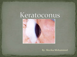
Keratoconus
- 2. Progressive, non-imflammatory ectatic disorder of the cornea Usually bilateral but asymmetric Paraxial stromal thinning and weakening leading to corneal surface distortion
- 4. Primary- irregular astigmatism - myopia Secondary- corneal scarring
- 5. Presents at puberty or early adulthood 50-230 per 100,000 Equal prevalence in both sexes and all races
- 6. Generally unknown, likely multifactorial Suspected: Family history in 6-8% of cases x15-67 higher incidence if first degree relative Eye rubbing Contact lens use Systemic disorders eg. Downs Syndrome, Ehlers-Danlos, Osteogenesis Imperfecta
- 7. All layers of the cornea believed to be affected Epithelial cells may be enlarged and elongated Early degeneration of basal epithelial cells Disruption of basement membrane
- 8. Growth of epithelium posterior to Bowman’s layer forming z-shaped interruptions or breaks Scarring of Bowman’s layer and anterior stroma Stromal thinning due to normal-sized fibres but low numbers of llamelae
- 9. Symptoms: Progression until 4th decade Asymmetric visual complaints Blur and distortions Glare/flare Monocular diplopia Photophobia Initial correction by spectacles then soft contact lenses
- 10. Signs: Slit lamp: Fleisher ring: Iron deposits in epithelial layer at cone base Vogt striae: Vertical stress lines at thinnest part of cornea Central and inferior paracentral corneal thinning Corneal scarring
- 11. Scissor reflex on retinoscopy due to irregular astigmatism Rizzutti’s sign: conical reflection on the nasal cornea when light is shone temporally Munson’s sign: corneal protrusion may cause angulation of the lower lid on downgaze (advanced) Corneal Hydrops: stromal edema due to leakage of aqueous through a tear in descemet membrane
- 12. Vogt Striae Corneal HydropsMunson’s Sign
- 13. Complete history and clinical examination Visual acuity testing Slit lamp examination Retinoscopy- for scissoring reflex Keratometry- may demonstrate irregular mires and progressive corneal steepening Diagnostic rigid contact lenses Corneal Topography
- 14. Maps the corneal curvature Indicates any distortions or scarring Common characteristics: Asymmetrical bowtie Inferior corneal steepening Skewed radia axes
- 15. K value – Measures central steepening of the cornea; ≥ 47.20 D suggests keratoconus I-S value – Measures inferior-versus-superior corneal dioptric asymmetry; ≥ 1.4 D suggests keratoconus KISA% - Incorporates K and I-S values quantifying regular and irregular astigmatism into a single index; 60-100% suggests keratoconus, ≥ 100% strongly suggests frank keratoconus
- 18. Non-Surgical: Spectacle correction- early, as long as visual acuity allows Contact lens- With progressive astigmatism Soft-tonic initially Rigid gas-permeable lenses most common Until corneal irregularity becomes too advanced Collagen cross-linking
- 19. Surgical: Intrastromal corneal ring segments: thin, semi-circular plastic inserts implanted into the mid-corneal layers to flatten the cornea Keratoplasty – 10-15% patients penetrating keratoplasty (full thickness corneal transplant) : most common Deep anterior lamellar keratoplasty (partial thickness corneal transplant)
- 20. Thank you!
- 21. References 1. Espandar L, Meyer J. Keratoconus: Overview and Update on Treatment. Middle East Afr J Ophthalmol [Internet]. 2010 [cited 9 January 2015];. Available from: http://www.ncbi.nlm.nih.gov/pmc/articles/PMC2880369/ 2. Wayman L, Trobe J, Park L. Keratoconus. [Internet]. 2014 [cited 9 January 2015];. Available from: http://www.uptodate.com.ezproxy.sastudents.uwi.tt:2048/contents/keratocon us?source=search_result&search=keratoconus&selectedTitle=1~13 3. Weissman B, Roy H. Keratoconus [Internet]. Medscape. 2014 [cited 9 January 2015]. Available from: http://emedicine.medscape.com/article/1194693- overview#showall 4. Romero-Jiménez M M, Santodomingo-Rubido J, Wolffsohn J. Keratoconus: a review. Elsevier [Internet]. 2010 [cited 9 January 2015];. Available from: http://www.ncbi.nlm.nih.gov/pubmed/20537579 5. OphthaClass. Amsler-Krumeich Classification for Grading Keratoconus - OphthaClass [Internet]. 2015 [cited 9 January 2015]. Available from: http://ophthaclassification.altervista.org/krumeichclass/ 6. Sinjab M. Quick Guide to the Management of Keratoconus A Systematic Step-by-Step Approach. New York: Springer; 2012.