Best Rate (Patna ) Call Girls Patna ⟟ 8617370543 ⟟ High Class Call Girl In 5 ...
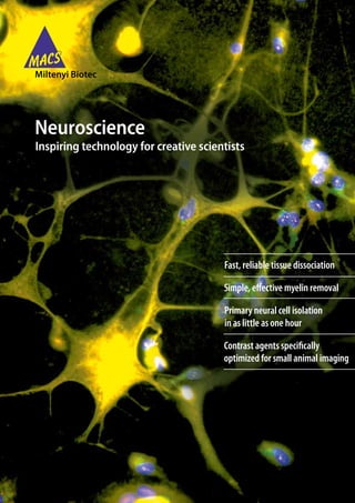
Neuroscience research brochure
1. Neuroscience
Inspiring technology for creative scientists
Fast, reliable tissue dissociation
Simple, effective myelin removal
Primary neural cell isolation
in as little as one hour
Contrast agents specifically
optimized for small animal imaging
2. Contents
Introducing a new milestone in cell analysis
The MACSQuant™ Analyzer paves the way to successful research
3 Sample preparation
• Time-saving and standardized dissociation of neural tissues
• Removal of myelin debris for better results from adult tissue samples
6 Cell separation
• MACS® Technology, the gold standard in cell separation
• Pure astrocytes, microglia, neurons and oligodendrocytes
• Standardization with automated cell separation
9 Cell analysis
• Titrated high-quality antibodies and brilliant fluorochromes
• Easy-to-use bench-top flow cytometers
10 Cell culture
• Serum-free medium and supplement for long-term viability
• Premium, GMP, and research grade cytokines
12 Neuroimaging
• Contrast agents optimized for small animal imaging
• MRI, optical imaging, CT and ultrasound
14 Molecular analysis
• Fast isolation of functional mitochondria
• Efficient mRNA amplification from small samples
• Genomic Services for mRNA and microRNA expression
2
3. Informational brochure
Sample preparation
Get a good start
Tissue dissociation kits Antigen compatibility of
The secret of success in any experiment lies in the preparation Neural Tissue Dissociation Kits
of the starting material. Get going fast with Miltenyi Biotec’s
Table 1 shows the recommended kit regarding antigen
Neural Tissue Dissociation Kits (NTDK) and the gentleMACS®
compatibility for subsequent cell separation or analysis.
Dissociators that help streamline and standardize the
Epitope sensitivities were tested with MACS® Antibodies.
generation of single-cell suspensions.
• Gentle and efficient: titrated enzymes and optimized buffers
Antigen Species Cell type
• Conserved epitopes for optimal downstream applications
Neural Tissue Dissociation Kits (P)
• Convenience: everything in one box, including detailed
protocols Prominin-1 Mouse Neural stem and
progenitor cells
• Reproducible results from standardized components A2B5 Human, mouse, rat Glial-restricted precursors
O4 Human, mouse, rat Immature oligodendrocytes
In order to conserve the epitopes of interest, it is important
to choose the correct protease because some epitopes are CD11b Human, mouse Microglia
degraded by trypsin, others by papain. Two kits were therefore CD81¹ Mouse, rat Microglia, macrophages,
developed, NTDK (T) and NDTK (P). endothelial cells, glia
CD31¹ Mouse Endothelial cells
CD133 Human Neural progenitor cells
AN2 Mouse NG2 glia
NTDK (T) NTDK (P)
(mouse NG2)²
1000 1000
20% 6%
Neural Tissue Dissociation Kits (T)
750 750
Prominin-1² Mouse Neural stem and progenitor
500 500 cells
O4 Human, mouse, rat Immature oligodendrocytes
250 250
PSA-NCAM Human, mouse, rat Neuronal precursors,
0 0 oligodendrocyte progenitors
-1 0 1 10¹ 10² 10³ -1 0 1 10¹ 10² 10³
CD24 Mouse Neuronal precursors,
ependymal cells
GLAST
A2B5³ Human, mouse, rat Glial-restricted precursors
CD11b Human, mouse Microglia
Figure 1: Flow cytometric analysis of cells dissociated with trypsin-
based NTDK (T) and papain-based NTDK (P) and then labeled with CD271 Human Schwann cells, motor neurons
Anti-GLAST-PE. After trypsin-based dissociation the epitope is conserved (LNGFR)
and 20% of the cells are positive for GLAST whereas after papain-based CD105 Mouse Endothelial cells
dissociation the antibody binds to only 6%. Thus, the NTDK (T) is
Ter-119 Mouse Erythrocytes
recommended for use with Anti-GLAST products. For detailed information
visit www.macsneuroscience.com/info and download poster 1. GLAST Human, mouse, rat Astrocytes, radial glia
CD133 Human Neural progenitor cells
If the epitope is not sensitive to proteases, we recommend AN2 Mouse NG2 glia
the papain-based kits. (mouse NG2)²
1 Slight epitope sensitivity with the use of the papain-based kit;
• Neural Tissue Dissociation Kit (T)* and (P)* therefore, a further dilution of the enzyme mix is recommended.
• Brain Tumor Dissociation Kit, human (T)* and (P)* (1:10; 5µL instead of 50 µL)
2 Incubation for re-expression of antigen necessary.
• Neural Tissue Dissociation Kit – Postnatal Neurons (P) 3 Slight epitope sensivity with the use of the trypsin-based kit; therefore,
use of the papain-based kit is recommended
• Neurosphere Dissociation Kit (T) and (P) Epitope sensitivities have been tested with antibodies available from Miltenyi Biotec.
• Embryoid Body Dissociation Kit, human and mouse ** Table 1: Antigen compatibility with papain (P) and trypsin (T)-based kits
*These kits can be used for even more reliable results with the gentleMACS
Dissociators. Protocols for manual dissociation using pipettes are also
available. ** Kit can only be used with the gentleMACS Dissociators.
3
4. Sample preparation
Get a good start
Automated tissue dissociation The gentleMACS Dissociators not only combine reliability and
reproducibility in an easy-to-use system, the resulting cells do
Team the tissue dissociation kits with the gentleMACS not look any different from cells prepared manually.
Dissociators for:
• Ultimate reproducibility, independent of user A
• A safe and sterile closed system
• Push-button technology instead of pipetting
• Processing multiple samples in parallel
“… one of our research focuses is related to the role of
brain-intrinsic immune cells in malignant brain tumors,
especially in the most malignant variant, the
glioblastoma …
The best results were obtained by using the gentleMACS®
system in combination with the Brain Tumor Dissociation
Kit. “ B
Michel Mittelbronn,Ph.D, Institute of Neurology Goethe-University
Frankfurt, Germany
Figure 3: Neuronal precursors in culture. The cells show the same
morphology in culture after (A) manual dissociation using NTDK (T) and
after (B) gentleMACS dissociation using NTDK (T). ßIII-Tubulin (green),
PSA-NCAM (red).
“Your digestion kit has not only improved our yields but
Figure 2: The gentleMACS and gentleMACS Octo Dissociators shown with a
tissue dissociation kit, C-Tubes, and M-Tubes. the preparation has much less debris. I wish I had used it
when we initiated these studies.”
The gentleMACS Dissociators provide different programs for Richard Ciavarra,PhD, Eastern Virginia Medical School, USA
gentle preparation of single-cell suspensions or for
homogenization.
Up to two samples can be processed with the gentleMACS
Dissociator and up to eight samples at a time with the
gentleMACS Octo Dissociator.
Visit www.macsneuroscience.com/videos to watch the
preparation of neural tissue with the gentleMACS
Dissociator.
4
5. Removal of myelin debris
A
Myelin Removal Beads II
before myelin removal
• Effectively removes myelin debris from single-cell
scatter
suspensions
Side scatter
• Use with rodent brain older than 1 week and human samples
Side
• Improves antibody binding in downstream applications
• No need for determination of cell/debris ratio or cell 4.2%
numbers Forward scatter
Forward scatter
“Myelin removal beads allow us to reliably and quickly
separate the “trash from the treasure”. B
Noel Derecki from the Kipnis lab., Department of Neuroscience, after myelin removal
University of Virginia, USA
Side scatter
Sidescatter
without myelin removal with myelin removal
89.0%
Anti-Sca-1-BTtiqa₃ / Anti-Biotin
Anti-Sca-1-BTtiqa₃ / Anti-Biotin
4.48% 16.67%
HK Secondp (g)₀₂βR₃ ow
HK Secondp (g)₀₂βR₃ ow
Forwardscatter
Forward
scatter
CD11b-APC
Figure 5: Removal of myelin debris from single-cell suspensions
with Myelin Removal Beads. Postnatal (P22) mouse brains were
dissociated using the Neural Tissue Dissociation Kit (P) and the resulting
single-cell suspensions analyzed by flow cytometry either before or after
Forward scatter Forward scatter treatment with Myelin Removal Beads. (A) Single-cell suspensions derived
from mouse brain consist of large amounts of myelin membrane fragments
Forward scatter and only 4% cells. (B) Myelin Removal Beads efficiently remove myelin
debris.
Figure 4: Staining of microglia from post-natal (P22) mouse brain with
CD11b-APC.
1×10⁶ cells from a single-cell suspension of P22 mouse brain were stained
with CD11b-APC without (left) and with (right) previous myelin removal
using Myelin Removal Beads. The dot plots show that in samples with
previous myelin removal, higher percentages of CD11b-positive cells are
stained. Dead cells were excluded using propidium iodide. Only the
positive cells along with positive debris are displayed in side and forward
scatters. For detailed information visit www.macsneuroscience.com/info
and download poster 2.
5
6. Cell separation
The fast track to pure, viable cells
Isolation of specific cell types from tissue samples or cell
cultures, e.g., ES or iPS cells, is a prerequisite for exact analysis, Magnetic labeling
efficient screening and optimal cell culture. Gentle to cells;
minimal influence
This is MACS® Technology: the gold standard in bench-top cell on downstream
separation with more than 14,500 publications to prove it. This experiments
renowned technology is also available for neuroscience
research:
• Preparation of astrocytes or microglia in 1 hour instead
of two weeks by the shake-off method
• Gentler procedure than flow sorting, simpler than immuno- Magnetic separation
panning MACS® Column Technology
provides a high-gradient
• Optimal recovery and high purity
N S magnetic field.
• Directly on your bench, no experience required • Gentle to cells
• Sterile sample handling • Thorough rinsing procedure
• High recovery
MicroBeads
• Small, non-toxic, biodegradable
Unlabeled cells are collected in
• Conjugated to highly specific monoclonal antibodies the flow-through
• Compatible with flow cytometry analysis
MACS Columns
• Amplify the magnetic field, only small amounts Elution of the labeled cell
fraction
of MicroBead labeling required
Optimal results–even for rare
• Cell-friendly steel matrix cells–by using positive selection
N S
• Depletion and enrichment of up to 2×10¹⁰ total cells
Watch the isolation of astrocytes with Anti-GLAST
(ACSA-1) MicroBeads on www.neuroscience.com/videos.
A B C
N
Figure 7: The principle of MACS Technology for depletion or enrichment of
Figure 6: The features of MicroBeads and MACS Columns cells.
(A) Scanning electron microscopy of a cell isolated with MACS MicroBeads
(B) 50 nm MicroBeads are so small they can only be seen on a Transmission
Electron Microscope (C). A cross section of a MACS Column showing
the steel ball matrix (gray) with the magnetic field in which labeled cells
(purple) are retained.
6
7. Direct conjugates of monoclonal antibodies and MicroBeads “We are studying neuron-glia interactions using primary
are optimized and already titrated for you cultures of highly purified neurons and glial cells.
Previously, we isolated cells by immunopanning.
Cell type Product Species
… we switched to magnetic cell isolation kits from
Neurons Miltenyi Biotec because of three advantages. First, the
cell preparation is more economical, second, it takes two
Retinal ganglion Retinal Ganglion Cell
cells Isolation Kit Rat instead of six hours, and third, it delivers very pure,
healthy cells.”
Neuronal Anti-PSA-NCAM-MicroBeads Human,
precursors mouse, rat Frank W. Pfrieger Ph.D. Institute of Cellular and Integrative
Neurons Neuron Isolation Kit Mouse Neurosciences (INCI), France
Astrocytes
Astrocytes and Anti-GLAST (ACSA-1) Human,
radial glia MicroBead Kit mouse, rat Original Negative Positive
fraction fraction fraction
Astrocytes Anti-ACSA-2 MicroBeads
9.1% 0.75% 98.7%
(coming soon)
CD11b
Oligodendrocytes
Immature Anti-O4 MicroBeads Human,
oligodendrocytes mouse, rat
Oligodendrocyte Anti-A2B5 MicroBeads Human,
precursors mouse, rat
Forward scatter
NG2 glia/ Anti-AN2 MicroBeads Mouse
Polydendrocytes
Figure 8: MACS Technology enables isolation of cells even from adult
Schwann cells CD271 (LNGFR) MicroBead Kit Human
tissue samples. Enrichment of human microglia from a glioblastoma
Neural progenitors sample achieved a purity of 99%. For detail information visit
www.macsneuroscience.com/info and download poster 3.
Neural progenitors CD133 MicroBead Kit Human
Neural progenitors Anti-Prominin-1 MicroBeads Mouse
A B
Neural crest cells CD271 (LNGFR) MicroBead Kit Human
ES/iPS derived Neural Crest Stem Cell
Neural crest cells MicroBeads (coming soon)
Microglia
Microglia CD11b (Microglia) MicroBeads Human,
mouse
T cells
T helper cells CD4 (L3T4) MicroBeads Mouse
Regulatory T cells CD4+CD25+ Regulatory T Cell Mouse Figure 9: MACS Technology is optimal for effective downstream
Isolation Kit, mouse applications. (A) Human brain biopsies were dissociated using the Brain
Tumor Dissociation Kit (P) and the gentleMACS Dissociator. Myelin was
Dendritic cells removed using Myelin Removal Beads, and cells isolated using CD11b
(Microglia) MicroBeads. The microglia show a normal morphology.
Dendritic cells CD11c MicroBeads, mouse Mouse (B) Astrocytes (7 DIV) isolated from P1 mice using the Anti-GLAST (ACSA-1)
Table 2: Examples of cell separation reagents available for neuroscience MicroBead Kit. Astrocytes were stained using Anti-GLAST (ACSA-1) (green)
research and Anti-GFAP (red). Cells were cultured in MACS® Neuro Medium and
MACS® Supplement B27 PLUS, 0.5 mM L-glutamine and 1% Pen/Strep on
poly-D-lysine-coated coverslips.
7
8. Cell separation
The fast track to pure, viable cells
Manual cell separation
All that is necessary for manual separation is a MultiStand with
a MACS Separator, columns and our separation reagents.
The Starter Kits are the easiest way to begin cell separation:
• Simple: the kit contains everything you need – separator,
columns and MicroBeads
• Flexible: order with antigen-specific MicroBeads of your
choice
• Value: save on the price of individual components
Automated cell separation
The autoMACS Pro® Separator is a benchtop automated Figure 10: OctoMACS Separator with MS Columns on MACS MultiStand
magnetic cell sorter for the isolation of virtually any cell type
from any species:
• Convenient: standardized walk-away cell isolation
• Versatile: isolate virtually any cell type, and also remove
myelin debris
• Simple: an intuitive touchscreen interface and preset
programs for optimal results
• Flexible: with high-speed sorting of more than 10 million
cells per second and volumes from 0.2 mL to 50 mL.
• Time-saving: Multiple samples with less hands-on time
“In contrast to very low numbers of microglia obtained
with conventional cellular depletion methods, we could
increase the purity to more than 95% by using CD11b
Microglia MicroBeads.
Figure 11: autoMACS Pro Separator
Based on our long-term experience, we can highly
recommend the products from Miltenyi Biotec. These
products allow a time-efficient and reproducible cell
separation, also in a high-throughput manner and with
an excellent quality.“
Michel Mittelbronn,Ph.D, Institute of Neurology Goethe-University
Frankfurt, Germany
8
9. Cell analysis
Analyze millions of cells in seconds
Revolutionize neural cell analysis with a range of premium
quality antibodies, brilliant fluorochromes, and novel
MACSQuant® Analyzers
instrumentation. Discover the benefits of flow cytometry. • Easy to use, best in class, flow cytometry for experts and
• As an alternative to immunohistochemistry and beginners alike
cytochemistry, a flow cytometer will measure millions of • 3 Lasers and up to 10 optical parameters
cells in seconds and enables the analysis of cell populations • Absolute cell counting
using multiple markers for a more accurate assessment of
the whole cell population • Rare cell analysis
• Complementing Western Blotting, a flow cytometer can • 96-well automation
analyze proteins with quantitative analysis on a cell for cell • Compact bench-top design also adds value to a core facility
basis, analyzing up to eight proteins at once rather than one • Live, around the clock, remote support
at a time
Flow cytometry enables the exact quantification of cell
populations and analysis of overlapping markers. Dot plots
depict cells and smaller particles as dots (events) and illustrate
a staining for a marker by a shift on the respective axis.
A B C
1.9% 4.8% 3.2% 4.9% 4.3% 0.4%
AN2-PE
14.0% 11.6% 4.4%
A2B5-APC
Figure 13: The MACSQuant Analyzer family. MACSQuant Analyzer:
a bench-top cell analyzer for highly sensitive multicolor flow analysis, with
Figure 12: Cells from P1 (A), P3 (B) and P7 (C) mouse cortex were
up to ten parameters. MACSQuant VYB: a versatile optical layout, powered
stained with Anti-AN2-PE and Anti-A2B5-APC. AN2+ cells are in the
by a 561 nm laser for fluorescent proteins like GFP, YFP and mCherry.
two upper quadrants, while A2B5+ cells are in the upper right and lower
right quadrants. The percentage of cells that are positive for both markers
is shown in the upper right quadrant. The dot plots show a significant See for yourself how MACS Cell Analysis can be at the
decrease of A2B5 positive cells with increasing animal age, so that the
heart of your experiments. Watch the video on
overlap with AN2 disappears.
www.macsneuroscience.com/videos
MACS Antibodies
Brilliant fluorochromes and high quality antibodies for brighter
staining and even better data, particularly for flow cytometry.
Choose from a large panel of antibodies against mouse, rat and
human antigens.
For neural markers please visit www.macsantibodies.com
to download the complete antibody list
9
10. Cell culture
The medium makes the difference
The MACS Cell Culture product line for neuroscience includes A
specially formulated cell culture medium, cytokines and growth
factors. Get the best for your cells and promote their growth
and differentiation in vitro , by working with a great team.
MACS Supplement B27 PLUS
and MACS Neuro Medium
• Serum-free supplement for astrocytes, neurons and
oligodendrocytes
• Based on B27 with optimized components for in vitro
propagation of all mouse, rat, or human neural cells
• Serum-free cell culture medium
• Promotes optimal growth and long-term survival of all
B
mouse, rat, or human neural cells
Related products:
Small molecules
Laminin
Stem cell-specific media
Visit www.macs-stemcells.com for more information
Basic media: Visit www.macscellculture.com
MACS Cytokines
A comprehensive range of cytokines is available for the
promotion of neural differentiation and cell maintenance. Figure 14: (A) Astrocytes and (B) oligodendrocytes show excellent
growth and morphology in MACS Neuro Medium and MACS
• Superior quality including premium and GMP-grade Supplement B27 PLUS, 0.5 mM L-glutamine and 1% PenStrep on
poly-D-lysine-coated coverslips. Astrocytes (4 DIV) were isolated from P1
• Standardized high biological activity
mice using Anti-ACSA-2 MicroBeads (coming soon), and oligodendrocytes
• Convenient packaging with small or bulk fillings were (5 DIV) isolated from P3 mice using Anti-O4-MicroBeads. Astrocytes
were stained using Anti-GLAST (ACSA-1) (green) and Anti-GFAP (red).
Visit www.macscytokines.com to download the cytokine
product list.
10
11. A
Figure 16: Neural/neuronal cells differentiation of human iPS cells to
B peripheral neurons.Pluripotent human iPS cells were isolated using the
TRA-1-60 MicroBead Kit prior to neural induction with dorsomorphin. At day
10 of differentiation, neural crest stem cells were enriched with Neural Crest
Stem Cell MicroBeads. Differentiation to peripheral neurons was performd
in MACS Neuro Medium with N2 Supplement, NGF, BDNF, dcAMP and
ascorbic acid for three weeks. For more detailed information visit
www.macsneuroscience.com/info and download poster 4.
Figure 15: Whole mouse brain was dissociated using the Neural Tissue
Dissociation Kit (P) and the gentleMACS Dissociator. Neural stem cells
were isolated with Anti-Prominin-1 MicroBeads and subsequently cultivated
in MACS Neuro Medium supplemented with MACS Supplement B27 PLUS,
penicillin/streptomycin, 2 mM L-glutamine, 20 ng/mL MACS Cytokines
Human EGF and Human FGF-2. Primary neurospheres formed after one
week in culture. (A) The Neurosphere Dissociation Kit (P) was used for the
dissociation of primary neurospheres. (B) Secondary neurospheres formed
in the same medium and cytokines.
11
12. Neuroimaging
Insights into neural function
Small animal imaging (SAI) techniques are used to investigate
models of human disease, such as Alzheimer’s Disease.
Viscover quality
State-of-the-art instrumentation coupled with sensitive and • Ready-to-use with step-by-step instructions
specific contrast agents, such as the Viscover™ range of • Excellent tolerability – only 100 μL injection volume
pre-clinical contrast agents, allow researches to obtain
high-fidelity anatomical and functional information from • Translatable to the clinic
animal studies in vivo. • Iso-osmolar sterile formulation
• Single dose bolus administration
Modality Product Applications SAI techniques have evolved from human applications in the
group
clinic, including magnetic resonance imaging (MRI), computer
MRI GadoSpin™ • Brain tumors tomography (CT), ultrasound (US) and optical imaging (OI). Of
• Compromised BBB these, MRI and OI are frequently used in neuroscience
• Neuroinflammation investigations in small animals although there are Viscover
• Stroke products to suit each application:
• MS
•
•
Angiography
DCE measurements
Magnetic resonance imaging
• Angiogenesis GadoSpin™ contrast agents are based on gadolinium chelate
• Molecular targeted imaging and can be used to examine angiogenesis in brain tumors and
FeraSpin™ • Different size-selected nanoparticles neuroinflammation where the blood-brain barrier has been
• CNS inflammation compromised.
• Osteomyelitis and aseptic vertebral
inflammation
• Stroke
• MS
• Alzheimer disease
• Angiography
• rCBV
Figure 17: Intracranial tumor in mouse exhibits contrast enhancement
• Microvessel density imaging after GadoSpin M has passed the compromised blood-brain barrier.
• Cell tracking The image series shows tumor progression.
• Molecular targeted imaging
FeraTrack™ Ready-to-use iron oxide particles for
ex vivo intracellular labelling of cells
prior to transplantation and tracking
CT ExiTron™ • Soft tissue contrast
• Angiography
• Angiogenesis
Ultrasound PolySon™ • Perfusion studies
• Molecular targeted imaging
(see figure 22)
Optical NiroWave™ • CNS inflammation
Imaging • Stroke
• MS
• Alzheimer's disease
• Angiography
• Vascular leakage Figure 18: MRI image of mouse after intracerebral injection of
GadoSpin F and intravenous injection of Gadospin D and FeraSpin XS.
• Angiogenesis
Image shows brain and spinal cord (purple) and the vasculature (red).
• Brain tumor
Table 3: SAI applications and Viscover imaging agents
12
13. FeraSpin™ contrast agents are based on iron oxide
nanoparticles and can be used for various neuroscience
Optical imaging
applications such as CNS inflammation studies, MRA, rCBV NiraWave™ optical imaging contrast reagents enable the study
measurements or cell tracking. of disease-related changes in vascular permeability, for
example, the breakdown of the blood-brain barrier. NiraWave
can also be conjugated to antibodies in the same way as
fluorochromes, to enable targeting.
Figure 19: Mouse whole-body T1-weighted MR angiography using FeraSpin
XS.
The FeraTrack tracking kit simplifies intracellular labeling of cells
Figure 21: Optical angiography in mouse injected with NiraWave nano
ex vivo. Cells labeled with FeraTrack can then be tracked in real
780. The fine structure of blood vessels is visible.
time by magnetic resonance imaging.
Ultrasound imaging
PolySon™ Ultrasound microbubbles have been used, for
example, to detect the distribution of lesions and to quantify
the expression of ICAM-1 in the study of experimental allergic
encephalitis in the mouse:
Figure 20: In vivo MRI imaging of mouse cortex: Extracellularly and
intracellularly FeraTrack™ labeled PSA-NCAM + cells were grafted into
the cortex of a mouse. A strong contrast was detected for the FeraTrack™
labeled cells in the left cortex at 7 Tesla MRI (Prof M. Hoehn, Max Planck
Institute for Neurological Research, Cologne).
Find out more at www.viscover.com
Figure 22: Detection of compromised BBB in EAE mouse model by i.v.
injection of anti-ICAM-1 conjugated PolySon H microbubbles using
Ultrasound Imaging: Anti-ICAM-1-PolySonH binds specifically on the
surface of lesions that express ICAM-1.
13
14. Molecular analysis
Look below the cell surface
Experience how MACSmolecular provides exquisite sensitivity
in cDNA synthesis, outstanding purity in protein isolation, high
Genomic Services - make the most of
recovery in mitochondria isolation, and enables expression small samples
profiling of both microRNA and mRNA.
If sample material is minimal or is only available as formalin-
fixed paraffin-embedded tissue, Genomic Services can extract
More than just a power house that valuable information and provide you with the data you
are seeking:
• SuperAmp: analysis of 10-10,000 cells by million-fold
amplification
• microRNA expression profiling based on
miRXplore™ Microarrays
• Agilent whole genome expression or array-CGH analysis
and bioinformatics
• Cell characterization, disease biology, biomarker
identification
Send us your sample and we’ll send you documented
gene expression data by return.
Visit us at www.miltenyibiotec.com/genomicservices.
Are you studying the role of mitochondria in
neurodegenerative disease or aging? The Mitochondria
Isolation Kit, human and the Mitochondria Isolation Kit,
mouse tissue, are based on renowned MACS Technology
(see page 6) and enable:
• Pure, intact, viable mitochondria in less than two hours
• Higher yields of mitochondria compared to differential
centrifugation or density gradient centrifugation
• Organelle integrity maintained for full functionality
Watch the Mitochondria Isolation Kit in action.
Visit www.macsneuroscience.com/videos.
μMACS™ and MultiMACS™ cDNA For more information on our entire molecular range visit
Synthesis Kits www.macsmolecular.com.
• Time-saving with one step instead of three: combination of
mRNA isolation with in-column cDNA synthesis. With the
cDNA magnetically bound to the column, no extra cDNA
purification step is needed.
• High yields
• Ready-to-use enzyme reaction mixes in the complete kit
14
15. Further information
Support and links
Technical support
We are well known for the high level of support our team of
experts provides our customers. We can help with any question
you have about our products by:
• On-line live technical support (New)
• Email
• Telephone
• On-line discussion forums
There are also many articles available for download including
• Scientific posters
• MACSmore – Neuroscience Special
• Datasheets
Simply visit www.miltenyibiotec.com/support
We also run customer training at our HQ near Cologne,
Germany and on-line customer webinars
For ordering information and the latest news on our
products:
macsneuroscience.com
info@macsneuroscience.com
The proof is in the publications!
Visit www.macsneuroscience.com/references for the
extensive list of literature on successful research conducted
with our neuroscience products.
15