SlideShare uma empresa Scribd logo
1 de 8
Baixar para ler offline
372       DUODENAL INJURIES

related to almost half the deaths in this series. In fact, delays                   6. Sinha R, Sharma N, Joshi M: Laparoscopic repair of small bowel perfora-
exceeding as little as 8 hours resulted in increased morbidity and                     tion. J Soc Laparoendosc Surg 9:399–402, 2005.
mortality.                                                                          7. Maull KI, Reath DB: Impact of early recognition on outcome in nonpen-
    Mortality after penetrating injury is most commonly related to                     etrating wounds of the small bowel. South Med J 77:1075–1077, 1984.
                                                                                    8. Brasel KJ, Olsen CJ, Stafford RE, Johnson TJ: Incidence and significance
injury to other intraperitoneal and/or retroperitoneal injuries.22                     of free fluid on abdominal computed tomographic scan in blunt trauma.
                                                                                       J Trauma 44:889–892, 1998.
                                                                                    9. Malhotra AK, Fabian TC, Katsis SB, et al: Blunt bowel and mesenteric
      CONCLUSIONS                                                                      injuries: the role of screening computed tomography. J Trauma 48:
                                                                                       991–1000, 2000.
Small bowel injuries may follow blunt or penetrating trauma. The                   10. Allen TL, Mueller MT, Bonk T, et al: Computed tomographic scanning
major concern in the patient who sustains blunt small bowel injury                     without oral contrast solution for blunt bowel and mesenteric injuries in
is recognizing the presence of the injury. In patients with associated                 abdominal trauma. J Trauma 56:314–322, 2004.
injuries requiring operation or in those sustaining gunshot wounds,                11. Gonzalez RP, Ickler J, Gachassin P: Complementary roles of diagnostic
                                                                                       peritoneal lavage and computed tomography in the evaluation of blunt
small bowel injuries are promptly discoverable. The trend toward                       abdominal trauma. J Trauma 51:1128–1136, 2001.
CT-based nonoperative management and the inaccuracy of early                       12. Mitsuhide K, Junichi S, Atsushi N et al: Computed tomography scanning
postinjury CT places patients with isolated blunt small bowel injury                   and selective laparoscopy in the diagnosis of blunt small bowel injury: a
at risk of delayed diagnosis and increased morbidity and mortality.                    prospective study. J Trauma 58:696–703, 2005.
The importance of injury mechanism and physical findings is often                   13. Brownstein MR, Bunting T, Meyer AA, Fakhry SM: Diagnosis and man-
overlooked. Patients who are neurologically intact will demonstrate                    agement of blunt small bowel injury: a survey of the membership of the
abdominal tenderness; many will have peritoneal findings at presen-                     American Association for the Surgery of Trauma. J Trauma 48:402–407,
tation. The presence of a seat-belt contusion should elevate concern.                  2000.
Injuries that can be repaired by lateral enterorrhaphy rarely cause                14. Kaban G, Somani RA, Carter J: Delayed presentation of small bowel
                                                                                       injury after blunt abdominal trauma. J Trauma 56:1144–1145, 2004.
postoperative complications, which are more commonly related to                    15. Hackam DJ, Ali J, Jastaniah SS: Effects of other intra-abdominal injuries
associated injuries after both blunt and penetrating trauma. Surgical                  on the diagnosis, management and outcome of small bowel injuries.
judgment is required to ensure early diagnosis and appropriate op-                     J Trauma 49:606–610, 2000.
erative management.                                                                16. Fakhry SM, Brownstein M, Watts DD, et al: Relatively short diagnostic
                                                                                       delays (Ͻ8 hours) produce morbidity and mortality in blunt small bowel
REFERENCES                                                                             injury: an analysis of time of operative intervention in 198 patients from
                                                                                       a multicenter experience. J Trauma 48:408–415, 2000.
 1. Nance ML, Peden GW, Shapiro MB, et al: Solid viscus injury predicts            17. Fang JF, Chen RJ, Lin BC, et al: Small bowel perforation: is urgent surgery
    major hollow viscus injury in blunt abdominal trauma. J Trauma 43:                 necessary? J Trauma 47:515–520, 1999.
    618–623, 1997.                                                                 18. Bensard DD, Beaver B, Besner GE, Cooney DR: Small bowel injury in
 2. Velitchkov NG, Losanoff JE, Kjossev KT, et al: Delayed small bowel injury          children after blunt abdominal trauma: is diagnostic delay important?
    as a result of penetrating extraperitoneal high-velocity ballistic trauma to       J Trauma 41:476–483, 1996.
    the abdomen. J Trauma 48:169–170, 2000.                                        19. Neugebauer H, Wallenboeck E, Hungerford M: Seventy cases of injury of
 3. Fakhry SM, Watts DD, Luchette FA: Current diagnostic approaches lack               the small intestine caused by blunt abdominal trauma: a retrospective
    sensitivity in the diagnosis of perforated small bowel injury: analysis from       study from 1970 to 1994. J Trauma 46:116–121, 1999.
    275,557 trauma admissions form the EAST multi-institutional HVI trial.         20. Maull KI: Stomach, small bowel and mesentery injury. In Dudley H,
    J Trauma 54:295–306, 2003.                                                         Carter D, Russell RC, editors: Operative Surgery. London, Butterworths,
 4. Shaftan GW: Indications for operation in abdominal trauma. Am J Surg               1989, pp. 401–413.
    99:657–664, 1960.                                                              21. Kirkpatrick AW, Baxter KA, Simons RK, et al: Intra-abdominal complica-
 5. Chiu WC, Shanmuganathan K, Mirvis SE, Scalea TM: Determining the                   tions after surgical repair of small bowel injuries: an international review.
    need for laparotomy in penetrating torso trauma: a prospective study               J Trauma 55:399–406, 2003.
    using triple-contrast enhanced abdominopelvic computed tomography.             22. Stevens SL, Maull KI: Small bowel injuries. Surg Clin North Am 70:
    J Trauma 51:860–869, 2001.                                                         541–560, 1990.


DUODENAL INJURIES                                                                  in published reports sustaining penetrating trauma.2 This figure may
                                                                                   primarily be a reflection of the experience of urban trauma centers
Gregory J. Jurkovich                                                               where penetrating mechanism are more prevalent, and of academic
                                                                                   centers that publish their results. Penetrating duodenal wounds are usu-
                                                                                   ally rapidly diagnosed as part of a laparotomy and evaluation of the
                                                                                   tract of the offending agent. But blunt duodenal injuries are often more


d
                                                                                   insidious in their presentation, making the initial diagnosis difficult.
       uodenal injuries are uncommon, but not so rare as to preclude a             Despite this well-known observation, delays in the diagnosis of duode-
       comprehensive understanding of treatment strategies by general              nal trauma continue to plague trauma surgeons and seriously compro-
surgeons. A 6-year statewide review in Pennsylvania documented a                   mise patient care.3
0.2% incidence of blunt duodenal injury (206 of 103,864 trauma regis-
try entries), and only 30 of these patients had full-thickness duodenal
injuries.1 Blunt duodenal injuries are the result of a direct blow to the               DETERMINANTS OF OUTCOME
epigastria, which in adults is usually from a steering wheel injury in an
unrestrained driver, and in children is the result of a direct blow from a         Directly attributable duodenal mortality ranges from 2%–5%, and
bicycle handlebar, fist, or similar mechanism. Penetrating wounds are               is the result of the common complications of wound dehiscence,
more common causes of duodenal injury, with about 75% of patients                  sepsis, and multiple organ failure.4–11 Associated causes of mortality
ABDOMINAL INJURIES          373

in patients with a duodenal injury can be garnered from large series         portion (and the remainder of the duodenum) is entirely retroperi-
of duodenal injuries reported during the late 20th century. These            toneal; this is the segment mobilized by a Kocher maneuver. The
reports demonstrated an average mortality in patients with a duo-            transverse or third portion of the duodenum runs 12 cm horizontally
denal injury of 18%, but with great individual report variability,           to the left in front of the ureter, inferior vena cava, lumbar column,
ranging from 6%–29%.6,12–14 Morbidity rates after duodenal injury            and aorta, and ends at just at the left edge of the third lumbar verte-
range from 30%–63%, although only about a third of these are di-             bra. The superior mesenteric artery runs downward over the anterior
rectly related to the duodenal injury itself.6,9,12 Reasons for this vari-   surface of the third portion of the duodenum. The ascending or
ability in morbidity and mortality statistics include the mechanism          fourth portion of the duodenum runs upward and slightly to the left
of injury, associated injuries, and time to initial diagnosis. For ex-       for only a short distance (2–3 cm) alongside the spine to the duode-
ample, Ivatury and colleagues’ review of 100 consecutive penetrat-           nal suspensory ligament of Treitz.
ing duodenal injuries documented a 25% mortality rate,6 compared                 The arterial blood supply of the duodenum is derived from the
with mortality rates of 12%–14% in patients with blunt injury                pancreaticoduodenal artery. The superior branch comes off the he-
mechanisms.3,9                                                               patic artery, and the inferior branch from the superior mesenteric
    Early death from a duodenal injury, particularly with penetrating        artery. These two arteries run in a groove between the descending
wounds, is caused by exsanguination from associated vascular, liver,         (second) and transverse (third) portions of the duodenum and the
or spleen injuries.15,16 The proximity of the duodenum to other vital        head of the pancreas, with well-developed collateralization via a con-
structures makes isolated injuries uncommon, but not unheard of.             tinuous marginal artery. The venous drainage parallels the arterial
While exsanguinating hemorrhage and associated injuries are re-              supply, with the posterosuperior arcade draining into the portal vein
sponsible for early deaths, infection and multiple-system organ fail-        and the anteroinferior arcade draining into the gastrocolic trunk.21
ure are responsible for most late deaths. Up to one-third of patients            The duodenal mucosa resembles that of the remainder of the
who survive the first 48 hours develop a complication related to the          small bowel, with the characteristic histologic feature of the submu-
duodenal injury. Anastomotic breakdown, fistula, intra-abdominal              cosal Brunner’s glands in the most proximal (first) portion. The vis-
abscess, pneumonia, septicemia, and organ failure are the common             cous, mucoid, alkaline secretion of these glands probably affords
complications. Late deaths in patients with a duodenal injury typi-          some protection to the duodenum from gastric acid and serves to
cally occur 1–2 weeks or more after the injury, with about one-third         begin neutralization of this acid. The mixing of pancreatic and bile
of the late deaths attributable to the injury itself.9,12                    juices with the gastric efflux also normally occurs in the duodenum.
    The time from injury to definitive treatment is also an important         The duodenum sees an average of 2500 ml gastric juice, 1000 ml of
factor in the development of late complications and subsequent               bile, 800–1000 ml of pancreatic juices, and 800 ml of saliva, for a
mortality. Roman and colleagues17 identified 10 patients in whom              total of about 5 liters of combined flow through the duodenum per
the diagnosis of duodenal injury was delayed over 24 hours; 4 of the         day. Such massive flow volumes make it clear that duodenal integrity
10 died, and 3 of the 10 had duodenal fistulas. In a true trauma clas-        is crucial, and help to explain why duodenal fistulae can be such a
sic report, Lucas and Ledgerwood18 demonstrated the remarkable               difficult complication of injury to this organ.
importance (and frequency) of a delay in diagnosis of duodenal in-
jury. In their report, a delay in diagnosis of more than 12 hours oc-
curred in 53% of their patients, and a delay of more than 24 hours in            DIAGNOSTIC ADJUVANTS
28%; mortality was 40% among the patients in whom the diagnosis
was delayed greater than 24 hours, as opposed to 11% in those un-            The radiologic signs of duodenal injury on the initial plain abdomi-
dergoing surgery within 24 hours. Snyder and coworkers9 confirmed             nal or upright chest radiograph are often quite subtle, with mild
these observations, noting that of the four patients with blunt duo-         spine scoliosis or obliteration of the right psoas muscle being occa-
denal trauma in their series in whom the diagnosis was delayed, two          sionally all that suggests a retroperitoneal duodenal injury. The pres-
died and the other two developed duodenal fistula. Cuddington and             ence of air in the retroperitoneum is a clear sign of duodenal injury,
associates3 also noted that 100% of the deaths directly attributable to      but this is often difficult to distinguish from the overlying transverse
duodenal injury occurred in patients in whom there was a delay in            colon. Computed tomography (CT) at the current time is the best
diagnosing such injury.                                                      method of early diagnosis of a duodenal injury, but it is not infallible.
    The implication of these observations is that the first priority in       In a 1997 report describing a 6-year statewide experience with
managing duodenal trauma should be control of hemorrhage. The                duodenal injuries, Ballard et al.1 reported that of 30 documented
next priority is limiting bacterial contamination from colon or other        blunt duodenal injuries, the initial CT scan missed 27%. The (CT)
bowel injury to prevent late infections. A clear identification of the        scan must be performed with both oral and intravenous contrast
extent of the duodenal injury should follow as the next priority, with       (Figure 1). The exam must be interpreted with great suspicion for
an emphasis on determining the status of the pancreas as well, as this       injury, and uncertainty in interpretation is adequate justification for
affects definitive treatment plans.13,19                                      operative exploration. False-negative exams are known to occur.22 In
                                                                             one careful study of the accuracy of CT in diagnosing duodenal and
                                                                             other small bowel injuries, only 59% (10 of 17) scans were prospec-
    ANATOMY AND PHYSIOLOGY                                                   tively (preoperatively) interpreted as suggestive for bowel injury,
                                                                             which increased to 88% (15 of 17 injuries) when evaluated retrospec-
The duodenum is the first portion of the small intestine, beginning           tively.23 These investigators emphasized that using CT for the diagno-
just to the right of the spine at the level of the first lumbar vertebra      sis of blunt bowel rupture requires careful inspection and technique
and extending from the pyloric ring to the duodenojejunal flexure,            to detect the often-subtle findings.
commonly known as the ligament of Treitz. The duodenum is named                  A more cumbersome alternative to CT is upper gastrointestinal
from the Latin word duodeni, which means “twelve each,” because it           series with water-soluble contrast medium followed by barium if the
is in total 25 to 30 cm, or about 12 fingerbreadths, in length. For           initial exam is negative. Some have advocated this study if the initial
convenience of description, the duodenum is arbitrarily divided into         CT is difficult to interpret, but I would argue that subtle findings on
four divisions, differentiated by the alteration in direction of the         CT are adequate justification for operative exploration. In a series of
organ.20 The superior or first portion of the duodenum passes back-           96 patients with CT findings suspicious for duodenal injury, the
ward and upward toward the neck of the gallbladder, and most                 sensitivity of a subsequent duodenography was 54% with a specific-
of this portion is intraperitoneal. The descending (vertical) or second      ity of 98%.24 For those injuries requiring operative repair, the sensi-
portion forms an acute angle with the first portion and descends              tivity was only 25%, with a 25% false-negative rate. Allen et al.25
7–8 cm. It contains the bile and pancreatic duct openings. This              demonstrated that 83% of the patients with a delay in the diagnosis
374      DUODENAL INJURIES


A                                                                                                                                                      B


    Figure 1    Duodenal perforation. Nineteen-year-old female dropped a 185-lb barbell on abdomen, presented to emergency department
    4 hours later with mild abdominal pain and nausea. Afebrile, hemodynamics stable, persistent epigastric tenderness without peritoneal signs,
    WCC 16 K, amylase 114. Arrow points to nonluminal air in the retroperitoneum and free extravasation of contrast.


of blunt duodenal injury had subtle CT findings, including pneumo-           Table 1: Determinants of Duodenal Injury Severity
peritoneum, unexplained fluid, and unusual bowel morphology
that were dismissed. These authors emphasized the point that subtle                                            Mild                     Severe
findings of duodenal injury on abdominal CT should mandate                   Determinants of Injury Severity
laparotomy.
    Diagnostic peritoneal lavage (DPL) is unreliable in detecting iso-       Agent                            Stab                 Blunt or missile
lated duodenal and other retroperitoneal injuries. Nevertheless, DPL         Size                           Ͻ75% wall                Ն75% wall
is often helpful because approximately 40% of patients with a duo-           Duodenal site                    3, 4                       1, 2
denal injury have associated intra-abdominal injuries that will result
in a positive peritoneal lavage. The findings of amylase or bile in the
                                                                             Injury-repair                    Ͻ24                       Ն24
lavage effluent are more specific indicators of possible duodenal in-           interval (hr)
jury. Serum amylase levels are nondiagnostic as well, but if elevated        Adjacent injury               No CBD                       CBD
additional investigation (CT or celiotomy) for the possibility of pan-                                No pancreatic injury         Pancreatic injury
creatic or duodenal injury is warranted. At celiotomy, the presence of      Outcome
any central upper abdominal retroperitoneal hematoma, bile stain-
ing, or air mandates visualization and a thorough examination of the         Mortality (%)                      6%                       16%
duodenum. Asensio and colleagues26 have published the technical              Duodenal                           6%                       14%
details of exposing the entire duodenum and pancreas.                         morbidity (%)

                                                                            CBD, Common bile duct.
    TREATMENT
Treatment principles are governed by the severity of duodenal injury
and the likelihood of postrepair complications. Approximately 70%           rithm approach to the management of duodenal injuries is provided
of duodenal wounds can be safely repaired primarily, and the re-            in Figure 2.27 A classification system for all organ injuries has been
maining 30% are “severe” injuries that require more complex proce-          developed by the American Association for the Surgery of Trauma
dures. Snyder and colleagues9 are credited with cataloging the factors      (AAST) and is known as the Organ Injury Scale (OIS).28 This classi-
that determine whether a duodenal wound can be primarily repaired.          fication for duodenal wounds is listed in Table 2, with duodenal
In their review of 247 patients treated for duodenal trauma, they re-       injuries graded from I to V, minor to major injury. In a review of
ported an overall duodenal fistula rate of 7% and a mortality rate of        164 duodenal trauma patients managed at eight trauma centers and
10.5% in the 228 patients surviving for greater than 72 hours. These        to whom the AAST/OIS classification scheme was applied, there were
investigators felt that five of the factors listed in Table 1 most signifi-   38 class I, 70 class II, 48 class III, four class IV, and four class V inju-
cantly correlate with the severity of duodenal injury and subsequent        ries. Primary repair alone was performed in 71% (117) of all cases.4
morbidity and mortality. A more recent addition, as noted in the            Primary duodenal repair was performed in 90 of 108 patients with
table, is the presence of a pancreatic injury, a significant predictor of    class I or II injuries. More complex duodenal treatment strategies,
late morbidity and mortality. Each of these factors, either individu-       including pyloric exclusion, duodenoduodenostomy, duodenojeju-
ally or in combination, has been used to develop a variety of duode-        nostomy, or pancreatoduodenectomy, were employed in 26 of
nal injury classification systems. Snyder and colleagues9 demon-             56 (46%) patients with class III to V injuries, or 29% of the total
strated that patients with “mild” duodenal trauma had 0% mortality          population.
and 2% duodenal fistula rate, as compared with 6% mortality and                  Primary repair is usually the simplest, fastest, and most appropri-
10% fistula rate among those with severe duodenal injuries. In gen-          ate way to manage duodenal injuries.29 Primary repair is appropriate
eral, patients with a “mild” duodenal injury and no pancreatic injury       for complete transection of the duodenum if there is little tissue loss,
can be primarily repaired. Patients with more severe duodenal inju-         if the ampulla is not involved, and if the mucosal edges can be de-
ries may require more complex treatment strategies. A useful algo-          brided and closed without tension. The repair is done as with any
ABDOMINAL INJURIES             375

                                                                                                             F
                                                                                                Detected at             Evacuation of
                                                                                                laparotomy               hematoma
                                                              Hematoma                                        G
                                              A                                                                          NG suction
                                                                                              Detected non-
                                                                                                                            TPN
           History and physical examination                                                   operatively
                                                                                                                         Observation
                  Abdominal trauma
                                                                                                                 H
                                                              Grade I/II simple
                                                                                              Suture closure            Pyloric exclusion
                                                                 laceration
                                                       E                                                                                     J
                       Duodenal          Inspection at                                                                   Primary repair or
                        Injury            laparotomy                                          Proximal to                   antrectomy
                                                                                               ampulla                   Gastojejunostomy
                                                                                     I
                                                                                                                          Stump closure
                                                              Grade III laceration
                                                                                                                           Primary repair or
                                        B                                                     Distal to
                         Labs                                                                                                 Roux-en-Y
                                                                                              ampulla
                    Abdominal x-ray     C                                                                                duodenojejunostomy
                    Contrast studies
                                        D
                                                                                         K    Complex biliary
                                                                                              reconstruction
                                                              Grade IV/V laceration
                                                                 or destruction
                                                                                               Pancreatico-
                                                                                              duodenectomy

           Figure 2     Algorithm for the management of duodenal injuries. (From Jurkovich G: Duodenal injury. In McIntyre R, Van
           Stiegmann G, Eiseman B, editors: Surgical Decision Making, 5th ed. Philadelphia, Elsevier, 2004, pp. 512–513.)


Table 2: AAST-OIS Grading of Duodenal Injury
Severity
Grade          Type                 Description
I              Hematoma             Single portion of duodenum
               Laceration           Partial thickness
II             Hematoma             More than one portion
               Laceration           Ͻ50% circumference
III            Laceration           50%–75% D2
                                    50%–100% D1, D3, D4
IV             Laceration           Ն75% D2
                                    Involves ampulla or distal CBD
V              Laceration           Massive disruption of duodeno-
                                     pancreatic complex
                                    Devascularization                         Figure 3 Extensive disruptions of the duodenum may be treated by
                                                                              resection with end-to-end Roux-en-Y duodenojejunostomy. (From
AAST-OIS, American Association for the Surgery of Trauma Organ Injury         Asensio J, Feliciano D, Britt L, Kerstein M: Management of duodenal injuries.
Severity scoring system.                                                      Curr Probl Surg 11, 1993, p. 1064, figure 9.)
Modified from American Association for the Surgery of Trauma (AAST).


small bowel repair. I prefer a two-layer closure, but a watertight,           ing a duodenal wound. In this report of 60 patients with penetrating
serosa-approximating single layer repair is equally acceptable. How-          duodenal injuries, there was a 64% incidence of abdominal sepsis
ever, if adequate mobilization for a tension-free repair is impossible,       and a 27% death rate in 11 patients with duodenal gunshot wounds,
or if the injury is very near the ampulla and mobilization risks com-         compared with a 7% abdominal sepsis and 0% mortality in
mon bile duct injury, a Roux-en-Y jejunal limb anastomosis to the             30 patients who had either primary repair of Roux-en-Y anastomotic
proximal duodenal injury with oversewing of the distal duodenal               repair of similar injuries. In 17 patients from that same series with
injury is the most reasonable option (Figure 3). Mucosal jejunal              duodenal stab wounds, the complication rate was also higher if the
patch repair as depicted in Figure 4 is rarely, if ever, used, and was not    “sucker patch” repair of a duodenal wound was used. It is usually
used in any patient in the most recent multicenter trial in which only        preferable to fully debride the wound and transect the duodenum,
five patients (3%) had duodenoduodenostomy or duodenojejunos-                  mobilize the edges, and perform a direct end-to-end duodeno–
tomy repairs.4 While historically utilized, a 1985 report by Ivatury          duodeno anastomosis. Because the duodenal mesentery is short, this
et al.30 demonstrates why this is generally a poor solution to manag-         is usually readily accomplished. Pancreatoduodenectomy is only
376        DUODENAL INJURIES


                                                                             required for duodenal injuries if there is uncontrollable pancreatic
                                                                             hemorrhage or combined duodenal and distal common bile duct or
                                                                             pancreatic duct injury. Most often this represents the completion of
                                                                             a debridement initiated by the injury forces.
                                                                                 Several techniques may help protect a tenuous duodenal repair.
                                                                             Buttressing the repair with omentum (my preference) or a “serosal
                                                                             patch” from a loop of jejunum seems logical, although the benefit of
                                                                             such techniques is unproven.6,31 Diversion of gastric contents is an-
                                                                             other option, most commonly accomplished by the Vaughan/Jordan
                                                                             pyloric exclusion technique.32 Probably first described by Summers in
                                                                             1904 as an adjunct to treatment of duodenal wounds,33 pyloric exclu-
                                                                             sion is a less disruptive procedure than true duodenal “diverticuliza-
                                                                             tion” advocated by Berne and Donovan and colleagues34,35 (Figure 5).
                                                                                 Duodenal “diverticulization” employs primary closure of the
                                                                             duodenal wound, antrectomy, vagotomy, end-to-side gastrojeju-
                                                                             nostomy, T-tube common bile duct drainage, and lateral tube duo-
Figure 4     Injuries in which loss of duodenal wall has occurred that       denostomy. The concept is to completely divert both gastric and
cannot be repaired, primarily without severe narrowing of the lumen,         biliary contents away from the duodenal injury, provide enteral
may be repaired by use of a serosal patch technique. The serosa of a         nutrition via the gastrojejunostomy, and convert a potential un-
loop of jejunum is sutured to the edges of the duodenal defect.              controlled lateral duodenal fistula to a controlled fistula. A less
Experimental studies have demonstrated that the serosa exposed to            formidable and less destructive alternative is the “pyloric exclu-
the duodenal lumen rapidly undergoes complete mucosal                        sion,” which does not employ antrectomy, biliary diversion, or
resurfacing. (From Asensio J, Feliciano D, Britt L, Kerstein M: Management   vagotomy32,34,36,37 (Figure 6). This procedure is performed through
of duodenal injuries. Curr Probl Surg 11, 1993, p. 1060, figure 6.)           a gastrotomy and consists of grasping the pylorus with a Babcock
                                                                             clamp and suturing closed the pylorus with absorbable size 0 poly-
                                                                             glycolic acid or polyglactin, or Maxon® (polyglyconate) or PDS®
                                                                             (polydioxanone) suture and construction of loop gastrojejunos-
 A                                                                           tomy. This diverts gastric flow away from the duodenum for sev-
                                         B                                   eral weeks while the duodenal and pancreatic injuries heal. The
                                                                             pylorus eventually opens (2 weeks to 2 months) and the gastroje-
                                                                             junostomy functionally closes. Fang and colleagues38 at Chang-
                                                                             Gung Memorial Hospital in Taiwan have described a technical
                                                                             method of a controlled release of the pyloric exclusion knot and
                                                                             thereby timing the opening of the pyloric occlusion. Marginal ul-
                                                                             ceration at the site of gastrojejunostomy has been reported in
                                                                             5%–33% of patients, prompting some to add truncal vagotomy to
                                                                             the procedure.14,32,36,38 Most surgeons do not add a truncal vagot-
                                                                             omy to pyloric exclusion, however, because nearly all of the pyloric
                                                                             closures open within a few weeks, regardless of the type of suture
                                                                             material used, and the occasional marginal ulcer can be medically
                                                                             managed in the interim. The data supports the use of pyloric ex-
                                                                             clusion and gastrojejunostomy in “severe” duodenal injuries7,14 or
                                                                             in cases of delayed diagnosis,36 although no prospective, random-
                                                                             ized trial has proven the true benefit of gastric diversion. In addi-
Figure 5      (A) Berne diverticulization and (B) pyloric                    tion, the added operating time and the extra anastomosis suggest
exclusion. (From Jurkovich GJ: Duodenum and pancreas. In Moore EE,           a good deal of selectivity should be applied to its use.
Feliciano DV, Mattox K, editors: Trauma, 5th ed. New York, McGraw-Hill,          Mortality directly related to the duodenal injury is the result
2004, p. 717.)                                                               of duodenal dehiscence, uncontrolled sepsis, and subsequent


                                                                                                      C
       A
                                                          B


        Figure 6      Pyloric exclusion technique.
ABDOMINAL INJURIES        377

multiple-system organ failure. Knowledge of the lethal nature
of duodenal dehiscence and duodenal fistula certainly tempts the
operating surgeon to add pyloric exclusion, anastomosis buttress-
ing, and duodenostomy to the repair of class III or IV duodenal
injuries. As noted previously, concomitant pancreatic injury should
also be included as a high-risk confounder that might warrant
pyloric exclusion added to the duodenal repair. In one report of
40 patients with penetrating duodenal injuries, there were 14 pa-
tients with combined duodenal and pancreatic wounds. Five
patients with this combination of injuries had primary duodenal
repair alone, and two incurred duodenal leaks. Three of the pa-
tients with combined injuries had pyloric exclusion as a treatment
adjunct, and none had duodenal leaks.39
    An alternative or addition to gastric diversion is duodenal
decompression via retrograde jejunostomy. Stone and Fabian10
reported a fistula rate of less than 0.5% (1 in 237 patients) in a
variety of duodenal injuries all treated by retrograde jejunostomy
tube drainage, in contrast to a 19.3% incidence of duodenal com-
plications when decompression was not used. Retrograde duode-
nodenal drainage is preferred to lateral duodenostomy. Direct
drainage with a tube through the suture line results in a high de-
hiscence or fistula rate of 23%. Hasson and colleagues40 reviewed          A
the literature up to 1984 on penetrating duodenal trauma and
tube duodenostomy, evaluating eight retrospective series and over
550 patients. They reported overall mortality of 19.4% and a fis-
tula rate of 11.8% without decompression, compared with 9%
mortality and 2.3% fistula rate with decompression. They too con-
cluded that tube drainage should be performed either via stomach
or retrograde jejunostomy, as these methods had a lower fistula
rate and less overall mortality than lateral tube duodenostomy.
Nonetheless, as is the case of pyloric exclusion for gastric diver-
sion, there has been no prospective, randomized analysis of the
efficacy of tube duodenal drainage techniques, and not all sur-
geons support use of decompression techniques.
    In very massive injuries of the proximal duodenum and head of
the pancreas, destruction of the ampulla and proximal pancreatic
duct or distal common bile duct may preclude reconstruction. In
addition, because the duodenum and the head of the pancreas have
a common arterial supply, it is essentially impossible to entirely
resect one without making the other ischemic. In this situation, a
pancreatoduodenectomy is required. Between 1961 and 1994, 184
Whipple procedures were reported for trauma, with 26 operative
deaths (14%) and 39 delayed deaths, for a 64% overall survival rate.41
With appropriate selection criteria, pancreatoduodenectomy for in-
jury can be performed with similar morbidity and mortality as de-
scribed in resections done for cancer.42–44


    DUODENAL HEMATOMA
Duodenal hematoma is generally considered an injury of childhood
play or child abuse, but can occur in adults as well. In one report,
50% of the cases of duodenal hematoma in children resulted from
child abuse.45 Remarkably, the duodenum is the fourth most com-
monly injured intra-abdominal organ after blunt abdominal trauma,         B
occurring in 2%–10% of children.46 Nearly one-third of the patients       Figure 7      Duodenal hematoma. The two images are sequential.
present with obstruction of insidious onset at least 48 hours after       (A) Initial showing near total duodenal obstruction. (B) Portrays
injury, presumably the result of fluid shift into the hyperosmotic         contrast passage into the jejunum. The hematoma has infiltrated the
duodenal hematoma. Duodenal hematoma in general represents a              wall, producing fold thickening, loop narrowing, and displacement.
nonsurgical injury, in that the best results are obtained with conser-    The mesentery is also involved, and there is a pronounced
vative or nonsurgical management.47 It can be diagnosed either by         hematoma component nearly occluding the first jejunal loop. This
contrast-enhanced CT scan or upper gastrointestinal (UGI) study           case shows the characteristic involvement of the duodenum as it
(Figure 7). The initial water-soluble contrast (meglumine diatri-         traverses the spine, sparing, but obstructing, the proximal duodenal
zoate) exam should be followed by barium to provide the greater           (1 and 2) segments.
detail needed to detect the so-called “coiled spring” or “stacked coin”
sign. Although characteristic of intramural duodenal hematoma, this
finding is present in only approximately one-quarter of patients with
hematoma.
378       DUODENAL INJURIES

    Although the initial treatment is nonoperative, associated injuries         12. Shorr R, Greaney G, Donovan A: Injuries of the duodenum. Am J Surg
should be excluded, particularly pancreatic injury. Desai et al.46 re-              154(7):93–98, 1987.
ported that 42% of pediatric patients with a duodenal injury (perfora-          13. Jurkovich GJ, Bulger E: Duodenum and pancreas. In Moore EE,
tion or hematoma) had a concomitant pancreatic injury, and Jewett                   Feliciano DV, Mattox K, editors: Trauma, 5th ed. New York, McGraw-Hill,
                                                                                    2004, pp. 709–733.
et al.47 found a 20% incidence of pancreatic injury in patients with a          14. Martin T, Feliciano D, Mattox K, Jordon G: Severe duodenal injuries:
duodenal hematoma. Continuous nasogastric suction should be em-                     treatment with pyloric exclusion and gastrojejunostomy. Arch Surg 118:
ployed and total parenteral nutrition begun. The patient should be                  631–635, 1983.
re-evaluated with UGI contrast studies at 5–7-day intervals if signs of         15. Heitsch R, Knutson C, Fulton R, et al: Delineation of critical factors in the
obstruction do not spontaneously abate. Ultrasound has also been                    treatment of pancreatic trauma. Surgery 80(4):523–529, 1976.
used to follow a resolving duodenal hematoma.48 Percutaneous drain-             16. Sukul K, Lont H, Johannes E: Management of pancreatic injuries. Hepa-
age of an unresolving duodenal hematoma has been reported,49,50 but                 togastroenterology 39:447–450, 1992.
operative exploration and evacuation of the hematoma is usually rec-            17. Roman E, Silva Y, Lucas C: Management of blunt duodenal injury. Surg
ommended after 2 weeks of conservative therapy to rule out stricture,               Gynecol Obstet 132:7–14, 1971.
                                                                                18. Lucas C, Ledgerwood A: Factors influencing outcome after blunt duode-
duodenal perforation, or injury to the head of the pancreas as factors              nal injury. J Trauma 15(10):839–846, 1975.
that might be contributing to the obstruction.51 One review of six cases        19. Smego DR, Richardson JD, Flint LM: Determinants of outcome in pan-
of duodenal and jejunal hematomas resulting from blunt trauma dem-                  creatic trauma. J Trauma 25(8):771–776, 1985.
onstrated resolution with nonoperative management in five of the six             20. Anatomy and physiology of the duodenum. In Shackelford R, Zuidema
patients, with an average hospital stay of 16 days (range, 10–23 days),             G, editors: Surgery of the Alimentary Tract. Philadelphia, WB Saunders,
and total parenteral nutrition of 9 days (range, 4–16 days). The sixth              1981, pp. 38–45.
case had evidence of complete bowel obstruction on UGI series, which            21. Edwards E, Malone P, MacArthur J: Operative Anatomy of Abdomen and
failed to resolve after 18 days of conservative management. Laparot-                Pelvis. Philadelphia, Lea & Febiger, 1975.
omy revealed jejunal and colonic strictures with fibrosis, which were            22. Sherck J, Oakes D: Intestinal injuries missed by computed tomography.
                                                                                    J Trauma 30(1):1–5, 1990.
successfully resected.52 Another report included 19 cases of duodenal           23. Mirvis S, Gens D, Shanmuganathan K: Rupture of the bowel after
hematoma in children, 17 (89%) managed nonoperatively and                           blunt abdominal trauma: diagnosis with CT. Am J Roentgenol 159(6):
2 patients in operative incision and drainage occurred within the first              1217–1221, 1992.
24 hours and never attempted nonoperative management.46 Nasogas-                24. Timaran CH, Daley BJ, Enderson BL: Role of duodenography in
tric decompression and total parenteral nutrition were employed for                 the diagnosis of blunt duodenal injuries. J Trauma 51(4):648–651,
an average of 9.3 (±7.7) days (range, 2–29 days), with an average hos-              2001.
pital stay of 16.4 (±17.8) days (range, 2–37 days).                             25. Allen G, Moore F, Cox CJ, Mehall J, Duke J: Delayed diagnosis of blunt
    If a duodenal hematoma is incidentally found at celiotomy, a                    duodenal injury: an avoidable complication. J Am Coll Surg 187:393–399,
thorough inspection must ensue to exclude perforation. This will                    1998.
                                                                                26. Asensio JA, Demetriades D, Berne JD, et al: A unified approach to the
require an extended Kocher maneuver, which usually successfully                     surgical exposure of pancreatic and duodenal injuries. Am J Surg
drains the subserosal hematoma. It is unclear whether the serosa of                 174(1):54–60, 1997.
the duodenum should intentionally be incised along its extent to                27. Jurkovich G: Duodenal injury. In McIntyre R, Van Stiegmann G,
“evacuate” the hematoma, or whether this in fact increases the likeli-              Eiseman B, editors: Surgical Decision Making, 5th ed. Philadelphia, Else-
hood of converting a partial duodenal wall tear into a complete                     vier, 2004, pp. 512–513.
perforation. Unless my index of suspicion is very high for a full-              28. Moore E, Cogbill T, Malangoni M, et al: Organ injury scaling II: pancreas,
thickness duodenal wall injury, I generally do not open a duodenal                  duodenum, small bowel, colon, and rectum. J Trauma 30(11):1427–1429,
hematoma found incidentally, although I do inspect it carefully. A                  1990.
feeding jejunostomy should be placed, because an extended period of             29. Asensio J, Feliciano D, Britt L, Kerstein M: Management of duodenal in-
                                                                                    juries. Curr Probl Surg 11:1021–1100, 1993.
gastric decompression will likely be required.                                  30. Ivatury R, Gaudino J, Ascer E, Nallathambi M, Ramirez-Schon G, Stahl
                                                                                    W: Treatment of penetrating duodenal injuries: primary repair vs. repair
REFERENCES                                                                          with decompressive enterostomy/serosal patch. J Trauma 25(4):337–341,
                                                                                    1985.
 1. Ballard RB, Badellino MM, Eynon CA, Spott MA, Staz CF, Buckman RF Jr:       31. McInnis W, Aust J, Cruz A, et al: Traumatic injuries of the duodenum: a
    Blunt duodenal rupture: a 6-year statewide experience. J Trauma 43(2):          comparison of primary closure and the jejunal patch. J Trauma 15:
    229–232discussion 33, 1997.                                                     847–858, 1975.
 2. Asensio J, Feliciano D, Britt L, Kerstein M: Management of duodenal in-     32. Vaughan G, Grazier O, Graham D, et al: The use of pyloric exclusion in
    juries. Curr Probl Surg 11:1021, 1993.                                          the management of severe duodenal injuries. Am J Surg 134:785–790,
 3. Cuddington G, Rusnak C, Cameron R, Carter J: Management of duode-               1977.
    nal injuries. Can J Surg 33(1):41–44, 1990.                                 33. Summers JJ: The treatment of posterior perforations of the fixed portions
 4. Cogbill T, Moore E, Feliciano D, et al: Conservative management of duode-       of the duodenum. Ann Surg 39:727, 1904.
    nal trauma: a multicenter perspective. J Trauma 30(22):1469–1475, 1990.     34. Berne C, Donovan A, White E, et al: Duodenal “diverticulization” for
 5. Flint L, McCoy M, Richardson J, Polk H: Duodenal injury: analysis of com-       duodenal and pancreatic injury. Am J Surg 127:503–505, 1974.
    mon misconceptions in diagnosis and treatment. Ann Surg 191(6):697–702,     35. Donovan A, Hagen W, Berne D: Traumatic perforations of the duode-
    1980.                                                                           num. Am J Surg 111:341–350, 1966.
 6. Ivatury R, Nallathambi M, Gaudino J, Rohman M, Stahl W: Penetrating         36. Buck JR, Sorensen VJ, Fath JJ, Horst HM, Obeid FN: Severe pancreatico-
    duodenal injuries: an analysis of 100 consecutive cases. Ann Surg               duodenal injuries: the effectiveness of pyloric exclusion with vagotomy.
    202(2):154–158, 1985.                                                           Am Surg 58(9):557–560discussion 561, 1992.
 7. Kashuk J, Moore E, Cogbill T: Management of the intermediate severity       37. Cogbill T, Moore E, Kashuk J: Changing trends in the management of
    duodenal injury. Surgery 92:758–764, 1982.                                      pancreatic trauma. Arch Surg 117:722–728, 1982.
 8. Levison M, Petersen S, Sheldon G: Duodenal trauma: experience of a          38. Fang JF, Chen RJ, Lin BC: Controlled reopen suture technique for pyloric
    trauma center. J Trauma 24(6):475–480, 1984.                                    exclusion. J Trauma 45(3):593–596, 1998.
 9. Snyder W, Weigelt J, Watkins W, Bietz D: The surgical management of         39. McKenney MG, Nir I, Levi DM, Martin L: Evaluation of minor penetrat-
    duodenal trauma. Arch Surg 115:422–429, 1980.                                   ing duodenal injuries. Am Surg 62(11):952–955, 1996.
10. Stone H, Fabian T: Management of duodenal wounds. J Trauma 19(5):           40. Hasson J, Stern D, Moss G: Penetrating duodenal trauma. J Trauma
    334–339, 1979.                                                                  24(6):471–474, 1984.
11. Vasquez JC, Coimbra R, Hoyt DB, Fortlage D: Management of penetrat-         41. Delcore R, Stauffer J, Thomas J, Pierce G: The role of pancreatogastros-
    ing pancreatic trauma: an 11-year experience of a level-1 trauma center.        tomy following pancreatoduodenectomy for trauma. J Trauma 37(3):
    Injury 32(10):753–759, 2001.                                                    395–400, 1994.
ABDOMINAL INJURIES          379

42. Heimansohn DA, Canal DF, McCarthy MC, Yaw PB, Madura JA, Broadie         48. Megremis S, Segkos N, Andrianaki A, et al: Sonographic diagnosis and
    TA: The role of pancreaticoduodenectomy in the management of traumatic       monitoring of an obstructing duodenal hematoma after blunt trauma:
    injuries to the pancreas and duodenum. Am Surg 56(8):511–514, 1990.          correlation with computed tomographic and surgical findings. J Ultra-
43. McKone T, Bursch L, Scholten D: Pancreaticoduodenectomy for trauma:          sound Med 23(12):1679–1683, 2004.
    a life saving procedure. Am Surg 54(6):361–364, 1988.                    49. Gullotto C, Paulson EK: CT-guided percutaneous drainage of a duodenal
44. Oreskovich M, Carrico C: Pancreaticoduodenectomy for trauma: a viable        hematoma. Am J Roentgenol 184(1):231–233, 2005.
    option? Am J Surg 147(5):618–623, 1984.                                  50. Kortbeek JB, Brown M, Steed B: Percutaneous drainage of a duodenal
45. Wooley M, Mahour G, Sloan T: Duodenal hematoma in infancy and                haematoma. Injury 28(5–6):419–420, 1997.
    childhood. Am J Surg 136:8–14, 1978.                                     51. Touloukian R: Protocol for the nonoperative treatment of obstructing
46. Desai KM, Dorward IG, Minkes RK, Dillon PA: Blunt duodenal inju-             intramural duodenal hematoma. Am J Surg 145:330–335, 1983.
    ries in children. J Trauma 54(4):640–645, discussion 645–646, 2003.      52. Czyrko C, Weltz C, Markowitz R, O’Neill J: Blunt abdominal trauma
47. Jewett TJ, Caldarola V, Karp M, Allen J, Cooney D: Intramural hematoma       resulting in intestinal obstruction: when to operate? J Trauma 30(12):
    of the duodenum. Arch Surg 123(1):54–58, 1988.                               1567–1571, 1990.


PANCREATIC INJURIES                                                          spleen. The splenic artery and vein can be found along the superior
                                                                             border of the pancreas. The superior mesenteric artery and vein re-
Louis J. Magnotti and Martin A. Croce                                        side just behind the neck of the pancreas and are enclosed posteriorly
                                                                             by the uncinate process. This process can be absent or can almost
                                                                             completely encircle the superior mesenteric artery and vein.
                                                                                 The head of the pancreas is suspended from the liver by the hepa-
                                                                             toduodenal ligament and is firmly fixed to the medial aspect of the

t      he pancreas is relatively protected deep within the confines of
       the retroperitoneum. As such, injuries to the pancreas are
uncommon, but not rare, and can present a diagnostic dilemma. In
                                                                             second and third portions of the duodenum. A line extending from
                                                                             the portal vein superiorly to the superior mesenteric vein inferiorly
                                                                             marks the division between the head and the neck of the gland. The
                                                                             neck of the pancreas measures approximately 1.5–2 cm in length and
fact, despite advances in modern trauma care, there remains signifi-
                                                                             lies at the level of the first lumbar vertebra. It overlies the superior
cant morbidity and mortality, with mortality rates ranging from
                                                                             mesenteric vessels and is fixed between them and the celiac trunk
9%–34%.1 Frequent complications are also common following pan-
                                                                             superiorly. The body of the pancreas is technically defined as that
creatic injuries, occurring in 30%–60% of patients. The high compli-
                                                                             portion of the pancreas that lies to the left of the superior mesenteric
cation rate associated with these injuries is primarily secondary to
                                                                             vessels. There is no true anatomic division between the body and the
diagnostic delays and missed injuries. When identified early, the
                                                                             tail, nor is there any imaginary dividing line as in the case of the head
treatment of most pancreatic injuries is straightforward. It is the
                                                                             and neck.
delayed recognition and/or treatment of these injuries that can result
                                                                                 The main pancreatic duct of Wirsung originates in the tail of the
in devastating outcomes.
                                                                             pancreas and typically traverses the entire length of the gland and
    There are few well-documented historical accounts about the
                                                                             joins the common bile duct before emptying into the duodenum.
management of pancreatic injuries. The first documented case of
                                                                             Throughout its course in the tail and body, the duct lies midway be-
pancreatic trauma was an autopsy report from St. Thomas Hospital
                                                                             tween the superior and inferior margins and slightly more posterior.
in London in 1827 in which a patient struck by the wheel of a stage-
                                                                             The accessory duct of Santorini usually branches out from the pan-
coach suffered a complete pancreatic body transection.2 Over the
                                                                             creatic duct in the neck of the pancreas and empties separately into
next several decades, reports of pancreatic injuries were scattered. In
                                                                             the duodenum. A significant number of anatomic variants exist and
1903, after extensive review of the literature, only 45 cases of pancre-
                                                                             must be recognized: (1) in 60% of individuals, the ducts open sepa-
atic trauma, 21 resulting from penetrating injuries and 24 from blunt
                                                                             rately into the duodenum; (2) in 30%, the duct of Wirsung carries
trauma could be identified.3 The occurrence of complications fol-
                                                                             the entire glandular secretion and the duct of Santorini ends blindly;
lowing pancreatic injury was also noted early. In 1905, Korte4 re-
                                                                             and (3) in 10%, the duct of Santorini carries the entire secretion of
ported a case of an isolated pancreatic transection with resultant
                                                                             the gland and the duct of Wirsung is either small or absent. In all
pancreatic fistula. The fistula closed spontaneously and the patient
                                                                             cases, the ducts lie anterior to the major pancreatic vessels.
survived.
                                                                                 The arterial and venous blood supply of the pancreas is relatively
    The following chapter attempts to clarify the anatomic and
                                                                             constant. The arterial blood supply of the pancreas originates from
physiologic basis for the concerns over injuries to the pancreas as well
                                                                             both the celiac trunk and the superior mesenteric artery. The blood
as elucidate specific diagnostic and therapeutic interventions after
                                                                             supply to the head of the pancreas appears to be the greatest, with less
traumatic injuries to the pancreas.
                                                                             flow to the body and tail and the least to the neck. The veins, like the
                                                                             arteries, are found posterior to the ducts, lie superficial to the arter-
                                                                             ies, and parallel the arteries for the most part throughout their
    ANATOMY                                                                  course. The venous drainage of the pancreas is to the portal, splenic,
                                                                             and superior mesenteric vein.
A complete understanding of pancreatic relational anatomy is es-
sential for providing appropriate treatment and understanding the
potential for associated injuries. The pancreas is about 15–20 cm in             PHYSIOLOGY
length, 3.1 cm wide, and 1–1.5 cm thick. The average mass is 90 g
(ranging from 40 to 180 g).5 The inferior vena cava, aorta, left kidney,     The pancreas is a compound tubuloalveolar gland with both endo-
both renal veins, and right renal artery lie posterior to the pancreas.      crine (insulin, glucagon, somatostatin) and exocrine (digestive en-
The head of the pancreas is nestled in the duodenal sweep, with the          zyme precursors, bicarbonate) function. The endocrine cells are
body crossing the spine and the tail resting within the hilum of the         separated histologically into nests of cells known as the islets of

Mais conteúdo relacionado

Mais procurados

Extra Levator Abdomino Perineal Resection
Extra Levator Abdomino Perineal Resection Extra Levator Abdomino Perineal Resection
Extra Levator Abdomino Perineal Resection Dr Harsh Shah
 
Bile Duct Injury and Post Cholecystectomy Biliary Stricture
Bile Duct Injury and Post Cholecystectomy Biliary StrictureBile Duct Injury and Post Cholecystectomy Biliary Stricture
Bile Duct Injury and Post Cholecystectomy Biliary StrictureArifuzzaman Shehab
 
Surgical Educator- How to use it effectively_withPageNumbers.pdf
Surgical Educator- How to use it effectively_withPageNumbers.pdfSurgical Educator- How to use it effectively_withPageNumbers.pdf
Surgical Educator- How to use it effectively_withPageNumbers.pdfSelvaraj Balasubramani
 
The Management of Pancreatic Trauma in the Modern Era
The Management of Pancreatic Trauma in the Modern EraThe Management of Pancreatic Trauma in the Modern Era
The Management of Pancreatic Trauma in the Modern EraSun Yai-Cheng
 
Ileo-sigmoid Knotting
Ileo-sigmoid KnottingIleo-sigmoid Knotting
Ileo-sigmoid KnottingHirwa Florent
 
Pancreas anatomy for experts by Dr Harsh Shah(www.gastroclinix.com)
Pancreas anatomy for experts by Dr Harsh Shah(www.gastroclinix.com)Pancreas anatomy for experts by Dr Harsh Shah(www.gastroclinix.com)
Pancreas anatomy for experts by Dr Harsh Shah(www.gastroclinix.com)Dr Harsh Shah
 
Metastatic liver disease (2)
Metastatic liver disease (2)Metastatic liver disease (2)
Metastatic liver disease (2)mostafa hegazy
 
Carcinoma rectum
Carcinoma   rectumCarcinoma   rectum
Carcinoma rectumbarun kumar
 
Tumors of small intestine
Tumors of small intestineTumors of small intestine
Tumors of small intestineVenkata Karthik
 
Minimally invasive necrosectomy
Minimally invasive necrosectomyMinimally invasive necrosectomy
Minimally invasive necrosectomyVivek Kaje
 
Splenic trauma - Causes, Complications, Management
Splenic trauma - Causes, Complications, ManagementSplenic trauma - Causes, Complications, Management
Splenic trauma - Causes, Complications, ManagementVikas V
 
Benign tumors of the Liver
Benign tumors of the LiverBenign tumors of the Liver
Benign tumors of the LiverPratap Tiwari
 
Traumatic Retroperitoneal Hematoma
Traumatic Retroperitoneal HematomaTraumatic Retroperitoneal Hematoma
Traumatic Retroperitoneal HematomaSun Yai-Cheng
 
Transanal total mesorectal excision
Transanal total mesorectal excisionTransanal total mesorectal excision
Transanal total mesorectal excisionAbhishek Thakur
 

Mais procurados (20)

Extra Levator Abdomino Perineal Resection
Extra Levator Abdomino Perineal Resection Extra Levator Abdomino Perineal Resection
Extra Levator Abdomino Perineal Resection
 
Bile Duct Injury and Post Cholecystectomy Biliary Stricture
Bile Duct Injury and Post Cholecystectomy Biliary StrictureBile Duct Injury and Post Cholecystectomy Biliary Stricture
Bile Duct Injury and Post Cholecystectomy Biliary Stricture
 
Surgical Educator- How to use it effectively_withPageNumbers.pdf
Surgical Educator- How to use it effectively_withPageNumbers.pdfSurgical Educator- How to use it effectively_withPageNumbers.pdf
Surgical Educator- How to use it effectively_withPageNumbers.pdf
 
The Management of Pancreatic Trauma in the Modern Era
The Management of Pancreatic Trauma in the Modern EraThe Management of Pancreatic Trauma in the Modern Era
The Management of Pancreatic Trauma in the Modern Era
 
Ileo-sigmoid Knotting
Ileo-sigmoid KnottingIleo-sigmoid Knotting
Ileo-sigmoid Knotting
 
Pancreas anatomy for experts by Dr Harsh Shah(www.gastroclinix.com)
Pancreas anatomy for experts by Dr Harsh Shah(www.gastroclinix.com)Pancreas anatomy for experts by Dr Harsh Shah(www.gastroclinix.com)
Pancreas anatomy for experts by Dr Harsh Shah(www.gastroclinix.com)
 
Pancreatic Trauma
Pancreatic TraumaPancreatic Trauma
Pancreatic Trauma
 
Liver trauma
Liver traumaLiver trauma
Liver trauma
 
Liver Trauma
Liver TraumaLiver Trauma
Liver Trauma
 
Ca rectum
Ca rectumCa rectum
Ca rectum
 
Metastatic liver disease (2)
Metastatic liver disease (2)Metastatic liver disease (2)
Metastatic liver disease (2)
 
Carcinoma rectum
Carcinoma   rectumCarcinoma   rectum
Carcinoma rectum
 
Tumors of small intestine
Tumors of small intestineTumors of small intestine
Tumors of small intestine
 
Minimally invasive necrosectomy
Minimally invasive necrosectomyMinimally invasive necrosectomy
Minimally invasive necrosectomy
 
Splenic trauma - Causes, Complications, Management
Splenic trauma - Causes, Complications, ManagementSplenic trauma - Causes, Complications, Management
Splenic trauma - Causes, Complications, Management
 
Benign tumors of the Liver
Benign tumors of the LiverBenign tumors of the Liver
Benign tumors of the Liver
 
CHOLANGIOCARCINOMA
CHOLANGIOCARCINOMA CHOLANGIOCARCINOMA
CHOLANGIOCARCINOMA
 
Traumatic Retroperitoneal Hematoma
Traumatic Retroperitoneal HematomaTraumatic Retroperitoneal Hematoma
Traumatic Retroperitoneal Hematoma
 
URETHRAL INJURY- Trauma Surgery
URETHRAL INJURY- Trauma SurgeryURETHRAL INJURY- Trauma Surgery
URETHRAL INJURY- Trauma Surgery
 
Transanal total mesorectal excision
Transanal total mesorectal excisionTransanal total mesorectal excision
Transanal total mesorectal excision
 

Destaque

ATLS (Advance Trauma Life Support)
ATLS (Advance Trauma Life Support)ATLS (Advance Trauma Life Support)
ATLS (Advance Trauma Life Support)Aamirr Xeb
 
ATLS 8e, The Evidence for Change
ATLS 8e,  The Evidence for ChangeATLS 8e,  The Evidence for Change
ATLS 8e, The Evidence for ChangeSun Yai-Cheng
 
ATLS 9E Major Changes
ATLS 9E Major ChangesATLS 9E Major Changes
ATLS 9E Major ChangesSun Yai-Cheng
 
Advanced trauma life support
Advanced trauma life supportAdvanced trauma life support
Advanced trauma life supportSunil Poonia
 
Advanced trauma and life support (atls)
Advanced trauma and life support (atls)Advanced trauma and life support (atls)
Advanced trauma and life support (atls)anu_sandhya
 
Atls advanced_trauma_life_support_for_doctors_eighth_edition
Atls  advanced_trauma_life_support_for_doctors_eighth_editionAtls  advanced_trauma_life_support_for_doctors_eighth_edition
Atls advanced_trauma_life_support_for_doctors_eighth_editionQasim Irshad
 
INITIAL ASSESSMENT OF TRAUMA PATIENTS....(INSPIRED FROM CTLS AND ATLS GUIDELI...
INITIAL ASSESSMENT OF TRAUMA PATIENTS....(INSPIRED FROM CTLS AND ATLS GUIDELI...INITIAL ASSESSMENT OF TRAUMA PATIENTS....(INSPIRED FROM CTLS AND ATLS GUIDELI...
INITIAL ASSESSMENT OF TRAUMA PATIENTS....(INSPIRED FROM CTLS AND ATLS GUIDELI...Prerna Biswal
 
advanced trauma life support
advanced trauma life supportadvanced trauma life support
advanced trauma life supportSitanshu Barik
 

Destaque (15)

ATLS (Advance Trauma Life Support)
ATLS (Advance Trauma Life Support)ATLS (Advance Trauma Life Support)
ATLS (Advance Trauma Life Support)
 
Trauma
Trauma  Trauma
Trauma
 
ATLS 8e, The Evidence for Change
ATLS 8e,  The Evidence for ChangeATLS 8e,  The Evidence for Change
ATLS 8e, The Evidence for Change
 
Resuscitation
ResuscitationResuscitation
Resuscitation
 
ATLS 9E Major Changes
ATLS 9E Major ChangesATLS 9E Major Changes
ATLS 9E Major Changes
 
Advanced trauma life support
Advanced trauma life supportAdvanced trauma life support
Advanced trauma life support
 
Trauma
TraumaTrauma
Trauma
 
Critical care units
Critical care unitsCritical care units
Critical care units
 
Advanced trauma and life support (atls)
Advanced trauma and life support (atls)Advanced trauma and life support (atls)
Advanced trauma and life support (atls)
 
ATLS- Advanced Trauma Life Support
ATLS- Advanced Trauma Life SupportATLS- Advanced Trauma Life Support
ATLS- Advanced Trauma Life Support
 
Serial numbers
Serial numbersSerial numbers
Serial numbers
 
Atls advanced_trauma_life_support_for_doctors_eighth_edition
Atls  advanced_trauma_life_support_for_doctors_eighth_editionAtls  advanced_trauma_life_support_for_doctors_eighth_edition
Atls advanced_trauma_life_support_for_doctors_eighth_edition
 
INITIAL ASSESSMENT OF TRAUMA PATIENTS....(INSPIRED FROM CTLS AND ATLS GUIDELI...
INITIAL ASSESSMENT OF TRAUMA PATIENTS....(INSPIRED FROM CTLS AND ATLS GUIDELI...INITIAL ASSESSMENT OF TRAUMA PATIENTS....(INSPIRED FROM CTLS AND ATLS GUIDELI...
INITIAL ASSESSMENT OF TRAUMA PATIENTS....(INSPIRED FROM CTLS AND ATLS GUIDELI...
 
Atls quemados
Atls quemadosAtls quemados
Atls quemados
 
advanced trauma life support
advanced trauma life supportadvanced trauma life support
advanced trauma life support
 

Semelhante a Duodenal injuries

Trauma de esófago y estómago
Trauma de esófago y estómagoTrauma de esófago y estómago
Trauma de esófago y estómagoEdd Vargas
 
Traumatic abdominal wall hernia ,rare case.pptx 1
Traumatic abdominal wall hernia ,rare case.pptx 1Traumatic abdominal wall hernia ,rare case.pptx 1
Traumatic abdominal wall hernia ,rare case.pptx 1chinmay gandhi
 
MANAGEMENT OF ABDOMINAL TRAUMA.pptx
MANAGEMENT OF ABDOMINAL TRAUMA.pptxMANAGEMENT OF ABDOMINAL TRAUMA.pptx
MANAGEMENT OF ABDOMINAL TRAUMA.pptxMikeAlen
 
Rev 10 AñOs Exclusion Pilorica
Rev 10 AñOs Exclusion PiloricaRev 10 AñOs Exclusion Pilorica
Rev 10 AñOs Exclusion Piloricaguest1c9ac82
 
Acs0708 Injuries To The Stomach, Small Bowel, Colon, And Rectum
Acs0708 Injuries To The Stomach, Small Bowel, Colon, And RectumAcs0708 Injuries To The Stomach, Small Bowel, Colon, And Rectum
Acs0708 Injuries To The Stomach, Small Bowel, Colon, And Rectummedbookonline
 
Dupuytren's Contracture By Ansarul Haq
Dupuytren's Contracture By Ansarul HaqDupuytren's Contracture By Ansarul Haq
Dupuytren's Contracture By Ansarul HaqAnsarul Haq
 
Pedi gu review renal trauma
Pedi gu review renal traumaPedi gu review renal trauma
Pedi gu review renal traumaGeorge Chiang
 
Isolated traumatic rupture of the duodenum: Case report - Perforations, prefe...
Isolated traumatic rupture of the duodenum: Case report - Perforations, prefe...Isolated traumatic rupture of the duodenum: Case report - Perforations, prefe...
Isolated traumatic rupture of the duodenum: Case report - Perforations, prefe...iosrjce
 
Practice management guidelines for selective nonoperative manegement of penet...
Practice management guidelines for selective nonoperative manegement of penet...Practice management guidelines for selective nonoperative manegement of penet...
Practice management guidelines for selective nonoperative manegement of penet...precirujanos
 
The Management of Enterocutaneous Fistulae
The Management of Enterocutaneous FistulaeThe Management of Enterocutaneous Fistulae
The Management of Enterocutaneous Fistulaeensteve
 
Tips and Tricks in Laparoscopic Dissection of Adhesions
Tips and Tricks in Laparoscopic Dissection of AdhesionsTips and Tricks in Laparoscopic Dissection of Adhesions
Tips and Tricks in Laparoscopic Dissection of AdhesionsGeorge S. Ferzli
 
Modified preauricular approach for treating intracapsular condylar fractures ...
Modified preauricular approach for treating intracapsular condylar fractures ...Modified preauricular approach for treating intracapsular condylar fractures ...
Modified preauricular approach for treating intracapsular condylar fractures ...Dr. SHEETAL KAPSE
 
Clavicle fractures
Clavicle fractures Clavicle fractures
Clavicle fractures Hardik Pawar
 
Kingsnorth comment-world-j-surg
Kingsnorth comment-world-j-surgKingsnorth comment-world-j-surg
Kingsnorth comment-world-j-surgR_Roumen
 
Radical Cholecystectomy.pptx
Radical Cholecystectomy.pptxRadical Cholecystectomy.pptx
Radical Cholecystectomy.pptxPushpa Lal Bhadel
 
Mx duod injuries curr probl surg
Mx duod injuries curr probl surgMx duod injuries curr probl surg
Mx duod injuries curr probl surgphongmachhoalu
 

Semelhante a Duodenal injuries (20)

Trauma gastrico
Trauma gastricoTrauma gastrico
Trauma gastrico
 
Trauma gastrico
Trauma gastricoTrauma gastrico
Trauma gastrico
 
Trauma de esófago y estómago
Trauma de esófago y estómagoTrauma de esófago y estómago
Trauma de esófago y estómago
 
Traumatic abdominal wall hernia ,rare case.pptx 1
Traumatic abdominal wall hernia ,rare case.pptx 1Traumatic abdominal wall hernia ,rare case.pptx 1
Traumatic abdominal wall hernia ,rare case.pptx 1
 
MANAGEMENT OF ABDOMINAL TRAUMA.pptx
MANAGEMENT OF ABDOMINAL TRAUMA.pptxMANAGEMENT OF ABDOMINAL TRAUMA.pptx
MANAGEMENT OF ABDOMINAL TRAUMA.pptx
 
Rev 10 AñOs Exclusion Pilorica
Rev 10 AñOs Exclusion PiloricaRev 10 AñOs Exclusion Pilorica
Rev 10 AñOs Exclusion Pilorica
 
Acs0708 Injuries To The Stomach, Small Bowel, Colon, And Rectum
Acs0708 Injuries To The Stomach, Small Bowel, Colon, And RectumAcs0708 Injuries To The Stomach, Small Bowel, Colon, And Rectum
Acs0708 Injuries To The Stomach, Small Bowel, Colon, And Rectum
 
Dupuytren's Contracture By Ansarul Haq
Dupuytren's Contracture By Ansarul HaqDupuytren's Contracture By Ansarul Haq
Dupuytren's Contracture By Ansarul Haq
 
Pedi gu review renal trauma
Pedi gu review renal traumaPedi gu review renal trauma
Pedi gu review renal trauma
 
Isolated traumatic rupture of the duodenum: Case report - Perforations, prefe...
Isolated traumatic rupture of the duodenum: Case report - Perforations, prefe...Isolated traumatic rupture of the duodenum: Case report - Perforations, prefe...
Isolated traumatic rupture of the duodenum: Case report - Perforations, prefe...
 
Practice management guidelines for selective nonoperative manegement of penet...
Practice management guidelines for selective nonoperative manegement of penet...Practice management guidelines for selective nonoperative manegement of penet...
Practice management guidelines for selective nonoperative manegement of penet...
 
The Management of Enterocutaneous Fistulae
The Management of Enterocutaneous FistulaeThe Management of Enterocutaneous Fistulae
The Management of Enterocutaneous Fistulae
 
Tips and Tricks in Laparoscopic Dissection of Adhesions
Tips and Tricks in Laparoscopic Dissection of AdhesionsTips and Tricks in Laparoscopic Dissection of Adhesions
Tips and Tricks in Laparoscopic Dissection of Adhesions
 
Modified preauricular approach for treating intracapsular condylar fractures ...
Modified preauricular approach for treating intracapsular condylar fractures ...Modified preauricular approach for treating intracapsular condylar fractures ...
Modified preauricular approach for treating intracapsular condylar fractures ...
 
Clavicle fractures
Clavicle fractures Clavicle fractures
Clavicle fractures
 
Kingsnorth comment-world-j-surg
Kingsnorth comment-world-j-surgKingsnorth comment-world-j-surg
Kingsnorth comment-world-j-surg
 
Abdominal trauma
Abdominal traumaAbdominal trauma
Abdominal trauma
 
Poster UPASICON 2016, Allahabad - Won Third Prize
Poster UPASICON 2016, Allahabad - Won Third PrizePoster UPASICON 2016, Allahabad - Won Third Prize
Poster UPASICON 2016, Allahabad - Won Third Prize
 
Radical Cholecystectomy.pptx
Radical Cholecystectomy.pptxRadical Cholecystectomy.pptx
Radical Cholecystectomy.pptx
 
Mx duod injuries curr probl surg
Mx duod injuries curr probl surgMx duod injuries curr probl surg
Mx duod injuries curr probl surg
 

Mais de Ferstman Duran

Gallstone ileus with cholecystoduodenal fistula
Gallstone ileus with cholecystoduodenal fistulaGallstone ileus with cholecystoduodenal fistula
Gallstone ileus with cholecystoduodenal fistulaFerstman Duran
 
Minimally invasive management of boerhaave´s syndrome
Minimally invasive management of boerhaave´s syndromeMinimally invasive management of boerhaave´s syndrome
Minimally invasive management of boerhaave´s syndromeFerstman Duran
 
Thoracoscopic primary esophageal repair in
Thoracoscopic primary esophageal repair inThoracoscopic primary esophageal repair in
Thoracoscopic primary esophageal repair inFerstman Duran
 
Current concepts in the management of esophagueal perforations
Current concepts in the management of esophagueal perforationsCurrent concepts in the management of esophagueal perforations
Current concepts in the management of esophagueal perforationsFerstman Duran
 
Bouveret's syndrome as an unusual cause of gastric outlet
Bouveret's syndrome as an unusual cause of gastric outletBouveret's syndrome as an unusual cause of gastric outlet
Bouveret's syndrome as an unusual cause of gastric outletFerstman Duran
 
Bouveret’s syndrome case report and review of the literature
Bouveret’s syndrome case report and review of the literatureBouveret’s syndrome case report and review of the literature
Bouveret’s syndrome case report and review of the literatureFerstman Duran
 
Sindrome de intestino corto
Sindrome de intestino cortoSindrome de intestino corto
Sindrome de intestino cortoFerstman Duran
 
Eco doppler y angiografía
Eco doppler y angiografíaEco doppler y angiografía
Eco doppler y angiografíaFerstman Duran
 
Calcio, fosforo y magnesio
Calcio, fosforo y magnesioCalcio, fosforo y magnesio
Calcio, fosforo y magnesioFerstman Duran
 
Appendicitis outcomes are better at resident teaching institutions a multi in...
Appendicitis outcomes are better at resident teaching institutions a multi in...Appendicitis outcomes are better at resident teaching institutions a multi in...
Appendicitis outcomes are better at resident teaching institutions a multi in...Ferstman Duran
 
Irrigation versus suction alone during laparoscopic
Irrigation versus suction alone during laparoscopicIrrigation versus suction alone during laparoscopic
Irrigation versus suction alone during laparoscopicFerstman Duran
 
Enfermedad pilonidal e hidradenitis
Enfermedad pilonidal e hidradenitisEnfermedad pilonidal e hidradenitis
Enfermedad pilonidal e hidradenitisFerstman Duran
 
Conceptos actuales en el melanoma cutaneo
Conceptos actuales en el melanoma cutaneoConceptos actuales en el melanoma cutaneo
Conceptos actuales en el melanoma cutaneoFerstman Duran
 
Lesiones beingnas de la piel quistes de inclusion, lipomas, biopsias muscular...
Lesiones beingnas de la piel quistes de inclusion, lipomas, biopsias muscular...Lesiones beingnas de la piel quistes de inclusion, lipomas, biopsias muscular...
Lesiones beingnas de la piel quistes de inclusion, lipomas, biopsias muscular...Ferstman Duran
 
Fisiologia de la cicatrizacion de la herida
Fisiologia de la cicatrizacion de la heridaFisiologia de la cicatrizacion de la herida
Fisiologia de la cicatrizacion de la heridaFerstman Duran
 
Selección de la sutura y otros metodos para el cierre de piel
Selección de la sutura y otros metodos para el cierre de pielSelección de la sutura y otros metodos para el cierre de piel
Selección de la sutura y otros metodos para el cierre de pielFerstman Duran
 
Reparación laparoscópica de la hernia inguinal
Reparación laparoscópica de la hernia inguinalReparación laparoscópica de la hernia inguinal
Reparación laparoscópica de la hernia inguinalFerstman Duran
 
Reparación abierta de hernias ventrales incisionales
Reparación abierta de hernias ventrales incisionalesReparación abierta de hernias ventrales incisionales
Reparación abierta de hernias ventrales incisionalesFerstman Duran
 
Material protésico en la reparación de la hernia ventral
Material protésico en la reparación de la hernia ventralMaterial protésico en la reparación de la hernia ventral
Material protésico en la reparación de la hernia ventralFerstman Duran
 

Mais de Ferstman Duran (20)

Gallstone ileus with cholecystoduodenal fistula
Gallstone ileus with cholecystoduodenal fistulaGallstone ileus with cholecystoduodenal fistula
Gallstone ileus with cholecystoduodenal fistula
 
Minimally invasive management of boerhaave´s syndrome
Minimally invasive management of boerhaave´s syndromeMinimally invasive management of boerhaave´s syndrome
Minimally invasive management of boerhaave´s syndrome
 
Thoracoscopic primary esophageal repair in
Thoracoscopic primary esophageal repair inThoracoscopic primary esophageal repair in
Thoracoscopic primary esophageal repair in
 
Current concepts in the management of esophagueal perforations
Current concepts in the management of esophagueal perforationsCurrent concepts in the management of esophagueal perforations
Current concepts in the management of esophagueal perforations
 
Bouveret's syndrome as an unusual cause of gastric outlet
Bouveret's syndrome as an unusual cause of gastric outletBouveret's syndrome as an unusual cause of gastric outlet
Bouveret's syndrome as an unusual cause of gastric outlet
 
Bouveret’s syndrome case report and review of the literature
Bouveret’s syndrome case report and review of the literatureBouveret’s syndrome case report and review of the literature
Bouveret’s syndrome case report and review of the literature
 
Sindrome de intestino corto
Sindrome de intestino cortoSindrome de intestino corto
Sindrome de intestino corto
 
Eco doppler y angiografía
Eco doppler y angiografíaEco doppler y angiografía
Eco doppler y angiografía
 
Calcio, fosforo y magnesio
Calcio, fosforo y magnesioCalcio, fosforo y magnesio
Calcio, fosforo y magnesio
 
Appendicitis outcomes are better at resident teaching institutions a multi in...
Appendicitis outcomes are better at resident teaching institutions a multi in...Appendicitis outcomes are better at resident teaching institutions a multi in...
Appendicitis outcomes are better at resident teaching institutions a multi in...
 
Irrigation versus suction alone during laparoscopic
Irrigation versus suction alone during laparoscopicIrrigation versus suction alone during laparoscopic
Irrigation versus suction alone during laparoscopic
 
Enfermedad pilonidal e hidradenitis
Enfermedad pilonidal e hidradenitisEnfermedad pilonidal e hidradenitis
Enfermedad pilonidal e hidradenitis
 
Conceptos actuales en el melanoma cutaneo
Conceptos actuales en el melanoma cutaneoConceptos actuales en el melanoma cutaneo
Conceptos actuales en el melanoma cutaneo
 
Lesiones beingnas de la piel quistes de inclusion, lipomas, biopsias muscular...
Lesiones beingnas de la piel quistes de inclusion, lipomas, biopsias muscular...Lesiones beingnas de la piel quistes de inclusion, lipomas, biopsias muscular...
Lesiones beingnas de la piel quistes de inclusion, lipomas, biopsias muscular...
 
Fisiologia de la cicatrizacion de la herida
Fisiologia de la cicatrizacion de la heridaFisiologia de la cicatrizacion de la herida
Fisiologia de la cicatrizacion de la herida
 
Colgajos cutaneos
Colgajos cutaneosColgajos cutaneos
Colgajos cutaneos
 
Selección de la sutura y otros metodos para el cierre de piel
Selección de la sutura y otros metodos para el cierre de pielSelección de la sutura y otros metodos para el cierre de piel
Selección de la sutura y otros metodos para el cierre de piel
 
Reparación laparoscópica de la hernia inguinal
Reparación laparoscópica de la hernia inguinalReparación laparoscópica de la hernia inguinal
Reparación laparoscópica de la hernia inguinal
 
Reparación abierta de hernias ventrales incisionales
Reparación abierta de hernias ventrales incisionalesReparación abierta de hernias ventrales incisionales
Reparación abierta de hernias ventrales incisionales
 
Material protésico en la reparación de la hernia ventral
Material protésico en la reparación de la hernia ventralMaterial protésico en la reparación de la hernia ventral
Material protésico en la reparación de la hernia ventral
 

Duodenal injuries

  • 1. 372 DUODENAL INJURIES related to almost half the deaths in this series. In fact, delays 6. Sinha R, Sharma N, Joshi M: Laparoscopic repair of small bowel perfora- exceeding as little as 8 hours resulted in increased morbidity and tion. J Soc Laparoendosc Surg 9:399–402, 2005. mortality. 7. Maull KI, Reath DB: Impact of early recognition on outcome in nonpen- Mortality after penetrating injury is most commonly related to etrating wounds of the small bowel. South Med J 77:1075–1077, 1984. 8. Brasel KJ, Olsen CJ, Stafford RE, Johnson TJ: Incidence and significance injury to other intraperitoneal and/or retroperitoneal injuries.22 of free fluid on abdominal computed tomographic scan in blunt trauma. J Trauma 44:889–892, 1998. 9. Malhotra AK, Fabian TC, Katsis SB, et al: Blunt bowel and mesenteric CONCLUSIONS injuries: the role of screening computed tomography. J Trauma 48: 991–1000, 2000. Small bowel injuries may follow blunt or penetrating trauma. The 10. Allen TL, Mueller MT, Bonk T, et al: Computed tomographic scanning major concern in the patient who sustains blunt small bowel injury without oral contrast solution for blunt bowel and mesenteric injuries in is recognizing the presence of the injury. In patients with associated abdominal trauma. J Trauma 56:314–322, 2004. injuries requiring operation or in those sustaining gunshot wounds, 11. Gonzalez RP, Ickler J, Gachassin P: Complementary roles of diagnostic peritoneal lavage and computed tomography in the evaluation of blunt small bowel injuries are promptly discoverable. The trend toward abdominal trauma. J Trauma 51:1128–1136, 2001. CT-based nonoperative management and the inaccuracy of early 12. Mitsuhide K, Junichi S, Atsushi N et al: Computed tomography scanning postinjury CT places patients with isolated blunt small bowel injury and selective laparoscopy in the diagnosis of blunt small bowel injury: a at risk of delayed diagnosis and increased morbidity and mortality. prospective study. J Trauma 58:696–703, 2005. The importance of injury mechanism and physical findings is often 13. Brownstein MR, Bunting T, Meyer AA, Fakhry SM: Diagnosis and man- overlooked. Patients who are neurologically intact will demonstrate agement of blunt small bowel injury: a survey of the membership of the abdominal tenderness; many will have peritoneal findings at presen- American Association for the Surgery of Trauma. J Trauma 48:402–407, tation. The presence of a seat-belt contusion should elevate concern. 2000. Injuries that can be repaired by lateral enterorrhaphy rarely cause 14. Kaban G, Somani RA, Carter J: Delayed presentation of small bowel injury after blunt abdominal trauma. J Trauma 56:1144–1145, 2004. postoperative complications, which are more commonly related to 15. Hackam DJ, Ali J, Jastaniah SS: Effects of other intra-abdominal injuries associated injuries after both blunt and penetrating trauma. Surgical on the diagnosis, management and outcome of small bowel injuries. judgment is required to ensure early diagnosis and appropriate op- J Trauma 49:606–610, 2000. erative management. 16. Fakhry SM, Brownstein M, Watts DD, et al: Relatively short diagnostic delays (Ͻ8 hours) produce morbidity and mortality in blunt small bowel REFERENCES injury: an analysis of time of operative intervention in 198 patients from a multicenter experience. J Trauma 48:408–415, 2000. 1. Nance ML, Peden GW, Shapiro MB, et al: Solid viscus injury predicts 17. Fang JF, Chen RJ, Lin BC, et al: Small bowel perforation: is urgent surgery major hollow viscus injury in blunt abdominal trauma. J Trauma 43: necessary? J Trauma 47:515–520, 1999. 618–623, 1997. 18. Bensard DD, Beaver B, Besner GE, Cooney DR: Small bowel injury in 2. Velitchkov NG, Losanoff JE, Kjossev KT, et al: Delayed small bowel injury children after blunt abdominal trauma: is diagnostic delay important? as a result of penetrating extraperitoneal high-velocity ballistic trauma to J Trauma 41:476–483, 1996. the abdomen. J Trauma 48:169–170, 2000. 19. Neugebauer H, Wallenboeck E, Hungerford M: Seventy cases of injury of 3. Fakhry SM, Watts DD, Luchette FA: Current diagnostic approaches lack the small intestine caused by blunt abdominal trauma: a retrospective sensitivity in the diagnosis of perforated small bowel injury: analysis from study from 1970 to 1994. J Trauma 46:116–121, 1999. 275,557 trauma admissions form the EAST multi-institutional HVI trial. 20. Maull KI: Stomach, small bowel and mesentery injury. In Dudley H, J Trauma 54:295–306, 2003. Carter D, Russell RC, editors: Operative Surgery. London, Butterworths, 4. Shaftan GW: Indications for operation in abdominal trauma. Am J Surg 1989, pp. 401–413. 99:657–664, 1960. 21. Kirkpatrick AW, Baxter KA, Simons RK, et al: Intra-abdominal complica- 5. Chiu WC, Shanmuganathan K, Mirvis SE, Scalea TM: Determining the tions after surgical repair of small bowel injuries: an international review. need for laparotomy in penetrating torso trauma: a prospective study J Trauma 55:399–406, 2003. using triple-contrast enhanced abdominopelvic computed tomography. 22. Stevens SL, Maull KI: Small bowel injuries. Surg Clin North Am 70: J Trauma 51:860–869, 2001. 541–560, 1990. DUODENAL INJURIES in published reports sustaining penetrating trauma.2 This figure may primarily be a reflection of the experience of urban trauma centers Gregory J. Jurkovich where penetrating mechanism are more prevalent, and of academic centers that publish their results. Penetrating duodenal wounds are usu- ally rapidly diagnosed as part of a laparotomy and evaluation of the tract of the offending agent. But blunt duodenal injuries are often more d insidious in their presentation, making the initial diagnosis difficult. uodenal injuries are uncommon, but not so rare as to preclude a Despite this well-known observation, delays in the diagnosis of duode- comprehensive understanding of treatment strategies by general nal trauma continue to plague trauma surgeons and seriously compro- surgeons. A 6-year statewide review in Pennsylvania documented a mise patient care.3 0.2% incidence of blunt duodenal injury (206 of 103,864 trauma regis- try entries), and only 30 of these patients had full-thickness duodenal injuries.1 Blunt duodenal injuries are the result of a direct blow to the DETERMINANTS OF OUTCOME epigastria, which in adults is usually from a steering wheel injury in an unrestrained driver, and in children is the result of a direct blow from a Directly attributable duodenal mortality ranges from 2%–5%, and bicycle handlebar, fist, or similar mechanism. Penetrating wounds are is the result of the common complications of wound dehiscence, more common causes of duodenal injury, with about 75% of patients sepsis, and multiple organ failure.4–11 Associated causes of mortality
  • 2. ABDOMINAL INJURIES 373 in patients with a duodenal injury can be garnered from large series portion (and the remainder of the duodenum) is entirely retroperi- of duodenal injuries reported during the late 20th century. These toneal; this is the segment mobilized by a Kocher maneuver. The reports demonstrated an average mortality in patients with a duo- transverse or third portion of the duodenum runs 12 cm horizontally denal injury of 18%, but with great individual report variability, to the left in front of the ureter, inferior vena cava, lumbar column, ranging from 6%–29%.6,12–14 Morbidity rates after duodenal injury and aorta, and ends at just at the left edge of the third lumbar verte- range from 30%–63%, although only about a third of these are di- bra. The superior mesenteric artery runs downward over the anterior rectly related to the duodenal injury itself.6,9,12 Reasons for this vari- surface of the third portion of the duodenum. The ascending or ability in morbidity and mortality statistics include the mechanism fourth portion of the duodenum runs upward and slightly to the left of injury, associated injuries, and time to initial diagnosis. For ex- for only a short distance (2–3 cm) alongside the spine to the duode- ample, Ivatury and colleagues’ review of 100 consecutive penetrat- nal suspensory ligament of Treitz. ing duodenal injuries documented a 25% mortality rate,6 compared The arterial blood supply of the duodenum is derived from the with mortality rates of 12%–14% in patients with blunt injury pancreaticoduodenal artery. The superior branch comes off the he- mechanisms.3,9 patic artery, and the inferior branch from the superior mesenteric Early death from a duodenal injury, particularly with penetrating artery. These two arteries run in a groove between the descending wounds, is caused by exsanguination from associated vascular, liver, (second) and transverse (third) portions of the duodenum and the or spleen injuries.15,16 The proximity of the duodenum to other vital head of the pancreas, with well-developed collateralization via a con- structures makes isolated injuries uncommon, but not unheard of. tinuous marginal artery. The venous drainage parallels the arterial While exsanguinating hemorrhage and associated injuries are re- supply, with the posterosuperior arcade draining into the portal vein sponsible for early deaths, infection and multiple-system organ fail- and the anteroinferior arcade draining into the gastrocolic trunk.21 ure are responsible for most late deaths. Up to one-third of patients The duodenal mucosa resembles that of the remainder of the who survive the first 48 hours develop a complication related to the small bowel, with the characteristic histologic feature of the submu- duodenal injury. Anastomotic breakdown, fistula, intra-abdominal cosal Brunner’s glands in the most proximal (first) portion. The vis- abscess, pneumonia, septicemia, and organ failure are the common cous, mucoid, alkaline secretion of these glands probably affords complications. Late deaths in patients with a duodenal injury typi- some protection to the duodenum from gastric acid and serves to cally occur 1–2 weeks or more after the injury, with about one-third begin neutralization of this acid. The mixing of pancreatic and bile of the late deaths attributable to the injury itself.9,12 juices with the gastric efflux also normally occurs in the duodenum. The time from injury to definitive treatment is also an important The duodenum sees an average of 2500 ml gastric juice, 1000 ml of factor in the development of late complications and subsequent bile, 800–1000 ml of pancreatic juices, and 800 ml of saliva, for a mortality. Roman and colleagues17 identified 10 patients in whom total of about 5 liters of combined flow through the duodenum per the diagnosis of duodenal injury was delayed over 24 hours; 4 of the day. Such massive flow volumes make it clear that duodenal integrity 10 died, and 3 of the 10 had duodenal fistulas. In a true trauma clas- is crucial, and help to explain why duodenal fistulae can be such a sic report, Lucas and Ledgerwood18 demonstrated the remarkable difficult complication of injury to this organ. importance (and frequency) of a delay in diagnosis of duodenal in- jury. In their report, a delay in diagnosis of more than 12 hours oc- curred in 53% of their patients, and a delay of more than 24 hours in DIAGNOSTIC ADJUVANTS 28%; mortality was 40% among the patients in whom the diagnosis was delayed greater than 24 hours, as opposed to 11% in those un- The radiologic signs of duodenal injury on the initial plain abdomi- dergoing surgery within 24 hours. Snyder and coworkers9 confirmed nal or upright chest radiograph are often quite subtle, with mild these observations, noting that of the four patients with blunt duo- spine scoliosis or obliteration of the right psoas muscle being occa- denal trauma in their series in whom the diagnosis was delayed, two sionally all that suggests a retroperitoneal duodenal injury. The pres- died and the other two developed duodenal fistula. Cuddington and ence of air in the retroperitoneum is a clear sign of duodenal injury, associates3 also noted that 100% of the deaths directly attributable to but this is often difficult to distinguish from the overlying transverse duodenal injury occurred in patients in whom there was a delay in colon. Computed tomography (CT) at the current time is the best diagnosing such injury. method of early diagnosis of a duodenal injury, but it is not infallible. The implication of these observations is that the first priority in In a 1997 report describing a 6-year statewide experience with managing duodenal trauma should be control of hemorrhage. The duodenal injuries, Ballard et al.1 reported that of 30 documented next priority is limiting bacterial contamination from colon or other blunt duodenal injuries, the initial CT scan missed 27%. The (CT) bowel injury to prevent late infections. A clear identification of the scan must be performed with both oral and intravenous contrast extent of the duodenal injury should follow as the next priority, with (Figure 1). The exam must be interpreted with great suspicion for an emphasis on determining the status of the pancreas as well, as this injury, and uncertainty in interpretation is adequate justification for affects definitive treatment plans.13,19 operative exploration. False-negative exams are known to occur.22 In one careful study of the accuracy of CT in diagnosing duodenal and other small bowel injuries, only 59% (10 of 17) scans were prospec- ANATOMY AND PHYSIOLOGY tively (preoperatively) interpreted as suggestive for bowel injury, which increased to 88% (15 of 17 injuries) when evaluated retrospec- The duodenum is the first portion of the small intestine, beginning tively.23 These investigators emphasized that using CT for the diagno- just to the right of the spine at the level of the first lumbar vertebra sis of blunt bowel rupture requires careful inspection and technique and extending from the pyloric ring to the duodenojejunal flexure, to detect the often-subtle findings. commonly known as the ligament of Treitz. The duodenum is named A more cumbersome alternative to CT is upper gastrointestinal from the Latin word duodeni, which means “twelve each,” because it series with water-soluble contrast medium followed by barium if the is in total 25 to 30 cm, or about 12 fingerbreadths, in length. For initial exam is negative. Some have advocated this study if the initial convenience of description, the duodenum is arbitrarily divided into CT is difficult to interpret, but I would argue that subtle findings on four divisions, differentiated by the alteration in direction of the CT are adequate justification for operative exploration. In a series of organ.20 The superior or first portion of the duodenum passes back- 96 patients with CT findings suspicious for duodenal injury, the ward and upward toward the neck of the gallbladder, and most sensitivity of a subsequent duodenography was 54% with a specific- of this portion is intraperitoneal. The descending (vertical) or second ity of 98%.24 For those injuries requiring operative repair, the sensi- portion forms an acute angle with the first portion and descends tivity was only 25%, with a 25% false-negative rate. Allen et al.25 7–8 cm. It contains the bile and pancreatic duct openings. This demonstrated that 83% of the patients with a delay in the diagnosis
  • 3. 374 DUODENAL INJURIES A B Figure 1 Duodenal perforation. Nineteen-year-old female dropped a 185-lb barbell on abdomen, presented to emergency department 4 hours later with mild abdominal pain and nausea. Afebrile, hemodynamics stable, persistent epigastric tenderness without peritoneal signs, WCC 16 K, amylase 114. Arrow points to nonluminal air in the retroperitoneum and free extravasation of contrast. of blunt duodenal injury had subtle CT findings, including pneumo- Table 1: Determinants of Duodenal Injury Severity peritoneum, unexplained fluid, and unusual bowel morphology that were dismissed. These authors emphasized the point that subtle Mild Severe findings of duodenal injury on abdominal CT should mandate Determinants of Injury Severity laparotomy. Diagnostic peritoneal lavage (DPL) is unreliable in detecting iso- Agent Stab Blunt or missile lated duodenal and other retroperitoneal injuries. Nevertheless, DPL Size Ͻ75% wall Ն75% wall is often helpful because approximately 40% of patients with a duo- Duodenal site 3, 4 1, 2 denal injury have associated intra-abdominal injuries that will result in a positive peritoneal lavage. The findings of amylase or bile in the Injury-repair Ͻ24 Ն24 lavage effluent are more specific indicators of possible duodenal in- interval (hr) jury. Serum amylase levels are nondiagnostic as well, but if elevated Adjacent injury No CBD CBD additional investigation (CT or celiotomy) for the possibility of pan- No pancreatic injury Pancreatic injury creatic or duodenal injury is warranted. At celiotomy, the presence of Outcome any central upper abdominal retroperitoneal hematoma, bile stain- ing, or air mandates visualization and a thorough examination of the Mortality (%) 6% 16% duodenum. Asensio and colleagues26 have published the technical Duodenal 6% 14% details of exposing the entire duodenum and pancreas. morbidity (%) CBD, Common bile duct. TREATMENT Treatment principles are governed by the severity of duodenal injury and the likelihood of postrepair complications. Approximately 70% rithm approach to the management of duodenal injuries is provided of duodenal wounds can be safely repaired primarily, and the re- in Figure 2.27 A classification system for all organ injuries has been maining 30% are “severe” injuries that require more complex proce- developed by the American Association for the Surgery of Trauma dures. Snyder and colleagues9 are credited with cataloging the factors (AAST) and is known as the Organ Injury Scale (OIS).28 This classi- that determine whether a duodenal wound can be primarily repaired. fication for duodenal wounds is listed in Table 2, with duodenal In their review of 247 patients treated for duodenal trauma, they re- injuries graded from I to V, minor to major injury. In a review of ported an overall duodenal fistula rate of 7% and a mortality rate of 164 duodenal trauma patients managed at eight trauma centers and 10.5% in the 228 patients surviving for greater than 72 hours. These to whom the AAST/OIS classification scheme was applied, there were investigators felt that five of the factors listed in Table 1 most signifi- 38 class I, 70 class II, 48 class III, four class IV, and four class V inju- cantly correlate with the severity of duodenal injury and subsequent ries. Primary repair alone was performed in 71% (117) of all cases.4 morbidity and mortality. A more recent addition, as noted in the Primary duodenal repair was performed in 90 of 108 patients with table, is the presence of a pancreatic injury, a significant predictor of class I or II injuries. More complex duodenal treatment strategies, late morbidity and mortality. Each of these factors, either individu- including pyloric exclusion, duodenoduodenostomy, duodenojeju- ally or in combination, has been used to develop a variety of duode- nostomy, or pancreatoduodenectomy, were employed in 26 of nal injury classification systems. Snyder and colleagues9 demon- 56 (46%) patients with class III to V injuries, or 29% of the total strated that patients with “mild” duodenal trauma had 0% mortality population. and 2% duodenal fistula rate, as compared with 6% mortality and Primary repair is usually the simplest, fastest, and most appropri- 10% fistula rate among those with severe duodenal injuries. In gen- ate way to manage duodenal injuries.29 Primary repair is appropriate eral, patients with a “mild” duodenal injury and no pancreatic injury for complete transection of the duodenum if there is little tissue loss, can be primarily repaired. Patients with more severe duodenal inju- if the ampulla is not involved, and if the mucosal edges can be de- ries may require more complex treatment strategies. A useful algo- brided and closed without tension. The repair is done as with any
  • 4. ABDOMINAL INJURIES 375 F Detected at Evacuation of laparotomy hematoma Hematoma G A NG suction Detected non- TPN History and physical examination operatively Observation Abdominal trauma H Grade I/II simple Suture closure Pyloric exclusion laceration E J Duodenal Inspection at Primary repair or Injury laparotomy Proximal to antrectomy ampulla Gastojejunostomy I Stump closure Grade III laceration Primary repair or B Distal to Labs Roux-en-Y ampulla Abdominal x-ray C duodenojejunostomy Contrast studies D K Complex biliary reconstruction Grade IV/V laceration or destruction Pancreatico- duodenectomy Figure 2 Algorithm for the management of duodenal injuries. (From Jurkovich G: Duodenal injury. In McIntyre R, Van Stiegmann G, Eiseman B, editors: Surgical Decision Making, 5th ed. Philadelphia, Elsevier, 2004, pp. 512–513.) Table 2: AAST-OIS Grading of Duodenal Injury Severity Grade Type Description I Hematoma Single portion of duodenum Laceration Partial thickness II Hematoma More than one portion Laceration Ͻ50% circumference III Laceration 50%–75% D2 50%–100% D1, D3, D4 IV Laceration Ն75% D2 Involves ampulla or distal CBD V Laceration Massive disruption of duodeno- pancreatic complex Devascularization Figure 3 Extensive disruptions of the duodenum may be treated by resection with end-to-end Roux-en-Y duodenojejunostomy. (From AAST-OIS, American Association for the Surgery of Trauma Organ Injury Asensio J, Feliciano D, Britt L, Kerstein M: Management of duodenal injuries. Severity scoring system. Curr Probl Surg 11, 1993, p. 1064, figure 9.) Modified from American Association for the Surgery of Trauma (AAST). small bowel repair. I prefer a two-layer closure, but a watertight, ing a duodenal wound. In this report of 60 patients with penetrating serosa-approximating single layer repair is equally acceptable. How- duodenal injuries, there was a 64% incidence of abdominal sepsis ever, if adequate mobilization for a tension-free repair is impossible, and a 27% death rate in 11 patients with duodenal gunshot wounds, or if the injury is very near the ampulla and mobilization risks com- compared with a 7% abdominal sepsis and 0% mortality in mon bile duct injury, a Roux-en-Y jejunal limb anastomosis to the 30 patients who had either primary repair of Roux-en-Y anastomotic proximal duodenal injury with oversewing of the distal duodenal repair of similar injuries. In 17 patients from that same series with injury is the most reasonable option (Figure 3). Mucosal jejunal duodenal stab wounds, the complication rate was also higher if the patch repair as depicted in Figure 4 is rarely, if ever, used, and was not “sucker patch” repair of a duodenal wound was used. It is usually used in any patient in the most recent multicenter trial in which only preferable to fully debride the wound and transect the duodenum, five patients (3%) had duodenoduodenostomy or duodenojejunos- mobilize the edges, and perform a direct end-to-end duodeno– tomy repairs.4 While historically utilized, a 1985 report by Ivatury duodeno anastomosis. Because the duodenal mesentery is short, this et al.30 demonstrates why this is generally a poor solution to manag- is usually readily accomplished. Pancreatoduodenectomy is only
  • 5. 376 DUODENAL INJURIES required for duodenal injuries if there is uncontrollable pancreatic hemorrhage or combined duodenal and distal common bile duct or pancreatic duct injury. Most often this represents the completion of a debridement initiated by the injury forces. Several techniques may help protect a tenuous duodenal repair. Buttressing the repair with omentum (my preference) or a “serosal patch” from a loop of jejunum seems logical, although the benefit of such techniques is unproven.6,31 Diversion of gastric contents is an- other option, most commonly accomplished by the Vaughan/Jordan pyloric exclusion technique.32 Probably first described by Summers in 1904 as an adjunct to treatment of duodenal wounds,33 pyloric exclu- sion is a less disruptive procedure than true duodenal “diverticuliza- tion” advocated by Berne and Donovan and colleagues34,35 (Figure 5). Duodenal “diverticulization” employs primary closure of the duodenal wound, antrectomy, vagotomy, end-to-side gastrojeju- nostomy, T-tube common bile duct drainage, and lateral tube duo- Figure 4 Injuries in which loss of duodenal wall has occurred that denostomy. The concept is to completely divert both gastric and cannot be repaired, primarily without severe narrowing of the lumen, biliary contents away from the duodenal injury, provide enteral may be repaired by use of a serosal patch technique. The serosa of a nutrition via the gastrojejunostomy, and convert a potential un- loop of jejunum is sutured to the edges of the duodenal defect. controlled lateral duodenal fistula to a controlled fistula. A less Experimental studies have demonstrated that the serosa exposed to formidable and less destructive alternative is the “pyloric exclu- the duodenal lumen rapidly undergoes complete mucosal sion,” which does not employ antrectomy, biliary diversion, or resurfacing. (From Asensio J, Feliciano D, Britt L, Kerstein M: Management vagotomy32,34,36,37 (Figure 6). This procedure is performed through of duodenal injuries. Curr Probl Surg 11, 1993, p. 1060, figure 6.) a gastrotomy and consists of grasping the pylorus with a Babcock clamp and suturing closed the pylorus with absorbable size 0 poly- glycolic acid or polyglactin, or Maxon® (polyglyconate) or PDS® (polydioxanone) suture and construction of loop gastrojejunos- A tomy. This diverts gastric flow away from the duodenum for sev- B eral weeks while the duodenal and pancreatic injuries heal. The pylorus eventually opens (2 weeks to 2 months) and the gastroje- junostomy functionally closes. Fang and colleagues38 at Chang- Gung Memorial Hospital in Taiwan have described a technical method of a controlled release of the pyloric exclusion knot and thereby timing the opening of the pyloric occlusion. Marginal ul- ceration at the site of gastrojejunostomy has been reported in 5%–33% of patients, prompting some to add truncal vagotomy to the procedure.14,32,36,38 Most surgeons do not add a truncal vagot- omy to pyloric exclusion, however, because nearly all of the pyloric closures open within a few weeks, regardless of the type of suture material used, and the occasional marginal ulcer can be medically managed in the interim. The data supports the use of pyloric ex- clusion and gastrojejunostomy in “severe” duodenal injuries7,14 or in cases of delayed diagnosis,36 although no prospective, random- ized trial has proven the true benefit of gastric diversion. In addi- Figure 5 (A) Berne diverticulization and (B) pyloric tion, the added operating time and the extra anastomosis suggest exclusion. (From Jurkovich GJ: Duodenum and pancreas. In Moore EE, a good deal of selectivity should be applied to its use. Feliciano DV, Mattox K, editors: Trauma, 5th ed. New York, McGraw-Hill, Mortality directly related to the duodenal injury is the result 2004, p. 717.) of duodenal dehiscence, uncontrolled sepsis, and subsequent C A B Figure 6 Pyloric exclusion technique.
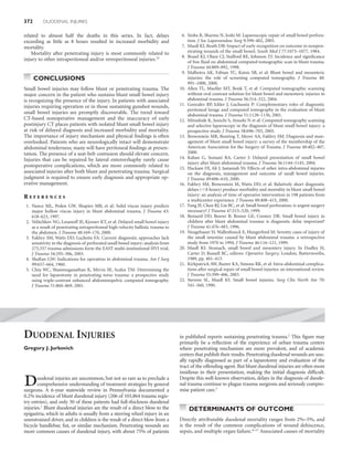
  • 6. ABDOMINAL INJURIES 377 multiple-system organ failure. Knowledge of the lethal nature of duodenal dehiscence and duodenal fistula certainly tempts the operating surgeon to add pyloric exclusion, anastomosis buttress- ing, and duodenostomy to the repair of class III or IV duodenal injuries. As noted previously, concomitant pancreatic injury should also be included as a high-risk confounder that might warrant pyloric exclusion added to the duodenal repair. In one report of 40 patients with penetrating duodenal injuries, there were 14 pa- tients with combined duodenal and pancreatic wounds. Five patients with this combination of injuries had primary duodenal repair alone, and two incurred duodenal leaks. Three of the pa- tients with combined injuries had pyloric exclusion as a treatment adjunct, and none had duodenal leaks.39 An alternative or addition to gastric diversion is duodenal decompression via retrograde jejunostomy. Stone and Fabian10 reported a fistula rate of less than 0.5% (1 in 237 patients) in a variety of duodenal injuries all treated by retrograde jejunostomy tube drainage, in contrast to a 19.3% incidence of duodenal com- plications when decompression was not used. Retrograde duode- nodenal drainage is preferred to lateral duodenostomy. Direct drainage with a tube through the suture line results in a high de- hiscence or fistula rate of 23%. Hasson and colleagues40 reviewed A the literature up to 1984 on penetrating duodenal trauma and tube duodenostomy, evaluating eight retrospective series and over 550 patients. They reported overall mortality of 19.4% and a fis- tula rate of 11.8% without decompression, compared with 9% mortality and 2.3% fistula rate with decompression. They too con- cluded that tube drainage should be performed either via stomach or retrograde jejunostomy, as these methods had a lower fistula rate and less overall mortality than lateral tube duodenostomy. Nonetheless, as is the case of pyloric exclusion for gastric diver- sion, there has been no prospective, randomized analysis of the efficacy of tube duodenal drainage techniques, and not all sur- geons support use of decompression techniques. In very massive injuries of the proximal duodenum and head of the pancreas, destruction of the ampulla and proximal pancreatic duct or distal common bile duct may preclude reconstruction. In addition, because the duodenum and the head of the pancreas have a common arterial supply, it is essentially impossible to entirely resect one without making the other ischemic. In this situation, a pancreatoduodenectomy is required. Between 1961 and 1994, 184 Whipple procedures were reported for trauma, with 26 operative deaths (14%) and 39 delayed deaths, for a 64% overall survival rate.41 With appropriate selection criteria, pancreatoduodenectomy for in- jury can be performed with similar morbidity and mortality as de- scribed in resections done for cancer.42–44 DUODENAL HEMATOMA Duodenal hematoma is generally considered an injury of childhood play or child abuse, but can occur in adults as well. In one report, 50% of the cases of duodenal hematoma in children resulted from child abuse.45 Remarkably, the duodenum is the fourth most com- monly injured intra-abdominal organ after blunt abdominal trauma, B occurring in 2%–10% of children.46 Nearly one-third of the patients Figure 7 Duodenal hematoma. The two images are sequential. present with obstruction of insidious onset at least 48 hours after (A) Initial showing near total duodenal obstruction. (B) Portrays injury, presumably the result of fluid shift into the hyperosmotic contrast passage into the jejunum. The hematoma has infiltrated the duodenal hematoma. Duodenal hematoma in general represents a wall, producing fold thickening, loop narrowing, and displacement. nonsurgical injury, in that the best results are obtained with conser- The mesentery is also involved, and there is a pronounced vative or nonsurgical management.47 It can be diagnosed either by hematoma component nearly occluding the first jejunal loop. This contrast-enhanced CT scan or upper gastrointestinal (UGI) study case shows the characteristic involvement of the duodenum as it (Figure 7). The initial water-soluble contrast (meglumine diatri- traverses the spine, sparing, but obstructing, the proximal duodenal zoate) exam should be followed by barium to provide the greater (1 and 2) segments. detail needed to detect the so-called “coiled spring” or “stacked coin” sign. Although characteristic of intramural duodenal hematoma, this finding is present in only approximately one-quarter of patients with hematoma.
  • 7. 378 DUODENAL INJURIES Although the initial treatment is nonoperative, associated injuries 12. Shorr R, Greaney G, Donovan A: Injuries of the duodenum. Am J Surg should be excluded, particularly pancreatic injury. Desai et al.46 re- 154(7):93–98, 1987. ported that 42% of pediatric patients with a duodenal injury (perfora- 13. Jurkovich GJ, Bulger E: Duodenum and pancreas. In Moore EE, tion or hematoma) had a concomitant pancreatic injury, and Jewett Feliciano DV, Mattox K, editors: Trauma, 5th ed. New York, McGraw-Hill, 2004, pp. 709–733. et al.47 found a 20% incidence of pancreatic injury in patients with a 14. Martin T, Feliciano D, Mattox K, Jordon G: Severe duodenal injuries: duodenal hematoma. Continuous nasogastric suction should be em- treatment with pyloric exclusion and gastrojejunostomy. Arch Surg 118: ployed and total parenteral nutrition begun. The patient should be 631–635, 1983. re-evaluated with UGI contrast studies at 5–7-day intervals if signs of 15. Heitsch R, Knutson C, Fulton R, et al: Delineation of critical factors in the obstruction do not spontaneously abate. Ultrasound has also been treatment of pancreatic trauma. Surgery 80(4):523–529, 1976. used to follow a resolving duodenal hematoma.48 Percutaneous drain- 16. Sukul K, Lont H, Johannes E: Management of pancreatic injuries. Hepa- age of an unresolving duodenal hematoma has been reported,49,50 but togastroenterology 39:447–450, 1992. operative exploration and evacuation of the hematoma is usually rec- 17. Roman E, Silva Y, Lucas C: Management of blunt duodenal injury. Surg ommended after 2 weeks of conservative therapy to rule out stricture, Gynecol Obstet 132:7–14, 1971. 18. Lucas C, Ledgerwood A: Factors influencing outcome after blunt duode- duodenal perforation, or injury to the head of the pancreas as factors nal injury. J Trauma 15(10):839–846, 1975. that might be contributing to the obstruction.51 One review of six cases 19. Smego DR, Richardson JD, Flint LM: Determinants of outcome in pan- of duodenal and jejunal hematomas resulting from blunt trauma dem- creatic trauma. J Trauma 25(8):771–776, 1985. onstrated resolution with nonoperative management in five of the six 20. Anatomy and physiology of the duodenum. In Shackelford R, Zuidema patients, with an average hospital stay of 16 days (range, 10–23 days), G, editors: Surgery of the Alimentary Tract. Philadelphia, WB Saunders, and total parenteral nutrition of 9 days (range, 4–16 days). The sixth 1981, pp. 38–45. case had evidence of complete bowel obstruction on UGI series, which 21. Edwards E, Malone P, MacArthur J: Operative Anatomy of Abdomen and failed to resolve after 18 days of conservative management. Laparot- Pelvis. Philadelphia, Lea & Febiger, 1975. omy revealed jejunal and colonic strictures with fibrosis, which were 22. Sherck J, Oakes D: Intestinal injuries missed by computed tomography. J Trauma 30(1):1–5, 1990. successfully resected.52 Another report included 19 cases of duodenal 23. Mirvis S, Gens D, Shanmuganathan K: Rupture of the bowel after hematoma in children, 17 (89%) managed nonoperatively and blunt abdominal trauma: diagnosis with CT. Am J Roentgenol 159(6): 2 patients in operative incision and drainage occurred within the first 1217–1221, 1992. 24 hours and never attempted nonoperative management.46 Nasogas- 24. Timaran CH, Daley BJ, Enderson BL: Role of duodenography in tric decompression and total parenteral nutrition were employed for the diagnosis of blunt duodenal injuries. J Trauma 51(4):648–651, an average of 9.3 (±7.7) days (range, 2–29 days), with an average hos- 2001. pital stay of 16.4 (±17.8) days (range, 2–37 days). 25. Allen G, Moore F, Cox CJ, Mehall J, Duke J: Delayed diagnosis of blunt If a duodenal hematoma is incidentally found at celiotomy, a duodenal injury: an avoidable complication. J Am Coll Surg 187:393–399, thorough inspection must ensue to exclude perforation. This will 1998. 26. Asensio JA, Demetriades D, Berne JD, et al: A unified approach to the require an extended Kocher maneuver, which usually successfully surgical exposure of pancreatic and duodenal injuries. Am J Surg drains the subserosal hematoma. It is unclear whether the serosa of 174(1):54–60, 1997. the duodenum should intentionally be incised along its extent to 27. Jurkovich G: Duodenal injury. In McIntyre R, Van Stiegmann G, “evacuate” the hematoma, or whether this in fact increases the likeli- Eiseman B, editors: Surgical Decision Making, 5th ed. Philadelphia, Else- hood of converting a partial duodenal wall tear into a complete vier, 2004, pp. 512–513. perforation. Unless my index of suspicion is very high for a full- 28. Moore E, Cogbill T, Malangoni M, et al: Organ injury scaling II: pancreas, thickness duodenal wall injury, I generally do not open a duodenal duodenum, small bowel, colon, and rectum. J Trauma 30(11):1427–1429, hematoma found incidentally, although I do inspect it carefully. A 1990. feeding jejunostomy should be placed, because an extended period of 29. Asensio J, Feliciano D, Britt L, Kerstein M: Management of duodenal in- juries. Curr Probl Surg 11:1021–1100, 1993. gastric decompression will likely be required. 30. Ivatury R, Gaudino J, Ascer E, Nallathambi M, Ramirez-Schon G, Stahl W: Treatment of penetrating duodenal injuries: primary repair vs. repair REFERENCES with decompressive enterostomy/serosal patch. J Trauma 25(4):337–341, 1985. 1. Ballard RB, Badellino MM, Eynon CA, Spott MA, Staz CF, Buckman RF Jr: 31. McInnis W, Aust J, Cruz A, et al: Traumatic injuries of the duodenum: a Blunt duodenal rupture: a 6-year statewide experience. J Trauma 43(2): comparison of primary closure and the jejunal patch. J Trauma 15: 229–232discussion 33, 1997. 847–858, 1975. 2. Asensio J, Feliciano D, Britt L, Kerstein M: Management of duodenal in- 32. Vaughan G, Grazier O, Graham D, et al: The use of pyloric exclusion in juries. Curr Probl Surg 11:1021, 1993. the management of severe duodenal injuries. Am J Surg 134:785–790, 3. Cuddington G, Rusnak C, Cameron R, Carter J: Management of duode- 1977. nal injuries. Can J Surg 33(1):41–44, 1990. 33. Summers JJ: The treatment of posterior perforations of the fixed portions 4. Cogbill T, Moore E, Feliciano D, et al: Conservative management of duode- of the duodenum. Ann Surg 39:727, 1904. nal trauma: a multicenter perspective. J Trauma 30(22):1469–1475, 1990. 34. Berne C, Donovan A, White E, et al: Duodenal “diverticulization” for 5. Flint L, McCoy M, Richardson J, Polk H: Duodenal injury: analysis of com- duodenal and pancreatic injury. Am J Surg 127:503–505, 1974. mon misconceptions in diagnosis and treatment. Ann Surg 191(6):697–702, 35. Donovan A, Hagen W, Berne D: Traumatic perforations of the duode- 1980. num. Am J Surg 111:341–350, 1966. 6. Ivatury R, Nallathambi M, Gaudino J, Rohman M, Stahl W: Penetrating 36. Buck JR, Sorensen VJ, Fath JJ, Horst HM, Obeid FN: Severe pancreatico- duodenal injuries: an analysis of 100 consecutive cases. Ann Surg duodenal injuries: the effectiveness of pyloric exclusion with vagotomy. 202(2):154–158, 1985. Am Surg 58(9):557–560discussion 561, 1992. 7. Kashuk J, Moore E, Cogbill T: Management of the intermediate severity 37. Cogbill T, Moore E, Kashuk J: Changing trends in the management of duodenal injury. Surgery 92:758–764, 1982. pancreatic trauma. Arch Surg 117:722–728, 1982. 8. Levison M, Petersen S, Sheldon G: Duodenal trauma: experience of a 38. Fang JF, Chen RJ, Lin BC: Controlled reopen suture technique for pyloric trauma center. J Trauma 24(6):475–480, 1984. exclusion. J Trauma 45(3):593–596, 1998. 9. Snyder W, Weigelt J, Watkins W, Bietz D: The surgical management of 39. McKenney MG, Nir I, Levi DM, Martin L: Evaluation of minor penetrat- duodenal trauma. Arch Surg 115:422–429, 1980. ing duodenal injuries. Am Surg 62(11):952–955, 1996. 10. Stone H, Fabian T: Management of duodenal wounds. J Trauma 19(5): 40. Hasson J, Stern D, Moss G: Penetrating duodenal trauma. J Trauma 334–339, 1979. 24(6):471–474, 1984. 11. Vasquez JC, Coimbra R, Hoyt DB, Fortlage D: Management of penetrat- 41. Delcore R, Stauffer J, Thomas J, Pierce G: The role of pancreatogastros- ing pancreatic trauma: an 11-year experience of a level-1 trauma center. tomy following pancreatoduodenectomy for trauma. J Trauma 37(3): Injury 32(10):753–759, 2001. 395–400, 1994.
  • 8. ABDOMINAL INJURIES 379 42. Heimansohn DA, Canal DF, McCarthy MC, Yaw PB, Madura JA, Broadie 48. Megremis S, Segkos N, Andrianaki A, et al: Sonographic diagnosis and TA: The role of pancreaticoduodenectomy in the management of traumatic monitoring of an obstructing duodenal hematoma after blunt trauma: injuries to the pancreas and duodenum. Am Surg 56(8):511–514, 1990. correlation with computed tomographic and surgical findings. J Ultra- 43. McKone T, Bursch L, Scholten D: Pancreaticoduodenectomy for trauma: sound Med 23(12):1679–1683, 2004. a life saving procedure. Am Surg 54(6):361–364, 1988. 49. Gullotto C, Paulson EK: CT-guided percutaneous drainage of a duodenal 44. Oreskovich M, Carrico C: Pancreaticoduodenectomy for trauma: a viable hematoma. Am J Roentgenol 184(1):231–233, 2005. option? Am J Surg 147(5):618–623, 1984. 50. Kortbeek JB, Brown M, Steed B: Percutaneous drainage of a duodenal 45. Wooley M, Mahour G, Sloan T: Duodenal hematoma in infancy and haematoma. Injury 28(5–6):419–420, 1997. childhood. Am J Surg 136:8–14, 1978. 51. Touloukian R: Protocol for the nonoperative treatment of obstructing 46. Desai KM, Dorward IG, Minkes RK, Dillon PA: Blunt duodenal inju- intramural duodenal hematoma. Am J Surg 145:330–335, 1983. ries in children. J Trauma 54(4):640–645, discussion 645–646, 2003. 52. Czyrko C, Weltz C, Markowitz R, O’Neill J: Blunt abdominal trauma 47. Jewett TJ, Caldarola V, Karp M, Allen J, Cooney D: Intramural hematoma resulting in intestinal obstruction: when to operate? J Trauma 30(12): of the duodenum. Arch Surg 123(1):54–58, 1988. 1567–1571, 1990. PANCREATIC INJURIES spleen. The splenic artery and vein can be found along the superior border of the pancreas. The superior mesenteric artery and vein re- Louis J. Magnotti and Martin A. Croce side just behind the neck of the pancreas and are enclosed posteriorly by the uncinate process. This process can be absent or can almost completely encircle the superior mesenteric artery and vein. The head of the pancreas is suspended from the liver by the hepa- toduodenal ligament and is firmly fixed to the medial aspect of the t he pancreas is relatively protected deep within the confines of the retroperitoneum. As such, injuries to the pancreas are uncommon, but not rare, and can present a diagnostic dilemma. In second and third portions of the duodenum. A line extending from the portal vein superiorly to the superior mesenteric vein inferiorly marks the division between the head and the neck of the gland. The neck of the pancreas measures approximately 1.5–2 cm in length and fact, despite advances in modern trauma care, there remains signifi- lies at the level of the first lumbar vertebra. It overlies the superior cant morbidity and mortality, with mortality rates ranging from mesenteric vessels and is fixed between them and the celiac trunk 9%–34%.1 Frequent complications are also common following pan- superiorly. The body of the pancreas is technically defined as that creatic injuries, occurring in 30%–60% of patients. The high compli- portion of the pancreas that lies to the left of the superior mesenteric cation rate associated with these injuries is primarily secondary to vessels. There is no true anatomic division between the body and the diagnostic delays and missed injuries. When identified early, the tail, nor is there any imaginary dividing line as in the case of the head treatment of most pancreatic injuries is straightforward. It is the and neck. delayed recognition and/or treatment of these injuries that can result The main pancreatic duct of Wirsung originates in the tail of the in devastating outcomes. pancreas and typically traverses the entire length of the gland and There are few well-documented historical accounts about the joins the common bile duct before emptying into the duodenum. management of pancreatic injuries. The first documented case of Throughout its course in the tail and body, the duct lies midway be- pancreatic trauma was an autopsy report from St. Thomas Hospital tween the superior and inferior margins and slightly more posterior. in London in 1827 in which a patient struck by the wheel of a stage- The accessory duct of Santorini usually branches out from the pan- coach suffered a complete pancreatic body transection.2 Over the creatic duct in the neck of the pancreas and empties separately into next several decades, reports of pancreatic injuries were scattered. In the duodenum. A significant number of anatomic variants exist and 1903, after extensive review of the literature, only 45 cases of pancre- must be recognized: (1) in 60% of individuals, the ducts open sepa- atic trauma, 21 resulting from penetrating injuries and 24 from blunt rately into the duodenum; (2) in 30%, the duct of Wirsung carries trauma could be identified.3 The occurrence of complications fol- the entire glandular secretion and the duct of Santorini ends blindly; lowing pancreatic injury was also noted early. In 1905, Korte4 re- and (3) in 10%, the duct of Santorini carries the entire secretion of ported a case of an isolated pancreatic transection with resultant the gland and the duct of Wirsung is either small or absent. In all pancreatic fistula. The fistula closed spontaneously and the patient cases, the ducts lie anterior to the major pancreatic vessels. survived. The arterial and venous blood supply of the pancreas is relatively The following chapter attempts to clarify the anatomic and constant. The arterial blood supply of the pancreas originates from physiologic basis for the concerns over injuries to the pancreas as well both the celiac trunk and the superior mesenteric artery. The blood as elucidate specific diagnostic and therapeutic interventions after supply to the head of the pancreas appears to be the greatest, with less traumatic injuries to the pancreas. flow to the body and tail and the least to the neck. The veins, like the arteries, are found posterior to the ducts, lie superficial to the arter- ies, and parallel the arteries for the most part throughout their ANATOMY course. The venous drainage of the pancreas is to the portal, splenic, and superior mesenteric vein. A complete understanding of pancreatic relational anatomy is es- sential for providing appropriate treatment and understanding the potential for associated injuries. The pancreas is about 15–20 cm in PHYSIOLOGY length, 3.1 cm wide, and 1–1.5 cm thick. The average mass is 90 g (ranging from 40 to 180 g).5 The inferior vena cava, aorta, left kidney, The pancreas is a compound tubuloalveolar gland with both endo- both renal veins, and right renal artery lie posterior to the pancreas. crine (insulin, glucagon, somatostatin) and exocrine (digestive en- The head of the pancreas is nestled in the duodenal sweep, with the zyme precursors, bicarbonate) function. The endocrine cells are body crossing the spine and the tail resting within the hilum of the separated histologically into nests of cells known as the islets of